Embed Size (px)
Citation preview
A C-terminal class I PDZ binding motif of EspI/NleAmodulates the virulence of attaching and effacingEscherichia coli and Citrobacter rodentium
Sau Fung Lee,1 Michelle Kelly,1 Adrian McAlister,1
Shelley N. Luck,1 Erin L. Garcia,2 Randy A. Hall,2
Roy M. Robins-Browne,3,4 Gad Frankel5 andElizabeth L. Hartland1,3,4*1Department of Microbiology, Monash University,Clayton, Victoria 3800, Australia.2Department of Pharmacology, Emory University Schoolof Medicine, Atlanta, GA 30322, USA.3Department of Microbiology and Immunology,University of Melbourne, Victoria 3010, Australia.4Murdoch Children’s Research Institute, RoyalChildren’s Hospital, Parkville, Victoria 3052, Australia.5Division of Cell and Molecular Biology, Imperial CollegeLondon, London SW7 2AZ, UK.
Summary
Enteropathogenic Escherichia coli induces character-istic attaching–effacing (A/E) lesions on the intestinalmucosa during infection. The locus of enterocyteeffacement is essential for A/E lesion formation andencodes a type III secretion system that translocatesmultiple effector proteins into the host cell. Followingtranslocation, EspI/NleA localizes to the Golgi. Usingthe yeast two-hybrid system (Y2HS) and PSD-95/Disk-large/ZO-1 (PDZ)-domain protein array overlays, weidentified 15 putative host-interacting partners ofEspI. All but two of the target proteins contained PDZdomains. Examination of the EspI amino acidsequence revealed a C-terminal consensus class IPDZ binding motif. Deletion of the last 7 amino acidsof EspI to generate EspIDC7 abrogated the Y2HS inter-action between EspI and 5 of the 6 putative host celltarget proteins tested. Deletion of the EspI PDZbinding motif also resulted in delayed trafficking ofEspI to the Golgi. Using a mouse model of infection,we showed that Citrobacter rodentium expressingtruncated EspIDC7 was attenuated when in competitionwith C. rodentium expressing full-length EspI.Overall, these results suggested that EspI may modu-
late the virulence of A/E pathogens by binding hostPDZ-domain proteins.
Introduction
Enteropathogenic Escherichia coli (EPEC) is an importanthuman pathogen that causes severe diarrhoea in youngchildren. A feature of EPEC colonization is the formationof attaching–effacing (A/E) lesions on the host gas-trointestinal tract during infection. EPEC is a member of agroup of A/E pathogens that carry the locus of enterocyteeffacement (LEE) pathogenicity island that is requiredfor A/E lesion formation (Elliott et al., 1998). The LEEencodes transcriptional regulators, the adhesin, intimin(Jerse et al., 1990), a type III secretion system (T3SS)(Jarvis and Kaper, 1996), translocators, chaperones andeffector proteins that are translocated by the T3SS intothe eukaryotic cell (Garmendia et al., 2005). A/E lesionsare characterized by localized effacement of the brush-border microvilli, intimate attachment of the bacteria to thehost cell plasma membrane, and the subsequent forma-tion of actin-rich pedestal-like structures adjacent toadherent bacteria (Garmendia et al., 2005). BecauseEPEC is a human-specific pathogen, infection of micewith the A/E pathogen, Citrobacter rodentium, is com-monly used as a model for EPEC infection (reviewed inMundy et al., 2005). C. rodentium induces A/E lesions inmice that are indistinguishable from those caused byother A/E pathogens (Schauer and Falkow, 1993).
Although EPEC and other A/E pathogens generallyremain extracellular and attached to the apical surfaceof enterocytes, all A/E pathogens are able to interferewith internal host cell processes through the LEE T3SS-dependent translocation of multiple effector proteins(Tobe et al., 2006). A minority of these effectors areencoded by genes within the LEE (Tobe et al., 2006). Oneof the LEE-encoded effector proteins, Tir (Kenny et al.,1997), is integrated into the host cell plasma membrane,where it adopts a hairpin loop topology (Hartland et al.,1999). Through the central extracellular domain, Tir actsas a receptor for intimin (Hartland et al., 1999; Kenny,1999). This specific interaction between intimin and Tir iscentral to intimate attachment of bacteria to the hostepithelium (Batchelor et al., 2000; Luo et al., 2000).
Received 6 February, 2007; revised 20 August, 2007; accepted 19September, 2007. *For correspondence. E-mail [email protected]; Tel. (+61) 3 8344 8041; Fax (+61) 9347 1540.
Cellular Microbiology (2008) 10(2), 499–513 doi:10.1111/j.1462-5822.2007.01065.xFirst published online 2 November 2007
© 2007 The AuthorsJournal compilation © 2007 Blackwell Publishing Ltd
Simultaneously, through the intracellular amino andcarboxy termini, Tir interacts with several cytoskeletal pro-teins, linking the extracellular bacterium to the host cellcytoskeleton (Goosney et al., 2000; Gruenheid et al.,2001; Batchelor et al., 2004).
EspI (also termed NleA) is a non-LEE-encoded effectorprotein that is not required for A/E lesion formation butcontributes to bacterial colonization and the induction ofhyperplasia in the colonic epithelium of mice infected withC. rodentium (Gruenheid et al., 2004; Mundy et al.,2004a). In EPEC and the human pathogen enterohaem-orrhagic E. coli (EHEC), the gene encoding EspI is morefrequent among strains associated with severe disease,suggesting that the protein also has an important role invirulence in these pathogens (Mundy et al., 2004b). Fol-lowing translocation, EspI is rapidly targeted to the Golgiapparatus of eukaryotic cells, where it colocalizes withmannosidase II and Golgin-97 (Gruenheid et al., 2004;Creuzburg et al., 2005), but it is not known whether thistrafficking event is important or relevant to the function ofEspI. EspI does not contain classical Golgi-targetingmotifs and may be targeted to the Golgi apparatus vianovel Golgi-targeting motifs or by interaction with otherGolgi-associated host cell proteins (Gruenheid et al.,2004).
Although espI contributes to the virulence ofC. rodentium and was identified twice in C. rodentiumsignature-tagged mutagenesis screens (Mundy et al.,2004a; Kelly et al., 2006), the host target of this proteinand its mechanism of action are unknown. To elucidatethe role of EspI in the host–pathogen interaction, we usedthe yeast two-hybrid system (Y2HS) to identify potentialhost cell binding partners that may be affected by EspIfunction. The Y2HS screen of a HeLa cell cDNA libraryyielded six putative interacting host proteins, four ofwhich contained reported PSD-95/Disk-large/ZO-1 (PDZ)domains. PDZ domains are common protein–proteininteraction domains in eukaryotic proteins that are presentin up to 0.5% of open reading frames. Typically PDZ-domain proteins are involved in the assembly of host cellmultiprotein signalling complexes (Harris and Lim, 2001).A PDZ-domain comprises ~90 amino acids that form aseries of six b-strands and two a-helices that fold into asix-stranded b-sandwich (Jelen et al., 2003; Piserchioet al., 2006). PDZ domains recognize a C-terminal aminoacid motif on target proteins comprising a ~5-amino-acidcore recognition motif that serves as an additional anti-parallel b-strand during a PDZ domain–ligand interaction(Jelen et al., 2003; Piserchio et al., 2006; Zhang et al.,2006). PDZ domains may be classified according to theC-terminal peptide they recognize. Class I PDZ domainsrecognize the consensus amino acid sequence x-[S/T]-x-[V/L/I]-COOH, class II PDZ domains recognize F-x-F-COOH,and class 3 PDZ domains recognize a target sequence
x-x-CCOOH, where x is any amino acid and F is a hydro-phobic amino acid (Songyang et al., 1997; Harris and Lim,2001; Jelen et al., 2003). Residues at positions 0 and -2(where the C-terminal amino acid is position 0) are criticalfor binding, but the specificity and affinity of binding maybe greatly influenced by upstream amino acids (Piserchioet al., 2006; Zhang et al., 2006). For example, aminoacids up to -10 have been reported to participate in PDZinteractions (Lim et al., 2002). Therefore, the specificityand affinity of PDZ interactions can vary considerably.While some PDZ domain–ligand interactions are highlyspecific, others are quite promiscuous (Lim et al., 2002;Zhang et al., 2006). In this study, we found that EspIcontained a class I consensus PDZ binding motif at theC-terminus, which was highly conserved among A/Epathogens. Here we examined the contribution of the PDZbinding ligand of EspI to host protein interactions, to Golgitrafficking and to the virulence of C. rodentium.
Results
Identification of putative host cell-interacting partnersof EspI
To identify potential eukaryotic interacting partners ofEspI, we performed a Y2HS screen of a pretransformedHeLa cDNA library using the full-length EspI from EPECE2348/69 as bait. Thirty-two yeast colonies were obtainedfrom the library screen following growth of the diploids onmedium that selects for protein–protein interactions.Sequencing of rescued cDNA plasmids from positiveyeast colonies yielded six different putative host cell-interacting partners of EspI. Five of the six targets wereidentified multiple times, with eight independent hits forSec24B and PDZK11 (Table 1). Four of the six putativeEspI-interacting proteins contained PDZ domains(Table 1). Expression of the Y2HS reporter, lacZ, wasassayed by measuring b-galactosidase activity in yeastclones carrying the bait and prey plasmids to assess therelative affinity of EspI binding to the various putative hostcell targets. Yeast strains expressing EspI and the respec-tive HeLa proteins showed a 5- to 40-fold increase inb-galactosidase activity compared with the negativecontrol yeast strains, thereby confirming a genuine inter-action of EspI with all six proteins in the Y2HS (Fig. 1A).
Although the Y2HS is a powerful screening tool todetect protein interactions, it has an inherent limitation assome interactions are not permissive in yeast, thus pro-ducing false-negative results. Given the observation thatEspI interacted with several host cell proteins containingPDZ domains, we hypothesized that EspI may recognizeadditional host PDZ-domain proteins. In order to addressthis experimentally, we fused the last 50 amino acids ofEspI to glutathione S-transferase (GST) and screened a
500 S. F. Lee et al.
© 2007 The AuthorsJournal compilation © 2007 Blackwell Publishing Ltd, Cellular Microbiology, 10, 499–513
PDZ-domain protein array for interacting partners. GST–EspI50 bound 13 of 96 possible PDZ domains, including 4isoforms of syntrophin and 2 isoforms of NHERF (Fig. 1Band C, Table 2). In general, the intensity of labelling on thePDZ array corresponds to the strength of the interaction.Thus, it appeared that EspI interactions with the syntro-phins, MAGI-3 and PSD-95 were the strongest interac-tions observed on the array (Fig. 1B). Interestingly, theEPEC effector protein Map was also recently found tobind PDZ1 of NHERF1 via the carboxy-terminal DTRL
PDZ binding motif (Alto et al., 2006; Simpson et al.,2006). Overall, we found that EspI had the potential tobind at least 15 host cell proteins.
Contribution of a putative C-terminal PDZ binding motifof EspI to protein–protein interactions
Because four of the six putative host cell binding partnersof EspI contained PDZ domains, we examined the aminoacid sequence of EspI for a C-terminal PDZ binding motif.
Table 1. Eukaryotic EspI binding proteins identified from Y2HS screen of a HeLa cDNA library.
EspI bindingprotein
GenBankaccessionnumber
Number oftimesidentified
PredictedPfamdomainsa
Totalaminoacids
Amino acids(and domains)in Y2HS clone Function
a1 syntrophin AAB36398 6 PDZ, PH, SU 505 79–300 (PDZ, PH) Targeting of signalling proteins andmaintaining membrane integrity
MALS3 BAD96876 5 PDZ, L27 197 1–194 (PDZ, L27) Protein targeting and traffickingPDZK11 AAH89433 8 PDZ 139 1–139 (PDZ) UnknownSNX27 EAW53427 1 PDZ, PX 508 56–167 (PDZ) Endocytosis and vesicle traffickingSec24B EAX06241 8 Sec23/24 1218 67–292 Protein trafficking from ER to the GolgiTCOF1 EAW61733 4 LisH 795 356–513 Associated with Treacher Collins–
Franceschetti syndrome
a. SU, syntrophin unique; L27, domain found in receptor targeting proteins Lin-2 and Lin-7; PX, phosphoinositide binding domain; Sec23/24,Sec23/24 helical domain; LisH, Lissencephaly type-1-like homology motif.
Fig. 1. A. Interaction of EspI and EspIDC7
from EPEC E2348/69 with a1 syntrophin,Sec24B, TCOF1, PDZK11, MALS3 andSNX27 in the Y2HS. Interactions wereanalysed by assessing b-galactosidaseactivity in S. cerevisiae strains carrying EspIand EspIDC7 fusions to the binding domain ofGAL4 and HeLa cDNA fusions to theactivation domain of GAL4 as indicated.Results are presented as mean � standarddeviation of at least three biological replicates.B. PDZ-domain protein array overlaid withpurified GST–EspI50 and detected withanti-GST antibodies.C. PDZ-domain protein array overlaid withpurified GST and detected with anti-GSTantibodies. PDZ domains included on thearray are listed in Table 2.E
sp
I
1 s
yn
tro
ph
inα
1 s
yn
tro
ph
inα
Esp
I-
1 s
yn
tro
ph
inα -
C7
ΔE
sp
I
Se
c2
4B
Esp
I-S
ec2
4B
-Se
c2
4B
C7
ΔE
sp
I
TC
OF
1
Esp
I-T
CO
F1
-TC
OF
1C
7Δ
Esp
I
PD
ZK
11
Esp
I-P
DZ
K1
1
-PD
ZK
11
C7
ΔE
sp
I
MA
LS
3
Esp
I-M
AL
S3
-MA
LS
3C
7Δ
Esp
I
SN
X2
7
Esp
I-S
NX
27
SN
X2
7−
C7
ΔE
sp
I
0
10
20
30
40
50
60
70
β - g
ala
cto
sid
ase a
ctivity
(Mill
er
Units)
A
GST-EspI50 GST onlyB C
EspI and the virulence of A/E pathogens 501
© 2007 The AuthorsJournal compilation © 2007 Blackwell Publishing Ltd, Cellular Microbiology, 10, 499–513
Alignment of the amino acid sequences of all EspI pro-teins available from public databases revealed a con-sensus class I PDZ binding motif, ETRV, which wasconserved among all A/E pathogens. In fact, theC-terminal 7 amino acids were highly conserved amongthe 18 EspI sequences examined (data not shown).Because up to 10 amino acids of a PDZ ligand maycontribute to the PDZ interaction (Lim et al., 2002), wedeleted the conserved C-terminal 7 amino acids to deter-mine whether the putative PDZ binding motif played adirect role in EspI–host protein interactions. The truncatedprotein from EPEC EspI was termed EspIDC7. EspIDC7 wasintroduced into the Y2HS and tested against the six hostproteins identified by screening with full-length EspI.b-Galactosidase assays showed that upon deletion of theC-terminal 7 amino acids, the interactions between EspIand five of the six host cell proteins were lost. The onlyinteraction to be maintained using EspIDC7 was withTCOF1, which does not have a PDZ domain. Interestingly,although Sec24B does not have a reported PDZ domain,EspI interacted with this target in a PDZ motif-dependentmanner (Fig. 1A).
In order to determine the specificity of the EspI PDZbinding motif, the 4 C-terminal amino acids of EspI(ETRV) were replaced with the PDZ binding motif of Map(DTRL). EspIDTRL was introduced into the Y2HS andtested for its ability to interact with the six putative Y2HSbinding partners of EspI. While interactions with TCOF1(which binds EspI independently of the ETRV motif) and
Sorting Nexin 27 (SNX27) were retained in the Y2HS,EspIDTRL did not support interactions with a1 syntrophin,Sec24B, Mammalian LIN Seven 3 (MALS3) and PDZK11(Fig. 2).
Table 2. PDZ domains included on protein array.
Position PDZ domaina Position PDZ domain Position PDZ domain Position PDZ domain
A1 MAGI-1 PDZ1 C1 INADL PDZ6 E1 RHOPHILIN-2 G1 PDZK1 DZ4A2 MAGI-1 PDZ2 C2 AP97 PDZ1 + 2 E2 HARMONIN PDZ1 G2 PDZK2 PDZ1b
A3 MAGI-1 PDZ3 C3 SAP97 PDZ3b E3 HARMONIN PDZ2 G3 PDZK2 PDZ2A4 MAGI-1 PDZ4 + 5 C4 SAP102 PDZ1 + 2 E4 NEURABIN PDZ G4 PDZK2 PDZ3A5 MAGI-2 PDZ1 C5 SAP102 PDZ3b E5 SPINOPHILIN PDZ G5 PDZK2 PDZ4A6 MAGI-2 PDZ2b C6 CHAP110 PDZ1 + 2 E6 a1 SYNTROPHINb G6 LNX1 PDZ1A7 MAGI-2 PDZ3 C7 CHAP110 PDZ3 E7 b1 SYNTROPHINb G7 LNX1 PDZ2A8 MAGI-2 PDZ4 C8 E6TP1 PDZ E8 b2 SYNTROPHINb G8 LNX1 PDZ3A9 MAGI-2 PDZ5 C9 ERBIN PDZ E9 g1 SYNTROPHIN G9 LNX1 PDZ4A10 MAGI-3 PDZ1b C10 ZO-1 PDZ1 E10 g2 SYNTROPHINb G10 LNX2 PDZ1A11 MAGI-3 PDZ2 C11 ZO-1 PDZ2 E11 PAPIN 1 G11 LNX2 PDZ2A12 MAGI-3 PDZ3 C12 ZO-1 PDZ3 E12 MUPP1 PDZ1 G12 LNX2 PDZ4B1 MAGI-3 PDZ4 D1 ZO-2 PDZ1 F1 MUPP1 PDZ6 H1 PTPN4 PDZB2 MAGI-3 PDZ5 D2 ZO-2 PDZ2 F2 MUPP1 PDZ7 H2 RHO-GEF PDZB3 NHERF1 PDZ1b D3 ZO-2 PDZ3 F3 MUPP1 PDZ8 H3 RA-GEF PDZB4 NHERF1 PDZ2 D4 ZO-3 PDZ1 F4 MUPP1 PDZ10 H4 ENIGMA PDZB5 HERF2 PDZ1 D5 ZO-3 PDZ2 F5 MUPP1 PDZ12 H5 LARG PDZB6 NHERF2 PDZ2b D6 ZO-3 PDZ3 F6 MUPP1 PDZ13 H6 MAST205 PDZB7 SD-95 PDZ1 + 2 D7 C2PA PDZ F7 PTPN13 PDZ1 H7 PTPN3 PDZB8 PSD-95 PDZ3b D8 GIPC PDZ F8 PTPN13 PDZ3 H8 SHANK1 PDZB9 PDZ-GEF1 PDZ D9 MALS1 PDZ F9 PTPN13 PDZ4 + 5 H9 TAMALIN PDZB10 CAL PDZ D10 MALS3 PDZ F10 PDZK1 PDZ1b H10 PAR-3 PDZ1B11 nNOS PDZ D11 DENSIN-180 F11 PDZK1 PDZ2 H11 PAR-3 PDZ2B12 INADL PDZ5 D12 RHOPHILIN-1 F12 PDZK1 PDZ3 H12 PAR-3 PDZ3
a. Full details of protein domains shown elsewhere (Fam et al., 2005; He et al., 2006).b. Positive interaction with GST–EspI50.
1 s
yn
tro
ph
inα -
DT
RL
Esp
I
-Se
c2
4B
DT
RL
Esp
I
TC
OF
1−
DT
RL
Esp
I
-PD
ZK
11
DT
RL
Esp
I
-MA
LS
3D
TR
LE
sp
I
-SN
X2
7D
TR
LE
sp
I
0
10
20
30
40
β -gala
cto
sid
ase a
ctivity
(Mill
er
Units)
Fig. 2. Interaction of EspIDTRL with a1 syntrophin, Sec24B, TCOF1,PDZK11, MALS3 and SNX27 in the Y2HS. Interactions wereanalysed by assessing b-galactosidase activity in S. cerevisiaestrains carrying EspIDTRL fusions to the binding domain of GAL4 andHeLa cDNA fusions to the activation domain of GAL4 as indicated.Results are presented as mean � standard deviation of at leastthree biological replicates.
502 S. F. Lee et al.
© 2007 The AuthorsJournal compilation © 2007 Blackwell Publishing Ltd, Cellular Microbiology, 10, 499–513
The PDZ binding motif of EspI is important for rapidtargeting to the Golgi apparatus
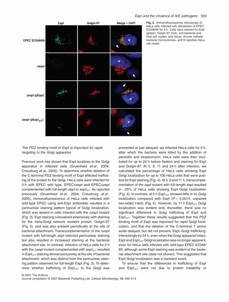
Previous work has shown that EspI localizes to the Golgiapparatus in infected cells (Gruenheid et al., 2004;Creuzburg et al., 2005). To determine whether deletion ofthe C-terminal PDZ binding motif of EspI affected traffick-ing of the protein to the Golgi, HeLa cells were infected for5 h with EPEC wild type, EPECDespI and EPECDespIcomplemented with full-length espI or espIDC7. As reportedpreviously (Gruenheid et al., 2004; Creuzburg et al.,2005), immunofluorescence of HeLa cells infected withwild-type EPEC using anti-EspI antibodies resulted in aperinuclear staining pattern typical of Golgi localization,which was absent in cells infected with the DespI mutant(Fig. 3). EspI staining colocalized extensively with stainingfor the trans-Golgi network resident protein, Golgin-97(Fig. 3), and was also present periodically at the site ofbacterial attachment. Transcomplementation of the DespImutant with full-length espI restored perinuclear stainingbut also resulted in increased staining at the bacterialattachment site. In contrast, infection of HeLa cells for 5 hwith the DespI mutant complemented with espIDC7 resultedin EspIDC7 staining almost exclusively at the site of bacterialattachment, which was distinct from the perinuclear stain-ing pattern observed for full-length EspI (Fig. 3). To deter-mine whether trafficking of EspIDC7 to the Golgi was
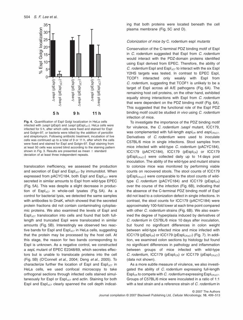
prevented or just delayed, we infected HeLa cells for 5 h,after which the bacteria were killed by the addition ofpenicillin and streptomycin. HeLa cells were then incu-bated for up to 24 h before fixation and staining for EspIand Golgin-97. At 5, 9, 11 and 24 h after infection, wecalculated the percentage of HeLa cells showing EspIGolgi localization for up to 100 HeLa cells that were posi-tive for EspI staining (Fig. 4).At 5, 9 and 11 h, transcomple-mentation of the espI mutant with full-length espI resultedin ~25% of HeLa cells showing EspI Golgi localization(Fig. 4). In contrast, at 5 h EspIDC7 showed little or no Golgilocalization compared with EspI (P = 0.0014, unpairedtwo-tailed t-test) (Fig. 4). However, by 11 h EspIDC7 Golgilocalization was evident and, thereafter, there was nosignificant difference in Golgi trafficking of EspI andEspIDC7. Together these results suggested that the PDZbinding motif of EspI was important for rapid Golgi local-ization, and that the deletion of the C-terminal 7 aminoacids delayed, but did not prevent, EspI–Golgi trafficking.Interestingly by 24 h, even when the Golgi appeared intact,EspI and EspIDC7 Golgi localization was no longer apparent,even for HeLa cells infected with wild-type EPEC E2348/69, although some EspI staining was evident at the bacte-rial attachment site (data not shown). This suggested thatEspI Golgi localization was a transient event.
To ensure that the differences in trafficking of EspIand EspIDC7 were not due to protein instability or
Fig. 3. Immunofluorescence microscopy ofHeLa cells infected with derivatives of EPECE2348/69 for 5 h. Cells were stained for EspI(green), Golgin-97 (red), and bacterial andhost cell nucleic acid (blue). Arrows indicatebacterial microcolonies, and N signifies HeLacell nuclei.
Merge + DAPI
ΔespI (pEspIΔC7)
Golgin-97EspI
ΔespI (pEspI)
ΔespI
EPEC E2348/69 N
N
N
N
Merge + DAPI
ΔespI (pEspIΔC7)
Golgin-97EspI
ΔespI (pEspI)
ΔespI
EPEC E2348/69 N
N
N
N
EspI and the virulence of A/E pathogens 503
© 2007 The AuthorsJournal compilation © 2007 Blackwell Publishing Ltd, Cellular Microbiology, 10, 499–513
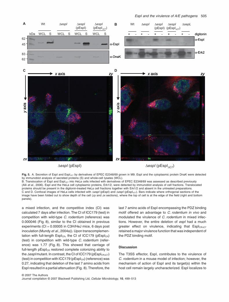
translocation inefficiency, we assessed the productionand secretion of EspI and EspIDC7 by immunoblot. Whenexpressed from pACYC184, both EspI and EspIDC7 weresecreted in similar amounts to EspI from wild-type EPEC(Fig. 5A). This was despite a slight decrease in produc-tion of EspIDC7 in whole-cell lysates (Fig. 5A). As acontrol for bacterial lysis, we detected the same sampleswith antibodies to DnaK, which showed that the secretedprotein fractions did not contain contaminating cytoplas-mic proteins. We also examined the levels of EspI andEspIDC7 translocation into cells and found that both full-length and truncated EspI were translocated in similaramounts (Fig. 5B). Interestingly we observed two reac-tive bands for EspI and EspIDC7 in HeLa cells, suggestingthat the protein may be processed by the host cell. Atthis stage, the reason for two bands corresponding toEspI is unknown. As a negative control, we constructeda sepL mutant of EPEC E2348/69, which secretes effec-tors but is unable to translocate proteins into the cell(Fig. 5B) (O’Connell et al., 2004; Deng et al., 2005). Tocharacterize further the location of EspI and EspIDC7 inHeLa cells, we used confocal microscopy to takeorthogonal sections through infected cells stained simul-taneously for EspI or EspIDC7 and actin. Staining for bothEspI and EspIDC7 clearly spanned the cell depth indicat-
ing that both proteins were located beneath the cellplasma membrane (Fig. 5C and D).
Colonization of mice by C. rodentium espI mutants
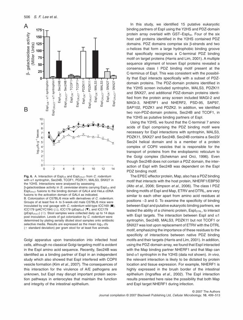
Conservation of the C-terminal PDZ binding motif of EspIin C. rodentium suggested that EspI from C. rodentiumwould interact with the PDZ-domain proteins identifiedusing EspI derived from EPEC. Therefore, the ability ofC. rodentium EspI and EspIDC7 to interact with the six EspIY2HS targets was tested. In contrast to EPEC EspI,TCOF1 interacted only weakly with EspI fromC. rodentium, suggesting that TCOF1 is unlikely to be atarget of EspI across all A/E pathogens (Fig. 6A). Theremaining host cell proteins, on the other hand, exhibitedequally strong interactions with EspI from C. rodentiumthat were dependent on the PDZ binding motif (Fig. 6A).This suggested that the functional role of the EspI PDZbinding motif could be studied in vivo using C. rodentiuminfection of mice.
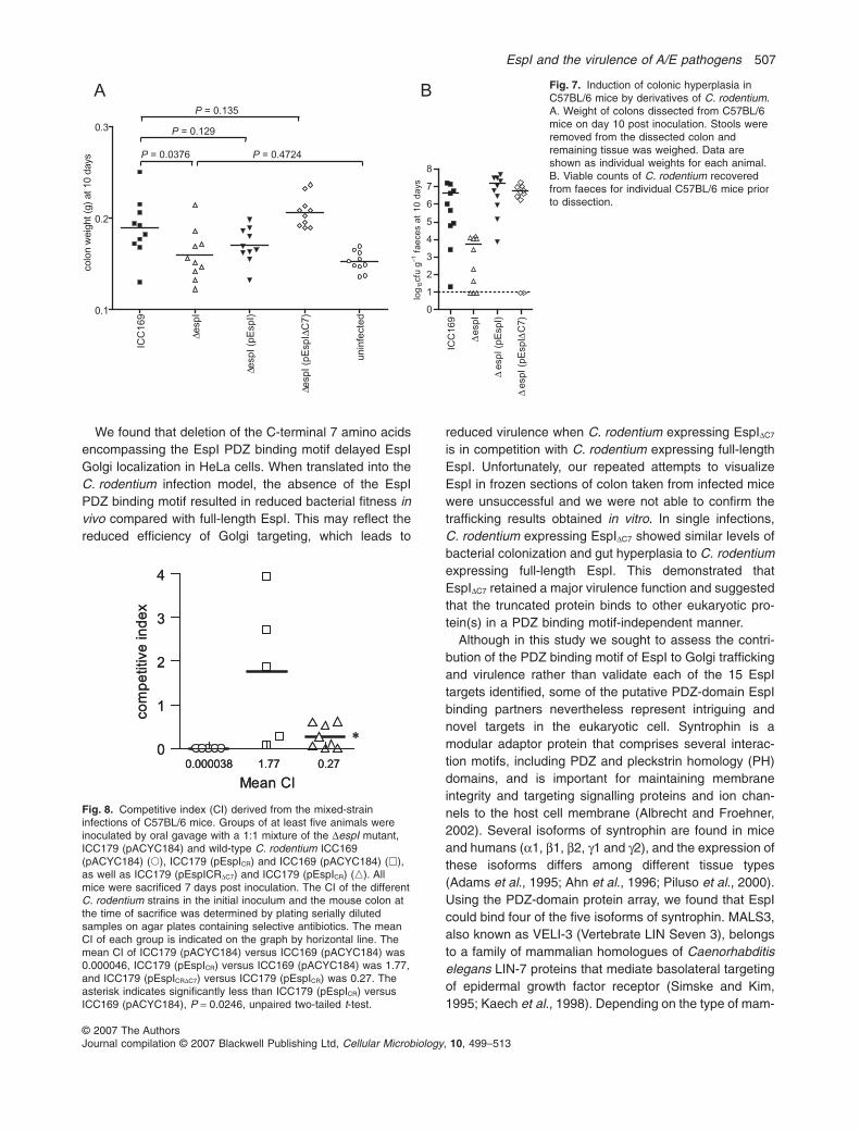
To investigate the importance of the PDZ binding motiffor virulence, the C. rodentium DespI mutant, ICC179,was complemented with full-length espICR and espICRDC7.Derivatives of C. rodentium were used to inoculateC57BL/6 mice in single infections. Stool samples frommice infected with wild-type C. rodentium (pACYC184),ICC179 (pACYC184), ICC179 (pEspICR) or ICC179(pEspICRDC7) were collected daily up to 14 days postinoculation. The ability of the wild-type and mutant strainsto colonize mice was monitored by performing viablecounts on recovered stools. The stool counts of ICC179(pEspICRDC7) were comparable to the stool counts of wild-type C. rodentium (pACYC184) and ICC179 (pEspICR)over the course of the infection (Fig. 6B), indicating thatthe absence of the C-terminal PDZ binding motif of EspIdid not lead to a colonization defect in single infections. Incontrast, the stool counts for ICC179 (pACYC184) wereapproximately 100-fold lower at each time point comparedwith other C. rodentium strains (Fig. 6B). We also exam-ined the degree of hyperplasia induced by derivatives ofC. rodentium in C57BL/6 mice 10 days after inoculation,but found no significant differences in colon weightbetween wild-type infected mice and mice infected withICC179 (pEspICR) or ICC179 (pEspICRDC7) (Fig. 7). In addi-tion, we examined colon sections by histology but foundno significant differences in pathology and inflammationbetween groups of mice infected with wild-typeC. rodentium, ICC179 (pEspICR) or ICC179 (pEspICRDC7)(data not shown).
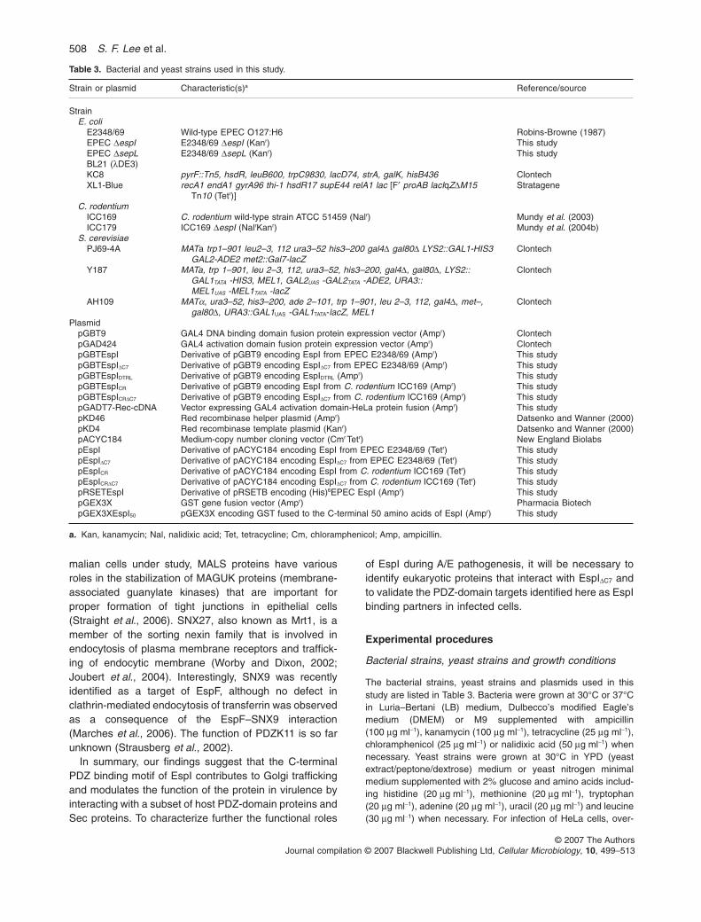
As a more subtle measure of virulence, we also investi-gated the ability of C. rodentium expressing full-lengthEspICR to compete with C. rodentium expressing EspICRDC7.Groups of C57BL/6 mice were inoculated in a ratio of 1:1with a test strain and a reference strain of C. rodentium in
espI (p
EspI)
Δ
)C
7Δ
espI (p
EspI
Δ
espI (p
EspI)
Δ
)C
7Δ
espI (p
EspI
Δ
espI (p
EspI)
Δ
)C
7Δ
espI (p
EspI
Δ
0
5
10
15
20
25
30
35
5 h 9 h 11 h
% E
spI
Golg
i lo
caliz
ation
Fig. 4. Quantification of EspI Golgi localization in HeLa cellsinfected with DespI (pEspI) and DespI (pEspIDC7). HeLa cells wereinfected for 5 h, after which cells were fixed and stained for EspIand Golgin-97, or bacteria were killed by the addition of penicillinand streptomycin. Following antibiotic treatment, incubation of livecells was continued up to a total of 9 or 11 h, after which the cellswere fixed and stained for EspI and Golgin-97. EspI staining fromat least 50 cells was scored blind according to the staining patternshown in Fig. 3. Results are presented as mean � standarddeviation of at least three independent repeats.
504 S. F. Lee et al.
© 2007 The AuthorsJournal compilation © 2007 Blackwell Publishing Ltd, Cellular Microbiology, 10, 499–513
a mixed infection, and the competitive index (CI) wascalculated 7 days after infection. The CI of ICC179 (test) incompetition with wild-type C. rodentium (reference) was0.000046 (Fig. 8), similar to the CI obtained in previousexperiments (CI = 0.00005 in C3H/HeJ mice, 6 days postinoculation (Mundy et al., 2004a)). Upon transcomplemen-tation with full-length EspICR, the CI of ICC179 (pEspICR)(test) in competition with wild-type C. rodentium (refer-ence) was 1.77 (Fig. 8). This showed that carriage offull-length pEspICR restored complete colonizing ability tothe DespI mutant. In contrast, the CI of ICC179 (pEspICRDC7)(test) in competition with ICC179 (pEspICR) (reference) was0.27, indicating that deletion of the last 7 amino acids fromEspI resulted in a partial attenuation (Fig. 8). Therefore, the
last 7 amino acids of EspI encompassing the PDZ bindingmotif offered an advantage to C. rodentium in vivo andmodulated the virulence of C. rodentium in mixed infec-tions. However, the entire deletion of espI had a muchgreater effect on virulence, indicating that EspICRDC7
retained a major virulence function that was independent ofthe PDZ binding motif.
Discussion
The T3SS effector, EspI, contributes to the virulence ofC. rodentium in a mouse model of infection; however, themechanism of action of EspI and its target(s) within thehost cell remain largely uncharacterized. EspI localizes to
α-EspI
α-DnaK
α-EspI
α-DnaK
kDa WCL S WCL S WCL S WCL S
62
45
83
62
Wt ΔespI ΔespI ΔespI(pEspI) (pEspIΔC7)
A
+ - + - + - + - + -
α-EspI
α-Erk2
digitonin+ - + - + - + - + -
α-EspI
α-Erk2
digitonin
B Wt ΔespI ΔespI ΔespI ΔsepL(pEspI) (pEspIΔC7)
C Dx axis
yaxis
zx
zy x axis
yaxis
zx
zyx axis
yaxis
zx
zyx axis
yaxis
zx
zy x axis
yaxis
zx
zyx axis
yaxis
zx
zy
ΔespI (pEspI) ΔespI (pEspIΔC7)
Fig. 5. A. Secretion of EspI and EspIDC7 by derivatives of EPEC E2348/69 grown in M9. EspI and the cytoplasmic protein DnaK were detectedby immunoblot analysis of secreted proteins (S) and whole-cell lysates (WCL).B. Translocation of EspI and EspIDC7 into HeLa cells infected with derivatives of EPEC E2348/69 was assessed as described previously(Aili et al., 2006). EspI and the HeLa cell cytoplasmic proteins, Erk1/2, were detected by immunoblot analysis of cell fractions. Translocatedproteins should be present in the digitonin-treated HeLa cell fractions together with Erk1/2 and absent in the untreated preparations.C and D. Confocal images of HeLa cells infected with DespI (pEspI) and DespI (pEspIDC7). Bars indicate where orthogonal sections of theimage have been folded out to show depth of the cell (zy and zx sections), where the top of cell is at the edge of the field (right and bottompanels).
EspI and the virulence of A/E pathogens 505
© 2007 The AuthorsJournal compilation © 2007 Blackwell Publishing Ltd, Cellular Microbiology, 10, 499–513
Golgi apparatus upon translocation into infected hostcells, although no classical Golgi-targeting motif is evidentin the EspI amino acid sequence. Recently, Sec24B wasidentified as a binding partner of EspI in an independentstudy which also showed that EspI interfered with COPIIvesicle formation (Kim et al., 2007). The consequences ofthis interaction for the virulence of A/E pathogens areunknown, but EspI may disrupt important protein secre-tion pathways in enterocytes that maintain the functionand integrity of the intestinal epithelium.
In this study, we identified 15 putative eukaryoticbinding partners of EspI using the Y2HS and PDZ-domainprotein array overlaid with GST–EspI50. Four of the sixhost cell proteins identified in the Y2HS contained PDZdomains. PDZ domains comprise six b-strands and twoa-helices that form a large hydrophobic binding groovethat specifically recognizes a C-terminal PDZ bindingmotif on target proteins (Harris and Lim, 2001). A multiplesequence alignment of known EspI proteins revealed aconsensus class I PDZ binding motif present at theC-terminus of EspI. This was consistent with the possibil-ity that EspI interacts specifically with a subset of PDZ-domain proteins. The PDZ-domain proteins identified inthe Y2HS screen included syntrophin, MALS3, PDZK11and SNX27, and additional PDZ-domain proteins identi-fied from the protein array screen included MAGI-2 andMAGI-3, NHERF1 and NHERF2, PSD-95, SAP97,SAP102, PDZK1 and PDZK2. In addition, we identifiedtwo non-PDZ-domain proteins, Sec24B and TCOF1, inthe Y2HS as putative binding partners of EspI.
Using the Y2HS, we found that the C-terminal 7 aminoacids of EspI comprising the PDZ binding motif werenecessary for EspI interactions with syntrophin, MALS3,PDZK11, SNX27 and Sec24B. Sec24B contains a Sec23/Sec24 helical domain and is a member of a proteincomplex of COPII vesicles that is responsible for thetransport of proteins from the endoplasmic reticulum tothe Golgi complex (Schekman and Orci, 1996). Eventhough Sec24B does not contain a PDZ domain, the inter-action of EspI with Sec24B was dependent on the EspIPDZ binding motif.
The EPEC effector protein, Map, also has a PDZ bindingmotif that interacts with the host protein, NHERF1/EBP50(Alto et al., 2006; Simpson et al., 2006). The class I PDZbinding motifs of EspI and Map, ETRV and DTRL, are verysimilar to each other apart from amino acid residues atpositions -3 and 0. To examine the specificity of bindingbetween EspI and putative eukaryotic binding partners, wetested the ability of a chimeric protein, EspIDTRL, to interactwith EspI targets. The interaction between EspI and a1syntrophin, Sec24B, MALS3, PDZK11 but not TCOF1 orSNX27 was lost upon replacement of ETRV with the DTRLmotif, emphasizing the importance of these residues to thespecificity of interactions between native PDZ bindingmotifs and their targets (Harris and Lim, 2001). In addition,using the PDZ-domain array, we found that EspI interactedwith the Map binding partner NHERF1 and that Map canbind a1 syntrophin in the Y2HS (data not shown). In vivo,the relevant interaction is likely to be dictated by proteinlocation and tissue expression. For example, NHERF1 ishighly expressed in the brush border of the intestinalepithelium (Ingraffea et al., 2002). The EspI interactionresults presented here raise the possibility that both Mapand EspI target NHERF1 during infection.
1 s
yn
tro
ph
inα
- C
R
Esp
I
1 s
yn
tro
ph
inα
- C
7
ΔC
RE
sp
I
- S
ec2
4B
CR
E
sp
I
- S
ec2
4B
C7
ΔC
RE
sp
I
- T
CO
F1
CR
E
sp
I
- T
CO
F1
C7
ΔC
RE
sp
I
- P
DZ
K1
1C
R
Esp
I
- P
DZ
K1
1C
7
ΔC
RE
sp
I
- M
AL
S3
CR
E
sp
I
- M
AL
S3
C7
ΔC
RE
sp
I
- S
NX
27
C
RE
sp
I
- S
NX
27
C7
ΔC
RE
sp
I
0
25
50
75
100
125
150
175β -
gala
cto
sid
ase a
ctivity
(Mill
er
Units)
A
0 2 4 6 8 10 12 14
0
1
2
3
4
5
6
7
8
9
10
log
10cfu
g–
1 f
aeces
B
Fig. 6. A. Interaction of EspICR and EspICRDC7 from C. rodentiumwith a1 syntrophin, Sec24B, TCOF1, PDZK11, MALS3, SNX27 inthe Y2HS. Interactions were analysed by assessingb-galactosidase activity in S. cerevisiae strains carrying EspICR andEspICRDC7 fusions to the binding domain of GAL4 and HeLa cDNAfusions to the activation domain of GAL4 as indicated.B. Colonization of C57BL/6 mice with derivatives of C. rodentium.Groups of at least five 4- to 5-week-old male C57BL/6 mice wereinoculated by oral gavage with C. rodentium wild-type ICC169 (�),ICC179 (pACYC184) (�), ICC179 (pEspICR) (�), and ICC179(pEspICRDC7) (�). Stool samples were collected daily up to 14 dayspost inoculation. Levels of gut colonization by C. rodentium weredetermined by plating serially diluted stool samples onto antibioticselective media. Results are expressed as the mean log10 cfu(� standard deviation) per gram stool for at least five animals.
506 S. F. Lee et al.
© 2007 The AuthorsJournal compilation © 2007 Blackwell Publishing Ltd, Cellular Microbiology, 10, 499–513
We found that deletion of the C-terminal 7 amino acidsencompassing the EspI PDZ binding motif delayed EspIGolgi localization in HeLa cells. When translated into theC. rodentium infection model, the absence of the EspIPDZ binding motif resulted in reduced bacterial fitness invivo compared with full-length EspI. This may reflect thereduced efficiency of Golgi targeting, which leads to
reduced virulence when C. rodentium expressing EspIDC7
is in competition with C. rodentium expressing full-lengthEspI. Unfortunately, our repeated attempts to visualizeEspI in frozen sections of colon taken from infected micewere unsuccessful and we were not able to confirm thetrafficking results obtained in vitro. In single infections,C. rodentium expressing EspIDC7 showed similar levels ofbacterial colonization and gut hyperplasia to C. rodentiumexpressing full-length EspI. This demonstrated thatEspIDC7 retained a major virulence function and suggestedthat the truncated protein binds to other eukaryotic pro-tein(s) in a PDZ binding motif-independent manner.
Although in this study we sought to assess the contri-bution of the PDZ binding motif of EspI to Golgi traffickingand virulence rather than validate each of the 15 EspItargets identified, some of the putative PDZ-domain EspIbinding partners nevertheless represent intriguing andnovel targets in the eukaryotic cell. Syntrophin is amodular adaptor protein that comprises several interac-tion motifs, including PDZ and pleckstrin homology (PH)domains, and is important for maintaining membraneintegrity and targeting signalling proteins and ion chan-nels to the host cell membrane (Albrecht and Froehner,2002). Several isoforms of syntrophin are found in miceand humans (a1, b1, b2, g1 and g2), and the expression ofthese isoforms differs among different tissue types(Adams et al., 1995; Ahn et al., 1996; Piluso et al., 2000).Using the PDZ-domain protein array, we found that EspIcould bind four of the five isoforms of syntrophin. MALS3,also known as VELI-3 (Vertebrate LIN Seven 3), belongsto a family of mammalian homologues of Caenorhabditiselegans LIN-7 proteins that mediate basolateral targetingof epidermal growth factor receptor (Simske and Kim,1995; Kaech et al., 1998). Depending on the type of mam-
Fig. 7. Induction of colonic hyperplasia inC57BL/6 mice by derivatives of C. rodentium.A. Weight of colons dissected from C57BL/6mice on day 10 post inoculation. Stools wereremoved from the dissected colon andremaining tissue was weighed. Data areshown as individual weights for each animal.B. Viable counts of C. rodentium recoveredfrom faeces for individual C57BL/6 mice priorto dissection.
ICC
169
esp
IΔ
esp
I (p
Esp
I)Δ
C7)
Δe
sp
I (p
Esp
IΔ
un
infe
cte
d
0.1
0.2
0.3
colo
n w
eig
ht (g
) at 10 d
ays P = 0.0376
P = 0.129
P = 0.135
P = 0.4724
ICC
169
espI
Δ
espI (p
EspI)
Δ
C7)
ΔespI (p
EspI
Δ
0
1
2
3
4
5
6
7
8
log
10cfu
g–1 f
ae
ce
s a
t 1
0 d
ays
A B
0
1
2
3
4
0.000038 1.77 0.27
Mean CI
co
mp
etitive
in
de
x
*0
1
2
3
4
0.000038 1.77 0.27
Mean CI
co
mp
etitive
in
de
x
*
Fig. 8. Competitive index (CI) derived from the mixed-straininfections of C57BL/6 mice. Groups of at least five animals wereinoculated by oral gavage with a 1:1 mixture of the DespI mutant,ICC179 (pACYC184) and wild-type C. rodentium ICC169(pACYC184) (�), ICC179 (pEspICR) and ICC169 (pACYC184) (�),as well as ICC179 (pEspICRDC7) and ICC179 (pEspICR) (�). Allmice were sacrificed 7 days post inoculation. The CI of the differentC. rodentium strains in the initial inoculum and the mouse colon atthe time of sacrifice was determined by plating serially dilutedsamples on agar plates containing selective antibiotics. The meanCI of each group is indicated on the graph by horizontal line. Themean CI of ICC179 (pACYC184) versus ICC169 (pACYC184) was0.000046, ICC179 (pEspICR) versus ICC169 (pACYC184) was 1.77,and ICC179 (pEspICRDC7) versus ICC179 (pEspICR) was 0.27. Theasterisk indicates significantly less than ICC179 (pEspICR) versusICC169 (pACYC184), P = 0.0246, unpaired two-tailed t-test.
EspI and the virulence of A/E pathogens 507
© 2007 The AuthorsJournal compilation © 2007 Blackwell Publishing Ltd, Cellular Microbiology, 10, 499–513
malian cells under study, MALS proteins have variousroles in the stabilization of MAGUK proteins (membrane-associated guanylate kinases) that are important forproper formation of tight junctions in epithelial cells(Straight et al., 2006). SNX27, also known as Mrt1, is amember of the sorting nexin family that is involved inendocytosis of plasma membrane receptors and traffick-ing of endocytic membrane (Worby and Dixon, 2002;Joubert et al., 2004). Interestingly, SNX9 was recentlyidentified as a target of EspF, although no defect inclathrin-mediated endocytosis of transferrin was observedas a consequence of the EspF–SNX9 interaction(Marches et al., 2006). The function of PDZK11 is so farunknown (Strausberg et al., 2002).
In summary, our findings suggest that the C-terminalPDZ binding motif of EspI contributes to Golgi traffickingand modulates the function of the protein in virulence byinteracting with a subset of host PDZ-domain proteins andSec proteins. To characterize further the functional roles
of EspI during A/E pathogenesis, it will be necessary toidentify eukaryotic proteins that interact with EspIDC7 andto validate the PDZ-domain targets identified here as EspIbinding partners in infected cells.
Experimental procedures
Bacterial strains, yeast strains and growth conditions
The bacterial strains, yeast strains and plasmids used in thisstudy are listed in Table 3. Bacteria were grown at 30°C or 37°Cin Luria–Bertani (LB) medium, Dulbecco’s modified Eagle’smedium (DMEM) or M9 supplemented with ampicillin(100 mg ml-1), kanamycin (100 mg ml-1), tetracycline (25 mg ml-1),chloramphenicol (25 mg ml-1) or nalidixic acid (50 mg ml-1) whennecessary. Yeast strains were grown at 30°C in YPD (yeastextract/peptone/dextrose) medium or yeast nitrogen minimalmedium supplemented with 2% glucose and amino acids includ-ing histidine (20 mg ml-1), methionine (20 mg ml-1), tryptophan(20 mg ml-1), adenine (20 mg ml-1), uracil (20 mg ml-1) and leucine(30 mg ml-1) when necessary. For infection of HeLa cells, over-
Table 3. Bacterial and yeast strains used in this study.
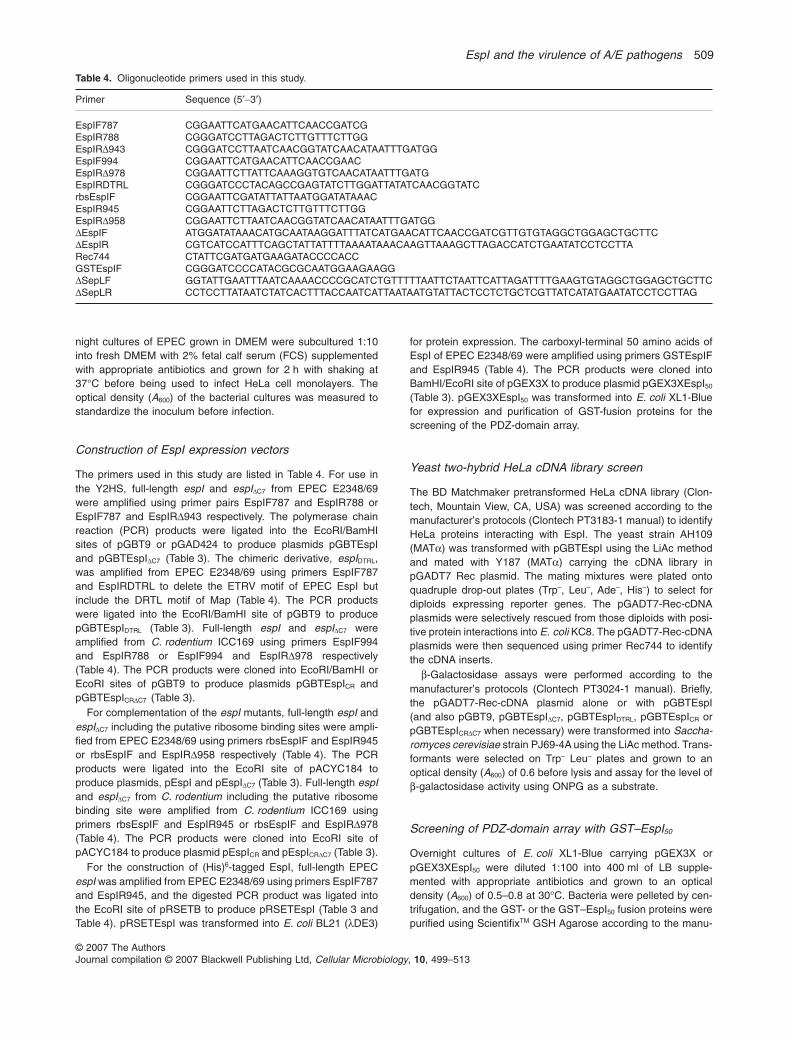
Strain or plasmid Characteristic(s)a Reference/source
StrainE. coli
E2348/69 Wild-type EPEC O127:H6 Robins-Browne (1987)EPEC DespI E2348/69 DespI (Kanr) This studyEPEC DsepL E2348/69 DsepL (Kanr) This studyBL21 (lDE3)KC8 pyrF::Tn5, hsdR, leuB600, trpC9830, lacD74, strA, galK, hisB436 ClontechXL1-Blue recA1 endA1 gyrA96 thi-1 hsdR17 supE44 relA1 lac [F′ proAB lacIqZDM15
Tn10 (Tetr)]Stratagene
C. rodentiumICC169 C. rodentium wild-type strain ATCC 51459 (Nalr) Mundy et al. (2003)ICC179 ICC169 DespI (NalrKanr) Mundy et al. (2004b)
S. cerevisiaePJ69-4A MATa trp1–901 leu2–3, 112 ura3–52 his3–200 gal4D gal80D LYS2::GAL1-HIS3
GAL2-ADE2 met2::Gal7-lacZClontech
Y187 MATa, trp 1–901, leu 2–3, 112, ura3–52, his3–200, gal4D, gal80D, LYS2::GAL1TATA -HIS3, MEL1, GAL2UAS -GAL2TATA -ADE2, URA3::MEL1UAS -MEL1TATA -lacZ
Clontech
AH109 MATa, ura3–52, his3–200, ade 2–101, trp 1–901, leu 2–3, 112, gal4D, met–,gal80D, URA3::GAL1UAS -GAL1TATA-lacZ, MEL1
Clontech
PlasmidpGBT9 GAL4 DNA binding domain fusion protein expression vector (Ampr) ClontechpGAD424 GAL4 activation domain fusion protein expression vector (Ampr) ClontechpGBTEspI Derivative of pGBT9 encoding EspI from EPEC E2348/69 (Ampr) This studypGBTEspIDC7 Derivative of pGBT9 encoding EspIDC7 from EPEC E2348/69 (Ampr) This studypGBTEspIDTRL Derivative of pGBT9 encoding EspIDTRL (Ampr) This studypGBTEspICR Derivative of pGBT9 encoding EspI from C. rodentium ICC169 (Ampr) This studypGBTEspICRDC7 Derivative of pGBT9 encoding EspIDC7 from C. rodentium ICC169 (Ampr) This studypGADT7-Rec-cDNA Vector expressing GAL4 activation domain-HeLa protein fusion (Ampr) This studypKD46 Red recombinase helper plasmid (Ampr) Datsenko and Wanner (2000)pKD4 Red recombinase template plasmid (Kanr) Datsenko and Wanner (2000)pACYC184 Medium-copy number cloning vector (Cmr Tetr) New England BiolabspEspI Derivative of pACYC184 encoding EspI from EPEC E2348/69 (Tetr) This studypEspIDC7 Derivative of pACYC184 encoding EspIDC7 from EPEC E2348/69 (Tetr) This studypEspICR Derivative of pACYC184 encoding EspI from C. rodentium ICC169 (Tetr) This studypEspICRDC7 Derivative of pACYC184 encoding EspIDC7 from C. rodentium ICC169 (Tetr) This studypRSETEspI Derivative of pRSETB encoding (His)6EPEC EspI (Ampr) This studypGEX3X GST gene fusion vector (Ampr) Pharmacia BiotechpGEX3XEspI50 pGEX3X encoding GST fused to the C-terminal 50 amino acids of EspI (Ampr) This study
a. Kan, kanamycin; Nal, nalidixic acid; Tet, tetracycline; Cm, chloramphenicol; Amp, ampicillin.
508 S. F. Lee et al.
© 2007 The AuthorsJournal compilation © 2007 Blackwell Publishing Ltd, Cellular Microbiology, 10, 499–513
night cultures of EPEC grown in DMEM were subcultured 1:10into fresh DMEM with 2% fetal calf serum (FCS) supplementedwith appropriate antibiotics and grown for 2 h with shaking at37°C before being used to infect HeLa cell monolayers. Theoptical density (A600) of the bacterial cultures was measured tostandardize the inoculum before infection.
Construction of EspI expression vectors
The primers used in this study are listed in Table 4. For use inthe Y2HS, full-length espI and espIDC7 from EPEC E2348/69were amplified using primer pairs EspIF787 and EspIR788 orEspIF787 and EspIRD943 respectively. The polymerase chainreaction (PCR) products were ligated into the EcoRI/BamHIsites of pGBT9 or pGAD424 to produce plasmids pGBTEspIand pGBTEspIDC7 (Table 3). The chimeric derivative, espIDTRL,was amplified from EPEC E2348/69 using primers EspIF787and EspIRDTRL to delete the ETRV motif of EPEC EspI butinclude the DRTL motif of Map (Table 4). The PCR productswere ligated into the EcoRI/BamHI site of pGBT9 to producepGBTEspIDTRL (Table 3). Full-length espI and espIDC7 wereamplified from C. rodentium ICC169 using primers EspIF994and EspIR788 or EspIF994 and EspIRD978 respectively(Table 4). The PCR products were cloned into EcoRI/BamHI orEcoRI sites of pGBT9 to produce plasmids pGBTEspICR andpGBTEspICRDC7 (Table 3).
For complementation of the espI mutants, full-length espI andespIDC7 including the putative ribosome binding sites were ampli-fied from EPEC E2348/69 using primers rbsEspIF and EspIR945or rbsEspIF and EspIRD958 respectively (Table 4). The PCRproducts were ligated into the EcoRI site of pACYC184 toproduce plasmids, pEspI and pEspIDC7 (Table 3). Full-length espIand espIDC7 from C. rodentium including the putative ribosomebinding site were amplified from C. rodentium ICC169 usingprimers rbsEspIF and EspIR945 or rbsEspIF and EspIRD978(Table 4). The PCR products were cloned into EcoRI site ofpACYC184 to produce plasmid pEspICR and pEspICRDC7 (Table 3).
For the construction of (His)6-tagged EspI, full-length EPECespI was amplified from EPEC E2348/69 using primers EspIF787and EspIR945, and the digested PCR product was ligated intothe EcoRI site of pRSETB to produce pRSETEspI (Table 3 andTable 4). pRSETEspI was transformed into E. coli BL21 (lDE3)
for protein expression. The carboxyl-terminal 50 amino acids ofEspI of EPEC E2348/69 were amplified using primers GSTEspIFand EspIR945 (Table 4). The PCR products were cloned intoBamHI/EcoRI site of pGEX3X to produce plasmid pGEX3XEspI50
(Table 3). pGEX3XEspI50 was transformed into E. coli XL1-Bluefor expression and purification of GST-fusion proteins for thescreening of the PDZ-domain array.
Yeast two-hybrid HeLa cDNA library screen
The BD Matchmaker pretransformed HeLa cDNA library (Clon-tech, Mountain View, CA, USA) was screened according to themanufacturer’s protocols (Clontech PT3183-1 manual) to identifyHeLa proteins interacting with EspI. The yeast strain AH109(MATa) was transformed with pGBTEspI using the LiAc methodand mated with Y187 (MATa) carrying the cDNA library inpGADT7 Rec plasmid. The mating mixtures were plated ontoquadruple drop-out plates (Trp–, Leu–, Ade–, His–) to select fordiploids expressing reporter genes. The pGADT7-Rec-cDNAplasmids were selectively rescued from those diploids with posi-tive protein interactions into E. coli KC8. The pGADT7-Rec-cDNAplasmids were then sequenced using primer Rec744 to identifythe cDNA inserts.
b-Galactosidase assays were performed according to themanufacturer’s protocols (Clontech PT3024-1 manual). Briefly,the pGADT7-Rec-cDNA plasmid alone or with pGBTEspI(and also pGBT9, pGBTEspIDC7, pGBTEspIDTRL, pGBTEspICR orpGBTEspICRDC7 when necessary) were transformed into Saccha-romyces cerevisiae strain PJ69-4A using the LiAc method. Trans-formants were selected on Trp– Leu– plates and grown to anoptical density (A600) of 0.6 before lysis and assay for the level ofb-galactosidase activity using ONPG as a substrate.
Screening of PDZ-domain array with GST–EspI50
Overnight cultures of E. coli XL1-Blue carrying pGEX3X orpGEX3XEspI50 were diluted 1:100 into 400 ml of LB supple-mented with appropriate antibiotics and grown to an opticaldensity (A600) of 0.5–0.8 at 30°C. Bacteria were pelleted by cen-trifugation, and the GST- or the GST–EspI50 fusion proteins werepurified using ScientifixTM GSH Agarose according to the manu-
Table 4. Oligonucleotide primers used in this study.
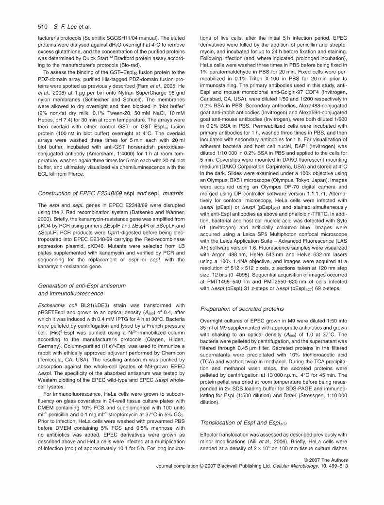
Primer Sequence (5′-3′)
EspIF787 CGGAATTCATGAACATTCAACCGATCGEspIR788 CGGGATCCTTAGACTCTTGTTTCTTGGEspIRD943 CGGGATCCTTAATCAACGGTATCAACATAATTTGATGGEspIF994 CGGAATTCATGAACATTCAACCGAACEspIRD978 CGGAATTCTTATTCAAAGGTGTCAACATAATTTGATGEspIRDTRL CGGGATCCCTACAGCCGAGTATCTTGGATTATATCAACGGTATCrbsEspIF CGGAATTCGATATTATTAATGGATATAAACEspIR945 CGGAATTCTTAGACTCTTGTTTCTTGGEspIRD958 CGGAATTCTTAATCAACGGTATCAACATAATTTGATGGDEspIF ATGGATATAAACATGCAATAAGGATTTATCATGAACATTCAACCGATCGTTGTGTAGGCTGGAGCTGCTTCDEspIR CGTCATCCATTTCAGCTATTATTTTAAAATAAACAAGTTAAAGCTTAGACCATCTGAATATCCTCCTTARec744 CTATTCGATGATGAAGATACCCCACCGSTEspIF CGGGATCCCCATACGCGCAATGGAAGAAGGDSepLF GGTATTGAATTTAATCAAAACCCCGCATCTGTTTTTAATTCTAATTCATTAGATTTTGAAGTGTAGGCTGGAGCTGCTTCDSepLR CCTCCTTATAATCTATCACTTTACCAATCATTAATAATGTATTACTCCTCTGCTCGTTATCATATGAATATCCTCCTTAG
EspI and the virulence of A/E pathogens 509
© 2007 The AuthorsJournal compilation © 2007 Blackwell Publishing Ltd, Cellular Microbiology, 10, 499–513
facturer’s protocols (Scientifix SGGSH11/04 manual). The elutedproteins were dialysed against dH2O overnight at 4°C to removeexcess glutathione, and the concentration of the purified proteinswas determined by Quick StartTM Bradford protein assay accord-ing to the manufacturer’s protocols (Bio-rad).
To assess the binding of the GST–EspI50 fusion protein to thePDZ-domain array, purified His-tagged PDZ-domain fusion pro-teins were spotted as previously described (Fam et al., 2005; Heet al., 2006) at 1 mg per bin onto Nytran SuperCharge 96-gridnylon membranes (Schleicher and Schuell). The membraneswere allowed to dry overnight and then blocked in ‘blot buffer’(2% non-fat dry milk, 0.1% Tween-20, 50 mM NaCl, 10 mMHepes, pH 7.4) for 30 min at room temperature. The arrays werethen overlaid with either control GST- or GST–EspI50 fusionprotein (100 nM in blot buffer) overnight at 4°C. The overlaidarrays were washed three times for 5 min each with 20 mlblot buffer, incubated with anti-GST horseradish peroxidase-conjugated antibody (Amersham, 1:4000) for 1 h at room tem-perature, washed again three times for 5 min each with 20 ml blotbuffer, and ultimately visualized via chemiluminescence with theECL kit from Pierce.
Construction of EPEC E2348/69 espI and sepL mutants
The espI and sepL genes in EPEC E2348/69 were disruptedusing the l Red recombination system (Datsenko and Wanner,2000). Briefly, the kanamycin-resistance gene was amplified frompKD4 by PCR using primers DEspIF and DEspIR or DSepLF andDSepLR. PCR products were Dpn1-digested before being elec-troporated into EPEC E2348/69 carrying the Red-recombinaseexpression plasmid, pKD46. Mutants were selected from LBplates supplemented with kanamycin and verified by PCR andsequencing for the replacement of espI or sepL with thekanamycin-resistance gene.
Generation of anti-EspI antiserumand immunofluorescence
Escherichia coli BL21(lDE3) strain was transformed withpRSETEspI and grown to an optical density (A600) of 0.4, afterwhich it was induced with 0.4 mM IPTG for 4 h at 30°C. Bacteriawere pelleted by centrifugation and lysed by a French pressurecell. (His)6-EspI was purified using a Ni2+-immobilized columnaccording to the manufacturer’s protocols (Qiagen, Hilden,Germany). Column-purified (His)6-EspI was used to immunize arabbit with ethically approved adjuvant performed by Chemicon(Temecula, CA, USA). The resulting antiserum was purified byabsorption against the whole-cell lysates of M9-grown EPECDespI. The specificity of the absorbed antiserum was tested byWestern blotting of the EPEC wild-type and EPEC DespI whole-cell lysates.
For immunofluorescence, HeLa cells were grown to subcon-fluency on glass coverslips in 24-well tissue culture plates withDMEM containing 10% FCS and supplemented with 100 unitsml-1 penicillin and 0.1 mg ml-1 streptomycin at 37°C in 5% CO2.Prior to infection, HeLa cells were washed with prewarmed PBSbefore DMEM containing 5% FCS and 0.5% mannose withno antibiotics was added. EPEC derivatives were grown asdescribed above and HeLa cells were infected at a multiplicationof infection (moi) of approximately 10:1 for 5 h. For long incuba-
tions of live cells, after the initial 5 h infection period, EPECderivatives were killed by the addition of penicillin and strepto-mycin, and incubated for up to 24 h before fixation and staining.Following infection (and, where indicated, prolonged incubation),HeLa cells were washed three times in PBS before being fixed in1% paraformaldehyde in PBS for 20 min. Fixed cells were per-meabilized in 0.1% Triton X-100 in PBS for 20 min prior toimmunostaining. The primary antibodies used in this study, anti-EspI and mouse monoclonal anti-Golgin-97 CDF4 (Invitrogen,Carlsbad, CA, USA), were diluted 1/50 and 1/200 respectively in0.2% BSA in PBS. Secondary antibodies, Alexa488-conjugatedgoat anti-rabbit antibodies (Invitrogen) and Alexa594-conjugatedgoat anti-mouse antibodies (Invitrogen), were both diluted 1/600in 0.2% BSA in PBS. Permeabilized cells were incubated withprimary antibodies for 1 h, washed three times in PBS, and thenincubated with secondary antibodies for 1 h. For visualization ofadherent bacteria and host cell nuclei, DAPI (Invitrogen) wasdiluted 1/10 000 in 0.2% BSA in PBS and applied to the cells for5 min. Coverslips were mounted in DAKO fluorescent mountingmedium (DAKO Corporation Carpinteria, USA) and stored at 4°Cin the dark. Slides were examined under a 100¥ objective usingan Olympus, BX51 microscope (Olympus, Tokyo, Japan). Imageswere acquired using an Olympus DP-70 digital camera andmerged using DP controller software version 1.1.1.71. Alterna-tively for confocal microscopy, HeLa cells were infected withDespI (pEspI) or DespI (pEspIDC7) and stained simultaneouslywith anti-EspI antibodies as above and phalloidin-TRITC. In addi-tion, bacterial and host cell nucleic acid was detected with Syto61 (Invitrogen) and artificially coloured blue. Images wereacquired using a Leica SP5 Multiphoton confocal microscopewith the Leica Application Suite – Advanced Fluorescence (LASAF) software version 1.6. Fluorescence samples were visualizedwith Argon 488 nm, HeNe 543 nm and HeNe 632 nm lasersusing a 100¥ 1.4NA objective, and images were acquired at aresolution of 512 ¥ 512 pixels, z sections taken at 120 nm stepsize, 12 bits (0–4095). Sequential acquisition of images occurredat PMT1495–540 nm and PMT2550–620 nm of cells infectedwith DespI (pEspI) 31 z-steps or DespI (pEspIDC7) 69 z-steps.
Preparation of secreted proteins
Overnight cultures of EPEC grown in M9 were diluted 1:50 into35 ml of M9 supplemented with appropriate antibiotics and grownwith shaking to an optical density (A600) of 1.0 at 37°C. Thebacteria were pelleted by centrifugation, and the supernatant wasfiltered through 0.45 mm filter. Secreted proteins in the filteredsupernatants were precipitated with 10% trichloroacetic acid(TCA) and washed twice in methanol. During the TCA precipita-tion and methanol wash steps, the secreted proteins werepelleted by centrifugation at 13 000 r.p.m., 4°C for 45 min. Theprotein pellet was dried at room temperature before being resus-pended in 2¥ SDS loading buffer for SDS-PAGE and immunob-lotting for EspI (1:500 dilution) and DnaK (Stressgen, 1:10 000dilution).
Translocation of EspI and EspIDC7
Effector translocation was assessed as described previously withminor modifications (Aili et al., 2006). Briefly, HeLa cells wereseeded at a density of 2 ¥ 106 on 100 mm tissue culture dishes
510 S. F. Lee et al.
© 2007 The AuthorsJournal compilation © 2007 Blackwell Publishing Ltd, Cellular Microbiology, 10, 499–513
approximately 40 h prior to infection. EPEC derivatives weregrown as previously described, and HeLa cells were infected atan moi of approximately 1000:1 for 4 h. Two dishes were used foreach strain, one for lysis with digitonin and the other to act as anunlysed control. After the infection period, the HeLa cells werewashed five times with PBS before the addition of 1 ml protein-ase K (250 mg ml-1) in order to remove extracellular proteins. Theproteinase K solution was removed after being evenly distributedon the dishes which were incubated at room temperature for20 min. In total, 0.5 ml of PMSF (6 mM) was added to stop theproteinase K enzymatic reaction, and infected cells were scrapedoff the dishes and collected in microfuge tubes. In total, 200 ml of2% digitonin was added to lyse the infected cells, and unbrokencells were slowly passaged through a 22-gauge needle using asyringe (approximately 6 times) for complete lysis. A total of200 ml of PBS was added to the unlysed-control tube. Bacteria,unbroken HeLa cells and ruptured HeLa cells’ debris were pel-leted by centrifugation at 5000 r.p.m. for 5 min, and the superna-tants were subjected to SDS-PAGE and immunoblotting for EspI(1:500 dilution) and Erk1/2 (BD Biosciences, 1:5000 dilution).
Infection of mice with derivatives of C. rodentium
In mixed-infection competition experiments, bacterial test strainswere grown to stationary phase in LB broth containing appropri-ate antibiotics. Overnight cultures of the bacterial strains werepelleted by centrifugation and resuspended in PBS. The twobacterial strains to be compared were combined in a ratio of 1:1(approximately 2 ¥ 109 cfu for each strain) in 200 ml PBS andused to infect 4- to 5-week-old male C57BL/6 mice by oralgavage. Dilutions of the inoculum were plated on respectiveantibiotic-containing plates to determine the ratio of the two bac-terial strains (test strain/reference strain) in the inoculum. Sevendays after inoculation, mice were killed by CO2 inhalation, andbacteria were recovered by plating dilutions of homogenizedcolon onto respective antibiotic plates to determine the ratio oftest strain cfu to reference strain cfu in the intestine. The CI wascalculated by dividing the ratio of test strain cfu and referencestrain cfu recovered from the colon by the ratio of test strain cfuto reference strain cfu in the inoculum (Mundy et al., 2003). A teststrain with a CI of < 0.5 was considered to be attenuated,whereas a CI � 1 indicated that the test strain colonized at leastas well as the reference strain. The CI was analysed using five ormore animals per group and assessed for significance using anunpaired Student’s two-tailed t-test.
In single-infection experiments, bacterial strains were pelletedby centrifugation and resuspended in PBS. At least four 4- to5-week-old male C57BL/6 mice were inoculated by oral gavagewith approximately 2 ¥ 109 cfu in 200 ml PBS. The viable count ofthe inoculum was determined retrospectively by plating dilutionsof the inoculum on plates with and without relevant antibiotics.Stool samples were collected daily up to 14 days after infection.The viable count per gram of stool was determined by platingserial dilutions of stool samples onto antibiotic selective media.Single-infection results were expressed as the mean log10cfu pergram feces from at least five animals for each bacterial strain.Hyperplasia was assessed 10 days after infection, whereuponmice were killed, and the distal section of colon from the cecumto the rectum was aseptically removed and weighed after theremoval of fecal pellets and cecal contents. A 1 cm section ofcolon was removed for fixation in 10% formalin and sectioning for
haematoxylin and eosin histology, and subsequent assessmentof gut pathology and inflammation. The remaining organ washomogenized mechanically in 5 ml of sterile PBS using a Seward80 stomacher, and the number of viable bacteria per gram oforgan homogenate was determined by plating onto LB agarcontaining the appropriate antibiotics.
Acknowledgements
We are grateful to Danni Krmek for her expert assistance withC. rodentium infections, and to Stephen Firth from the MonashMicroImaging Facility for help with confocal microscopy. We arealso indebted to Margareta Aili for advice on translocationassays, to Vicki Bennett-Wood for her help with haematoxylinand eosin sections, and to Dorit Becher for her advice and helpwith EspI staining of mouse frozen tissue sections. This work wassupported by grants from the Australian Research Council andthe Australian National Health and Medical Research Council.S.F.L. is the recipient of a Monash Graduate Scholarship and aMonash International Postgraduate Research Scholarship.R.A.H. is supported by grants from the National Institutes ofHealth and W. M. Keck Foundation.
References
Adams, M.E., Dwyer, T.M., Dowler, L.L., White, R.A., andFroehner, S.C. (1995) Mouse alpha 1- and beta2-syntrophin gene structure, chromosome localization, andhomology with a discs large domain. J Biol Chem 270:25859–25865.
Ahn, A.H., Freener, C.A., Gussoni, E., Yoshida, M., Ozawa,E., and Kunkel, L.M. (1996) The three human syntrophingenes are expressed in diverse tissues, have distinct chro-mosomal locations, and each bind to dystrophin and itsrelatives. J Biol Chem 271: 2724–2730.
Aili, M., Isaksson, E.L., Hallberg, B., Wolf-Watz, H., andRosqvist, R. (2006) Functional analysis of the YopEGTPase-activating protein (GAP) activity of Yersiniapseudotuberculosis. Cell Microbiol 8: 1020–1033.
Albrecht, D.E., and Froehner, S.C. (2002) Syntrophins anddystrobrevins: defining the dystrophin scaffold atsynapses. Neurosignals 11: 123–129.
Alto, N.M., Shao, F., Lazar, C.S., Brost, R.L., Chua, G.,Mattoo, S., et al. (2006) Identification of a bacterial type IIIeffector family with G protein mimicry functions. Cell 124:133–145.
Batchelor, M., Prasannan, S., Daniell, S., Reece, S., Conner-ton, I., Bloomberg, G., et al. (2000) Structural basis forrecognition of the translocated intimin receptor (Tir) byintimin from enteropathogenic Escherichia coli. EMBO J19: 2452–2464.
Batchelor, M., Guignot, J., Patel, A., Cummings, N., Cleary,J., Knutton, S., et al. (2004) Involvement of the intermedi-ate filament protein cytokeratin-18 in actin pedestal forma-tion during EPEC infection. EMBO Rep 5: 219.
Creuzburg, K., Recktenwald, J., Kuhle, V., Herold, S.,Hensel, M., and Schmidt, H. (2005) The Shiga toxin1-converting bacteriophage BP-4795 encodes an NleA-liketype III effector protein. J Bacteriol 187: 8494–8498.
Datsenko, K.A., and Wanner, B.L. (2000) One-step inactiva-
EspI and the virulence of A/E pathogens 511
© 2007 The AuthorsJournal compilation © 2007 Blackwell Publishing Ltd, Cellular Microbiology, 10, 499–513
tion of chromosomal genes in Escherichia coli K-12 usingPCR products. Proc Natl Acad Sci USA 97: 6640–6645.
Deng, W., Li, Y., Hardwidge, P.R., Frey, E.A., Pfuetzner,R.A., Lee, S., et al. (2005) Regulation of type III secretionhierarchy of translocators and effectors in attaching andeffacing bacterial pathogens. Infect Immun 73: 2135–2146.
Elliott, S.J., Wainwright, L.A., McDaniel, T.K., Jarvis, K.G.,Deng, Y.K., Lai, L.C., et al. (1998) The complete sequenceof the locus of enterocyte effacement (LEE) from entero-pathogenic Escherichia coli E2348/69. Mol Microbiol 28:1–4.
Fam, S.R., Paquet, M., Castleberry, A.M., Oller, H., Lee, C.J.,Traynelis, S.F., et al. (2005) P2Y1 receptor signaling iscontrolled by interaction with the PDZ scaffold NHERF-2.Proc Natl Acad Sci USA 102: 8042–8047.
Garmendia, J., Frankel, G., and Crepin, V.F. (2005) Entero-pathogenic and enterohemorrhagic Escherichia coli infec-tions: translocation, translocation, translocation. InfectImmun 73: 2573–2585.
Goosney, D.L., DeVinney, R., Pfuetzner, R.A., Frey, E.A.,Strynadka, N.C., and Finlay, B.B. (2000) EnteropathogenicE. coli translocated intimin receptor, Tir, interacts directlywith alpha-actinin. Curr Biol 10: 735–738.
Gruenheid, S., DeVinney, R., Bladt, F., Goosney, D., Gelkop,S., Gish, G.D., et al. (2001) Enteropathogenic E. coli Tirbinds Nck to initiate actin pedestal formation in host cells.Nat Cell Biol 3: 856–859.
Gruenheid, S., Sekirov, I., Thomas, N.A., Deng, W.,O’Donnell, P., Goode, D., et al. (2004) Identification andcharacterization of NleA, a non-LEE-encoded type III trans-located virulence factor of enterohaemorrhagic Escherichiacoli O157: H7. Mol Microbiol 51: 1233–1249.
Harris, B.Z., and Lim, W.A. (2001) Mechanism and role ofPDZ domains in signaling complex assembly. J Cell Sci114: 3219–3231.
Hartland, E.L., Batchelor, M., Delahay, R.M., Hale, C., Mat-thews, S., Dougan, G., et al. (1999) Binding of intimin fromenteropathogenic Escherichia coli to Tir and to host cells.Mol Microbiol 32: 151–158.
He, J., Bellini, M., Inuzuka, H., Xu, J., Xiong, Y., Yang, X.,et al. (2006) Proteomic analysis of beta1–adrenergicreceptor interactions with PDZ scaffold proteins. J BiolChem 281: 2820–2827.
Ingraffea, J., Reczek, D., and Bretscher, A. (2002) Distinctcell type-specific expression of scaffolding proteins EBP50and E3KARP: EBP50 is generally expressed with ezrin inspecific epithelia, whereas E3KARP is not. Eur J Cell Biol81: 61–68.
Jarvis, K.G., and Kaper, J.B. (1996) Secretion of extracellularproteins by enterohemorrhagic Escherichia coli via aputative type III secretion system. Infect Immun 64: 4826–4829.
Jelen, F., Oleksy, A., Smietana, K., and Otlewski, J. (2003)PDZ domains – common players in the cell signaling. ActaBiochim Pol 50: 985–1017.
Jerse, A.E., Yu, J., Tall, B.D. and Kaper, J.B. (1990) A geneticlocus of enteropathogenic Escherichia coli necessary forthe production of attaching and effacing lesions on tissueculture cell. Proc Natl Acad Sci USA 87: 7839–7843.
Joubert, L., Hanson, B., Barthet, G., Sebben, M., Claeysen,
S., Hong, W., et al. (2004) New sorting nexin (SNX27) andNHERF specifically interact with the 5-HT4a receptorsplice variant: roles in receptor targeting. J Cell Sci 117:5367–5379.
Kaech, S.M., Whitfield, C.W., and Kim, S.K. (1998) The LIN-2/LIN-7/LIN-10 complex mediates basolateral membranelocalization of the C. elegans EGF receptor LET-23 invulval epithelial cells. Cell 94: 761–771.
Kelly, M., Hart, E., Mundy, R., Marches, O., Wiles, S., Badea,L., et al. (2006) Essential role of the type III secretionsystem effector NleB in colonization of mice by Citrobacterrodentium. Infect Immun 74: 2328–2337.
Kenny, B. (1999) Phosphorylation of tyrosine 474 of theenteropathogenic Escherichia coli (EPEC) Tir receptormolecule is essential for actin nucleating activity and ispreceded by additional host modifications. Mol Microbiol31: 1229–1241.
Kenny, B., DeVinney, R., Stein, M., Reinscheid, D.J., Frey,E.A., and Finlay, B.B. (1997) Enteropathogenic E. coli(EPEC) transfers its receptor for intimate adherence intomammalian cells. Cell 91: 511–520.
Kim, J., Thanabalasuriar, A., Chaworth-Musters, T., Fromme,J.C., Frey, E.A., Lario, P.I., et al. (2007) The bacterial viru-lence factor NleA binds and disrupts function of mamma-lian COPII. Cell Host Microbe 2: 160–171.
Lim, I.A., Hall, D.D., and Hell, J.W. (2002) Selectivity andpromiscuity of the first and second PDZ domains ofPSD-95 and synapse-associated protein 102. J Biol Chem277: 21697–21711.
Luo, Y., Frey, E.A., Pfuetzner, R.A., Creagh, A.L., Knoechel,D.G., Haynes, C.A., et al. (2000) Crystal structure ofenteropathogenic Escherichia coli intimin-receptorcomplex. Nature 405: 1073–1077.
Marches, O., Batchelor, M., Shaw, R.K., Patel, A., Cum-mings, N., Nagai, T., et al. (2006) EspF of enteropatho-genic Escherichia coli binds sorting nexin 9. J Bacteriol188: 3110–3115.
Mundy, R., Pickard, D., Wilson, R.K., Simmons, C.P.,Dougan, G., and Frankel, G. (2003) Identification of a noveltype IV pilus gene cluster required for gastrointestinal colo-nization of Citrobacter rodentium. Mol Microbiol 48: 795–809.
Mundy, R., Petrovska, L., Smollett, K., Simpson, N., Wilson,R.K., Yu, J., et al. (2004a) Identification of a novel Citro-bacter rodentium type III secreted protein, EspI, and rolesof this and other secreted proteins in infection. InfectImmun 72: 2288–2302.
Mundy, R., and Jenkins, C., Yu, J., Smith, H., and Frankel, G.(2004b) Distribution of espI among clinical enterohaemor-rhagic and enteropathogenic Escherichia coli isolates.J Med Microbiol 53: 1145–1149.
Mundy, R., MacDonald, T.T., Dougan, G., Frankel, G., andWiles, S. (2005) Citrobacter rodentium of mice and man.Cell Microbiol 7: 1697–1706.
O’Connell, C.B., Creasey, E.A., Knutton, S., Elliott, S.,Crowther, L.J., Luo, W., et al. (2004) SepL, a proteinrequired for enteropathogenic Escherichia coli type IIItranslocation, interacts with secretion component SepD.Mol Microbiol 52: 1613–1625.
Piluso, G., Mirabella, M., Ricci, E., Belsito, A., Abbondanza,C., Servidei, S., et al. (2000) Gamma1- and gamma2-
512 S. F. Lee et al.
© 2007 The AuthorsJournal compilation © 2007 Blackwell Publishing Ltd, Cellular Microbiology, 10, 499–513
syntrophins, two novel dystrophin-binding proteins local-ized in neuronal cells. J Biol Chem 275: 15851–15860.
Piserchio, A., Spaller, M., and Mierke, D.F. (2006) Targetingthe PDZ domains of molecular scaffolds of transmembraneion channels. Aaps J 8: E396–E401.
Robins-Browne, R.M. (1987) Traditional enteropathogenicEscherichia coli of infantile diarrhea. Rev Infect Dis 9:28–53.
Schauer, D.B., and Falkow, S. (1993) Attaching and effacinglocus of a Citrobacter freundii biotype that causes trans-missible murine colonic hyperplasia. Infect Immun 61:2486–2492.
Schekman, R., and Orci, L. (1996) Coat proteins and vesiclebudding. Science 271: 1526–1533.
Simpson, N., Shaw, R., Crepin, V.F., Mundy, R., FitzGerald,A.J., Cummings, N., et al. (2006) The enteropathogenicEscherichia coli type III secretion system effector Mapbinds EBP50/NHERF1: implication for cell signalling anddiarrhoea. Mol Microbiol 60: 349–363.
Simske, J.S., and Kim, S.K. (1995) Sequential signallingduring Caenorhabditis elegans vulval induction. Nature375: 142–146.
Songyang, Z., Fanning, A.S., Fu, C., Xu, J., Marfatia, S.M.,
Chishti, A.H., et al. (1997) Recognition of unique carboxyl-terminal motifs by distinct PDZ domains. Science 275:73–77.
Straight, S.W., Pieczynski, J.N., Whiteman, E.L., Liu, C.J.,and Margolis, B. (2006) Mammalian lin-7 stabilizes polarityprotein complexes. J Biol Chem 281: 37738–37747.
Strausberg, R.L., Feingold, E.A., Grouse, L.H., Derge, J.G.,Klausner, R.D., Collins, F.S., et al. (2002) Generation andinitial analysis of more than 15,000 full-length human andmouse cDNA sequences. Proc Natl Acad Sci USA 99:16899–16903.
Tobe, T., Beatson, S.A., Taniguchi, H., Abe, H., Bailey, C.M.,Fivian, A., et al. (2006) An extensive repertoire of type IIIsecretion effectors in Escherichia coli O157 and the role oflambdoid phages in their dissemination. Proc Natl Acad SciUSA 103: 14941–14946.
Worby, C.A., and Dixon, J.E. (2002) Sorting out the cellularfunctions of sorting nexins. Nat Rev Mol Cell Biol 3: 919–931.
Zhang, Y., Yeh, S., Appleton, B.A., Held, H.A., Kausalya, P.J.,Phua, D.C., et al. (2006) Convergent and divergent ligandspecificity among PDZ domains of the LAP and zonulaoccludens (ZO) families. J Biol Chem 281: 22299–22311.
EspI and the virulence of A/E pathogens 513
© 2007 The AuthorsJournal compilation © 2007 Blackwell Publishing Ltd, Cellular Microbiology, 10, 499–513