Embed Size (px)
Citation preview

A Brief Intro to Cortical Neuroanatomy
Last Update: November 29, 2008Last Course, Louvain, Belgium, 2008
http://www.fmri4newbies.com/
Jody CulhamBrain and Mind Institute
Department of PsychologyUniversity of Western Ontario

14 Major Sulci
Main sulci are formed early in developmentFissures are really deep sulci
Typically continuous sulci•Interhemispheric fissure•Sylvian fissure•Parieto-occipital fissure •Collateral sulcus•Central sulcus•Calcarine Sulcus
Typically discontinuous sulci•Superior frontal sulcus•Inferior frontal sulcus•Postcentral sulcus•Intraparietal sulcus•Superior temporal sulcus•Inferior temporal sulcus•Cingulate sulcus•Precentral sulcus
Other minor sulci are much less reliableSource: Ono, 1990

Interhemispheric Fissure
-hugely deep (down to corpus callosum)-divides brain into 2 hemispheres

Sylvian Fissure-hugely deep-mostly horizontal-insula (purple) is buried within it-separates temporal lobe from parietal and frontal lobes
Sylvian Fissure

Parieto-occipital Fissure and Calcarine SulcusParieto-occipital fissure (red)-very deep-often Y-shaped from sagittal view, X-shaped in horizontal and coronal views
Calcarine sulcus (blue)-contains V1
Cuneus (pink)-visual areas on medial side above calcarine (lower visual field)
Lingual gyrus (yellow)-visual areas on medial side below calcarine and above collateral sulcus (upper visual field)

Collateral Sulcus-divides lingual (yellow) and parahippocampal (green) gyri from fusiform gyrus (pink)

Cingulate Sulcus-divides cingulate gyrus (turquoise) from precuneus (purple) and paracentral lobule (gold)
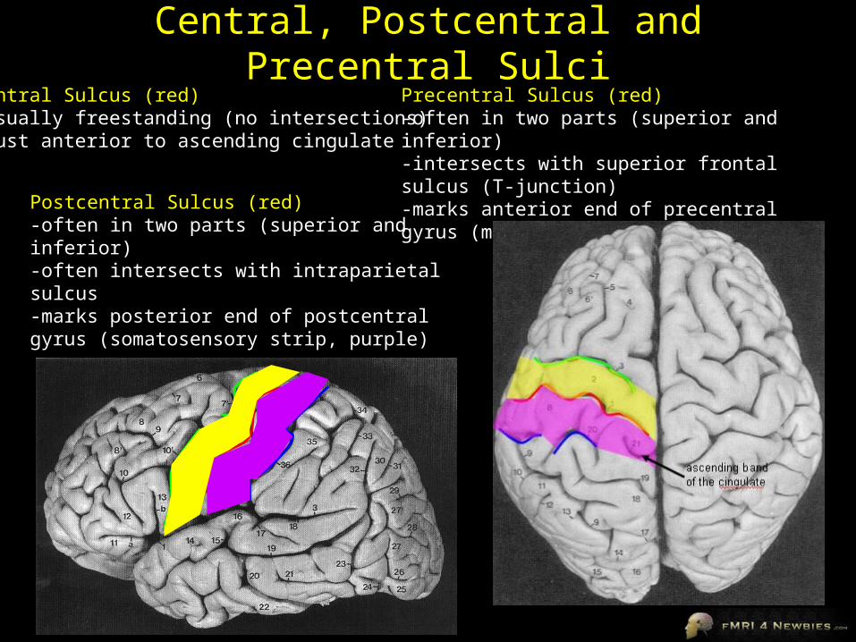
Central, Postcentral and Precentral SulciCentral Sulcus (red)-usually freestanding (no intersections)-just anterior to ascending cingulate
Postcentral Sulcus (red)-often in two parts (superior and inferior)-often intersects with intraparietal sulcus-marks posterior end of postcentral gyrus (somatosensory strip, purple)
Precentral Sulcus (red)-often in two parts (superior and inferior)-intersects with superior frontal sulcus (T-junction)-marks anterior end of precentral gyrus (motor strip, yellow)

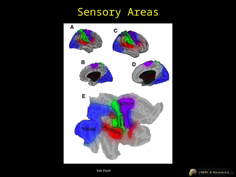
Intraparietal Sulcus-anterior end usually intersects with inferior postcentral (some texts call inferior postcentral the ascending intraparietal sulcus)-posterior end usually forms a T-junction with the transverse occipital sulcus (just posterior to the parieto-occipital fissure)-IPS divides the superior parietal lobule from the inferior parietal lobule (angular gyrus, gold, and supramarginal gyrus, lime)
POF
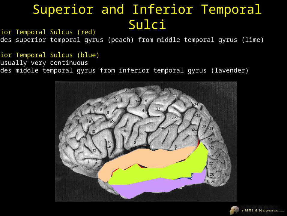
Superior and Inferior Temporal SulciSuperior Temporal Sulcus (red)-divides superior temporal gyrus (peach) from middle temporal gyrus (lime)
Inferior Temporal Sulcus (blue)-not usually very continuous-divides middle temporal gyrus from inferior temporal gyrus (lavender)

Superior and Inferior Frontal SulciSuperior Frontal Sulcus (red)-divides superior frontal gyrus (mocha) from middle frontal gyrus (pink)
Inferior Frontal Sulcus (blue)-divides middle frontal gyrus from inferior frontal gyrus (gold)
orbital gyrus (green) and frontal pole (gray) also shown
Frontal Eye fields lie at this junction
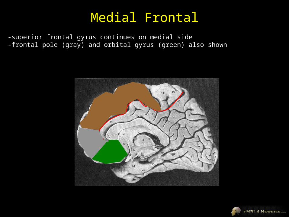
Medial Frontal-superior frontal gyrus continues on medial side-frontal pole (gray) and orbital gyrus (green) also shown

Learning More AnatomyDuvernoy, 1999, The Human Brain: Surface, Blood Supply, and Three-Dimensional Sectional Anatomy• beautiful pictures• good schematic diagrams• clear anatomy• slices of real brain• Springer, US$326
Ono, 1990, Atlas of the Cerebral Sulci• great for showing intersubject variability• gives probabilities of configurations and stats on sulci• Theime, US$199
Damasio,1995, Human Brain Anatomy in Computerized Images• good for showing sulci across wide range of slice planes• really crappy reconstructions in first edition• second edition available April 2005 with new images• Oxford University Press, US$125
Tamraz & Comair, 2000, Atlas of Regional Anatomy of the Brain Using MRI with Functional Correlations• good overview• Springer, US$203
Talairach & Tournoux, 1988. Co-Planar Stereotaxic Atlas of the Human Brain• just because it’s the standard doesn’t mean it’s good (see also PC vs. Mac, VHS vs. betamax)• Theime, US$240
Wanna get rich? Publish a brain atlas.Sheesh, these are expensive!

Anatomical and Functional Neuroanatomy
Images from Brain Voyager Brain Tutor
(Freeware)
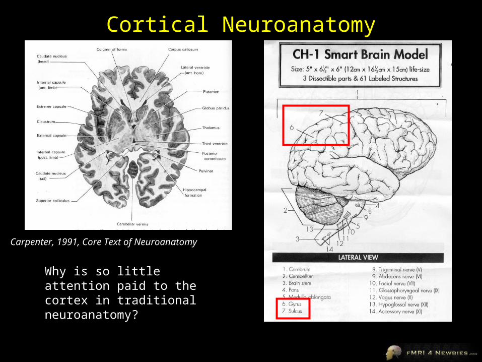
Why is so little attention paid to the cortex in traditional neuroanatomy?
Cortical Neuroanatomy
Carpenter, 1991, Core Text of Neuroanatomy
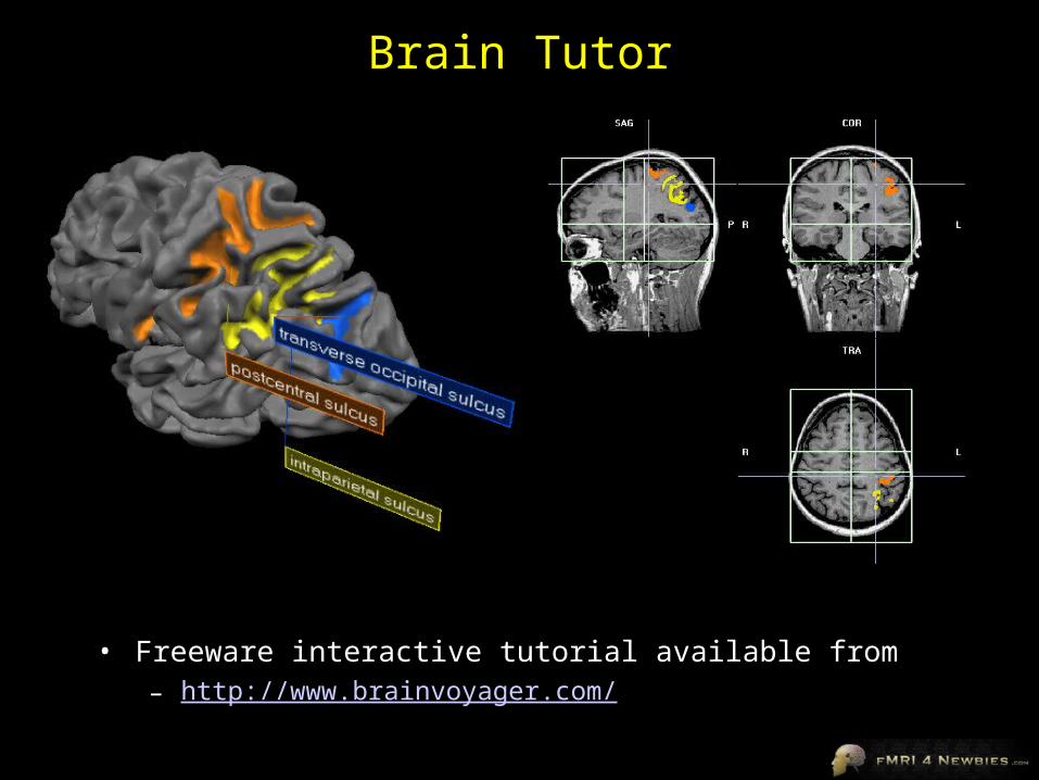
Brain Tutor
• Freeware interactive tutorial available from– http://www.brainvoyager.com/

UNDER CONSTRUCTION
• I’m still working on the “areas” slides and they are admittedly on the lame side, especially for temporal, frontal and subcortical areas
• Suggestions/images welcome

Category-Specific Visual Areas
• Lateral Occipital (LO)– object-selective
– objects > (faces & scenes)
faces
places
objects
Malach, 2002, TICS
• Fusiform Face Area (FFA)– face-selective– faces > (objects & scenes)
• Parahippocampal Place Area (PPA)– place-selective– places > (objects and faces)
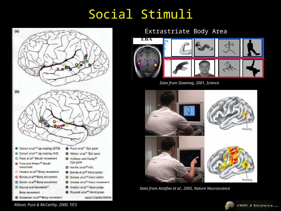
Social StimuliExtrastriate Body Area
Allison, Puce & McCarthy, 2000, TICS
Data from Astafiev et al., 2005, Nature Neuroscience
Data from Downing, 2001, Science
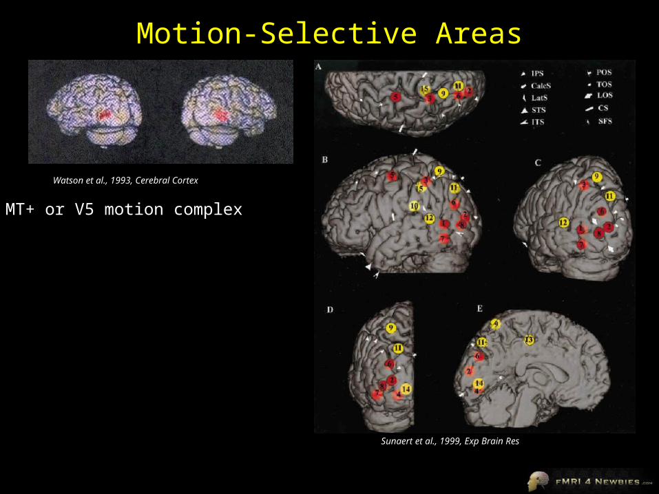
Motion-Selective Areas
MT+ or V5 motion complex
Sunaert et al., 1999, Exp Brain Res
Watson et al., 1993, Cerebral Cortex
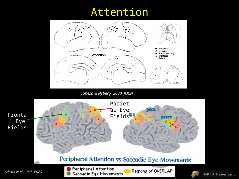
Attention
Corbetta et al., 1998, PNAS
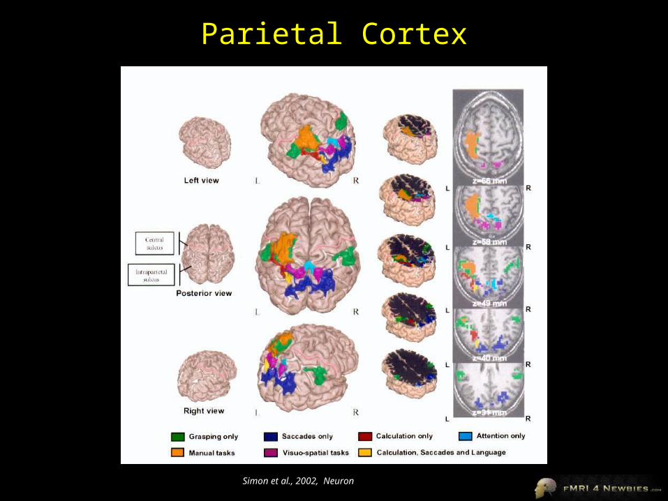
Cabeza & Nyberg, 2000, JOCN
Frontal Eye
Fields
Parietal Eye
Fields
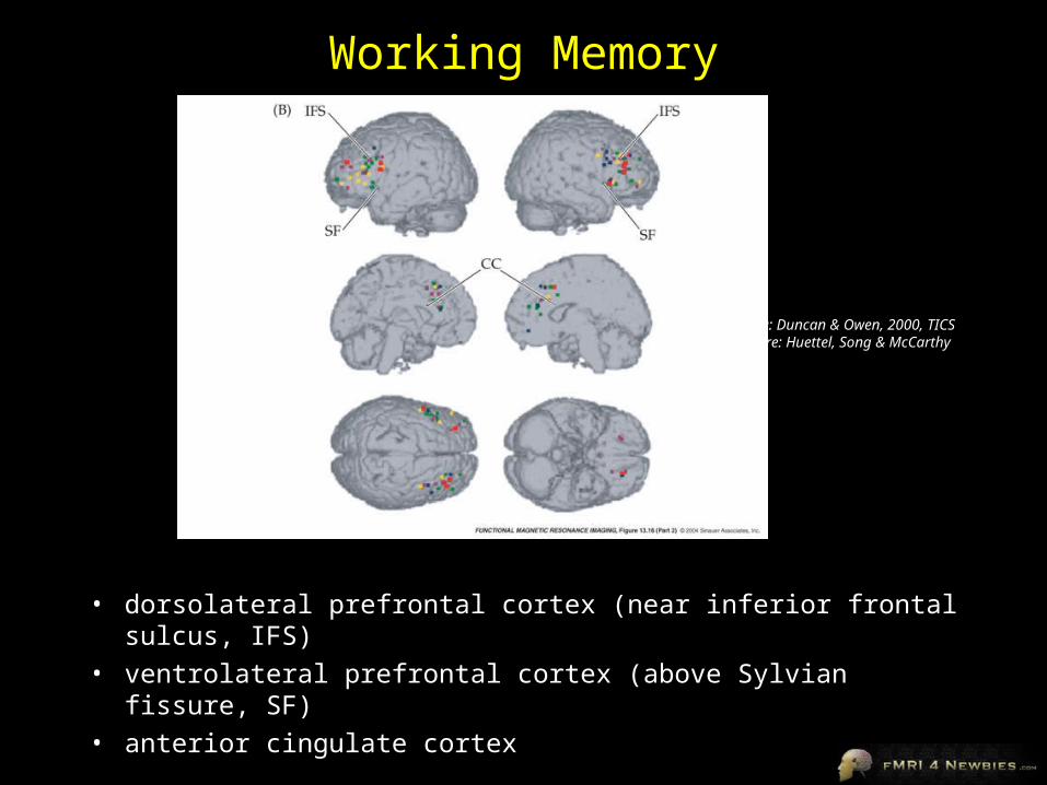
Working Memory
• dorsolateral prefrontal cortex (near inferior frontal sulcus, IFS)• ventrolateral prefrontal cortex (above Sylvian fissure, SF)• anterior cingulate cortex
Data: Duncan & Owen, 2000, TICSFigure: Huettel, Song & McCarthy

Motor RegionsHand area of Motor Cortex
Premotor Cortex
Supplementary Motor Area
Picard & Strick, 2001, CONB
Yousry et al, 1997, Brain


![UNIVERSITE CATHOLIQUE DE LOUVAIN ECOLE POLYTECHNIQUE …pvr/MemoireRaphaelBauduin.pdf · the Oz programming language[Moz], the last stable release dating from 2008. It was deemed](https://img.pdfslide.us/doc/110x75/5f0a5afc7e708231d42b3cf1/universite-catholique-de-louvain-ecole-polytechnique-pvrm-the-oz-programming.jpg)