Embed Size (px)
Citation preview

�9 1995 by Humana Press Inc. All rights of any nature whatsoever reserved. 1044-7393195/2402-03--0179 $06.80
A Biochemical and (lltrastructural Evaluation of the Type 2 Gaucher Mouse
R. WILLEMSEN, 1 V. TYBULEWlCZ, 2 E. SIDRANSKY, 3 W. K. ELIASON, 3 B. M. MARTIN, 3 M. E. LAMARC^, 3
A . J . J . REUSER, 1 M . TREMBLAY, 4 H. WESTPHAL, 5
R. C . MULLIGAN, 6 AND E . I. GINNS *'3
t Department of Cell Biology and Genetics, Erasmus University, Rotterdam 3000 DR, The Netherlands; 2Medical Research Council, NIMR, London, UK;
3 Clinical Neuroscience Branch, National Institute of Mental Health, Bldg. 49, Rm. B1EE16, Bethesda, MD: 4McGill University,
Montreal, Canada; 5National Institute of Child Health and Human Development, Bethesda, MD; and 6 Whitehead Institute
for Biomedical Research, Cambridge, MA
Received June 29, 1994; Accepted October 10, 1994
ABSTRACT
Gaucher mice, created by targeted disruption of the glucocere- brosidase gene, are totally deficient in glucocerebrosidase and have a rapidly deteriorating clinical course analogous to the most severely aG fected type 2 human patients. An ultrastructural study of tissues from these mice revealed glucocerebroside accumulation in bone marrow, liver, spleen, and brain. This glycolipid had a characteristic elongated tubular structure and was contained in lysosomes, as demonstrated by colocalization with both ingested carbon particles and cathepsin D. In the central nervous system (CNS), glucocerebroside was diffusely stored in microglia cells and in brainstem and spinal cord neurons, but not in neurons of the cerebellum or cerebral cortex. This rostral- caudal pattern of neuronal lipid storage in these Gaucher mice repli- cates the pattern seen in type 2 human Gaucher patients and clearly
*Author to whom all correspondence and reprint requests should be addressed.
Molecular and Chemical Neuropathology ] 79 Vol. 24, 1995
180 Willemsen et al.
demonstrates that glycosphingolipid catabolism and/or accumulation varies within different brain regions. Surprisingly, the cellular pathol- ogy of tissue from these Gaucher mice was relatively mild, and sug- gests that the early and rapid demise of both Gaucher mice and severely affected type 2 human neonates may be the result of both a neurotoxic metabolite, such as glucosylsphingosine, and other fac- tors, such as skin water barrier dysfunction secondary to the absence of glucocerebrosidase activity.
Index Entries: Type 2 Gaucher mice; targeted disruption; gluco- cerebrosidase; glucocerebroside; lysosomes; microglia; neurons.
INTRODUCTION
Gaucher disease in humans and null-allele mice is caused by the inherited deficiency of glucocerebrosidase, the lysosomal enzyme that hydrolyzes the fl-glucosidic bond in glucocerebroside. The major source of this glycosphingolipid is the turnover of senescent white and red blood cell membranes. Although glucocerebroside is predominantly degraded within lysosomes of normal macrophages, accumulation of this lipid in Gaucher disease transforms macrophages throughout the reticuloendo- thelial system into the characteristic "Gaucher cells." The Gaucher cells have a relatively small eccentrically located nucleus, and the cytoplasm contains numerous irregularly shaped or branching lysosomes filled with twisted tubular deposits of glucocerebroside (Barranger and Ginns, 1989; Martin et al., 1989).
Gaucher disease has been classified into three subtypes, type 1 (chronic, non-neuronopathic), type 2 (acute, neuronopathic), and type 3 (subacute, neuronopathic) on the basis of the presence and degree of neurologic in- volvement (Barranger and Ginns, 1989; Martin et al., 1989). The clinical manifestations encountered in type 2 Gaucher disease include failure to thrive, hepatosplenomegaly, and neurologic deterioration, which often includes the triad of strabismus, trismus, and retroflexion of the head. The most severely affected type 2 patients may also have ichthyotic skin and/or hydrops fetalis as associated symptoms (Sidransky et al., 1992). In brains from the few Gaucher patients with neurologic involvement that have been studied, perivascular Gaucher cells are found and a variable degree of neuronal loss is observed. Neurons with the characteristic lyso- somal inclusions typically observed in Gaucher cells are encountered only sporadically (Banker et al., 1962; Adachi et al., 1967, Conradi et al., 1984; Kaye et al., 1986; Hernandez and Bueno, 1973). The pathophysiologic mechanisms leading to the neurologic symptoms observed in these patients remain unknown, largely because of the rarity of severely affected type 2 human neonates and the very limited opportunities for extensive patho- logical studies in these patients.
Molecular and Chemical Neuropathology Vol. 24, 1995

Ultrastructural Pathology of Gaucher Mice 181
A Gaucher mouse line recently created by targeted disruption of the glucocerebrosidase gene (Tybulewicz et al., 1992) has provided a relevant animal model for investigation of the pathophysiologic mechanisms occur- ring in type 2 Gaucher disease. In the initial report we presented limited pathologic results on these Gaucher mice, demonstrating that the defi- ciency of glucocerebrosidase did result in accumulation of glucocerebro- side in macrophages. The present manuscript describes the more detailed ultrastructural pathology in a variety of tissues, including the liver, spleen, bone marrow, and brain from type 2 Gaucher mice having clinical features characteristic of the more severely affected type 2 Gaucher patients. Particular attention is focused on the CNS, including frontal and occipital cortex, brainstem, cerebellum, and spinal cord, in an attempt to explain the rapid postnatal demise of these type 2 Gaucher mice and severely affected type 2 human neonates.
MATERIALS AND METHODS
The Gaucher Mouse
A Gaucher mouse line was developed by targeted disruption of the mouse glucocerebrosidase gene via homologous recombination in embry- onic stem (ES) cells (Tybulewicz et al., 1992). The neomycin resistance cassette replaces part of the exons 9 and 10 and abolishes glucocerebro- sidase activity in the homozygous mutant mice.
Western Blot Analyses Tissue samples were stored at -20~ until use. Prior to extraction,
the tissue samples were frozen in liquid nitrogen and then crushed into a fine powder. The powdered tissue was resuspended in 200-250/~L of 60- mM potassium phosphate, pH 5.9, containing 0.1% Triton X-100, and sonicated three times each for 10 s (50 W, Heat Systems Ultrasonics Inc., Cell Disrupter Model W225R) at 4~ After sonication, the samples were centrifuged at 12,000g at 4~ for 5 min, and the supernatant used for enzyme assay and Western blot analyses. Protein concentration was deter- mined with the Pierce BCA reagent using the recommended protocol with BSA as a standard. Enzyme assay was performed using 4-methyl- umbelliferyl-~-D-glucopyranoside as substrate (Beutler and Kuhl, 1970).
Polyacrylamide gel electrophoresis was performed using precast 12% minigels (NOVEX, 1.0 mm thickness) at constant current of 40 ma using reducing, denaturing conditions. Each lane contained 36 #g of tissue. After electrophoretic separation, the proteins were transferred to Immobilon-P (Millipore, Bedford, MA) using 25 mM tris, 192 mM glycine, and 20 mM methanol as buffer. The membrane was briefly rinsed with water and incubated in a blocking solution (5% milk powder, 0.05%
Molecular and Chemical Neuropathology Vol. 24, 1995
182 Wil lemsen et al.
Tween-20, PBS pH 7.2) at 37~ for 1 h. The membrane was then in- cubated with rabbit antibody specific for mouse glucocerebrosidase (diluted 1:5000 in blocking solution) for I h at 37~ washed five times for 5 min at 37~ with PBS pH 7.2 containing 0.05% Tween-20, and finally incubated with goat antirabbit IgG-HRP (human serum adsorbed, Sigma, St. Louis, MO) at a dilution of 1:5000 in blocking solution for 30 rain at 37~ The wash was then repeated five times each for 5 rain in PBS pH 7.2 containing 0.05% Tween-20 at 37~ The antibody-protein conjugate was detected with the ECL Detection Kit (Amersham, Arlington Heights, IL) using the recommended protocol. After the antibody-protein signals were developed, the membrane was rinsed with water, and protein stan- dards (Nova wide range SDS-PAGE protein standards) were visualized with Sulforhodamine B (50 mg/L in 30% methanol, 2% acetic acid, [Molec- ular Probes Inc. Eugene, OR] $1307).
Glycosphingolipid Analyses Liver and brain tissue from normal, heterozygous, and mutant homo-
zygous mice were extracted with 18 v/w of 2:1 chloroform-methanol by sonication for 10 min at RT, followed by addition of 0.2 vol of water. After vortexing, samples were centrifuged for 2 min at 12,000g. A 100-uL aliquot of the lower phase was evaporated to dryness, resuspended in 2:1 chloro- form-methanol, and applied to precoated silica gel-60 high-performance thin- layer chromatography (HPLC) plates (Merck, Darmstadt, Germany) that had been activated at 100~ for I h (Kundu, 1981). Development was per- formed with a solvent mixture of chloroform-methanol-water, 65:25:4 (v/v/v) for 45 rain. The plates were then air dried and the glycolipids visualized with orcinol-ferric chloride (Bial's) reagent.
Tissue Processing for Epon Embedding Liver, spleen, bone marrow, and brain from normal and homozygous
mutant mice were fixed in 0.1M phosphate buffer (PB), pH 7.3, contain- ing 1% acrolein and 0.4% glutaraldehyde. After fixation for 24 h at 4~ the tissues were stored in 0.1M PB, containing 2% paraformaldehyde. Postfixation was performed according to De Bruyn and DenBreejen (1976). Specific areas of the brain were selected for epon embedding, i.e., the frontal and occipital cerebral cortex, the cerebellum, the brainstem, and the cervical part of the spinal cord. After epon embedding, a further selection was made for the red nucleus, the vestibular nucleus, and the facial nerve nucleus of the brainstem. Ultrathin sections were cut with a LKB ultratome Nova and stained with uranylacetate and lead citrate.
Macrophage Marking A suspension of carbon particles (India ink) was injected intravenously
into normal and homozygous glucocerebrosidase deficient mice seconds prior to sacrificing the animals. The liver was fixed as described earlier.
Molecular and Chemical Neuropathology Vol. 24, 1995

Ultrastructural Pathology of Gaucher Mice 183
A B
m ~
kDa
-97
-45
-31
C
1 2 3 1 2 3 Std 1 2 3 Std
Fig. 1. (A) Western blot analysis of glucocerebrosidase in brain of normal (lane 1), heterozygote (lane 2), and homozygous mutant mice (lane 3). Sample preparation, electrophoresis, and Western blot analyses were performed as described in Materials and Methods. Each lane contains 36 ug of protein; (B and C) Thin layer chromatography of neutral glycosphingolipids from brain (B) and liver (C) of normal (lane 1), heterozygote (lane 2), and homozygous mutant (and 3) mice. The lane marked Std contains 4 ug of glucocerebroside (GL1) standard.
lmmunocytochemistry Spleen from normal and affected mice was fixed and stored as
described earlier, but no postfixation was performed. Specimens were embedded in Lowicryl K4M according to standard procedures. Im- munolabeling of cathepsin D on ultrathin sections was performed as previously described (Armbruster et al., 1982). Sections were stained with uranylacetate and lead citrate. Sections incubated with normal rabbit serum showed negligible labeling.
RESULTS
Biochemical Findings The type 2 Gaucher mouse described in this work was produced by
targeted disruption of the glucocerebrosidase gene. The affected mice are "null mutants" of Gaucher disease, where glucocerebrosidase is com- pletely deficient. This is illustrated in Fig. 1A by the absence of glucocere- brosidase protein in brain tissue of affected animals. Western blot analyses of brain and liver (not shown) from normal and heterozygous mice (Fig. 1A) show that the major form of crossreacting material (CRM) to mouse glucocerebrosidase has an apparent mol wt of 56 kDa.
Thin Layer Chromatographic Analyses for Glucocerebroside Elevated glucocerebroside levels were demonstrated in brain (Fig. 1B)
and liver (Fig. 1C) of homozygous mutant mice. The difference in mobility
Molecular and Chemical Neuropathology Vol. 24, 1995

184 Willemsen et al.
of glucocerebroside from brain compared to that from liver or the standard is consistent with the heterogeneity in fatty acid moieties of the lipid, being C24 in liver or standard and C18 in brain (Svennerholm et al., 1982). No elevation of glucocerebroside is observed in heterozygous mice.
Macrophages in Bone Marrow, Liver, and Spleen
Except for the dramatic changes observed in the skin (Sidransky et al., 1992) at the light-microscopic level, no definite pathological changes were observed. Typical Gaucher cells, easily observed in bone marrow, liver, and spleen of affected humans were not readily identified in the neonatal mouse organs. Therefore, we performed electronmicroscopy to obtain information about the presence and precise localization of stored glycolipid. The overall cellularity of the bone marrow was identical in normal and mutant mice. Bone marrow from both normal and mutant mice is shown in Fig. 2. Storage of glucocerebroside was observed only in macrophages from homozygous mutant mice, where the glucocerebro- side is deposited in elongated tubular assays that give the lipid laden lysosomes their characteristic irregular shape.
In these type 2 Gaucher mice, the lysosomal accumulation of glyco- lipid was most obvious in the liver. A high power magnification of a Kupffer cell is shown in Fig. 3. The lysosomes were marked in this experi- ment by intravenous injection of carbon particles prior to sacrificing the animals. In Fig. 4, the lysosomal nature of the storage organelles is demon- strated by the colocalization of the lipid deposits and the lysosomal marker enzyme cathepsin D. The figure shows a Lowicryl K4M ultrathin section of the spleen from a Gaucher mouse on which immunocytochem- istry was performed. The gold particles marking cathepsin D were located along the twisted glycolipid tubules in the lysosomes in liver. The accumulation of lipid was seen consistently in homozygous mutant mice (n = 10), but never observed in macrophages from normal (n = 5) or heterozygous (carrier) mice (n = 2).
Macrophages in the CNS Typical Gaucher cells were not observed in histological serial sections
of the brain of glucocerebrosidase deficient mice. However, examination of the selected areas of the brain (n = 6; see Materials and Methods), revealed at the subcellular level microglia with lysosomal accumulation of lipid in tubular arrays that was not seen in normal mice (Figs. 5A, B). Macrophages throughout the CNS did contain dense material, but in this respect the glucocerebrosidase-deficient mice were not different from the normal. No lipid storage was observed in either astroglia cells or oligo- dendrocytes. The various cell types were distinguished by morphological criteria.
Molecular and Chemical Neuropathology Vol. 24, 1995

Ultrastructural Pathology o f Gaucher Mice 185
Fig. 2. (,6) Ultrathin epon section of bone marrow ot homozygous mutant glucocerebrosidase deficient mice; (B) Stored glucocerebroside (SP) is observed within a lysosome (arrow). N, nucleus; M, mitochondrion; E, erythrocyte. Bar represents 1.0 #m in A and 0.25/~m in B.
Molecular and Chemical Neuropathology Vol. 24, 1995

186 W i l l e m s e n e t al.
Fig. 3. Ultrathin epon section of liver of homozygous mutant glucocere- brosidase deficient mice sacrificed after intravenous injection of carbon particles. The lysosomes (L) of the Kupffer cells are marked by the ingested carbon par- ticles (arrows) that colocalize with the stored material. G, Golgi complex; N, nucleus; M, mitochondrion. Bar represents 0.2 #m.
Fig. 4. Ultrathin Lowicryl K4M section of spleen of homozygous mutant mice immunogold labeled for lysosomal cathepsin D. The labeled structure is an elongated lysosome (L). Cathepsin D is associated with the twisted tubular deposits, N, nucleus. Bar represents 0.2 #m.
Molecular and Chemical Neuropathology Vol. 24, 1995
Ultrastructural Pathology of Gaucher Mice 187
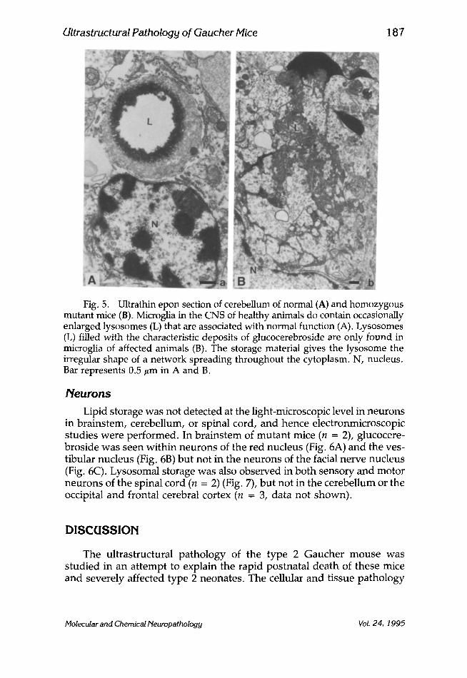
Fig. 5. Ultrathin epon section of cerebellum of normal (A) and homozygous mutant mice (B). Microglia in the CNS of healthy animals do contain occasionally enlarged lysosomes (L) that are associated with normal function (A). Lysosomes (L) filled with the characteristic deposits of glucocerebroside are only found in microglia of affected animals (B). The storage material gives the lysosome the irregular shape of a network spreading throughout the cytoplasm. N, nucleus. Bar represents 0.5 #m in A and B.
Neurons
Lipid storage was not detected at the light-microscopic level in neurons in brainstem, cerebellum, or spinal cord, and hence electronmicroscopic studies were performed. In brainstem of mutant mice (n = 2), glucocere- broside was seen within neurons of the red nucleus (Fig. 6A) and the ves- tibular nucleus (Fig. 6B) but not in the neurons of the facial nerve nucleus (Fig. 6C). Lysosomal storage was also observed in both sensory and motor neurons of the spinal cord (n = 2) (Fig. 7), but not in the cerebellum or the occipital and frontal cerebral cortex (n = 3, data not shown).
DISCUSSION
The ultrastructural pathology of the type 2 Gaucher mouse was studied in an attempt to explain the rapid postnatal death of these mice and severely affected type 2 neonates. The cellular and tissue pathology
Molecular and Chemical Neuropathology Vol. 24, 1995

188 Willernsen et al.
Fig. 6. Ultrathin epon section of brainstem of homozygous mutant mice. Neurons of the red nucleus (A) and vestibular nucleus (B) have lysosomes (L) with storage material, but lysosomal storage was not observed in neurons of the facial nerve nucleus (C). M, mitochondrion; G, Golgi complex; N, nucleus. Bar represents 0.2/~m in A, B, and C.
was remarkably mild, despite the complete deficiency of glucocerebro- sidase. Although the overall architecture of the liver, bone marrow, spleen, and brain appeared appropriate for age as judged by light micro- scopy, accumulation of glucocerebroside was clearly demonstrated by electronmicroscopy. The stored lipid in macrophages of the liver and spleen had the characteristic elongated tubular structure (Lee et al., 1973; Lee, 1982; Willemsen et al., 1988) and was contained in organelles identi- fied as lysosomes by the presence of ingested carbon particles and lyso- somal cathepsin D. Interestingly, even though these storage vacuoles occasionally did occupy significant cellular volume, typical Gaucher cells were not observed by light microscopy.
Molecular and Chemical Neuropathology Vol. 24, 1995

Ultrastructural Pathology of Uaucher Mice 189
Fig. 7. Ultrathin epon sections of sensory (A) and motor (B) neurons in the spinal cord of homozygous mutant mice. The arrows indicate the array of lipid tubules present in the lysosomes (L). G, Golgi complex; N, nucleus. Bar represents 0.2/~m in A and B.
The cellular pathology in the CNS of the homozygous mutant mice similarly is surprisingly limited, particularly in light of the rapid clinical deterioration that occurs. Glucocerebroside is stored predominantly in microglia cells and only to a very limited extent in neurons. Although cer- tain sensory and motor neurons of the brainstem and spinal cord were affected, Purkinje cells and neurons of the cerebellar and cerebral cortex were free of storage product. This rostral-caudal pattern of neuronal lipid storage suggests that glycosphingolipid catabolism and/or accumulation varies between neurons in different brain regions.
A comparison of the neuropathology of the glucocerebrosidase-deficient mouse with the human type 2 and 3 patients suggests that in the Gaucher patients there is also more deposition of glucocerebroside in microglia than in neurons (Banker et al., 1962; Adachi et al., 1967; Conradi et al., 1984; Kaye et al., 1986). The limited number of neuropathology studies in Gaucher patients consistently demonstrate that Gaucher cells are predom- inantly located in the adventitia of vessels in the cerebral and cerebellar subcortical white matter, but the reported involvement of microglia and neurons in the different regions of the brain is more varied. An increasing gradient of Gaucher cells was observed from frontal to occipital cerebral cortex by Kaye et al. (1986), whereas Banker et al. (1962) report a compar- able density of Gaucher cells in the frontal, temporal, parietal, and occipital cortex. A similar comparison could not be made in the mouse model because of the lack of histologically detectable Gaucher cells. Neuronal storage was only sporadically found in humans throughout the CNS, albeit with an increasing gradient from rostral to caudal, as in the Gaucher mice (Banker et al., 1962; Adachi et al., 1967; Hernandez and Bueno, 1973; Conradi, 1984).
The degree of neuronal loss and neuronophagia in humans varied in different nuclei (Banker et al., 1962; Adachi et al., 1967; Hernandez and
Molecular and Chemical Neuropathology Vol. 24, 1995

190 Willemsen et al.
Bueno, 1973; Conradi et al., 1984; Kaye et al., 1986). No such dramatic pathological changes were observed in the mouse.
This Gaucher mouse line with a complete deficiency of glucocerebro- sidase, provides us with a relevant animal model of the most severe neo- natal form of Gaucher disease in which we can conveniently study the clinical-pathological progression of this disease. From the studies reported here, we conclude that the early demise of affected mice is probably not caused by liver, spleen, or bone marrow dysfunction, nor by neuronal degeneration, unless the observed amount of lysosomal glucocerebroside accumulation results in neuronal dysfunction before gross cellular pathol- ogy occurs. Nilsson and Svennerholm have suggested that the neurotoxin glucosylsphingosine (glucosylpsychosine) is elevated in the nervous sys- tem of type 2 patients (Nilsson and Svennerholm, 1982). Hannun and Bell (1987) have recently reported that lysophinogolipids, including glucosyl- sphingosine, are potent inhibitors of both protein kinase C activity and phorboldiester binding, suggesting that these sphingolipid derivatives can interfere with signal transduction and cellular differentiation (Ballou, 1992; Zeller and Marchase, 1992) and thus disrupt neuronal function.
The unusual skin texture, turgor, and lipid composition of type 2 Gaucher skin (Sidransky et al., 1992; Tybulewicz et al., 1992; Holleran et al., 1993b) may also be an important factor contributing to the rapid deteri- oration observed in severely affected mice and human neonates. Ceramides are critical components in skin permeability barrier homeostasis (Holleran et al., 1993b), and the functional disruption of the skin barrier as a consequence of the absence of glucocerebrosidase could result in fluid or electrolyte imbalance (Holleran et al., 1993a,b; Holleran et al., 1994). Lastly, the homozygous mutant mice are rapidly identified by their mothers and are ejected from the nest. Thus they have been poorly fed, and, as a consequence, dehydration and hypoglycemia, may further hasten their demise.
The opportunity to perform further biochemical and pathological studies using this mouse model of type 2 Gaucher disease should further our understanding of the basis of neurologic involvement observed in these Gaucher mice and type 2 infants. Investigations using these mice may also provide more details of the pathophysiologic mechanisms of systemic involvement seen in Gaucher disease and permit the more rational development of successful therapeutic interventions.
ACKNOWLEDGMENTS
The authors thank L. Carmon (NIMH) for technical assistance with animal procedures, J. M. Tager (University of Amsterdam) and coworkers for providing rabbit polyclonal antibodies against cathepsin D, and J. C. Holstege (Department of Anatomy, Erasmus University, Rotterdam) for
Molecular and Chemical Neuropathology Vol. 24, 1995

Ultrastructural Pathology of Gaucher Mice 191
stimulating discussion. R. M. Koppenol, M. Kuit, and J. de Vries Lentsch are acknowledged for photography, and J. Lokker, L. Syed, and E. Alzona for manuscript preparation.
REFERENCES
Adachi M., Wallace B. J., Schneck L., and Volk B. W. (1967) Fine structure of central nervous system in early infantile Gaucher's disease. Arch. Pathol. 83, 513-526.
Armbruster B. L., Carlemalm E., and Chiovetti R. (1982) Specimen preparation for electron microscopy using low temperature embedding resins. J. Microsc. 126, 77-85.
Ballou L. R. (1992) Sphingolipids and cell function. Immunol. Today 13, 339-341. Banker B. Q., Miller J. O., and Crocker A. C. (1962) The cerebral pathology of
infantile Gaucher's disease, in Cerebral Sphingolipidosis (Aronson S. M. and Volk B. W., eds.), pp. 73-99, Academic, New York.
Barranger J. A., and Ginns E. I. (1989) Glucosylceramide lipidosis: Gaucher disease, in The Metabolic Basis of Inherited Disease II, vol. 6 (Scriver C. R., Beaudet A. L., Sly W. S., and Valle D., eds.), pp. 1677-1698, McGraw-Hill, New York.
Beutler E. and Kuhl W. L. (1970) Detection of the defect of Gaucher's disease and its carrier state in peripheral blood leukocytes. Lancet 1, 612,613.
Conradi N. G., Sourander P., Nilsson O., Svennerholm L., and Erikson A. (1984) Neuropathology of the Norrbottnian type of Gaucher disease: morpholog- ical and biochemical studies. Acta Neuropathol. (Berl) 65, 99-109.
DeBruyn W. C. and DenBreejen P. (1976) Glycogen, its chemistry and morpho- logical appearance in the electron microscope. Histochem. J. 8, 121-142.
Hannun Y. A. and Bell R. M. (1987) Lysosphingolipids inhibit protein kinase C: implications for the Sphingolipidoses. Science 235, 670-674.
Hernandez F. and Bueno M. (1973) Infantile neurological Gaucher's disease in three siblings: an ultrastructural study. Virchows Arch. (Pathol. Anat.) 360, 27-32.
Holleran W. M., Ginns E. I., Menon G. K., Grundmann J. U., Fartasch M., McKinney C. E., Elias P. M., and Sidransky E. (1994) Consequences of fl- glucocerebrosidase deficiency in epidermis: ultrastructure and permeability barrier alterations in Gaucher disease. J. Clin. Invest. 93, 1756-1764.
Holleran W. M., Menon G. K., Elias P. M., Ginns E. I., and Sidransky E. (1983a) fl-glucocerebrosidase-deficient transgenic mice have abnormal stratum cor- neum lamellar bilayers. Clin. Res. 41(2), 402A.
Holleran W. M., Takagi Y., Menon G. K., Legler G., Feingold K. R., and Elias P. M. (1993b) Processing of epidermal glucosylceramides is required for optimal mammalian cutaneous permeability barrier function. J. Clin. Invest. 91, 1656-1664.
Kaye E. M., Ullman M. D., Wilson E. R., and Barranger J. A. (1986) Type 2 and type 3 Gaucher disease: a morphological and biochemical study. Ann. Neurol. 20, 223-230.
Molecular and Chernical Neuropathology Vol. 24, 1995

192 Willernsen et al.
Kundu S. K. (1981) In Methods ofEnzymology (Lowenstein J. M., ed.), pp. 185-204, Academic, New York.
Lee R. E. (1982) The pathology of Gaucher disease, in Gaucher's Disease: A Century of Delineation and Research (Desnick R. J., Gatt S., and Grabowski G. A., eds.), pp. 177-218, Liss, New York.
Lee, R. E., Worthington C. R., and Glew R. H. (1973) The bilayer nature of deposits occurring in Gaucher's disease. Arch. Biochem. Biophys. 159, 259-266.
Martin B. M., Sidransky E., and Ginns E. I. (1989) Gaucher disease: advances and challenges. Adv. Pediatr. 36, 277-306.
Nilsson O. and Svennerholm L. (1982) Accumulation of glucosylceramide and glucosylsphingosine in cerebrum and cerebellum in infantile and juvenile Gaucher disease. J. Neurochem. 39, 709-718.
Sidransky E., Sherer D. M., and Ginns E. I. (1992) Gaucher disease in the neo- nate: a distinct Gaucher phenotype is analogous to a mouse model created by targeted disruption of the glucocerebrosidase gene. Pediatric Res. 32, 494-498.
Svennerholm L., Hakansson G., Mansson J. E., and Nilsson O. (1982) Chemical differentiation of the Gaucher subtypes, in Gaucher's Disease: A Century of Delineation and Research (Desnick R. J., Gatt S., and Grabowski G. A., eds.), pp. 231-252, Liss, New York.
Tybulewicz, V. L. J., Tremblay M. L., LaMarca M. E., Willemsen R., Stubblefield B. K., Winfield S., Zablocka B., Sidransky E., Martin B. M., Huang, S. P., Mintzer K. A., Westphal H., Mulligan R. C., and Ginns E. I. (1992) Animal model of Gaucher's disease from targeted disruption of the mouse gluco- cerebrosidase gene. Nature 357, 407-410.
Willemsen R., VanDongen J., Aerts J. M. F. G., et al. (1988) An immunoelectron microscopic study of glucocerebrosidase in type 1 Gaucher's disease spleen. Ultrastruct. Pathol. 12, 471-478.
Zeller C. B. and Marchase R. B. (1992) Gangliosides as modulators of cell func- tion. Am. J. Physiol. 262, C1341-C1355.
Molecular and Chemical Neuropathology Vol. 24, 1995
![Skeletal Muscle Ultrastructural and Plasma Biochemical ...several plasmatic substances [28], the activity of tissue-type plasminogen activator (tPA) and plasminogen acti-vator inhibitor](https://img.pdfslide.us/doc/110x75/60b8618b56dc3872d86fbc25/skeletal-muscle-ultrastructural-and-plasma-biochemical-several-plasmatic-substances.jpg)