Embed Size (px)
Citation preview
JOURNAL OF VIROLOGY,0022-538X/00/$04.0010
Dec. 2000, p. 11873–11880 Vol. 74, No. 24
Copyright © 2000, American Society for Microbiology. All Rights Reserved.
A Baculovirus Superinfection System: Efficient Vehicle forGene Transfer into Drosophila S2 Cells
DUNG-FANG LEE,1 CHUN-CHEN CHEN,1 TSU-AN HSU,2 AND JYH-LYH JUANG1*
Division of Molecular and Genomic Medicine1 and Division of Biotechnology and Pharmaceutical Research,2
National Health Research Institutes, Taipei 11529, Taiwan
Received 10 February 2000/Accepted 18 September 2000
The baculovirus expression vector system is considered to be a safe, powerful, but cell-lytic heterologousprotein expression system in insect cells. We show here that there is a new baculovirus system for efficient genetransfer and expression using the popular and genetically well-understood Drosophila S2 cells. The recombi-nant baculovirus was constructed to carry an enhanced green fluorescent protein under the control ofpolyhedrin promoter as a fluorescent selection marker in the Sf21 cell line. Recombinant baculoviruses werethen used to transduce S2 cells with target gene expression cassettes containing a Drosophila heat shock protein70, an actin 5C, or a metallothionein promoter. Nearly 100% of the S2 cells showed evidence of gene expressionafter infection. The time course for the optimal protein expression peaked at 24 to 36 h postinfection, whichis significantly earlier than a polyhedrin-driven protein expression in Sf21 cells. Importantly, S2 cells did notappear to be lysed after infection, and the protein expression levels are comparable to those of proteins underthe control of polyhedrin promoter in several lepidopteran cell lines. Most surprisingly, S2 cells permitrepetitive infections of multiple baculoviruses over time. These findings clearly suggest that this baculovirus-S2system may effect the efficient gene transfer and expression system of the well-characterized Drosophila S2 cells.
Drosophila S2 cells have been proven to be a useful experi-mental system for a high level of protein expression (23). Inparticular, previous studies have shown that protein process-ing, such as glycosylation (2, 25, 36, 41) and amidation (27), inS2 cells is basically similar to that in mammalian cells. Thus, S2cells provide an attractive alternative for mammalian recom-binant protein expression. S2 cells have also been used toexpress and study recombinant proteins, including cell adhe-sion molecules (3), oncogenes (14, 21), antibodies (22), recep-tors (13, 20, 29, 41), transcription factors (10, 38), and viralantigens (6, 7). In most cases, the test proteins were reliablyprocessed and biologically active. Up until now, there has beena major restraint in using S2 cells for protein expression, i.e.,the low transfection efficiency attained by various standardDNA transfection methods (17, 18). Therefore, it is gratifyingto introduce a far more efficient and expeditious method thatdelivers target genes into S2 cells.
Baculovirus is one of the most powerful vehicles for foreigngene expression at extremely high levels. In nature, baculovirusonly replicates in insect (lepidopteran) host cells and was con-sidered to be nonpermissive in other insect cells (such as Dro-sophila cell lines) (35). Nevertheless, Miller and colleaguesdemonstrated that baculovirus could in deed be used to trans-duce Drosophila DL-1 and DM cells under well-specified lab-oratory conditions (8, 30, 35, 37). It was also found that foreigngene expression in other nonpermissive cells (including insectand mammalian cells) is promoter dependent and that thepolyhedrin (PH) promoter of baculovirus has little or no ac-tivity in these cell lines (4, 8, 19, 30, 31). Of special note was theapplication of recombinant baculovirus with mammalian ex-pression cassettes in the delivery and expression of foreigngenes in such mammalian cell lines as HeLa, CHO, BHK, andCOS-7 (4, 9, 19, 39, 42). Accordingly, we explored the possi-
bility of utilizing recombinant baculovirus as a gene deliveryvehicle for target gene expression in the widely employed Dro-sophila S2 cells, whose genetic background is well understood.
Here we describe a baculovirus-mediated gene expressionsystem in Drosophila S2 cells which we call the “baculovirus-S2system”. This system provides several advantages over the clas-sical baculovirus system in lepidopteran host cells. First, thebaculovirus-S2 system does not cause cell lysis after S2 cells areinfected with virus, and the protein expression level in S2 cellsis comparable with that in Sf9, Sf21, and High Five cells. Thus,the baculovirus-S2 system provides a valuable alternative forlarge-scale protein production. Second, baculovirus can be suc-cessively used to deliver multiple exogenous genes into S2 cells.Third, this system can be incorporated into the method (12)used in viral plaque screening and viral titer determinationusing enhanced green fluorescent protein (EGFP) as a visiblemarker. Our findings significantly expand the applications ofDrosophila S2 cells for protein expression and genetic studies.
MATERIALS AND METHODS
Construction and production of recombinant baculoviruses. All viruses uti-lized were constructed by using shuttle vectors derived from pBacPAK8 (Clon-tech). pBacEGFP (Fig. 1A) was constructed by cloning an EGFP PCR productinto pBacPAK8 using BamHI and PacI sites. The EGFP fragment was PCRamplified from pEGFP-1 (Clontech) with primers 59-CAGGATCCGCCACCATGGTGAGCAAGGGCG-39 and 59-AGCAATTAATTAATGAACATGTCGAGCAGGTAC-39. The constructs of pBacEGFP/hsp70, pBacEGFP/actin 5C,and pBacEGFP/MT were derived from pBacEGFP through cloning PCR frag-ments of Drosophila heat shock protein 70 (hsp70), actin 5C, and metallotheio-nein (MT) promoters in combination with simian virus 40 polyadenylation se-quences from pUAST (5), pAc5.1/V5/HisA, and pMT/V5/HisA (Invitrogen) intothe EcoRV site. These promoters were located next to the baculovirus PHpromoter, but in the opposite direction. For the convenience of assay, the sec-ond EGFP was cloned into pBacEGFP/hsp70, pBacEGFP/actin 5C, andpBacEGFP/MT at EcoRI and NotI sites as the foreign target gene. The resultingconstructs were named pBacEGFP/hsp70 EGFP, pBacEGFP/actin 5C EGFP,and pBacEGFP/MT EGFP, respectively (Fig. 1B, C, and D). EGFP under thecontrol of the PH promoter was used as a selection marker for plaque selectionand virus titer assay. The vector pBacEGFP/hsp70 ena, which contains ena(encoding an Abl kinase substrate) as the target gene was cut from pPAC ena(21) and inserted into the NotI site of pBac EGFP/hsp70. Another target gene,HA-dok (another kinase substrate of Abl), was also constructed into NotI site of
* Corresponding author. Mailing address: Division of Molecular andGenomic Medicine, National Health Research Institutes, 128 Yen-Chiu-Yuan Road, Sec. 2, Taipei 11529, Taiwan. Phone: 886-2-2653-4401, ext. 6520. Fax: 886-2-2789-0484. E-mail: [email protected].
11873
on Decem
ber 19, 2018 by guesthttp://jvi.asm
.org/D
ownloaded from
pBacEGFP/hsp70. The pBacEGFP/hsp70 His-EGFP was constructed by cloningHis-EGFP PCR product into pBacEGFP/hsp70 using EcoRI and NotI sites. TheHis-EGFP fragment was PCR amplified from pEGFP-1 with primers 59-CAGGAATTCCCACCATGCATCATCACCATCACCATGTGAGCAAGGGCG-39and 59-AGCGCGGCCGCAATGAACATGTCGAGCAGGTAC-39. The vectorpBacEGFP/hsp70 HA-dcdc2 was constructed by cutting dcdc2 (Drosophila cdc2)from pPAC HA-dcdc2 and inserted into the XbaI site of pBacEGFP/MT. Thevector pBacEGFP/hsp70 DsRed was engineered to carry a red fluorescent targetgene DsRed from pDsRed-N1 (Clontech). Recombinant viruses were generatedby using standard protocols of the BacPAK system (Clontech).
Cell culture. S2 cells were maintained at 23.5°C in a modified M3 mediumsupplemented with 10% fetal bovine serum (Life Technologies). Spodopterafrugiperda (fall armyworm) Sf21 and Sf9 cells were maintained at 27°C in Grace’smedium (Life Technologies) with 10% fetal bovine serum (Life Technologies).High Five cells were maintained at 27°C in High Five cell culture medium(Invitrogen).
Infection of S2 cells by recombinant baculovirus. S2 cells were seeded in24-well culture dishes at 106 cells per well. Culture medium was removed andreplaced with virus inoculum at the indicated multiplicities of infection (MOIs),and S2 cells were incubated with 40 rpm shaking for 1 h at room temperature.After removal of the virus, fresh medium was added, and S2 cells were incubatedat 23.5°C. For Western blot analysis, cell pellets were added with sample buffer,boiled, and analyzed by polyacrylamide gels under denaturing and reducingconditions. Proteins were transferred to Immobilon-P transfer membranes (Mil-lipore) by Semidrier (Bio-Rad) and immunoblotted by standard protocols.EGFP was detected by the Living Colors Peptide Antibody (Clontech). Hemag-glutinin (HA) epitope was detected by HA monoclonal antibody (BAbCO). Histag was detected by His tag monoclonal antibody (Clontech). Ena was detectedby Ena monoclonal antibody (21).
Flow cytometry. Infected S2 cells were collected, washed, and resuspended inphosphate-buffered saline (PBS). Data collection was performed on a Becton
Dickinson FACSCalibur flow cytometer. EGFP was used as a reporter gene toassess transfection efficiencies and relative protein expression levels. To quanti-tate the relative EGFP expressions of infected S2 cells, the LinearFlow GreenFlow Cytometry Intensity Calibration Kit (Molecular Probes, Ltd.) was used tocalibrate the EGFP intensity. The fluorescence intensity standard was generatedby six precisely determined intensity level of fluorescent microspheres. Six sus-pensions of fluorescent polystyrene microspheres were mixed in PBS and excitedin a flow cytometer with a 488-nm excitation. These microspheres were used forcalibrating the FL1 channel of a flow cytometer and as reference standards.Relative EGFP expression values were estimated by intrapolation from the linearfluorescence intensity standard.
Confocal microscopy. S2 cells were infected with BacEGFP/hsp70 EGFP atMOIs of 1, 10, 100, and 200. At 36 h postinfection, cells were harvested andplated on polylysin-coated glass slides for 30 min. Cells were washed twice withPBS, covered with cover slips, and sealed by using mounting medium. EGFPexpressions were detected by a Leica TCS NT confocal microscope.
Relative protein quantification assay. Cells were seeded at different densitiesin six-well dishes with 1 3 106 S2 cells and 2 3 105 cells of Sf9, Sf21, and HighFive to make up for the cell size difference (S2 cells were about one-fifth of thevolume of Sf or High Five cells) for the optimal virus infection and culturingconditions. S2 cells were infected by BacEGFP/hsp70 EGFP with an MOI of 100PFU per cell and incubated for 36 h until assay. The same titer of BacEGFP wasused to infect Sf9, Sf21, and High Five cells and then incubated for 72 h. At theindicated postinfection times, cells were harvested, washed twice with PBS, andlysed on ice for 30 min in 1 ml of cold lysis buffer. Protease inhibitors (100 mMN-acetyl-L-leucinyl-L-leucinyl-methioninal, 100 mM N-acetyl-L-leucinyl-L-leuci-nyl-norleucinal, 100 mM ALLN, 1 mM pefabloc, and 1 mg of pepstatin, 1 mg ofaprotinin, and 1 mg of leupeptin per ml) were added into the lysis buffer. Totalproteins of individual cell types were quantified by DC Protein Assay (Bio-Rad).Cell lysates of equal amount of total protein from different cell lines weresubjected to sodium dodecyl sulfate-polyacrylamide gel electrophoresis analysis.
FIG. 1. Maps of the transfer vectors for constructing recombinant baculoviruses (detailed construct information described in text). (A) pBacEGFP. (B) pBacEGFP/hsp70 EGFP. (C) pBacEGFP/actin 5C EGFP. (D) pBacEGFP/MT EGFP. The PH promoter-driven EGFP selection marker was used to isolate a single virus plaquefor amplification and to determine the virus titer.
11874 LEE ET AL. J. VIROL.
on Decem
ber 19, 2018 by guesthttp://jvi.asm
.org/D
ownloaded from

Proteins were transferred to nitrocellulose membranes and immunoblotted byusing Living Colors Peptide Antibody against EGFP. Relative EGFP protein ofeach cell extract was quantified by using a FLA-2000 phosphorimager (Fujifilm).The experimental means were obtained by averaging data from three indepen-dent experiments.
RESULTS AND DISCUSSION
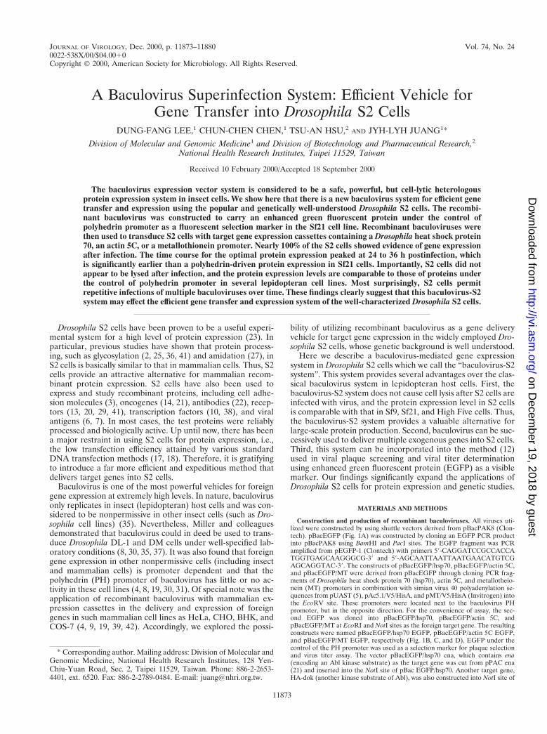
Baculovirus-mediated gene expression in S2 cells. Autogra-pha californica nuclear polyhedrosis virus (AcNPV) is currentlythe most commonly used baculovirus for foreign gene expres-sion. To examine if AcNPV is able to deliver an effective geneexpression cassette into S2 cells, we constructed recombinantbaculoviruses coding for EGFP under the control of four dif-ferent promoters. One (pBacEGFP) was derived from thebaculovirus PH promoter and three (pBacEGFP/hsp70,pBacEGFP/actin 5C, and pBacEGFP/MT) were obtained fromthe Drosophila hsp70, actin 5C, and MT promoters, respec-tively (Fig. 1). Gene expression levels by the four recombinantbaculoviruses were examined at 36 h postinfection. Westernblots of cell extracts were probed with antibody directedagainst EGFP. When S2 cells were mock infected or infectedwith BacEGFP, no EGFP expression could be detected (Fig.2A, lanes 1 and 2). Expression of EGFP could only be observedin S2 cells infected with baculovirus containing Drosophila pro-moters (hsp70, actin 5C, and MT) (Fig. 2A, lanes 3, 4, and 5).These results support previous studies that the virus PH pro-moter is functionally restrained in nonpermissive cells (8, 37).Thus, baculovirus-mediated gene expression in S2 cells is alsopromoter dependent. Our data suggest that modified recom-
binant baculovirus containing a Drosophila expression cassettecan transduce gene expression in S2 cells.
To further assess the efficacy of these promoters in S2 cells,flow cytometry analysis was conducted to determine the ex-pression levels of EGFP. As shown in Fig. 2B, EGFP expres-sion by recombinant virus containing hsp70 promoter gave thehighest level among three different Drosophila expression cas-settes. We then investigated whether we could manipulateEGFP expression by heat shock treatments since the Drosoph-ila hsp70 promoter is known as a temperature-inducible pro-moter in various systems (11, 40). With various heat shocktreatments, the EGFP expression levels in S2 cells were exam-ined by Western blot analysis. No significant difference ofEGFP expression levels among the various treatments wasnoted (data not shown). Therefore, hsp70 may be a tempera-ture-independent and constitutively active promoter in thebaculovirus-S2 system. Similarly, the inducibility of the MTpromoter in the baculovirus-S2 system was also tested with therecombinant baculovirus of BacEGFP/MT EGFP containing aCu21-inducible MT promoter. The results show that the MTpromoter is weak and leaky in the baculovirus-S2 system (Fig.2B). Collectively, these data suggest that the baculovirus-S2system can serve as an efficient gene delivery vehicle for thetarget gene expression in Drosophila S2 cells. Among variousDrosophila promoters tested, hsp70 functioned as the mostpotent and constitutively active promoter.
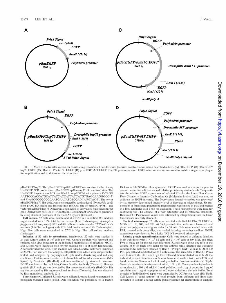
Plaque selection by a fluorescent marker. GFP has beenused as a tool for screening recombinant baculoviruses in Sf9cells (12, 33). Taking advantage of the promoter-dependentphenomenon that virus PH promoter is active in Sf21 cells butnot in S2 cells, we constructed EGFP under the control of thePH promoter as a visible selection marker for virus plaqueassay in Sf21 cells. To investigate the use of EGFP for recom-binant virus selection, a gene called ena that encodes a kinasesubstrate that is phosphorylated by the Abl proto-oncogenefound in mammalian and Drosophila cells (14, 21) was clonedunder the hsp70 promoter into a vector with EGFP under thePH promoter. After cotransfection of Sf21 cells, fluorescentplaques were selected and characterized to identify those ex-pressing Ena. The resulting recombinant baculovirus was thenused to infect S2 cells for exogenous gene expression. Whilethe autofluorescent signal of the EGFP marker was below thedetection level in S2 cells (Fig. 3Ab), the exogenous geneexpression of Ena was detected by antibody directed againstEna (Fig. 3Ac). The finding that the selection marker was onlyexpressed in Sf9 and Sf21 cells but not in S2 cells eliminates thepossible competition and interference with the target geneexpression in S2 cells. This design also excludes the possibilityof target gene mutation from the traditional chemical plaqueselection procedure by neutral red in Sf21 cells (26).
High infection efficiency and gene expression levels. Theresults described above show the capability of baculovirus as avehicle for gene delivery and expression in Drosophila S2 cells.Previous researchers observed that the MOI is critical inachieving baculovirus-mediated protein expression in mamma-lian cells (9). The optimal virus MOI for the highest infectionefficiency in S2 cells was examined. S2 cells were transducedwith an increasing titer of BacEGFP/hsp70 EGFP viruses. At36 h postinfection, the infection efficiency was examined byconfocal microscopy (Fig. 3B). For accurate calculation, infec-tion efficiency was determined by flow cytometry studies. At anMOI of 1 PFU per cell, approximately 17% of the cells showedEGFP expression. The “infection efficiency” increased steadilywith higher MOIs (39, 51, 73, and 85% for MOIs of 5, 10, 25,and 50, respectively) and reached close to 100% at an MOI of100. Therefore, the infection efficiency of S2 cell is virus dose
FIG. 2. Susceptibility of Drosophila S2 cells to baculovirus-mediated foreigngene transduction. (A) Western blot analysis of EGFP expression in S2 cells at36 h postinfection. Lane 1, uninfected cells; lane 2, cells infected with BacEGFP(with PH promoter), showing no EGFP expression; lanes 3 and lane 4, cellsinfected with BacEGFP/hsp70 EGFP and BacEGFP/actin 5C EGFP, respec-tively: lane 5, S2 cells infected with recombinant virus BacEGFP/MT EGFP, andEGFP expression was induced by 1 mM CuSO4 at 24 h postinfection. (B) Efficacyof EGFP expression by these promoters as analyzed by flow cytometry (theEGFP quantification method is described in detail in the text). Results are theaverages of three independent infections.
VOL. 74, 2000 BACULOVIRUS SYSTEM FOR DROSOPHILA S2 CELLS 11875
on Decem
ber 19, 2018 by guesthttp://jvi.asm
.org/D
ownloaded from

FIG. 3. Confocal microscopic analysis of the gene expression and the infection efficiency. (A) Confocal microscopic analysis of EGFP selection marker and targetgene (ena) expression in the baculovirus-S2 system. Mid-exponentially grown S2 cells (106 cells/well) were seeded into a 24-well petri dish and exposed to an MOI forBacEGFP/hsp70 ena of 100. At 36 h postinfection, cells were harvested, attached to poly-L-lysine coated slides, stained with anti-Ena polyclonal antibody, and thenanalyzed by confocal microscopy (Leica TCS NT). (Aa) Phase image of BacEGFP/hsp70 ena-infected S2 cells. (Ab). No detectable EGFP signal in S2 cells. (Ac) Enaprotein detected in S2 cells by immunocytochemistry with polyclonal antibody directed against Ena. (B) Confocal microscopy analysis of the dose-response infectionefficiency. S2 cells transduced with BacEGFP/hsp70 EGFP at the indicated MOIs were examined by confocal microscopy to determine infection efficiencies.
11876
on Decem
ber 19, 2018 by guesthttp://jvi.asm
.org/D
ownloaded from
dependent at from 1 to 100 MOI. However, at an MOI of from100 to 800, the infection efficiency remained optimal.
Using the PH promoter, protein expression by the conven-tional baculovirus expression system usually reaches its maxi-mum expression level by 48 to 72 h postinfection. To explorethe optimal timing for baculovirus-mediated protein expres-sion in S2 cells, a time course study of virus-mediated geneexpression was done. S2 cells were transduced with BacEGFP/hsp70 EGFP at an MOI of 100. Infection efficiencies and totalgene expression levels were examined at 12, 24, 36, 48, 60, and72 h postinfection by flow cytometry analysis. The relativeEGFP expressions were calibrated to relative fluorescence in-tensities by using the LinearFlow Green Flow Cytometry In-tensity Calibration Kit (Molecular Probes, Inc.). The optimalefficiency was achieved by as early as 24 to 36 h postinfectionat an MOI of 100 (Table 1). The total EGFP level of theinfected cells increased steadily over the 36 h of baculovirustransduction and started to decline by 36 to 72 h postinfection(Table 1). These results suggest that the optimal protein ex-pression driven by the hsp70 promoter in the baculovirus-S2system is about 12 to 36 h earlier than that by the PH promoterin lepidopteran cells.
Nonlytic system for S2 cells. In the classical baculovirussystem, lepidopteran cells such as Sf9, Sf21, and High Five cellsundergo a lytic cycle after virus infection. Such a lethal viralinfection in cells might trigger the cellular protein processingmachinery to deteriorate several days postinfection. This eventmight lead to improper posttranscriptional and/or posttransla-tional modifications of gene products. To investigate whetherS2 cells are destined for the same lytic pathway, S2 cells wereexposed to the BacEGFP/hsp70 EGFP virus at various MOIs,and viabilities were determined by trypan blue exclusion assay.The percent viabilities of S2 cells transduced with BacEGFP/hsp70 EGFP baculovirus at various MOIs were as follows(MOI, percent viability [mean 6 standard deviation]): 1,99.2 6 0.4; 5, 98.6 6 0.2; 10, 96.2 6 0.5; 25, 95.2 6 0.7; 50,95.5 6 0.3; 100, 93.1 6 0.7; 200, 94.6 6 0.5; 400, 93.2 6 0.6; and800, 95.8 6 0.4. The viability of the infected S2 cells wasdetermined by staining the cells with 0.4% trypan blue solution(Sigma) at 72 h postinfection. These results are the averages ofthree independent experiments. The result indicates that thereare no apparent cytopathic effects on S2 cells by 72-h postin-fection, even at an MOI of 800. Thus, this expression systemdoes not include the lytic cycle of baculovirus.
There are two putative explanations for this nonlytic viruscycle in S2 cells. First, protein expression by this baculovirussystem might be a persistent infection in S2 cells. However,data from our aforementioned time course studies do notreveal a persistent expression of the exogenous genes in S2
cells. The total EGFP level of the entire infected cells startedto decline by 36 to 72 h postinfection. Thus, it is likely that thetransduced viral DNA might be degraded or diluted uponcellular replication over time. To test the second hypothesis,we isolated total DNA from the infected cells at 30 days postin-fection and tested for the virus genomic DNA by PCR analysis.Neither EGFP cDNA nor baculovirus genomic DNAs could bedetected in the cells (data not shown). These data suggest thatthe S2 cell line is not a persistent infection target for baculo-virus. In nature, baculovirus DNA replicates by 6 h postinfec-tion and virions bud from the infected cell’s surface to infectother host cells by 10 to 12 h postinfection (15). Studies byothers (8) also showed that baculovirus-mediated foreign geneexpression in Drosophila DL-1 cells has limited viral DNAreplication and undergoes DNA degradation sometime afterinfection.
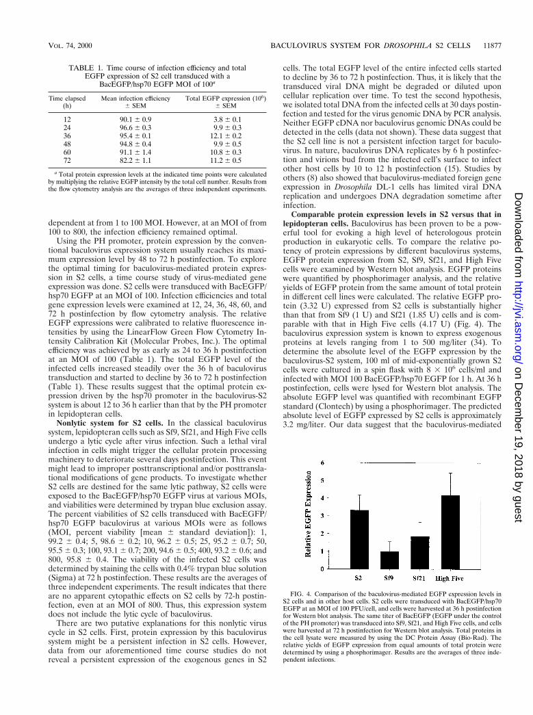
Comparable protein expression levels in S2 versus that inlepidopteran cells. Baculovirus has been proven to be a pow-erful tool for evoking a high level of heterologous proteinproduction in eukaryotic cells. To compare the relative po-tency of protein expressions by different baculovirus systems,EGFP protein expression from S2, Sf9, Sf21, and High Fivecells were examined by Western blot analysis. EGFP proteinswere quantified by phosphorimager analysis, and the relativeyields of EGFP protein from the same amount of total proteinin different cell lines were calculated. The relative EGFP pro-tein (3.32 U) expressed from S2 cells is substantially higherthan that from Sf9 (1 U) and Sf21 (1.85 U) cells and is com-parable with that in High Five cells (4.17 U) (Fig. 4). Thebaculovirus expression system is known to express exogenousproteins at levels ranging from 1 to 500 mg/liter (34). Todetermine the absolute level of the EGFP expression by thebaculovirus-S2 system, 100 ml of mid-exponentially grown S2cells were cultured in a spin flask with 8 3 106 cells/ml andinfected with MOI 100 BacEGFP/hsp70 EGFP for 1 h. At 36 hpostinfection, cells were lysed for Western blot analysis. Theabsolute EGFP level was quantified with recombinant EGFPstandard (Clontech) by using a phosphorimager. The predictedabsolute level of EGFP expressed by S2 cells is approximately3.2 mg/liter. Our data suggest that the baculovirus-mediated
FIG. 4. Comparison of the baculovirus-mediated EGFP expression levels inS2 cells and in other host cells. S2 cells were transduced with BacEGFP/hsp70EGFP at an MOI of 100 PFU/cell, and cells were harvested at 36 h postinfectionfor Western blot analysis. The same titer of BacEGFP (EGFP under the controlof the PH promoter) was transduced into Sf9, Sf21, and High Five cells, and cellswere harvested at 72 h postinfection for Western blot analysis. Total proteins inthe cell lysate were measured by using the DC Protein Assay (Bio-Rad). Therelative yields of EGFP expression from equal amounts of total protein weredetermined by using a phosphorimager. Results are the averages of three inde-pendent infections.
TABLE 1. Time course of infection efficiency and totalEGFP expression of S2 cell transduced with a
BacEGFP/hsp70 EGFP MOI of 100a
Time elapsed(h)
Mean infection efficiency6 SEM
Total EGFP expression (106)6 SEM
12 90.1 6 0.9 3.8 6 0.124 96.6 6 0.3 9.9 6 0.336 95.4 6 0.1 12.1 6 0.248 94.8 6 0.4 9.9 6 0.560 91.1 6 1.4 10.8 6 0.372 82.2 6 1.1 11.2 6 0.5
a Total protein expression levels at the indicated time points were calculatedby multiplying the relative EGFP intensity by the total cell number. Results fromthe flow cytometry analysis are the averages of three independent experiments.
VOL. 74, 2000 BACULOVIRUS SYSTEM FOR DROSOPHILA S2 CELLS 11877
on Decem
ber 19, 2018 by guesthttp://jvi.asm
.org/D
ownloaded from
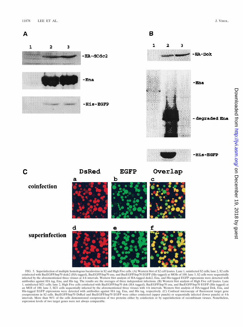
FIG. 5. Superinfection of multiple homologous baculovirus in S2 and High Five cells. (A) Western blot of S2 cell lysates. Lane 1, uninfected S2 cells; lane 2, S2 cellscoinfected with BacEGFP/hsp70 dcdc2 (HA tagged), BacEGFP/hsp70 ena, and BacEGFP/hsp70 EGFP (His tagged) at MOIs of 100; lane 3, S2 cells were sequentiallyinfected by the aforementioned three viruses at 4-h intervals. Western blot analysis of HA-tagged dcdc2, Ena, and His-tagged EGFP expressions were detected withantibodies against HA tag, Ena, and His tag. The results are the averages of three independent infections. (B) Western blot analysis of High Five cell lysates. Lane1, uninfected Sf21 cells; lane 2, High Five cells coinfected with BacEGFP/hsp70 dok (HA tagged), BacEGFP/hsp70 ena, and BacEGFP/hsp70 EGFP (His tagged) atan MOI of 100; lane 3, Sf21 cells sequentially infected by the aforementioned three viruses with 4-h intervals. Western blot analysis of HA-tagged Dok, Ena, andHis-tagged EGFP expressions were detected with antibodies against HA tag, Ena, and His tag, respectively. (C) Confocal microscopy of fluorescent target genecoexpressions in S2 cells. BacEGFP/hsp70 DsRed and BacEGFP/hsp70 EGFP were either coinfected (upper panels) or sequentially infected (lower panels) at 4-hintervals. More than 96% of the cells demonstrated coexpression of two proteins either by coinfection or by superinfection of recombinant viruses. Nonetheless,expression levels of two target genes were not always comparable.
11878 LEE ET AL. J. VIROL.
on Decem
ber 19, 2018 by guesthttp://jvi.asm
.org/D
ownloaded from
gene expressions in S2 cells might be an alternative for large-scale protein expressions.
Superinfection in S2 cells. Superinfection is defined as aninfected cell that is capable of being infected by another virusat subsequent time points. For the classical baculovirus expres-sion system, multiple recombinant baculoviruses are capable ofcoinfecting into the same cell concurrently. However, the samehost cell becomes nonpermissive to another baculovirus infec-tion once it is first infected by a homologous (the same species)recombinant baculovirus (16, 24) at an earlier time point. Oth-ers have shown that it is possible for an infected Sf cell to beinfected with another heterologous virus (24). However, thereis no report describing the prospect of homologous baculovirussuperinfection in any applied cell lines. To explore the feasi-bility of utilizing baculovirus to express two or more genes inone cell at different infection times, S2 cells were either con-currently coinfected with 100-MOI virus doses of BacEGFP/hsp70 HA-dcdc2, BacEGFP/hsp70 ena, and BacEGFP/hsp70His6-EGFP in combination or were individually infected withthese three viruses sequentially at intervals of 4 h. At 36 hpostinfection, the three Western blots of cell extract wereprobed, respectively, with antibodies directed against HA-tagged dCdc2, Ena, and His-tagged EGFP and subsequentlydetected with alkaline phosphatase-conjugated goat anti-mouse secondary antibody. As shown in Fig. 5A, three pro-teins, which were either transduced concurrently or sequen-tially, were expressed equally well in the S2 cells. The samesuperinfection studies were also done with High Five cells.Three different recombinant baculoviruses (BacEGFP/hsp70HA-dok, BacEGFP/hsp70 ena, and BacEGFP/hsp70 His6-EGFP) were used for this experiment. In contrast to the S2cells being repeatedly transduced with multiple recombinantbaculovirus, lepidopteran High Five cells can only be infectedonce by baculovirus (Fig. 5B). To confirm that the coexpres-sion of multiple target genes by superinfection is not due todifferent populations of infected cells each infected with adifferent virus, confocal microscopic analysis of the coexpres-sion of two fluorescent target genes in S2 cells was conducted.BacEGFP/hsp70 DsRed (a red fluorescent protein from seaanemone) (28) and BacEGFP/hsp70 EGFP were either coin-fected or sequentially infected at 4-h intervals. Figure 5Cshows that more than 96% of the cells coexpressed both fluo-rescent proteins either by coinfection or by superinfection ofrecombinant viruses. It is evident that the initial entry of therecombinant baculovirus did not prohibit the transduction ofanother homologous virus into the same S2 cells over time. Wehave also noted that the expression levels of two heterologousgenes in the same cell were not always equivalent. The expres-sion level of the second transduced target gene is not neces-sarily lower than that of the first target gene. Together, thesedata strongly suggest that the baculovirus-S2 system may be analternative approach to coinfect two or more recombinantbaculoviruses concurrently or sequentially over time for theproduction of biologically active proteins.
Cell lysis-associated proteolysis is also considered as themajor drawback for the baculovirus expression system (32).For instance, if the protein of interest is a secreted protein,proteinases from the lysed cells could severely compromise theyield of protein production. In the present study we also notedthat Ena was degraded in High Five cells but not in S2 cells bythe baculovirus expression system (Fig. 5B, lane 2). Accordingto our previous studies (21), Ena is a relatively stable proteinwhile it was transiently expressed in S2 cells and some mam-malian cells by the liposome transfection method. In addition,we know of no report that suggests Ena could interact with theother two proteins (EGFP and Dok) used in the study. Thus,
ena was considered to be an appropriate independent targetgene for the study of superinfection in our system. However,we were surprised that Ena was actually relatively degradablewhile it was expressed by the conventional baculovirus systemin the High Five cell line, which permits the lytic cycle ofbaculovirus. In contrast, the putative cell lysis associated pro-teolysis of Ena had not occurred in our nonlytic baculovirus-S2system. Consequently, the protein expression level and qualitymight be severely compromised in the conventional baculovi-rus protein expression system. The baculovirus-S2 system maybe considered as an alternative to solve such a problem. Thus,this baculovirus-S2 system provides a very useful and flexiblesuperinfection vehicle in order to analyze protein-protein orprotein-drug interactions in intact cells.
Among currently known viral expression systems in eukary-otic cells, baculovirus is a relatively safe expression system withoptimal infection efficiency and high expression levels for het-erologous protein production. Nonetheless, some features ofbaculovirus expression are considered to be inconvenient formany common applications. Restricted host range and cell lysisoccurring after infection are the two major limiting factors forthe application of the conventional baculovirus system in mod-ern biomedical research. We report here a baculovirus-S2 sys-tem that mediates protein expression in Drosophila S2 cellswithout the major drawbacks of the classical baculovirus ex-pression. In addition, this alternative system may also providea desirable device for the superinfection of multiple targetgenes at the appropriate stoichiometric ratios over an experi-mental time course. In particular, S2 cells share several generalfeatures of baculovirus AcNPV host cells that are favorable forthe mass production of proteins. For example, the cells adapteasily to the spinner flask and can achieve high densities in aninexpensive media cultured at room temperature without CO2supplementation. In addition, Drosophila S2 cells with a com-pletely sequenced genome (1) could be treated as a knownbackground for the expression and purification of heterologousmammalian proteins without the interference of the nativeprotein in cells.
In summary, our study describes a novel virus expressionsystem in S2 cells with several favorable attributes. First, bac-ulovirus can serve as an efficient gene transfer vehicle in thewidely used Drosophila S2 cell line. Protein expression in thisnonpermissive insect cell line is promoter dependent, and theDrosophila hsp70 promoter in the expression cassette functionsas a constitutively active promoter in S2 cells. Second, a visibleEGFP selection maker in this system can facilitate the opera-tion of plaque selection and virus titer determination. Third, aninfection efficiency of up to 100% can be reached, and thisvalue is critical in some applications that require a homoge-neous background (e.g., microarray analysis). Fourth, compa-rable transient protein expression levels with Sf9, Sf21, andHigh Five cells are not compromised by the proteolysis effectresulting from the viral lytic cycle. Last, and perhaps mostimportantly, our experimental results reveal the first charac-terized homologous superinfection system for sequential mul-tiple target gene expressions in eukaryotic cells. We are nowinvestigating whether the same superinfection phenomenamight take place in mammalian cells as well.
ACKNOWLEDGMENTS
We thank Tsu-Fen Huang for assistance with the confocal micros-copy. We also thank Stanley D. Carlson, Entomology Department andNeuroscience Training Program, University of Wisconsin–Madison,and Michael Betenbaugh, Department of Chemical Engineering, TheJohns Hopkins University, for reviewing the manuscript.
VOL. 74, 2000 BACULOVIRUS SYSTEM FOR DROSOPHILA S2 CELLS 11879
on Decem
ber 19, 2018 by guesthttp://jvi.asm
.org/D
ownloaded from
REFERENCES
1. Adams, M. D., S. E. Celniker, R. A. Holt, et al. 2000. The genome sequenceof Drosophila melanogaster. Science 287:2185–2195.
2. Aldecoa, A., R. Gujer, J. A. Fischer, and W. Born. 2000. Mammalian calci-tonin receptor-like receptor/receptor activity modifying protein complexesdefine calcitonin gene-related peptide and adrenomedullin receptors in Dro-sophila Schneider 2 cells. FEBS Lett. 471:156–160.
3. Bellosta, P., M. Costa, D. A. Lin, and C. Basilico. 1995. The receptor tyrosinekinase ARK mediates cell aggregation by homophilic binding. Mol. Cell.Biol. 15:614–625.
4. Boyce, F. M., and N. L. Bucher. 1996. Baculovirus-mediated gene transferinto mammalian cells. Proc. Natl. Acad. Sci. USA 93:12348–12352.
5. Brand, A. H., and N. Perrimon. 1993. Targeted gene expression as a meansof altering cell fates and generating dominant phenotypes. Development118:401–415.
6. Brighty, D. W., M. Rosenberg, I. S. Chen, and M. Ivey-Hoyle. 1991. Envelopeproteins from clinical isolates of human immunodeficiency virus type 1 thatare refractory to neutralization by soluble CD4 possess high affinity for theCD4 receptor. Proc. Natl. Acad. Sci. USA 88:7802–7805.
7. Brighty, D. W., and M. Rosenberg. 1994. A cis-acting repressive sequencethat overlaps the Rev-responsive element of human immunodeficiency virustype 1 regulates nuclear retention of env mRNAs independently of knownsplice signals. Proc. Natl. Acad. Sci. USA 91:8314–8318.
8. Carbonell, L. F., M. J. Klowden, and L. K. Miller. 1985. Baculovirus-medi-ated expression of bacterial genes in dipteran and mammalian cells. J. Virol.56:153–160.
9. Condreay, J. P., S. M. Witherspoon, W. C. Clay, and T. A. Kost. 1999.Transient and stable gene expression in mammalian cells transduced with arecombinant baculovirus vector. Proc. Natl. Acad. Sci. USA 96:127–132.
10. Courey, A. J., and R. Tjian. 1998. Analysis of Sp1 in vivo reveals multipletranscriptional domains, including a novel glutamine-rich activation motif.Cell 55:887–898.
11. Elkins, T., M. Hortsch, A. J. Bieber, P. M. Snow, and C. S. Goodman. 1990.Drosophila fasciclin I is a novel homophilic adhesion molecule that alongwith fasciclin III can mediate cell sorting. J. Cell Biol. 110:1825–1832.
12. Eriksson, S., E. Raivio, J. P. Kukkonen, K. Eriksson, and C. Lindqvist. 1996.Green fluorescent protein as a tool for screening recombinant baculoviruses.J. Virol. Methods 59:127–133.
13. Fehon, R. G., P. J. Kooh, I. Rebay, C. L. Regan, T. Xu, M. A. T. Muskavitch,and S. Artavanis-Tsakonas. 1990. Molecular interactions between the pro-tein products of the neurogenic loci Notch and Delta, two EGF-homologousgenes in Drosophila. Cell 61:523–534.
14. Getler, F. B., A. R. Comer, J. L. Juang, S. M. Ahern, M. J. Clark, E. C. Liebl,and F. M. Hoffmann. 1995. Enabled, a dosage-sensitive suppressor of mu-tations in the Drosophila Abl tyrosine kinase, encodes an Abl substrate withSH3 domain-binding properties. Genes Dev. 9:521–533.
15. Granados, R. R., and B. A. Federici. 1986. In vivo infection and replicationof baculoviruses, p. 89–108. In R. R. Granados and K. A. Williams (ed.), Thebiology of baculovirus, vol. I. Biological properties and molecular biology.CRC Press, Boca Raton, Fla.
16. Granados, R. R., and B. A. Federici. 1986. Persistent baculovirus infection, p.159–175. In J. P. Burand, C. Y. Kawanishi, and Y. S. Huang (ed.), Thebiology of baculovirus, vol. I. Biological properties and molecular biology.CRC Press, Boca Raton, Fla.
17. Han, K., M. S. Levine, and J. L. Manley. 1989. Synergistic activation andrepression of transcription by Drosophila homeobox proteins. Cell 56:573–583.
18. Han, K. 1996. An efficient DDAB-mediated transfection of Drosophila S2cells. Nucleic Acids Res. 24:4362–4363.
19. Hofmann, C., V. Sandig, G. Jennings, M. Rudolph, P. Schlag, and M.Strauss. 1995. Efficient gene transfer into human hepatocytes by baculovirusvectors. Proc. Natl. Acad. Sci. USA 92:10099–10103.
20. Johanson, K., E. Appelbaum, M. Doyle, P. Hensley, B. Zhao, S. S. Abdel-Mequid, P. Young, R. Cook, S. Carr, R. Matico, D. Cusimano, E. Dul, M.Angelichio, I. Brooks, E. Winborne, P. McDonnell, T. Morton, D. Bennett, T.Sokolski, D. McNulty, M. Rosenberg, and I. Chaiken. 1995. Binding inter-actions of human interleukin 5 with its receptor alpha subunit. J. Biol. Chem.270:9459–9471.
21. Juang, J. L., and F. M. Hoffmann. 1999. Drosophila Abelson interacting
protein (dAbi) is a positive regulator of Abelson tyrosine kinase activity.Oncogene 37:5138–5147.
22. Kirkpatrick, R. B., S. Ganguly, M. Angelichio, S. Griego, A. Shatzman, C.Silverman, and M. Rosenberg. 1995. Heavy chain dimers as well as completeantibodies are efficiently formed and secreted from Drosophila via a Bip-mediated pathway. J. Biol. Chem. 270:19800–19805.
23. Kirkpatrick, R. B., and A. Shatzman. 1999. Drosophila S2 system for heter-ologous gene expression, p. 289–331. In J. M. Fernandez and J. P. Hoeffler(ed.), Gene expression systems: Drosophila S2 system for heterologous geneexpression. Academic Press, San Diego, Calif.
24. Lee, J.-C., H.-H. Chen, H.-L. Wei, and Y.-C. Chao. 1993. Superinfection-induced apoptosis and its correlation with the reduction of viral progeny incells persistently infected with Hz-1 baculovirus. J. Virol. 67:6989–6994.
25. Li, B., S. Tsing, A. H. Kosaka, B. Nguyen, E. G. Osen, C. Bach, H. Chan, andJ. Barnett. 1996. Protein expression of human dopamine beta-hydroxylase inDrosophila Schneider 2 cells. Biochem. J. 69:125–133.
26. Longnecker, D. S., T. J. Curphey, and D. S. Daninel. 1977. Mutagenicity ofneutral red. Mutat. Res. 48:109–111.
27. Matsumura, M., Y. Saito, M. R. Jackson, E. S. Song, and P. A. Peterson.1992. In vitro peptide binding to soluble empty class I major histocompati-bility complex molecules isolated from transfected Drosophila melanogastercells. J. Biol. Chem. 267:23589–23595.
28. Matz, M. V., A. F. Fradkov, Y. A. Labas, A. P. Savitsky, A. G. Zaraisky, andM. L. Markelov. 1999. Fluorescent proteins from nonbioluminescent Antho-zoa. Nat. Biotechnol. 17:969–973.
29. Millar, N., and S. Buckingham. 1994. Stable expression of a functionalhomo-oligomeric Drosophila GABA receptor in a Drosophila cell line. Proc.R. Soc. London Ser. B 258:307–314.
30. Morris, T. D., and L. K. Miller. 1992. Promoter influence on baculovirus-mediated gene expression in permissive and nonpermissive insect cell lines.J. Virol. 66:7397–7405.
31. Morris, T. D., and L. K. Miller. 1993. Characterization of productive andnon-productive AcMNPV infection in selected insect cell line. Virology197:339–348.
32. Naggie, S., and W. E. Bentley. 1998. Appearance of protease activities co-incides with p10 and polyhedrin-driven protein production in the baculovirusexpression system: effects on yield. Biotechnol. Prog. 14:227–232.
33. Nordstrom, T., A. Senkas, S. Eriksson, N. Pontynen, E. Nordstrom, and C.Lindqvist. 1999. Generation of a new protein purification matrix by loadingceramic hydroxyapatite with metal ions—demonstration with polyhistidine-tagged green fluorescent protein. J. Biotechnol. 69:125–133.
34. O’Reilly, D. R., L. K. Miller, and V. A. Luckow. 1992. Baculovirus expressionvectors: a laboratory manual, p. 28. W. H. Freeman and Company, NewYork, N.Y.
35. Pennock, G. D., C. Shoemaker, and L. K. Miller. 1984. Strong and regulatedexpression of Escherichia coli b-galactosidase in insect cells with a baculovi-rus vector. Mol. Cell. Biol. 4:399–406.
36. Percival, M. D., L. Bastien, P. R. Griffin, S. Kargman, M. Ouellet, and G. P.O’Neill. 1997. Investigation of human cyclooxygenase-2 glycosylation heter-ogeneity and protein expression in insect and mammalian cell expressionsystems. Protein Expr. Purif. 9:388–398.
37. Rice, W. C., and L. K. Miller. 1986. Baculovirus transcription in the presenceof inhibitors and in nonpermissive Drosophila cells. Virus Res. 6:155–172.
38. Santoro, C., N. Mermod, P. C. Andrews, and R. Tjian. 1998. A family ofhuman CCAAT-box-binding proteins active in transcription and DNA rep-lication: cloning and expression of multiple cDNAs. Nature 334:218–224.
39. Shoji, I., H. Aizaki, H. Tani, K. Ishii, T. Chiba, I. Saito, I. Miyamura, and Y.Matsuura. 1997. Efficient gene transfer into various mammalian cells, in-cluding non-hepatic cells, by baculovirus vectors. J. Gen. Virol. 78:2657–2664.
40. Snow, P. M., A. J. Bieber, and C. S. Goodman. 1989. Fasciclin III: a novelhomophilic adhesion molecule in Drosophila. Cell 59:313–323.
41. Tota, M. R., L. Xu, A. Sirotina, C. D. Strader, and M. P. Graziano. 1995.Interaction of [fluorescein-Trp]glucagon with the human glucagon receptorexpressed in Drosophila Schneider 2 cells. J. Biol. Chem. 270:26466–26472.
42. Yap, C.-C., K. Ishii, Y. Aoki, H. Aizaki, H. Tani, H. Shimizu, Y. Ueno, T.Miyamura, and Y. Matsuura. 1997. A hybrid baculovirus-T7 RNA polymer-ase system for recovery of an infectious virus from cDNA. Virology 231:192–200.
11880 LEE ET AL. J. VIROL.
on Decem
ber 19, 2018 by guesthttp://jvi.asm
.org/D
ownloaded from