Embed Size (px)
Citation preview
EXTRACTION SITE PRESERVATION USING FDBA WITH BIOXCLUDE VS.
FDBA WITH BIO-GIDE: A RANDOMIZED CLINICAL TRIAL.
by
Barak A. Wray, DMD Lieutenant, Dental Corps
United States Navy
A thesis submitted to the Faculty of the Periodontics Graduate Program
Naval Postgraduate Dental School Uniformed Services University of the Health Sciences
in partial fulfillment of the requirements for the degree of Master of Science in Oral Biology
June 2015
Naval Postgraduate Dental School Uniformed Services University of the Health Sciences
Bethesda, Maryland
CERTIFICATE OF APPROVAL
MASTER'S THESIS
This is to ce1tify that the Master's thesis of
Barak A. Wray
has been approved by the Examining Committee for the thesis requirement for the Master of Science degree in Oral Biology at the Ju e 2013 a uation.
Thesis Committee: f /} ,, ) l
Peter M. Be11rand, DDS CAPT (Ret), DC, USN, Professor, Dental Research Thesis Supervisor
Thu P. Getka, DDS, MS, BS CAPT, DC, USN Program Director Periodontics Department
Glenn A. u ro, DDS, MBA CAPT, DC, USN Dean, Naval postgraduate Dental School
The author hereby ce1tifies that the use of any copyrighted material in the thesis manuscript titled:
EXTRACTION SITE PRESERVATION USING FDBA WITH BIOXCLUDE VS FDBA WITH BIO-GIDE: A RANDOMIZED CLINICAL TRIAL.
is appropriately acknowledged and, beyond brief excerpts, is with the permission of the copyright owner.
~~ Periodontics Depmiment Naval Postgraduate Dental School March20!5
NAVAL POSTGRADUATE DENTAL SCHOOL BARAK A. WRAY
2015
This thesis may not be re-printed without the expressed written permission of the author.
ii
ABSTRACT
EXTRACTION SITE PRESERVATION USING FDBA WITH BIOXCLUDE VS. FDBA WITH BIO-GIDE: A RANDOMIZED CLINICAL TRIAL.
BARAK A. WRAY, DDS PERIODONTICS 2015
Thesis directed by: Peter M. Bertrand, DDS CAPT (Ret), DC, USN Professor, Dental Research
Introduction:
Naval Postgraduate Dental School
Thu P. Getka, DMD, MS, BS CAPT, DC, USN Naval Postgraduate Dental School
This randomized, controlled, blinded, clinical investigation will compare
extraction site preservation performed with freeze dried bone allograft (FDBA) and either
the Bio-Gide membrane or the BioXclude membrane. Alveolar ridge dimensional
' changes from the time of extraction until implant placement at 6 months will be assessed.
Up to 70 patients treatment planned for an extraction with site preservation and implant
placement will be enrolled. Participants will be randomized to receive Bio-Gide or
BioXclude. Each patticipant will have a jaw impression made of the extraction site to
provide a stone model for the fabrication of a customized acrylic stent. The stent will be
used by blinded investigators to obtain standardized clinical measurements of the alveolar
ridge at 6 points around the extraction site, immediately after the extraction, and at 6
months right before implant placement. In addition to the pre-extraction impression,
impressions will also be taken at 4 and 12 weeks post-extraction, and at the time of
iii
implant placement. The four sets of models from these impressions will be used to
measure dimensional changes in the alveolar ridge. Soft tissue closure across the
membranes will also be assessed. Study investigators, blinded to the membrane material,
will make all clinical measurements. Standard clinical procedure evaluates postoperative
healing at 1, 2, 4, 6, 8, 12 and 16 weeks and again at 6 months. For study participants,
impressions and research measurements will be made only during these post-operative
visits. Data analysis will compare changes in site dimension and soft tissue closure.
Methods:
Up to 70 patients treatment planned for an extraction with site preservation and
implant placement will be included in the study. Up to 35 patients will receive FDBA
and Bio-Gide and up to 35 patients receive FDBA and BioXclude . In both groups, no
attempt will be made to obtain primary closure. Participants will have impressions made
of the anticipated extraction site to provide stone models for the fabrication of a
customized acrylic stent. Acrylic stents will be used by blinded investigators to obtain
standardized clinical measurements of the alveolar ridge at 6 points around the extraction
site following extraction and 6 months later at implant placement. In addition to the pre
extraction impression, impressions will also be taken at 4 and 12 weeks post-extraction
and at the time of implant placement. The four sets of models from these impressions
will be used to measure dimensional changes in the alveolar ridge. Soft tissue closure
across the membranes will also be assessed. Standard clinical procedure evaluates
postoperative healing at I, 2, 4, 6, 8, 12 and 16 weeks and again at 6 months. For study
patticipants, impressions and research measurements will be made only during these
iv

post-operative visits. Data analysis will compare changes in ridge dimension and soft
tissue closure.
Results:
Currently this research protocol has been prepared to be submitted to the !RB.
Following results of correspondence with Snoasis, the makers ofBioXclude, the protocol
will be submitted.
Discussion:
Research will commence following !RB approval.
v
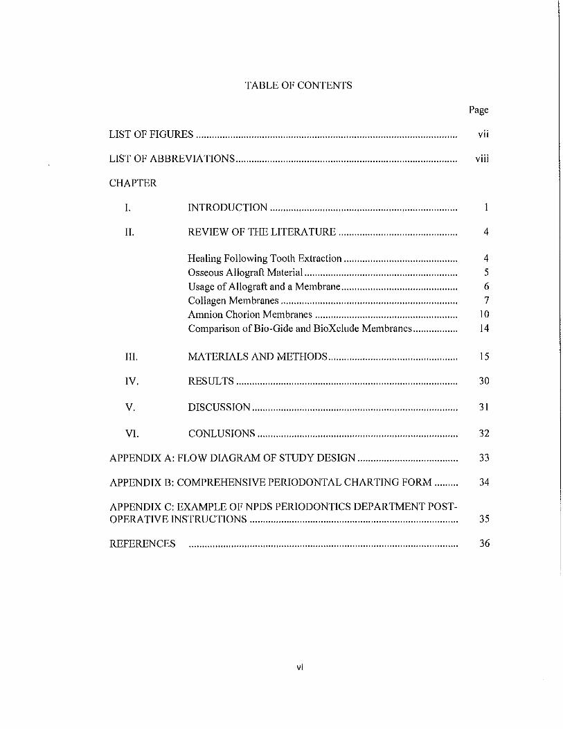
TABLE OF CONTENTS
Page
LIST OF FIGURES ................................................................................................... vii
LIST OF ABBREVIATIONS.................................................................................... viii
CHAPTER
I. INTRODUCTION ...................................................................... .
II. REVIEW OF THE LITERATURE ........................................... .. 4
Healing Following Tooth Extraction........................................... 4
Osseous Allograft Material.......................................................... 5
Usage of Allograft and a Membrane............................................ 6 Collagen Membranes ................................................................... 7 Amnion Chorion Membranes ...................................................... 10
Comparison of Bio-Gide and BioXclude Membranes................. 14
Ill. MATERIALS AND METHODS ................................................ . 15
IV. RESULTS ................................................................................... . 30
v. DISCUSSION ............................................................................. . 31
VI. CONLUSIONS ........................................................................... . 32
APPENDIX A: FLOW DIAGRAM OF STUDY DESIGN...................................... 33
APPENDIX B: COMPREHENSIVE PERIODONTAL CHARTING FORM......... 34
APPENDIX C: EXAMPLE OF NPDS PERIODONTICS DEPARTMENT POST-OPERATIVE INSTRUCTIONS............................................................................... 35
REFERENCES 36
vi
Figure
I.
2.
3.
4.
LIST OF FIGURES
Implant placed with insufficient alveolar bone .......................................... .
Bone graft in extraction site ...................................................................... ..
Bone graft covered with a membrane ....................................................... ..
Stone Model example ................................................................................ .
Page
2
2
2
16
5. Cast after removing the tooth planned for extraction (#19) at the height of
contour, occlusal (top-down) view. ............................................................ 18
6. Cast after removing the tooth planned for extraction (#19) at the height of
7.
8.
9.
10.
contour, buccal (side) view......................................................................... 18
Mo ldable acrylic applied and cured ........................................................... .
Acrylic trimmed ......................................................................................... .
Measuring grooves created ....................................................................... ..
Membrane cards ......................................................................................... .
vii
19
19
20
22
LIST OF ABBREVIATIONS
GBR Guided Bone Regeneration
FDBA Freeze-dried Bone Allograft
PD Probing Depth
CAL Clinical Attachment Level
CEJ Cementoenamel Junction
WRNMMC Walter Reed National Medical Military Center
PO Post-Operative
NSAIDS Nonsteroidal Anti-Inflammatory Drugs
q6h Every 6 Hours
q8h Every 8 Hours
mg Milligram
TBSP Tablespoon
VAS Visual Analog Scale
viii
CHAPTER I: INTRODUCTION
Both the esthetic and functional outcomes of dental implant therapy are highly
dependent on sufficient alveolar bone volume following tooth extraction. Although bone
loss prior to extraction may have occurred due to trauma, periapical pathosis, or
periodontal disease, it is important to understand that alveolar ridge bone loss occurs after
extraction. In the normal post extraction remodeling process, without an intervention to
preserve alveolar ridge contours, bone loss occurs in both horizontal and vettical
dimensions. 1 Considering that the dimensions of the alveolar ridge are critical for implant
placement, it is imperative to recognize that site (ridge) preservation following tooth
extraction is needed to maintain as ideal an alveolar ridge
anatomy as possible. If an implant is placed at a site in the jaw
with less ideal jaw dimension, then the implant may not be
completely integrated (embedded) in bone. When this occurs,
and implant threads are not covered as seen in Figure 1, the
threads become locations where bacterial plaque can grow.
Such a situation clinically exposes the patient to recurrent
infection around the implant and increased risk to implant loss.
Ridge Preservation Therapy
Figure 1: Implant placed with insufficient alveolar
bone.
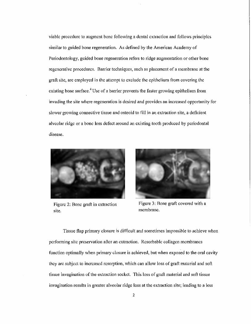
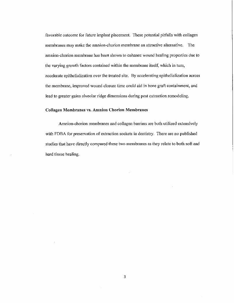
Bone loss following tooth extraction can be minimized by ridge preservation
therapy utilizing an osseous graft material and membrane over the grafted bone. Without
site (ridge) preservation interventions such as bone grafting with a membrane that try to
minimize post extraction bone loss (Figures 2 and 3), a significant reduction in both
horizontal and vettical bone volume can be witnessed. Ridge preservation therapy is a
1
viable procedure to augment bone following a dental extraction and follows principles
similar to guided bone regeneration. As defined by the American Academy of
Periodontology, guided bone regeneration refers to ridge augmentation or other bone
regenerative procedures. Barrier techniques, such as placement of a membrane at the
graft site, are employed in the attempt to exclude the epithelium from covering the
existing bone surface.4 Use of a barrier prevents the faster growing epithelium from
invading the site where regeneration is desired and provides an increased oppotiunity for
slower growing connective tissue and osteoid to fill in an extraction site, a deficient
alveolar ridge or a bone loss defect around an existing tooth produced by periodontal
disease.
Figure 2: Bone graft in extraction site.
Figure 3: Bone graft covered with a
membrane.
Tissue flap primary closure is difficult and sometimes impossible to achieve when
performing site preservation after an extraction. Resorbable collagen membranes
function optimally when primary closure is achieved, but when exposed to the oral cavity
they are subject to increased resorption, which can allow loss of graft material and soft
tissue invagination of the extraction socket. This loss of graft material and soft tissue
invagination results in greater alveolar ridge loss at the extraction site; leading to a less
2
favorable outcome for future implant placement. These potential pitfalls with collagen
membranes may make the amnion-chorion membrane an attractive alternative. The
amnion-chorion membrane has been shown to enhance wound healing prope1ties due to
the varying growth factors contained within the membrane itself, which in turn,
accelerate epithelialization over the treated site. By accelerating epithelialization across
the membrane, improved wound closure time could aid in bone graft containment, and
lead to greater gains alveolar ridge dimensions dming post extraction remodeling.
Collagen Membranes vs. Amnion Chorion Membranes
Amnion-chorion membranes and collagen barriers are both utilized extensively
with FDBA for preservation of extraction sockets in dentistry. There are no published
studies that have directly compared these two membranes as they relate to both soft and
hard tissue healing.
3
CHAPTER II: REVIEW OF THE LITERATURE
Healing Following Tooth Extraction
Both the esthetic and functional outcomes of dental implant therapy are highly
dependent on sufficient alveolar bone volume following tooth extraction. Although bone
loss prior to extraction may have occurred due to trauma, periapical pathosis, or
periodontal disease, it is important to understand that alveolar ridge bone loss occurs after
extraction. In the normal post extraction remodeling process, without an intervention to
preserve alveolar ridge contours, bone loss occurs in both horizontal and vet1ical
dimensions. 1 Considering that the dimensions of the alveolar ridge are critical for implant
placement, it is imperative to recognize that site (ridge) preservation following tooth
extraction is needed to maintain as ideal an alveolar ridge anatomy as possible. If an
implant is placed at a site in the jaw with less ideal jaw dimension, then the implant may
not be completely integrated (embedded) in bone. When this occurs, and implant threads
are not covered by bone as seen in the photograph to the right, the threads become
locations where bacterial plaque can grow. Such a situation clinically exposes the patient
to recurrent infection around the implant and increased risk to implant loss.
Wound healing following extraction has been evaluated extensively in humans.
Shottly following extraction, clot formation occurs that is replaced by granulation tissue
within seven days. By the twentieth day, the granulation tissue is replaced by connective
tissue. While this connective tissue replacement is occurring, osteoid formation begins at
the base of the socket by seven days, and eventually, two thirds of the original bony
socket that held the tooth will be filled with bony trabeculae at thirty-eight days. During
healing, epithelium from the gingiva adjacent to the extracted tooth quickly migrates into
4

the extraction site. This process is called apical proliferation and it occurs much more
quickly than bone deposition. It can be seen as early as four days after extraction. 2
Inhibition of epithelial apical proliferation into the extraction site affords connective
tissue and bone greater opportunity to regenerate the extraction site.
Following extraction, dimensional changes in the bone surrounding the extraction
site occur and this resorption of alveolar crest can adversely influence subsequent
restorative treatment.3 Without site (ridge) preservation interventions such as bone
grafting (Figure 2) that try to minimize post extraction bone loss, a significant reduction
in both horizontal and ve1tical bone volume can be witnessed. In non-bone grafted
extraction sites, a reduction in ridge height of 1 mm and nearly a 50% of alveolar ridge
width loss can be measured clinically over a twelve month period. However, most of the
alveolar ridge resorption occurs within the first three months following extraction. 1
Osseous Allograft Material
Osseous allograft material has become the gold standard when preserving the
dimensions of an edentulous area that is intended to receive future implant placement.
Bone allograft is defined by the American Academy of Periodontology as a graft between
genetically dissimilar members of the same species.4 Bone allograft can be cadaver in
origin and can also be obtained from living donors undergoing such procedures as hip
replacement surgery. Allograft material is available in both block and particulate form.
A commonly utilized particulate osseous allograft is freeze-dried bone allograft (FDBA).
FDBA is a versatile graft material that is used extensively in dentistry and medicine for
many different procedures.
5
Allograft materials are safe. Mellonig's comprehensive review of bone graft
donor selection, and the testing and inactivation of transmissible diseases in bone
allograft material, concluded that the chance of obtaining a bone graft from an HIV
infective donor is I in 1.67 million. Furthermore, the calculated risk of HIV transmission
after receiving a bone graft was I in 2.8 billion. At the time ofMellonig's 1995 article,
FDBA had been used for over 25 years and there had been no documented cases of any
type of disease transmission.5 This finding ofFDBA safety was re-enforced by a 25
October 2013 posting by the Centers for Disease Control and Prevention. 6 It is
recommended that only allografts from sources accredited by the American Association
of Tissue Banks be used.7
FDBA has proven to be osteoconductive. Osteoconduction is the physical effect
in which the matrix of the graft material forms a scaffold allowing cells from the
recipient to penetrate the graft and form new bone. The mineralized natme of freeze
dried bone allows for excellent space maintenance propetties at graft sites compared to
demineralized FDBA. Moreover, freeze-dried bone allograft may stimulate earlier, more
rapid and substantial new bone formation. 8 Due to its mineralized nature, freeze-dried
bone allograft maintains a grafted area with a denser allograft material as seen in Beck &
Mealey's study in 2010. Histologic samples of extraction sites grafted with mineralized
freeze-dried bone allograft at three and six months post operatively depicted how residual
grafting material was smrounded by new woven bone.9
Usage of Allograft and a Membrane
As shown by Hammerle and Jung in 2003, using a bone graft material in
6

conjunction with a membrane has a higher success rate in the treatment of horizontal
ridge defects when compared to bone grafting without the use of an epithelial excluding
membrane. 10 The ideal barrier membrane for use with guided bone regeneration in
extraction sites should have several characteristics. The membrane should be
biologically inert, help stabilize the clot formation, and be able to contain the graft
material at the surgical site. It should have sufficient rigidity to maintain space in the
extraction site, but should be flexible enough to allow for ease in adaption to the
treatment area. The membrane should produce predictable results and be cost effective.
Further, it should be resorbable, eliminating the need for surgical re-entry to remove the
membrane. Most importantly, the membrane should last long enough before it resorbs to
permit epithelium to grow over the healing surface, yet still prevent the epithelium from
growing into the grafted site. This epithelial exclusion prope1ty allows for selective cell
repopulation in which cells associated with bone healing can predominate in the location
where bone deposition is needed.
In extraction sites, grafting with an allograft and a membrane enhances horizontal
ridge preservation by significantly reducing the loss of the bucco-lingual (cheek-tongue)
dimension at extraction sites. Ridge preservation techniques minimize horizontal ridge
dimension loss to 27 to 30% as opposed to nearly a 50% loss following extractions
without ridge preservation. 11· 1 When a membrane is used in studies on guided tissue
regeneration, fibroblasts and epithelial cells are precluded from entering the grafted site
which allows the more slowly migrating osteogenic cells to produce bone in the defect
area. 12
Collagen Membranes
7

Many studies have utilized a collagen material, such as Bio-Gide, when
performing site preservation procedures. Since collagen is a resorbable, naturally
occurring biological substance, collagen membranes are highly versatile and used
extensively in dentistry and medicine. Collagen is a protein composed of three
polypeptide chains. Each polypeptide chain contains nearly one thousand amino acids.
Collagen can be assembled into various larger molecules that have different properties.
This functional diversity allows collagen to be prepared into lattice-like gels or cross
linked compacted solids that can be fabricated into membranes. 13
Many collagen membranes in various shapes and sizes are available to the
practitioner. These membranes are defined as xenograft materials because the collagen is
derived from other species. Although membranes authorized for commercial use by the
FDA are derived mostly from porcine and bovine collagen sources, membranes made
with bovine collagen are predominantly used. Collagen membranes are resorbable and
possess the ability to promote wound healing through stabilization of the clot. In
addition, they enhance wound closure by attracting fibroblasts and providing a scaffold
for cellular growth and movement. 14
Different collagen membranes demonstrate varying degrees of longevity before
they are fully resorbed or degraded during the healing process. Cross-linked, resorbable
collagen membranes can last up to 24 weeks after their application to a surgical site while
non-cross-linked collagen membranes persist from eight to twelve weeks. As epithelium
covers the outer surface of the collagen membrane a layer of dense fibrous tissue
infiltrate forms below the membrane. Therefore, during the optimal healing process as the
membrane is resorbed the grafted bone will be covered by an intervening layer of
8

connective tissue below the outer epithelium. 15
However, when collagen membranes are not completely covered by soft tissue
and are exposed to the oral cavity, they resorb at an increased rate. The differences in the
degradation rates of collagen membranes upon exposure may be dependent on the
crosslinking level of the collagen in a given membrane, as was recently shown in an in
vitro study by Sela and colleagues. 16 When premature exposure of a collagen membrane
to the oral environment occurs, oral bacteria adhere to the membrane surface. 17 Bacteria
of the oral flora produce proteinases with the ability to degrade collagen membranes
leading to premature loss of the membrane and exposure of the underlying tissue. 14 Jn
respect to guided bone regeneration, this exposure can cause loss of the grafted material
and reduced gains from the attempted ridge preservation procedure. 18
As a type of collagen membrane, Bio-Gide is a non-cross-linked porcine derived
collagen membrane containing both type I and III collagen. It contains a bilayer structure
with a cell occlusive outer surface and a fibrous surface. Studies have shown that these
qualities of the Bio-Gide membrane contribute to early vascularization and bone
formation. 19• 20 The overlying cell occlusive layer allows for fibroblast attachment leading
to favorable soft tissue healing. The underlying fibrous layer facing the grafted site also
functions a guide for angiogenic and osteoblastic cells.21 Bio-Gide has been shown to
protect the initial blood clot and then degrade over time. Bio-Gide is indicated for use in
extraction sockets, ridge augmentation, periodontal defects and sinus floor elevation.
Ridge preservation using freeze-dried bone allograft along with a collagen
membrane clinically improves ridge height and width when compared to non-grafted
9
extraction sites alone. In 2003, Iasella and colleagues examined trephined cores of
grafted extraction sites prior to implant placement. They found that the quantity of bone
in sites treated with a bone graft and membrane was greater than in sites not treated with
a membrane or graft. They also found when using a collagen membrane along with
freeze dried bone allograft for extraction sites, that there was greater preservation of ridge
height as well as ridge width.22
Amnion Chorion Membranes
A new class membranes, sourced from human amniotic tissue, have recently
become available for use in guided tissue and guided bone regeneration procedures.
Amnion layer contains collagen types III, IV, and VI 23 and the chorion layer contains
collagen Types I, III, IV, V, and VI.24 Allograft membranes that only, utilized the
amnion layer of the amniotic sac, have been used successfully since the early 1900s in
skin wound applications25, and subsequently used in numerous applications including
ophthalmologic surger/6, and vaginal repair27
, and specific to periodontal and oral
maxillofacial surgery, in vestibuloplast/8, periodontal surgery29
, nerve repair30, and as
interpositional material to prevent adhesions in temporomandibular joint atihroplasty.31
Amniotic tissue is considered to be an immunologically privileged tissue. In being
immunologically privileged, it will not cause an immune reaction or contribute to a graft
versus host rejection.32 Amnion tissue displays an anti-inflammatory effect evidenced by
reduced numbers of inflammatory cells at the healing site. Further, amnion tissue
exhibits antibacterial properties by precluding bacteria from entering the grafted site
which results in lower post-surgical infection rates.33 Additionally, growth factors
present in amnion include epidermal growth factor, transforming growth factor-p,
10
transforming growth factor-a., basic fibroblast growth factor, and keratinocyte growth
factor34, which promote and accelerate epithelialization.35 Amnion contains
metalloproteinase-1, which reduces the degradation of collagen and results in the
promotion of epithelial wound healing35• Collectively, the propetties in dental surgery
have shown to decrease patient pain36 and more generally, amnion's ability to promote
accelerated healing.37
The tissue used in this class of membrane are obtained from mothers who donate
their placenta following elective cesarean section in compliance with Food and Drug
Administration regulations and tissue standards of the American Association of Tissue
Banks. The amnion chorion membrane (ACM), used in the study, is composed of both
the amnion and chorion layers which comprise the amniotic sac. Prior to the availability
of amnion chorion membrane (ACM) in 2010, only amnion membranes were
commercially available, primarily for use in ophthalmologic surgery. The amnion chorion
tissues are prepared using a proprietary process (Purion®) that cleanses and preserves the
biological properties of the allograft. The dehydrated ACM is then packaged, terminally
sterilized (SAL 10 -6), and stored at ambient temperature. A comparison of three
commercially available amnion only membranes, demonstrated ACM is 4-5x thicker than
amnion only membranes, contain 20x more growth factors, and the chorion layer, on
average, contributes 82% of the growth factors found in ACM.38 ACM contains a vast
array of extracellular matrix (ECM) proteins that enhance wound healing. These ECM
proteins include fibronectin and laminin. The extracellular matrix proteins positively
influence cell differentiation, migration, adhesion and growth adjacent to ACM.24 Of
pmticular impmtance is laminin-5. This ECM protein possess a high affinity for
11
adhesion of gingival epithelial cells39 and aids the migration of direct-attachment tooth
(DAT) cells.40 Immunohistochemical analysis showed ACM contains high
concentrations of laminin 5, especially in the chorion layer, which is not present in the
bovine collagen membrane (Bio-Gide®).41 The absence of this critical ECM protein
may explain why in the treatment of periodontal defects using guided tissue regeneration,
sites covered with bovine collagen membrane (Bio-Gide®) had on average 2 mm more
post-operative gingival recession at six months, compared to amnion membrane treated
sites.42 Moreover, the high concentration of laminin-5 within ACM suppo1ts why the
use of ACM has been shown to hasten gingival flap re-attachment following surgery.43
Bioactivity of the biological factors within ACM, following Purion® tissue processing,
has been confirmed by endogenous growth factor production, by human dermal
fibroblasts, after exposure to amnion chorion extracts.44 These soluble growth factors
have shown to stimulate the proliferation and migration of human microvascular
endothelial cells, and caused these cells to produce and release angiogenic growth
factors45 . (REF). In two different in-vitro tests, these soluble growth factors were shown
to have a chemotactic effect on the migration of mesenchymal stem cells and human
umbilical vein endothelial cells.45'46 Neovascularization of amnion chorion membrane
implanted subcutaneously in-vivo displayed continual increase of microvessel formation
at all-time points, suggesting an active intra-implant neovascular process.45 Results from
two murine pre-clinical models showed amnion-chorion membrane effectively recruited
circulating hematopoietic progenitor cells and bone marrow progenitor cells to the site of
implantation, with parabiosis modeling confirming the circulatory origin of recruited
cells.47
12
The FDA classifies ACM as minimally manipulated allograft for use as a wound
covering, and today, these Purion processed allografts are used in numerous medical
applications including wound care, surgical, 01thopedic, spinal, sports medicine, and
ophthalmic surgery, as well as dental applications. In the treatment of chronic diabetic
lower extremity ulcers, a prospective, randomized, controlled, multicenter clinical study,
showed that after 4 weeks, 85% of ACM patients achieved complete healing compared to
only 35% of patients treated with a bi-layered bioengineered skin substitute, increasing to
95% and 45% respectively at 6 weeks. 48 In robot assisted laparoscopic radical
prostatectomy, only 50% of the control group recovered erectile function following
surgery, compared to 95% when ACM was used.49 In dental surgery, a retrospective
study was conducted, consisting of 64 patients (range 34-92) involving 78 sites with
moderate to severe chronic periodontitis.50 There was no exclusion criteria and the group
included 14 smokers, two on oral bisphosphonates, and one on immunomodulative
medication. On the day of surgery, average probing depth (PD) was 8.5 mm, and clinical
attachment loss (CAL) was 9 mm. At 12 months, the average PD was 3.5 mm, and CAL
was 4.7 mm, demonstrating that the combination ofBioXclude and FDBA decreased
average PD by 5 mm and reduced CAL by 4.7 mm. There were no complications from
surgery and all sites exhibited excellent early healing.
ACM are easily managed in the oral cavity. The membrane material, upon
removal from its packaging, can be folded in half, does not require trimming, and is
placed directly at the surgical site without having to be rehydrated. ACM is self-adhering
and does not require sutures. When adapting the amnion-chorion membrane, it may fold
over itself, and lay over adjacent exposed roots.51 This feature allows the membrane to
13
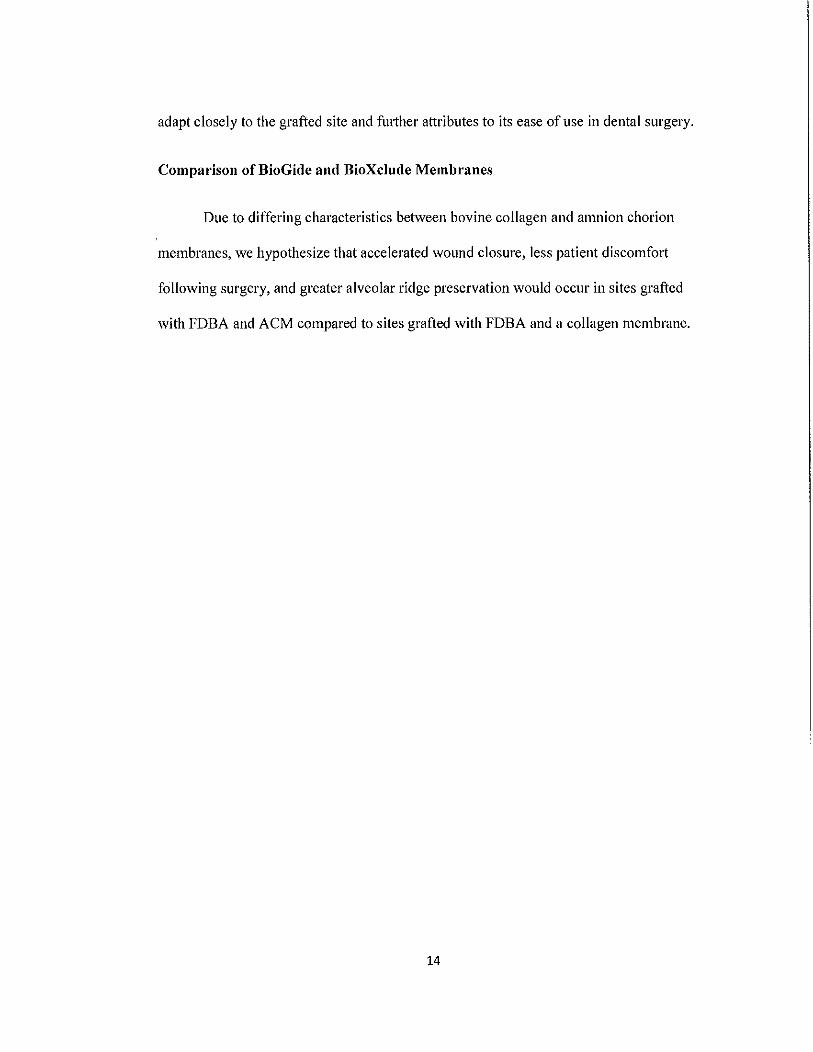
adapt closely to the grafted site and fmiher attributes to its ease of use in dental surgery.
Comparison of BioGicle ancl BioXclucle Membranes
Due to differing characteristics between bovine collagen and amnion chorion
membranes, we hypothesize that accelerated wound closure, less patient discomfort
following surgery, and greater alveolar ridge preservation would occur in sites grafted
with FDBA and ACM compared to sites grafted with FDBA and a collagen membrane.
14
CHAPTER III: MATERIALS AND METHODS
70 patients requiring dental extractions and subsequent site preservation for future
dental implants will be enrolled in the study. Please see Appendix A for a flow diagram
of the study. The findings oftheit· comprehensive periodontal evaluation such as probing
depths (PD), clinical attachment levels (CAL), and recession will have been recorded on
the Navy Periodontal Chart Form - NAVMED 6660/2 (Appendix B) by the subject's
provider.
A study investigator will initiate the consent process as described within this
protocol after being contacted by a provider whose patient is treatment planned for an
extraction with site preservation in preparation for an implant and has expressed possible
interest in study patticipation.
Standard Clinical Sequence (with introduction of study and consent):
I. Patient is referred for a periodontal evaluation.
2. A treatment plan including a dental extraction with site preservation and
eventual implant therapy is developed for the patient by their provider.
3. The provider asks their patient if he/she would like to learn about the
study being conducted at WRNMMC and will be provided a one page
brief about the study.
4. If the patient is not interested in the study the provider pursues the
established treatment plan.
5. If the patient expresses interest in participation, an investigator is asked to
discuss the study and consent process with the patient.
15
6. If the patient does not consent to be in the study, therapy will continue as
planned by the patient's surgeon.
7. If the patient consents, the following process ensues.
Following Consent:
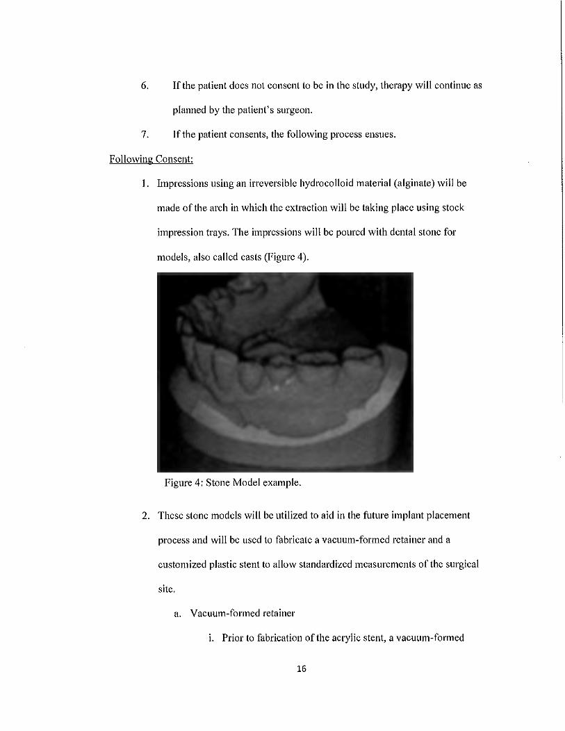
I. Impressions using an irreversible hydrocolloid material (alginate) will be
made of the arch in which the extraction will be taking place using stock
impression trays. The impressions will be poured with dental stone for
models, also called casts (Figure 4).
Figure 4: Stone Model example.
2. These stone models will be utilized to aid in the future implant placement
process and will be used to fabricate a vacuum-formed retainer and a
customized plastic stent to allow standardized measurements of the surgical
site.
a. Vacuum-formed retainer
i. Prior to fabrication of the acrylic stent, a vacuum-formed
16
retainer will be made from 0.020 inch clear plastic as per the
manufacturers guidelines
ii. The vacuum-formed retainer will be trimmed and delivered to
the patient following the extraction
iii. The vacuum-formed retainer will function to:
1. Provide esthetics for the edentulous site from the
extraction
2. Maintain space for future implant placement and crown
delivery.
3. Maintain space to allow for stent utilization
b. Laboratory acrylic stent:
i. An acrylic stent for making measurements at the extraction site
will be fabricated.
ii. Preserving the soft tissue margins on the cast, the tooth to be
extracted will be removed from the cast using a slow speed
laboratory handpiece (Figures 5 and 6).
17
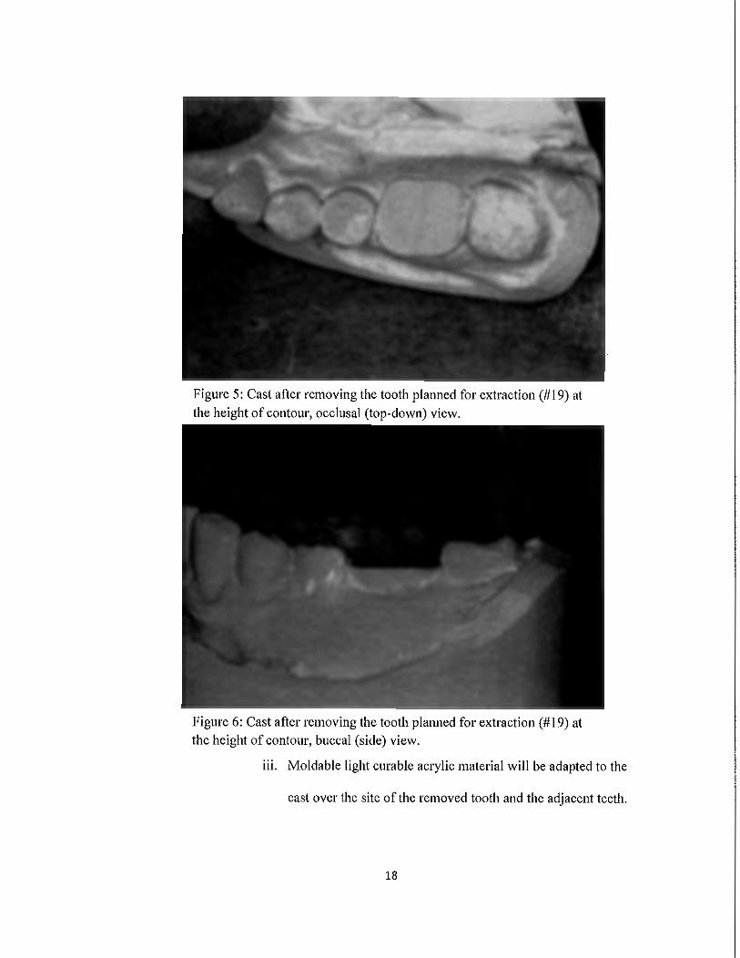
Figure 5: Cast after removing the tooth planned for extraction(# 19) at the height of contour, occlusal (top-down) view.
Figure 6: Cast after removing the tooth planned for extraction (#19) at the height of contour, buccal (side) view.
iii. Moldable light curable acrylic material will be adapted to the
cast over the site of the removed tooth and the adjacent teeth.
18
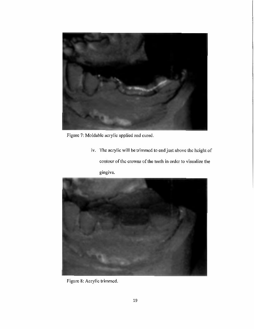
Figure 7: Moldable acrylic applied and cured.
iv. The acrylic will be trimmed to end just above the height of
contour of the crowns of the teeth in order to visualize the
gingiva.
Figure 8: Acrylic trimmed.
19
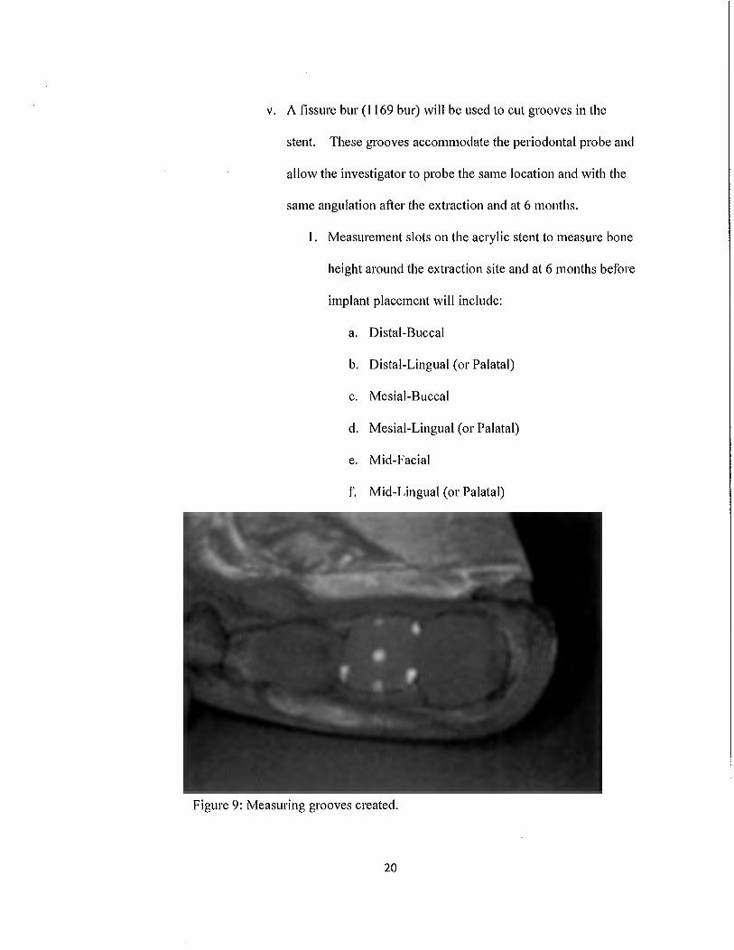
v. A fissure bur (1169 bur) will be used to cut grooves in the
stent. These grooves accommodate the periodontal probe and
allow the investigator to probe the same location and with the
same angulation after the extraction and at 6 months.
1. Measurement slots on the acrylic stent to measure bone
height around the extraction site and at 6 months before
implant placement will include:
a. Distal-Buccal
b. Distal-Lingual (or Palatal)
c. Mesial-Buccal
d. Mesial-Lingual (or Palatal)
e. Mid-Facial
f. Mid-Lingual (or Palatal)
Figure 9: Measuring grooves created.
20
v1. Following use the stent will be cleaned and disinfected with
Dispatch spray and stored in a ziplock plastic bag labeled with
the subject's study number. The bag will be locked in a
secured drawer maintained by the primary examiner; and then
retrieved for measurements at 6 months.
c. Also at baseline, buccal-lingual measurements of the cast will be made
at the direct mid-point of the future extraction site 4mm apical to the
cementoenamel junctions of the adjacent teeth.
Randomization Procedure:
I. Subjects will be stratified by location of the tooth in the mouth (i.e. maxillary
anterior, maxillary posterior, mandibular anterior or mandibular posterior) and
randomized to either Bio-Gide or BioXclude in a I: l ratio. Both treatments will
appear in blocks of2 or blocks of 4, in a random order. A computer program will
randomly sequence each subject's study enrollment numbers (1-70) as in the
example below.
a. For each of the 4 jaw locations, a random sequence table will be generated
by the research coordinator following !RB approval in order to maintain
blinding of investigators.
2. For each location, seventy envelopes marked I -70 will contain either a card
stating Bio-Gide or BioXclude. Thereafter, the random sequence table will be
placed in a sealed envelope that will not be opened until all data has been
collected. Sealed envelopes (1-70) will be stored by the principal investigator in a
locked drawer.
21
3. When each patticipant goes to surgery the investigator will provide the surgical
team the envelope corresponding to that subject's enrollment number and tooth
location. The surgical team will open the envelope and remove a card which will
state which membrane material to place following extraction.
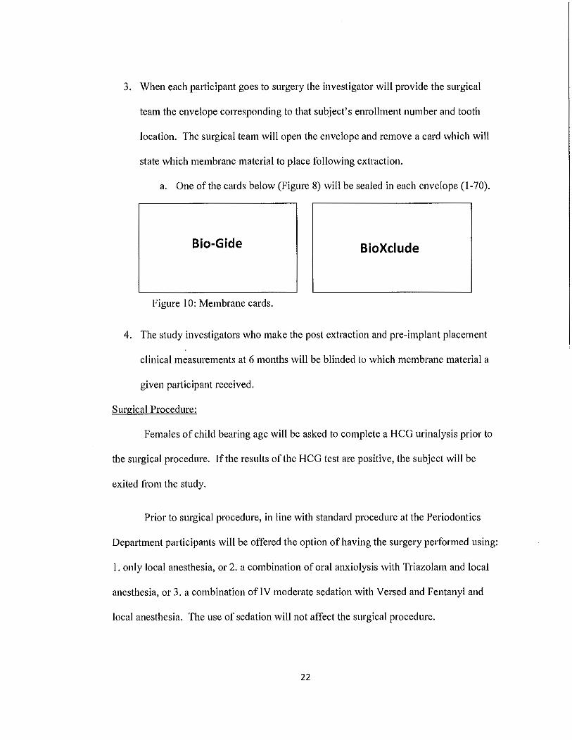
a. One of the cards below (Figure 8) will be sealed in each envelope (1-70).
Bio-Gide BioXclude
Figure 10: Membrane cards.
4. The study investigators who make the post extraction and pre-implant placement
clinical measurements at 6 months will be blinded to which membrane material a
given patticipant received.
Surgical Procedure:
Females of child bearing age will be asked to complete a HCG urinalysis prior to
the surgical procedure. If the results of the HCG test are positive, the subject will be
exited from the study.
Prior to surgical procedure, in line with standard procedure at the Periodontics
Department paiticipants will be offered the option of having the surgery performed using:
I. only local anesthesia, or 2. a combination of oral anxiolysis with Triazolam and local
anesthesia, or 3. a combination of IV moderate sedation with Versed and Fentanyl and
local anesthesia. The use of sedation will not affect the surgical procedure.
22
1. The surgical provider will be either a board cettified staff periodontist or a 2nd or
3rd year periodontal resident. All surgical providers will be briefed in the
protocol. All surgeries will follow the same steps listed below.
a. Surgical set-up is standardized for all surgeries done at the Naval
Postgraduate Dental School Periodontics Department.
b. Both the experimental (BioXclude) and control (Bio-Gide) materials will
be available to the surgeon. The membrane material used will be
determined when the sealed envelope is opened by a surgical team
member following the dental extraction.
c. Surgical Procedure Steps:
i. Administration of oral anxiolysis or IV moderate sedation if
patient desired and indicated
ii. Administration of topical and local anesthetic with any
combination of2% Lidocaine with l:lOOK epinephrine, 4%
Articane with 1: 1 OOK epinephrine, and 0.5% Marcaine with
1 :200K epinephrine
iii. Sulcular incision around the tooth to be extracted
iv. Periotomes utilized when required to minimize trauma to the
surrounding bone, teeth elevated, and atraumatically extracted with
forceps was performed. When required, teeth were sectioned
within the socket to preserve all socket walls.
v. The extraction socket was then curetted to remove all soft tissue
from the extraction site.
23
v1. The adjacent soft tissue will be undennined to allow for proper
membrane placement and no ve1tical incisions will be utilized.
vii. Characterization of the defect by a study investigator.
1. Presence or absence of a buccal dehiscence recorded as:
a. >50% of the apical-coronal dimension of the
extraction site
b. <50% of the apical-coronal dimension of the
extractiqn site
2. Measurement utilizing slots on the acryclic stent to the
alveolar crest of the extraction site will include:
a. Distal-Buccal
b. Distal-Lingual (or Palatal)
c. Mesial-Lingual (or Palatal)
d. Mesial-Buccal
e. Mid-Lingual (or Palatal)
f. Mid-Buccal
ii. FDBA will be hydrated with sterile saline as per the
manufacturer's instructions and placed into the extraction site
viii. Membrane material (determined from the sealed envelope)
ix. For BioXclude, the untrimmed membrane will be placed dry UP or
DOWN, allowed to hydrate, passively covering the graft material,
such that the membrane extends at least 3 mm beyond the alveolar
crest buccally and lingually, and may fold over itself and lay over
24
adjacent exposed root surfaces. For Bio-Gide, membrane will be
trimmed to size and shape of the extraction socket.
x. The membranes will be secured using a non-resorbable
monofilament suture (ie. Gore-tex)
xi. Following suturing, the remaining exposed membrane over the
extraction site will be measured horizontally in a mesial-distal and
a buccal-palatal/lingual direction over the midpoint of the alveolar
crest using a periodontal probe to function as a base-line
measurement for soft tissue healing until closure.
xii. Vacuum-formed retainers will be given to the patients
1. Patients will be instructed to wear the vacuum-formed
retainers during the day unless when eating and nightly
Post-operative Care (measurements and impressions shown):
I. All pa1ticipants receive the following post-operative regimen:
a. Pain medication consisting of any of the following alone or in
combination:
i. Ibuprofen 800 mg , Take I tab PO q6-8h for moderate pain or
ii. Hydrocodone/Acetaminophen 5/325 mg, Take 1-2 tab PO q6h
prn severe/breakthrough pain or
iii. Oxycodone/Acetaminophen 5/325mg, Take 1-2 tab PO q6h prn
severe/breakthrough pain
b. Pain medication for patients who cannot take NSAIDS will be
prescribed any of the following alone or in combinations:
25
i. Acetaminophen 325 mg, Take 1-2 tabs PO q4h for moderate
pain
ii. Oxycodone 5mg, Take 1 tab PO q4h prn severe/breakthrough
pain
c. Antibiotics consisting of either of the following:
i. Amoxicilin 500mg, Take 1 tab PO q8h for 10 days
11. Clindamycin 300 mg, Take 1 tab PO q8h for 10 days
d. Bio-Gide patients, 12% Chlorhexidine, 1 bottle, Rinse and spit bid
with l TBSP as directed on the bottle. BioXclude patients will be
instructed to avoid using Chlorhexidine-based oral rinses and not
given any following surgery. Patients should rinse with water,
gently rolling their head side to side and let the water passively
leave the mouth.
2. All patients are provided with the standard post-operative instructions (See
Appendix C for an example of the standard postoperative care instruction
form).
3. All patients will receive VAS forms. Every time patients feel the need to
take pain medication, they will record the type of medication (if taken),
time, and log their perceived pain level in the VAS form.
4. Patients are recalled at 1 week to assess post-operative healing and remove
plaque/deposits from the adjacent teeth. Degree of soft tissue closure over
membrane will be measured.
5. Patients recalled at 2 weeks post-operative to assess healing, remove
26
plaque from adjacent teeth, and remove sutures at the surgical site. Degree
of soft tissue closure over membrane will be measured.
6. Patients recalled approximately at weeks 4, 6, 8, 12, and 16 to assess
healing, remove plaque, and reinforce oral hygiene. Degree of soft tissue
closure over membrane will be measured. At 4 weeks closure has probably
occurred.
7. Impressions for dental stone models will be made in the same manner they
were made at baseline at 4 and 12 weeks post-extraction.
8. Patients recalled approximately at 6 months following the surgical
procedure to continue with implant therapy.
a. Prior to implant placement, impressions of the extraction site and study
models were created in the same manner as they were at baseline.
b. After full thickness flap reflection and prior to creation of an osteotomy
for implant placement, all measurements were completed using the same
acrylic stent by a blinded investigator. Measurements will also be
completed on the study model as previously described.
c. Implants will be placed base on their manufacturer's guidelines.
i. Standard post-operative care will be provided.
9. Patient will be exited from the study and followed by the periodontist and
primary care dentist for subsequent maintenance therapy and implant
restoration.
Analysis of Data:
1. Measurement of horizontal alveolar ridge dimensional changes using the stone
27
models made from impressions made at baseline, 4 weeks, 12 weeks and at time
of implant placement at six months.
2. Measurement of vertical changes in alveolar ridge height at 6 locations at the
extraction site using a customized acrylic stent immediately following the
extraction and at 6 months right before implant placement.
3. Measurement of soft tissue closure over the membranes. This will be measured in
a mesial distal and buccal-lingual (or palatal) direction over the midpoint of the
alveolar crest using a periodontal probe at I week, 2 weeks, and 4 weeks or until
the site has fully healed (when all soft tissue margins have approximated with
each other).
4. Clinical attachment levels and probing depths of the teeth adjacent to the
extraction site before the extraction and at 6 months before implant placement
will be compared.
5. Compare postoperative discomfort data collected in the VAS forms.
6. Statistical analysis will assess pre and post-test differences.
Human Subjects Justification
The use of human subjects is warranted because patients will be receiving
standard of care therapy for their dental extractions with site preservation. All patients
participating in the study will be receiving the same surgical care they would receive if
they were not included in the study except for the randomization of the membrane and
the measurements made with the stent and on jaw models in order to assess degree of
ridge preservation with the two membranes.
Possible risks of participation in this study are no different than the risks of
28
receiving surgical periodontal treatment outside the parameters of the study. These risks
include infection, abscess, bleeding, and post-treatment pain. Other adverse effects
include poor bone fill of the extraction site or failure of the bone allograft to integrate in
the grafted site. In the event that adverse effects occur in conjunction with the surgical
procedure, subjects will be able to call the Pl and receive follow-up treatment at the
NPDS Periodontics Clinic.
29
CHAPTERIV:RESULTS
Currently this research protocol has been prepared to be submitted to the IRB.
Following results of correspondence with Snoasis, the makers ofBioXclude, the protocol
will be submitted.
30

CHAPTER V: DISCUSSION
Resem·ch will commence following !RB approval.
31
CHAPTER VI: CONLUSIONS
Research will commence following !RB approval.
32
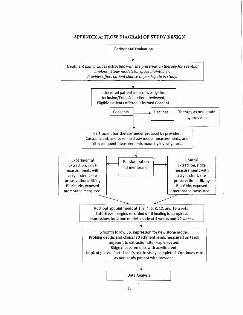
APPENDIX A: FLOW DIAGRAM OF STUDY DESIGN
Periodontal Evaluation J
Treatment plan includes extraction with site preservation therapy for eventual implant. Study models for space maintainer.
Provider offers patient chance to participate in study.
t Interested patient meets investigator. Inclusion/Exclusion criteria reviewed.
Eligible patients offered Informed Consent.
I Consents I Declines Therapy as non-study
by provider.
Participant has therapy under protocol by provider. Custom stent, and baseline study model measurements, and
all subsequent measurements made by investigators.
1 Experimental Randomization Control
Extraction, ridge of membrane ~ Extraction, ridge
measurements with measurements with acrylic stent, site acrylic stent, site
preservation utilizing preservation utilizing BioXclude, exposed Bio-Gide, exposed
membrane measured. membrane measured.
~ ~ Post opt appointments at 1, 2, 4, 6, 8, 12, and 16 weeks. Soft tissue margins recorded until healing is complete.
Impressions for stone models made at 4 weeks and 12 weeks.
:I 6 month follow up, impression for new stone model.
Probing depths and clinical attachment levels measured on teeth adjacent to extraction site. Flap elevated. Ridge measurements with acrylic stent.
Implant placed. Participant's role in study completed. Continues care as non-study patient with provider.
Data Analysis
33
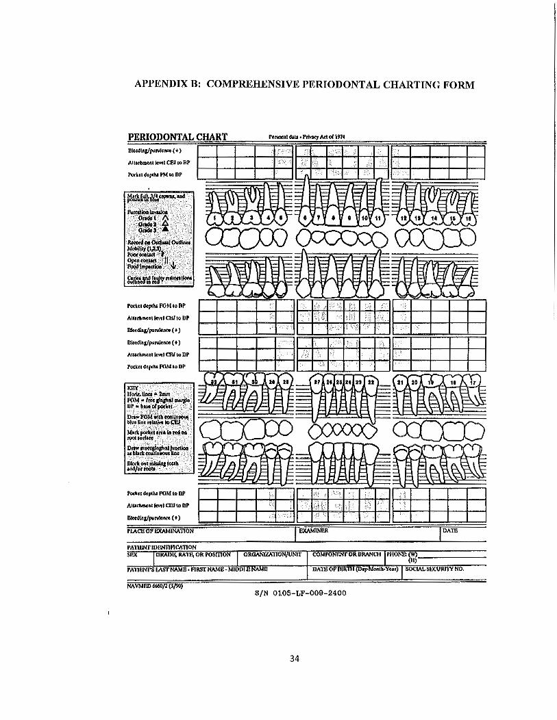
APPENDIX B: COMPREHENSIVE PERIODONTAL CHARTING FORM
PERIODONTAL CHART '""""' .. ~ · '""'"" o11m
:::::::::·· I I I I- I '~~m~I: I ~' I~' I ~U'!kW.'.....,7'0
~;g~f~ Or*S>.&._,,
~~·9'J~'~ttir.u U~.~ity{l,2~f.·.:: _.,. .... ,~,' ~~~ilL,i, ~111'1~-
l'«ttl ckpthi FOM lo PP
AttJduM111 k-o'tl a!J' lo lJP
IU«dinf/pqrvktK>C ( +)
mC>C<1l11ifpi:iNknoe ( +)
A1Ud!me111 k~I CliJ to DP
~tel lkplhl POM lo DI
Atta!iuM.at~<c.ICl!Jto DP
PAlUWf IDENl1PICh110N
I llXAM!h'l!R IDATn
snx ORAD.E{ RA.11!, Ok POS ON ORCIM'lV.t'fO /Ut.lr <X>Mf'ONP.Hi' OR DRANtll l'HON'B: (W) ______ _ OQ
i'A11l!Nl'S LASJ'NAMB •FIRST NAMH- MIOl'>l.ll NAMB DA'r8 OPDIP.TII (DqMonth·Yt-u SOCIALS&-"URllY NO.
Nt\VMPDWl)/2 (J}JO} S/N 0105-LF-009-2400
34
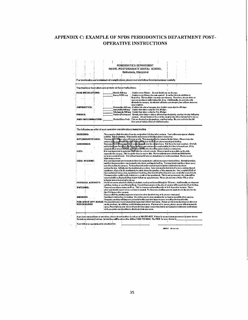
APPENDIX C: EXAMPLE OF NPDS PERIODONTICS DEPARTMENT POSTOPERATIVE INSTRUCTIONS
PERIODOUTICS DEPARTMEllT UAVAL POSTGRADUATE OEllTAL SCHOOL
Bethesda1 Maryland
! Yoo ma~ have bun Qlven one Of mote of tMse m~tlons:
'PAIN M.EDICAT~ON.S: ;_______;.M«silt\OOaq: U*kl.ti•~q6~1'. Oo~®"~t'Po~donq~
_, ---; Nw<o st;l~S"<i= : ~~(7{~=~="'~~ !o<,,~,~t~~,l:~::.~'!'1.: :nt w
: c-~ut~•KM..utwli!~t•~ tk.i$dm!. A~~. ®Mtt•h 'lfflh ~°"~ i>tHnqt~ u,i, m~vi'l 1»kt ~w1ll«r*1, tm vii M-t dtc1n;<{t fO"'<f(~91t.
ANTIBIOTICS: -' ___ :0o1M"<llia.-t l00ao.q: .tt->Muac,;,,<;1-~ Unlt..bl«~Nq.,n,/«$()<kf,1. -· ___ ·/\.lt.oJk&500111.-q;; lf~/1J-W6'NS'lt/qf«lfckJt/qF,
C&d_,a,:.$00.nQ lt#-t/t>UIM.t:>t/q{t;t l't" Jl)-th;-F. RINSES: :::::J P~ildu fPttlc.qnrd' l/>c.tik, tifuflfk~~-tf>f~.f.fN«fM-NtkWlk; stN~.tkdql~~
' 1
, sw.:xr,. Do~bi'•:lo<>fflo~~""~sw4c..!W.tHk~~i>rn..:t~dtod<>..o. ANTl-INflAMMATION: ___ : Mi:drol Oo:H: PKk: . TUt ttd'rtctl:d °"' lh-tptth<tt,~twti.lK! tMw. Bt .:wt u<I hh tlt fvl
· ' f11,;t row ol hbkt~ (f11,;t m hW~~l tod•l·
The I~ a1e a fist of post·opH ~tlve consldtr atlons durinq healinq:
'BLEEDING: T~rt ,.~ta ~t b«t<litqfre-a ~ w(tl(lllor H! d~?Hft~t«t<l«f- Yow .ain 111-l'I ~put A!qWf ltddlsl Tkhb C~ ti fOt i.e-lk~ It.lo b.<fOH b b«:t<fuqpln:~ COl\ll(t ·~
'SlJTURESfSTITCHES: Yo,l'llf db ~ f1.ttD»iou/}obltnri,ottdh~ffA~:t. Plo:;1lnvtt-" YOA of '~~10 aw IApW ln~q.
DRESSINGS: tlt tfqk~ ~n. It l:tltttfot fOU coalo1t. If lthll: totlhcolllorlM,ltbf'"tolnult~ lftlt d<t$~ rti>W;td i>1u1t c<>i.l'lct EM>.
'DIET: :1tb•WJ1~lutto~lb ttfor~kuhv«k. Clttiui.«•upw~toaUt~l<Jt Ol'>PO"'.ftt ~-q~•f. Tl#b i.e-i ~ lb!.tto ~;,t ~ ~t. Pkut rlilitW. 'i'<>W c'l!«k~d ftfld ktlh
· u ~ prt·wQk~ k•tb. Y <>• 11·lll l<>t lnhrtl If fO'I ~ft dd.ydnttd <>r udu»'>flfMd. Pln:HE do ~ dlk.li •fW:i ~ w~.
i OR Al H'tQIEHE: : II: bttt'f i=J;ortut ~to bl...-..t. or &ntltAtqkll ffltWil~«i oprt1,lutnctlo.u. floral! bl.mJ,,,.q j ttd fl~,b.q pr.xtdwtt ca lll~tt tlt tln'" ~lm9W litlil<I- Y<>'l r>'l'! l>roia ud &:$ tl<>:t l•tH i o"Ot ..tltcltd O, tlt WQttf. T<> kup 1>1.cwlli udu <«lloh p:uc1it>Uo~ l'!.Ototl.1lv..cl1,u bt«i 1 w1ittui tor to'I· b.IU~. irn tl.t ""°"°'~a '1'$~ rb:t. htu '°'""''bit bru1Ktt<:lto ,,a~ <<>Ito.a.·~ ' oppic~or, ~td l.t tl.t n<>"'1r.a, t<> ~vK> ~ tlt (J<'::t. it<\ of Wt 'tlqttf ffl~ lkt l opld (UN} ol ! tl.t l'!.01U.1na t1'kt ~ <lli't.•<>rloh<i ud bt<ffilot, ..tw bl<1Ab.Qlflo:U..q 'l'O'<• ~--qk~ llot•<:I tttd.. ' y <>• .. ,, &<Otkt a1i<:I tO<>lll. ~U!thl.q u ~ IH<ill ol tlu1 .. ::AA•1na. T~ la0ot p{/lllUU.\; tl<I ~w. ril bit rta.<>1«l 11itli «~polklk.<i ~ ~ folow·vp ~P~™•· f'Hut do lr<>I 11tt ~ V~u-Plii or <>Uu 'ltrlq~Of llLkH lv..tr«t«l lo do«>.
i PHY81CllL ACTIVITY: 'A.1<»d ~u ... .,o•• p\.yW:ll «thitt ftohdNt nu.!l;q ud Ml'1' llfu.Ql for ?at.<>wo. A~,» ~Ott ~th-q, r~..kq. or 'Pttikq ff«!il.q\. F orcdd ""°"~~ it t1t ~itt of 'W'l{l9 d ~~thdt \l/¢(t k:i&q..
!8VELLING: iYO'llllllfUl><lk«t~, ... tl&q. nbl:c~u.dorni:i!,pttb~2·3dl-f$:HtH-qtfl. TWt· ~tu'*" MW! Upt(t to tt~ l lttWA to &Ofl!lll To dtCIOttHctlW.q "°'cu ~l)t\lkt to 1.lol: ril:t for till fir,t 3·4 "°"'~!ti , ... Qt!,, f'Hy..c c'llJ if w $Wt~ ~'11$ l<> il.(1tut Uttr tlt tllrd dw, Of if ''°"'~It (<>l;(Ullll.d.
i &MOICING i :Sa.<>kl!.qb ddttttloff to l-t~ Vt ~drlttt<>t to~op1AOilt.<:i lor ttiof;q u ~t :Nm n1quy. , :St~ molir.Q ril jm,p:on pot™1ll ltt&<1 w:I '1w bP!'ott "°'' O'l<oll p{1lod<>i.I~ M~
'f'OR SINUS LlfT SURGE You:.~molout1tctht4fM.lldtc~Wtttnl>kow:l~1f. Pkutwi:ttl«ta«lkWoutt&«t«l PROCEDURES OJJtl.t ~cklqt. ki ~~ u®~toil lt<>ff. If N1 ~t¢<:1 to n«tt, pltut ~"'tt~ viUI toW ~
i opu .. f'ky~ irJOf,. t<>W do<. tor if "°"' dndop .:.il.u c<>«1t,tloA t>M. I< »t R.hk!.i.."f.d villi~ atdkW.0.110 orll N't &<Otkt~"I bk«l>lq or diHttfqt f101ll you Mott.
It f<>'I htt uy prot./t.,.~ Of <ptstiou, pl.tut do ir.<>t kl'il.~t~ to c~ M ~ 301·235·0011. If tlut b u tl!l.-l.IQtl«f f().Q "'"l'I P1Qt 'fC'it do<tor tlc~ lll ~<>aNt,d ~m. brtr«Uo"'o vii bt ql1t- :Ntu di~ 1·&00·153'·&Ua:. ntPIHI for '~r doctOf ~--------
Yow f<!l<>v'<l)~l:ttl.tddtdfor:
35
REFERENCES
I. Schropp L, Wenzel A, Kostopoulos L, Karring T. Bone healing and soft tissue
contour changes following single tooth extraction: A clinical and radiographic 12-
month prospective study. International Journal of Periodontics and Restorative
Dentistry 2003;23 (4):313-323.
2. Amler MH, Johnson PL, Salman I. Histological and histochemical investigation
of human alveolar socket healing in undisturbed extraction wounds. Journal of the
American Dental Association 1960; 61 :32-44.
3. Pietrokovski J, Massler M. Alveolar ridge resorption following tooth extraction.
Journal of Prosthetic Dentistry 1967; 17(1 ):21-27.
4. The American Academy of Periodontology. Glossary of Periodontal Terms, 4th
ed. Chicago: The American Academy of Periodontology; 200 I.
5. Mellonig JT. Donor selection, testing, and inactivation of the HIV virus in freeze
dried bone allografts. Pract Periodontics Aesthet Dent. 1995;7:13-22.
6. CDC. Infection control, frequently asked questions - Bone allografts. Centers for
Disease Control and Prevention. 25 October 2013. Web. 27 April 2014.
7. Holtzclaw D, Toscano, N, Eisenlohr L, Callan D. The Safety of Bone Allografts
Used in Dentistry: A Review. JADA. 2008; 139(9):1192-1199.
8. Yukna A, Vastardis S. Comparative Evaluation ofDecalcified and Non
Decalcified Freeze-Dried Bone Allografts in Rhesus Monkeys. I. Histologic
Findings. Journal of Periodontology 2005; 76(1): 57-65.
36
9. Beck TM, Mealey BL. Histologic analysis of healing after tooth extraction with
ridge preservation using mineralized human bone allograft. Journal of
Periodonto/ogy 20 IO; 81 (12): 1765-1772.
I 0. Hammerle CH, Jung RE. Bone augmentation by means of barrier membranes.
Periodontology 2000 2003; 33(1 ): 36-53.
11. Engler-Hamm D, Cheung WS, Yen A, Stark PC, Griffin T. Ridge preservation
using a composite bone graft and a bioabsorbable membrane with and without
primary wound closure-A comparative clinical trial. Journal of Periodontology
2011; 82(3): 377-387.
12. Dahlin C, Linde A, Gottlow J, Nyman S. Healing of bone defects by guided tissue
regeneration. Plastic and Reconstructive Surgery 1988; 81(5):672-676.
13. Patino MG, Neiders ME, Andreana S, Noble B, Cohen RE. Collagen as an
Implantable Material in Medicine and Dentistry. Journal of Oral Implantology
2002; 28, (5): 220-225.
14. Bunyaratavej P, Wang HL. Collagen membranes: A review. Journal of
Periodontology 2001;72(2): 215-229.
15. Mardas N, Chadha V, Donos N. Alveolar ridge preservation with guided bone
regeneration and a synthetic bone substitute or a bovine-derived xenograft: A
randomized, controlled clinical trial. Clinical Oral Implants Research 2010;
21(7): 688-698.
16. Sela, M.N., Babitski, E., Steinberg, D., Kohavi, D. & Rosen, G. Degradation of
collagen guided tissue regeneration membranes by proteolytic enzymes of
37
Porphyromonas gingivalis and its inhibition by antibacterial agents. Clinical Oral
Implants Research. 2009. 20: 496-502.
17. Sela, M.N.C., Steinberg, D.C., Klinger, A., Krausz, A.A.S. & Kohavi, D.C.
Adherence of periodontopathic bacteria to bioabsorbable and non-absorbable
barrier membranes in vitro. Clinical Oral Implants Research. 1999. 10: 445-452.
18. Klinger A, Asad R, Shapira L, Zubery Y. In vivo degradation of collagen barrier
membranes exposed to the oral cavity. Clinical oral implants research 2010;
22(8): 873.
19. Schwarz F, Rothamel D, Herten M, Sager M, Becker J. Angiogenesis pattern of
native and cross-linked collagen membranes: an immunohistochemical study in
the rat. Clin Oral Implants Res. 2006 Aug;l7(4):403-9.
20. Zitzmann NUl, NaefR, Scharer P. Resorbable versus nonresorbable membranes
in combination with Bio-Oss for guided bone regeneration. Int J Oral Maxillofac
Implants. 1997 Nov-Dec;l2(6):844-52.
21. Schwarz F, Rothamel D, Herten M, Wlistefeld M, Sager M, Ferrari D, Becker J.
Immunohistochemical characterization of guided bone regeneration at a
dehiscence-type defect using different barrier membranes: an experimental study
in dogs. Clin Oral Implants Res. 2008 Apr;l9(4):402-15.
22. Iasella JM, Greenwell H, Miller RL, Hill M, Drisko C, Bohrn AA, Scheetz JP.
Ridge preservation with freeze-dried bone allograft and a collagen membrane
compared to extraction alone for implant site development: a clinical and
histologic study in humans. Journal of Periodonto/ogy 2003; 74(7): 990-999.
38
23. Hodde J. Naturally occurring scaffolds for soft tissue repair and regeneration. Tiss
Eng 2002; 8(2): 295-308.
24. Niknejad H, Peirovi H, Jmjani M, et al. Properties of the amniotic membrane for
potential use in tissue engineering. Euro Cells Mat 2008; 15: 88-89.
25. Davis J. Skin transplantation with a review of 550 cases at the Johns Hopkins
Hospital. Johns Hopkins Medical Journal 191 O; 15: 307-396.
26. De Rotth A. Plastic repair of conjunctiva! defects with fetal membrane. Arch
Opthalmol 1940; 23:522-525.
27. Tancer ML, Katz M, Veridiano NP. Vaginal epithelialization with human amnion.
Obstet Gynecol 1979; 54(3): 345-349.
28. Guler R, Ercan MT, Ulutuncel M, et al. Measurement of blood flow by the 133Xe
clearance technique to grafts of amnion used in vestibuloplasty. Brit J Oral Max
Surg 1997; 35: 280-283.Holtzclaw DJ, Toscano NJ. Amnion-Chorion Allograft
Barrier Used for Guided Tissue Regeneration Treatment of Periodontal Intrabony
Defects: A Retrospective Observational Report. Clinical Advances in
Periodontics 2012; 0: 1-7.
29. Shaila V, Kothiwale L, Anuroopa A, et al. A clinical and radiological evaluation
ofDFDBA with amniotic membrane versus bovine derived xenograft with
amniotic membrane in human periodontal grade II furcation defects. Cell Tissue
Bank 2009; 10:317-326.
30. Karaman M, Tuncel A, Sheidaei S, ~enol M, et al. Amniotic membrane covering
for facial nerve repair. Neural Regen Res 2013; 8(11): 975-982.
39
31. Bauer F, Hingsammer L, Wolff K, et al. Temporomandibular joint at1hroplasty
with human amniotic membrane: a case report. Eplasty 2013; 13: el7. Epub Mar
18.
32. Kubo M, Sonoda Y, Muramatsu R, Usui M. Immunogenicity of Human Amniotic
Membrane in Experimental Xenotransplantion. IOVS 2001; 42(7): 1539-1546.
33. Rinastit M, Harijadi, Santoso ALS, Sosroseno W. Histological evaluation of
rabbit gingival wound healing transplanted with human amniotic membrane. In J
Oral Maxillofac Surg 2006; 35: 247-251.
34. Koizumi N, Inatomi T, Sotozono C, Fullwood NJ, et al. Growth factor mRNA
and protein in preserved human amniotic membrane. Curr Eye Res 2000; 20(3):
173-177.
35. Koh JW, Shin YJ, Oh JY et al. The expression ofTIMPs in cryo-preserved and
freeze-dried amniotic membrane. Curr Eye Res 2007; 32(7-8): 611-616.
36. Velez I, Parker W, Siege M, et al. Cryopreserved Amniotic Membrane for
Modulation of Periodontal Soft Tissue Healing: A Pilot Study. J Peria 201 O; 81:
1797-1804.
37. Chen A, Tofe A. A literature review of the safety and biocompatibility of amnion
tissue. J Imp Adv Clin Dent 2010; 2(3):67-75.
38. Koob, T; Lim, J; Zabek, N; and Massee, M "Cytokines in single layer amnion
allografts compared to multi-layer amnion/chorion allografts for wound healing."
J Biomed Mater Res B 2014: DOI: 10.1002/jbm.b.33265.
40
39. Pakkala T, Vittanen I, Oksanen J et al. Function of laminins and laminin-5
binding integrins in gingival epithelial cell adhesion. J Perio 2002; 73(7): 709-
719.
40. Kinumatsu T, Hashimoto S, Muramatsu T, et al. Involvement of laminin-5 and
integrins in adhesion and migration of junctional epithelium cells. J Perio Res
2009; 44: 13-20.
41. Xenoudi P, Lucas M. Comparison of porcine and amnion chorion resorbable
collagen membranes using immunohistochemistry. Journal of Dental Research
International Association for Dental Research 2011; March; Abstract # 146797.
42. Yazdi K, Moloudi F, Heslunat B. Comparison of Amnion Membrane with Bio
Gide in Conjunction with Bio-Oss in Treatment of Intrabony Periodontal Defects.
Sadra Med Sci J 2013; 1(4): 1-18.
43. Holtzclaw D, Hinze F, Toscano N. Gingival flap attachment healing with
amnion-chorion allograft membrane: A controlled, split mouth case report
replication of the classic 1968 Hiatt study. J Imp Adv Clin Dent 2012; 4(5): 19-25.
44. Koob T, Zabek N, Massee Net al. Prope1ties of Dehydrated Human
Amnion/Chorion Composite Grafts: Implications for Wound Repair and Soft
Tissue Regeneration. J BioMed Mat Res Patt B 2014:008:000-000.
45. Koob T, PhD; Li W, Gurtner Get al. Angiogenic Properties of Dehydrated
Human Amnion/Chorion Allografts: Therapeutic Potential for Soft Tissue Repair
and Regeneration. Vas Cell 2014; 4(10): doi: IO. l l 86/2045-824X-6-l 0.
41
46. Koob T, Renne1t R, Zabek Net al. Biological properties of dehydrated human
amnion/chorion composite graft: implications for chronic wound healing. Int
Wound J 2013; 10(5):493-500.
47. Maan Z, Rennert R, Koob T, et al. Cell recruitment by amnion chorion grafts
promotes neovascularization. J Surgical Res 2015; 193(2): 953-962
48. Zelen CM, Gould L, Serena TE, et al. A prospective, randomized, controlled,
multi-center comparative effectiveness study of healing using dehydrated human
amnion/chorion membrane allograft, bioengineered skin substitute or standard of
care for treatment of chronic lower extremity diabetic ulcers. Int Wound J 2014
Nov 26. doi: 10.l l l l/iwj.12395.
49. Razdan S, Razdan S, Johnson J. Use of dehydrated human amniotic membrane
during robot assisted laparoscopic prostatectomy in early return of erectile
function: A retrospective study. Joumal ofEndourology 2014; 20(Sup 1): Poster#
MP24-03.
50. Amnion---<:horion allograft barrier used for guided tissue regeneration treatment of
periodontal intrabony defects: A retrospective observational report of 64 patients.
Holtzclaw D, Toscano N. Clin Adv Perio 2013; 3(3):131-137.
51. Holtzclaw D, Toscano N. Amnion chorion allograft barrier: indications and
techniques update. J Imp Adv Clin Dent 2012; 4(2):25-38.
42