Embed Size (px)
Citation preview
A 6-week teaching module for undergraduate biology majors on light microscopy using Chlamydomonas reinhardtii
N. T. Ahmed
Biology Department, St. John Fisher College, Rochester, NY, 14618
Chlamydomonas reinhardtii is a biflagellate, unicellular eukaryote that is a good model organism in an undergraduate setting because it is inexpensive to maintain, easy for students to manipulate, and is amenable to a variety to cytological and biochemical techniques, including immunofluorescence. This paper outlines a 6-week teaching module in which students are exposed to the theory and practice of light microscopy using C. reinhardtii. Each week includes a lecture component, breakout sessions, and regrouping for exhibition and reflection. At the end of six weeks, students are expected to know the theory of light microscopy, including contrast and fluorescence microscopy, and how images are captured and processed. Students must also demonstrate proficiency in setting up Köhler illumination, Nomarski optics and fluorescence microscopy. Lastly, students are graded on their portfolio, which consists of an original figure of the images they generate from their immunofluorescence experiments. This paper includes examples of student work, an example of a guided-reading worksheet, an immunofluorescence protocol and rubrics for assessed learning outcomes.
Keywords: light microscopy; Köhler illumination; Nomarski optics; differential interference contrast; fluorescence microscopy; teaching module; assessment rubrics; immunofluorescence; Chlamydomonas reinhardtii
1. Introduction
There has been a paradigm shift in the last decade in regards to the focus of undergraduate biology education from being primarily content-driven to a combination of concepts and competencies. As articulated by initiatives like Vision and Change in Undergraduate Biology Education, sponsored by the American Association for the Advancement of Science (AAAS), and the National Science Foundation (NSF), there is a greater emphasis on student-centered learning environments [1]. In particular, there is a call to move away from cookie-cutter lab experiences to inquiry-based labs that more closely resemble the way actual scientists do research. The focus should be on teaching a few techniques and allowing students the time to master these skills at their own pace. This paper outlines a 6-week inquiry-based laboratory experience that meets this goal in which students characterize novel C. reinhardtii motility mutants using DIC and fluorescence microscopy. Students are introduced to the theory of light microscopy as they learn and practice the techniques. Students generate original data and spend their time trouble-shooting the techniques, as opposed to recapitulating results.
1.1 Chlamydomonas reinhardtii
Chlamydomonas reinhardtii is unicellular green alga that has several characteristics that make it an excellent organism to use in an undergraduate laboratory setting. This organism has a 5-hour generation time and can be easily maintained as it grows on solid media and in liquid culture. C. reinhardtii can grow in the presence of light on minimal media and also grow in darkness in presence of acetate-containing media. During the vegetative portion of the organisms’ life cycle, they are haploid and easily amenable to genetic manipulation. Growth in nitrogen-free media induces these organisms to become gametes. Gametes of opposite mating types fuse to produce a diploid zygospore, allowing for tetrad analysis. The genome has been completely sequenced [2] and a stock center maintains strains and libraries [3]. C. reinhardtii has been used as a model organism to answer some basic questions in cell biology. Since C. reinhardtii is capable of photosynthesis, the chloroplast of this organism has been extensively studied. This organism has two anterior flagella that are capable of switching between an asymmetric (ciliary) waveform and a symmetric (flagellar) waveform, a capacity that has been lost in most other ciliated organisms [4]. These flagella are assembled using the same process needed for assembly of all cilia and flagella in humans and other ciliated organisms [5]. These flagella allow the organism to display both phototaxic and photoshock responses. Light sensitivity is due to an eyespot that contains opsin proteins [6], making this a good organism for studying phototaxis as well as circadian rhythm regulation. More recently, research efforts have focused on the potential use of this organism as a source for biofuel production. In an undergraduate biology laboratory, this organism can be used as the basis for experiments ranging from studying metabolic pathways to the evolutionary divergence of plant and animals. The experimental question driving the use of C. reinhardtii in this paper is the study of flagellar assembly via characterizing the phenotype of two novel motility mutants, F3 and F5, using immunofluorescence microscopy.
Microscopy: advances in scientific research and education (A. Méndez-Vilas, Ed.)__________________________________________________________________
1123© FORMATEX 2014
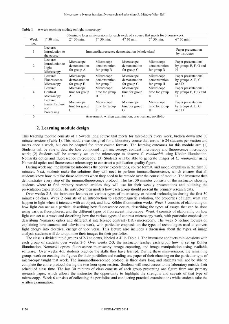
Table 1 6-week teaching module on light microscopy
30-minute long mini-sessions for each week of a course that meets for 3 hours/week Week
no. 1st 30 min. 2nd 30 min. 3rd 30 min. 4th 30 min. 5th 30 min. 6th 30 min.
1 Lecture: Introduction to the course
Immunofluorescence demonstration (whole class) Paper presentation by instructor
2
Lecture: Introduction to Light Microscopy
Microscope demonstration for group A
Microscope demonstration for group B
Microscope demonstration for group C
Microscope demonstration for group D
Paper presentations by groups E, F, G and H
3 Lecture: Fluorescence Microscopy
Microscope demonstration for group E
Microscope demonstration for group F
Microscope demonstration for group G
Microscope demonstration for group H
Paper presentations by groups A, B, C and D
4 Lecture: Contrast Microscopy
Microscope time for group A
Microscope time for group B
Microscope time for group C
Microscope time for group D
Paper presentations by groups E, F, G and H
5
Lecture: Image Capture and Processing
Microscope time for group E
Microscope time for group F
Microscope time for group G
Microscope time for group H
Paper presentations by groups A, B, C and D
6 Assessment: written examination, practical and portfolio
2. Learning module design
This teaching module consists of a 6-week long course that meets for three-hours every week, broken down into 30 minute sessions (Table 1). This module was designed for a laboratory course that enrols 16-24 students per section and meets once a week, but can be adapted for other course formats. The learning outcomes for this module are: (1) Students will be able to describe how compound light microscopy, contrast microscopy and fluorescence microscopy work; (2) Students will be correctly set up the microscope to observe C. reinhardtii using Köhler illumination, Nomarski optics and fluorescence microscopy; (3) Students will be able to generate images of C. reinhardtii using Nomarski optics and fluorescence microscopy to construct a publication-quality figure. During week one, the instructor introduces the course expectations, course format, and model organism in the first 30 minutes. Next, students make the solutions they will need to perform immunofluorescence, which ensures that all students know how to make these solutions when they need to be remade over the course of module. The instructor then demonstrates every step of the immunofluorescence protocol. The last 30 minutes consists of the instructor showing students where to find primary research articles they will use for their weekly presentations and outlining the presentation expectations. The instructor then models how each group should present the primary research data. Over weeks 2-5, the instructor lectures on various types of microscopy or related technologies during the first 30 minutes of class. Week 2 consists of an introduction to electromagnetic radiation, the properties of light, what can happen to light when it interacts with an object, and how Köhler illumination works. Week 3 consists of elaborating on how light can act as a particle, describing how fluorescence occurs, describing the types of assays that can be done using various fluorophores, and the different types of fluorescent microscopy. Week 4 consists of elaborating on how light can act as a wave and describing how the various types of contrast microscopy work, with particular emphasis on describing Nomarski optics and differential interference contrast (DIC) microscopy. The week 5 lecture focuses on explaining how cameras and televisions work, with particular emphasis on the types of technologies used to convert light energy into electrical energy or vice versa. This lecture also includes a discussion about the types of image analysis students will do to optimize their images for their portfolios. The class is divided into 8 groups of 2-3 students, labeled A-H in Table 1. The instructor conducts mini-sessions with each group of students over weeks 2-5. Over weeks 2-3, the instructor teaches each group how to set up Köhler illumination, Nomarski optics, fluorescence microscopy, image capturing, and image manipulation using available software. Over weeks 4-5, students practice the skills they have learned. During these mini-sessions, the remaining groups work on creating the figures for their portfolios and reading one paper of their choosing on the particular type of microscopy taught that week. The immunofluorescence protocol is three days long and students will not be able to complete the entire protocol during the two-hour open session. Students will need access to the laboratory outside their scheduled class time. The last 30 minutes of class consists of each group presenting one figure from one primary research paper, which allows the instructor the opportunity to highlight the strengths and caveats of that type of microscopy. Week 6 consists of collecting the portfolios and conducting practical examinations while students take the written examination.
Microscopy: advances in scientific research and education (A. Méndez-Vilas, Ed.)__________________________________________________________________
© FORMATEX 20141124
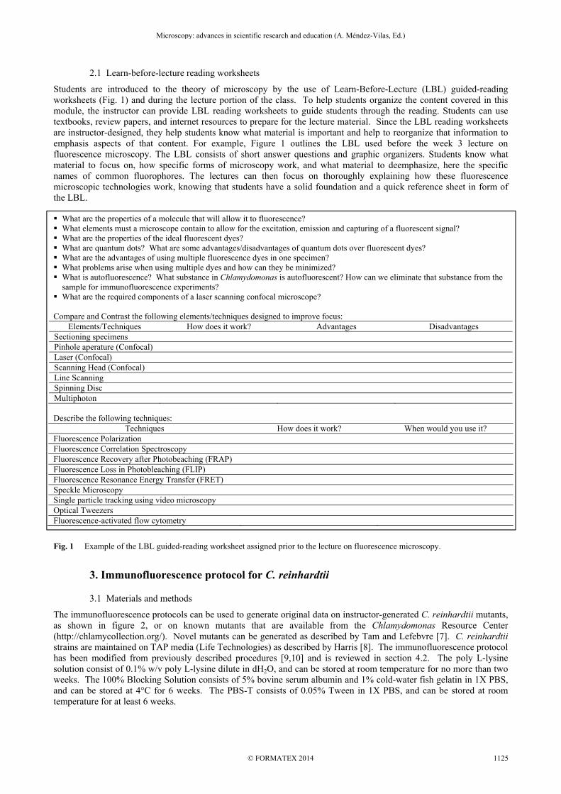
2.1 Learn-before-lecture reading worksheets
Students are introduced to the theory of microscopy by the use of Learn-Before-Lecture (LBL) guided-reading worksheets (Fig. 1) and during the lecture portion of the class. To help students organize the content covered in this module, the instructor can provide LBL reading worksheets to guide students through the reading. Students can use textbooks, review papers, and internet resources to prepare for the lecture material. Since the LBL reading worksheets are instructor-designed, they help students know what material is important and help to reorganize that information to emphasis aspects of that content. For example, Figure 1 outlines the LBL used before the week 3 lecture on fluorescence microscopy. The LBL consists of short answer questions and graphic organizers. Students know what material to focus on, how specific forms of microscopy work, and what material to deemphasize, here the specific names of common fluorophores. The lectures can then focus on thoroughly explaining how these fluorescence microscopic technologies work, knowing that students have a solid foundation and a quick reference sheet in form of the LBL. What are the properties of a molecule that will allow it to fluorescence? What elements must a microscope contain to allow for the excitation, emission and capturing of a fluorescent signal? What are the properties of the ideal fluorescent dyes? What are quantum dots? What are some advantages/disadvantages of quantum dots over fluorescent dyes? What are the advantages of using multiple fluorescence dyes in one specimen? What problems arise when using multiple dyes and how can they be minimized? What is autofluorescence? What substance in Chlamydomonas is autofluorescent? How can we eliminate that substance from the
sample for immunofluorescence experiments? What are the required components of a laser scanning confocal microscope? Compare and Contrast the following elements/techniques designed to improve focus:
Elements/Techniques How does it work? Advantages Disadvantages Sectioning specimens Pinhole aperature (Confocal) Laser (Confocal) Scanning Head (Confocal) Line Scanning Spinning Disc Multiphoton Describe the following techniques:
Techniques How does it work? When would you use it? Fluorescence Polarization Fluorescence Correlation Spectroscopy Fluorescence Recovery after Photobeaching (FRAP) Fluorescence Loss in Photobleaching (FLIP) Fluorescence Resonance Energy Transfer (FRET) Speckle Microscopy Single particle tracking using video microscopy Optical Tweezers Fluorescence-activated flow cytometry
Fig. 1 Example of the LBL guided-reading worksheet assigned prior to the lecture on fluorescence microscopy.
3. Immunofluorescence protocol for C. reinhardtii
3.1 Materials and methods
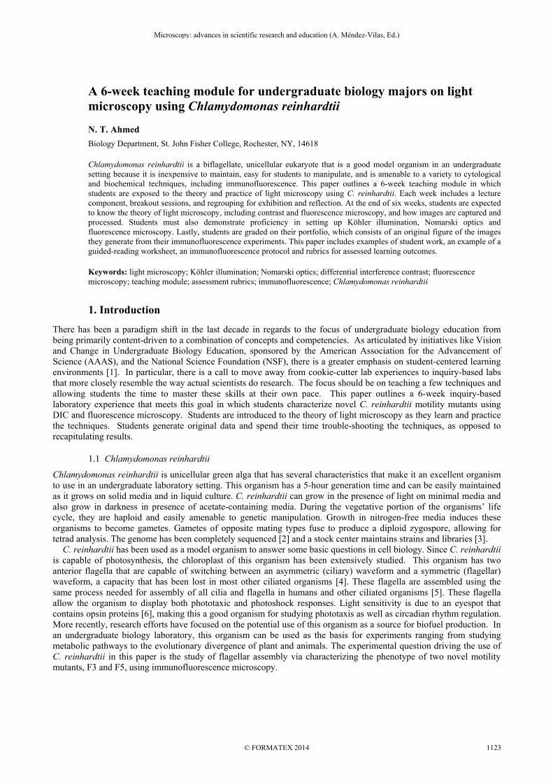
The immunofluorescence protocols can be used to generate original data on instructor-generated C. reinhardtii mutants, as shown in figure 2, or on known mutants that are available from the Chlamydomonas Resource Center (http://chlamycollection.org/). Novel mutants can be generated as described by Tam and Lefebvre [7]. C. reinhardtii strains are maintained on TAP media (Life Technologies) as described by Harris [8]. The immunofluorescence protocol has been modified from previously described procedures [9,10] and is reviewed in section 4.2. The poly L-lysine solution consist of 0.1% w/v poly L-lysine dilute in dH2O, and can be stored at room temperature for no more than two weeks. The 100% Blocking Solution consists of 5% bovine serum albumin and 1% cold-water fish gelatin in 1X PBS, and can be stored at 4°C for 6 weeks. The PBS-T consists of 0.05% Tween in 1X PBS, and can be stored at room temperature for at least 6 weeks.
Microscopy: advances in scientific research and education (A. Méndez-Vilas, Ed.)__________________________________________________________________
1125© FORMATEX 2014
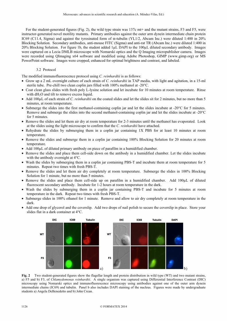
For the student-generated figures (Fig. 2), the wild type strain was 137c mt+ and the mutant strains, F3 and F5, were instructor-generated novel motility mutants. Primary antibodies against the outer arm dynein intermediate chain protein IC69 (C11.4, Sigma) and against the tyrosinated form of α-tubulin (YL1/2, Abcam Inc.) were diluted 1:400 in 20% Blocking Solution. Secondary antibodies, anti-mouse FITC (Sigma) and anti-rat TR (Abcam Inc.) were diluted 1:400 in 20% Blocking Solution. For figure 1b, the student added 1µL DAPI to the 100µL diluted secondary antibody. Images were captured on a Lecia DMLB microscope with Nomarski optics and the Q Imaging micropublisher camera. Images were recorded using QImaging x64 software and modified using Adobe Photoshop, GIMP (www.gimp.org) or MS PowerPoint software. Images were cropped, enhanced for optimal brightness and contrast, and labeled.
3.2 Protocol
The modified immunofluorescence protocol using C. reinhardtii is as follows: • Grow up a 2 mL overnight culture of each strain of C. reinhardtii in TAP media, with light and agitation, in a 15-ml
sterile tube. Pre-chill two clean coplin jars filled with 100% methanol at -20°C. • Coat clean glass slides with fresh poly L-lysine solution and let incubate for 10 minutes at room temperature. Rinse
with dH2O and tilt to remove excess liquid. • Add 100µL of each strain of C. reinhardtii on the coated slides and let the slides sit for 2 minutes, but no more than 5
minutes, at room temperature. • Submerge the slides into the first methanol-containing coplin jar and let the slides incubate at -20°C for 5 minutes.
Remove and submerge the slides into the second methanol-containing coplin jar and let the slides incubate at -20°C for 5 minutes.
• Remove the slides and let them air dry at room temperature for 2-3 minutes until the methanol has evaporated. Look at the slides using the light microscope to confirm that the C. reinhardtii have attached.
• Rehydrate the slides by submerging them in a coplin jar containing 1X PBS for at least 10 minutes at room temperature.
• Remove the slides and submerge them in a coplin jar containing 100% Blocking Solution for 20 minutes at room temperature.
• Add 100µL of diluted primary antibody on piece of parafilm in a humidified chamber. • Remove the slides and place them cell-side down on the antibody in a humidified chamber. Let the slides incubate
with the antibody overnight at 4°C. • Wash the slides by submerging them in a coplin jar containing PBS-T and incubate them at room temperature for 5
minutes. Repeat two times with fresh PBS-T. • Remove the slides and let them air dry completely at room temperature. Submerge the slides in 100% Blocking
Solution for 1 minute, but no more than 5 minutes. • Remove the slides and place them cell-side up on parafilm in a humidified chamber. Add 100µL of diluted
fluorescent secondary antibody. Incubate for 1-2 hours at room temperature in the dark. • Wash the slides by submerging them in a coplin jar containing PBS-T and incubate for 5 minutes at room
temperature in the dark. Repeat two times with fresh PBS-T. • Submerge slides in 100% ethanol for 1 minute. Remove and allow to air dry completely at room temperature in the
dark. • Add one drop of glycerol and the coverslip. Add two drops of nail polish to secure the coverslip in place. Store your
slides flat in a dark container at 4°C.
Fig. 2 Two student-generated figures show the flagellar length and protein distribution in wild type (WT) and two mutant strains, a) F5 and b) F3, of Chlamydomonas reinhardtii. A single organism was captured using Differential Interference Contrast (DIC) microscopy using Nomarski optics and immunofluorescence microscopy using antibodies against one of the outer arm dynein intermediate chains (IC69) and tubulin. Panel b also includes DAPI staining of the nucleus. Figures were made by undergraduate students a) Angela DeBenedetto and b) John Crean.
Microscopy: advances in scientific research and education (A. Méndez-Vilas, Ed.)__________________________________________________________________
© FORMATEX 20141126
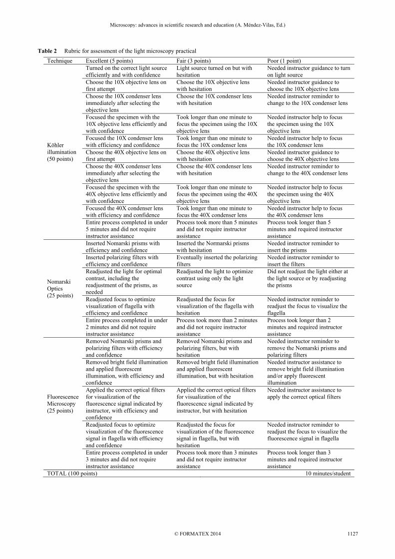
Table 2 Rubric for assessment of the light microscopy practical
Technique Excellent (5 points) Fair (3 points) Poor (1 point)
Köhler illumination (50 points)
Turned on the correct light source efficiently and with confidence
Light source turned on but with hesitation
Needed instructor guidance to turn on light source
Choose the 10X objective lens on first attempt
Choose the 10X objective lens with hesitation
Needed instructor guidance to choose the 10X objective lens
Choose the 10X condenser lens immediately after selecting the objective lens
Choose the 10X condenser lens with hesitation
Needed instructor reminder to change to the 10X condenser lens
Focused the specimen with the 10X objective lens efficiently and with confidence
Took longer than one minute to focus the specimen using the 10X objective lens
Needed instructor help to focus the specimen using the 10X objective lens
Focused the 10X condenser lens with efficiency and confidence
Took longer than one minute to focus the 10X condenser lens
Needed instructor help to focus the 10X condenser lens
Choose the 40X objective lens on first attempt
Choose the 40X objective lens with hesitation
Needed instructor guidance to choose the 40X objective lens
Choose the 40X condenser lens immediately after selecting the objective lens
Choose the 40X condenser lens with hesitation
Needed instructor reminder to change to the 40X condenser lens
Focused the specimen with the 40X objective lens efficiently and with confidence
Took longer than one minute to focus the specimen using the 40X objective lens
Needed instructor help to focus the specimen using the 40X objective lens
Focused the 40X condenser lens with efficiency and confidence
Took longer than one minute to focus the 40X condenser lens
Needed instructor help to focus the 40X condenser lens
Entire process completed in under 5 minutes and did not require instructor assistance
Process took more than 5 minutes and did not require instructor assistance
Process took longer than 5 minutes and required instructor assistance
Nomarski Optics (25 points)
Inserted Nomarski prisms with efficiency and confidence
Inserted the Normarski prisms with hesitation
Needed instructor reminder to insert the prisms
Inserted polarizing filters with efficiency and confidence
Eventually inserted the polarizing filters
Needed instructor reminder to insert the filters
Readjusted the light for optimal contrast, including the readjustment of the prisms, as needed
Readjusted the light to optimize contrast using only the light source
Did not readjust the light either at the light source or by readjusting the prisms
Readjusted focus to optimize visualization of flagella with efficiency and confidence
Readjusted the focus for visualization of the flagella with hesitation
Needed instructor reminder to readjust the focus to visualize the flagella
Entire process completed in under 2 minutes and did not require instructor assistance
Process took more than 2 minutes and did not require instructor assistance
Process took longer than 2 minutes and required instructor assistance
Fluorescence Microscopy (25 points)
Removed Nomarski prisms and polarizing filters with efficiency and confidence
Removed Nomarski prisms and polarizing filters, but with hesitation
Needed instructor reminder to remove the Nomarski prisms and polarizing filters
Removed bright field illumination and applied fluorescent illumination, with efficiency and confidence
Removed bright field illumination and applied fluorescent illumination, but with hesitation
Needed instructor assistance to remove bright field illumination and/or apply fluorescent illumination
Applied the correct optical filters for visualization of the fluorescence signal indicated by instructor, with efficiency and confidence
Applied the correct optical filters for visualization of the fluorescence signal indicated by instructor, but with hesitation
Needed instructor assistance to apply the correct optical filters
Readjusted focus to optimize visualization of the fluorescence signal in flagella with efficiency and confidence
Readjusted the focus for visualization of the fluorescence signal in flagella, but with hesitation
Needed instructor reminder to readjust the focus to visualize the fluorescence signal in flagella
Entire process completed in under 3 minutes and did not require instructor assistance
Process took more than 3 minutes and did not require instructor assistance
Process took longer than 3 minutes and required instructor assistance
TOTAL (100 points) 10 minutes/student
Microscopy: advances in scientific research and education (A. Méndez-Vilas, Ed.)__________________________________________________________________
1127© FORMATEX 2014
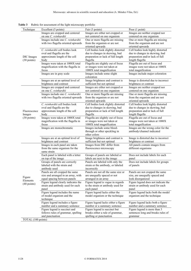
Table 3 Rubric for assessment of the light microscopy portfolio
Technique Excellent (5 points) Fair (3 points) Poor (1 point)
DIC Images (30 points)
Images are cropped and centered on one C. reinhardtii
Images are either not cropped or not centered on one organism
Images are neither cropped nor centered on one organism
Images include one C. reinhardtii with two flagella oriented upwards
One or more flagella are missing from the organism or are not oriented upwards
One or more flagella are missing from the organism and are not oriented upwards
C. reinhardtii cell bodies look oval and flagella are the approximate length of the cell body
Cell bodies look slightly distorted due to changes in skewing, bad preparation or lack of full length flagella
Cell bodies look highly distorted due to changes in skewing, bad preparation and/or lack of full length flagella
Images were taken at 1000X total magnification with the flagella in focus
Flagella are slightly out of focus or images were not taken at 1000X total magnification
Flagella are out of focus and images were not taken at 1000X total magnification
Images are in gray scale Images include some slight coloration
Images include major coloration
Images are at an optimal level of brightness and contrast
Image brightness and contrast is sufficient but not optimal
Image is distorted due to incorrect brightness or contrast
Fluorescence Images (30 points)
Images are cropped and centered on one C. reinhardtii
Images are either not cropped or not centered on one organism
Images are neither cropped nor centered on one organism
Images include one C. reinhardtii with two flagella oriented upwards
One or more flagella are missing from the organism or are not oriented upwards
One or more flagella are missing from the organism and are not oriented upwards
C. reinhardtii cell bodies look oval and flagella are the approximate length of the cell body
Cell bodies look slightly distorted due to changes in skewing, bad preparation or lack of full length flagella
Cell bodies look highly distorted due to changes in skewing, bad preparation and/or lack of full length flagella
Images were taken at 1000X total magnification with the flagella in focus
Flagella are slightly out of focus or images were not taken at 1000X total magnification
Flagella are out of focus and images were not taken at 1000X total magnification
Images are monochromatic Images include some bleed-through or other speckling in other colors
Images are the wrong color for the antibody/channel indicated
Images are at an optimal level of brightness and contrast
Image brightness and contrast is sufficient but not optimal
Image is distorted due to incorrect brightness or contrast
Figure Elements (40 points)
Images in each panel are taken from the same organism for the same strain
Images from DIC differ from fluorescence microscopy
All panels contain images from different organisms
Each panel is labeled with a letter on top of the image
Groups of panels are labeled or labels are next to the image
Does not include labels for each panel
Groups of panels are correctly labeled with the strain and antibody used
Panels are labeled with only the strain or the antibody, or labeled incorrectly
Does not include labels for groups of panels
Panels are all cropped the same size and arranged in an array, with equal spacing between panels
Panels are not all the same size or are unequally spaced or not arranged in an array
Panels are not cropped the same size, are unequally spaced and look disorganized
Figure legend clearly indicates the strain and antibody used for each panel
Figure legend is vague in regards to the strain or antibody used for each panel
Figure legend does not indicate the strain or antibody used for each panel
Figure legend includes the name of model organism and the technique
Figure legend lacks either the model organism or the technique
Figure legend lacks both the model organism and the technique
Figure legend includes a figure number and a summary sentence
Figure legend lacks either a figure number or a summary sentence
Figure legend lacks both a figure number and a summary sentence
Figure legend is succinct and follows rules of grammar, spelling and punctuation
Figure legend is succinct but breaks either a rule of grammar, spelling or punctuation
Figure legend is more than 3 sentences long and breaks rules of grammar
TOTAL (100 points)
Microscopy: advances in scientific research and education (A. Méndez-Vilas, Ed.)__________________________________________________________________
© FORMATEX 20141128
4. Assessment
Three different assessments are used to determine if students achieved the learning objectives of this module. To determine if students are able to describe how compound light microscopy, contrast microscopy and fluorescence microscopy work, a written examination is given on week 6 of this module. The written examination format is short answer and mirrors the questions asked by the instructor on the LBL reading worksheets. Students are given a practical examination the last week of this module to assess whether students can correctly set up the microscope to observe C. reinhardtii using Köhler illumination, Nomarski optics and fluorescence microscopy. Lastly, students are assessed on the quality of the figure they generate.
4.1 Practical
During the second and third weeks of this module the instructor demonstrates how to set up Köhler illumination, Nomarski optics and fluorescence microscopy at 400X and 1000X total magnification. Students have 3-4 weeks to practice the microscopy techniques. On the last week of this module, students are observed for their efficiency and confidence in performing these techniques in the form of a practical examination. Students have ten minutes to set up the microscope for Köhler illumination at 400X and 1000X total magnification, Nomarski optics, at 1000X and fluorescence microscopy at 1000X using one of their prepared slides. Assessment is scored based on the rubric described in table 2. The instructor should be able to assess 18 students over a three-hour class period. For larger classes, the instructor can start assessing students in groups A-D in week 5.
4.2 Portfolio
Students learn how to perform immunofluorescence using C. reinhardtii on week 1 of this module. This includes learning how to make all the solutions, maintain the strains, and grow overnight cultures. Students practice these techniques for the following 4 weeks in their groups during the breakout sessions and on their own time outside of class, as needed. During the instructor demonstration sessions in weeks 2 and 3, the instructor also demonstrates how to capture digital images and manipulate them using available software, such Adobe Photoshop or GIMP. To determine if students can generate images of C. reinhardtii using Nomarski optics and fluorescence microscopy to construct a publication-quality figure, students are asked to create a portfolio. This consists of a multiple-paneled figure of student-generate images. The figure must include DIC and immunofluorescence images using two antibodies on wild type and one mutant strain. The images should C. reinhardtii with two full-length flagella oriented upwards. A publication quality figure should contain panels that are uniformly cropped. Panels should be arranged by strain and microscopy or antibody used. The images used to generate each panel should be optimized for brightness and contrast. DIC and immunofluorescence images for each strain should be taken of the same organism. For all images, the flagella should be in focus. The portfolio also includes a figure legend. The figure legend should be a succinct description of the figure, and include a one-sentence summary of results, the species name, the name of strains used, and the name of microscopy used. The figure legend should follow the rules of grammar, spelling and punctuation. Students are assessed on their portfolio as described in Table 3. Two examples of student-generated portfolios have been included (Fig. 2).
Acknowledgements The support by St. John Fisher College is gratefully acknowledged. I would like to thank Angela DeBenedetto and John Crean for the use of their images.
References
[1] Conference AA for the A of S (AAAS). Vision and Change in Undergraduate Biology Education: View for the 21st Century [Internet]. American Association for the Advancement of Science (AAAS) Conference Homepage. 2009 [cited 2014 May 4]. Available from: www.visionandchnage.org
[2] Merchant SS, Prochnik SE, Vallon O, Harris EH, Karpowicz SJ, Witman GB, Terry A, Salamov A, Fritz-Laylin LK, Maréchal-Drouard L, Marshall WF, Qu L-H, Nelson DR, Sanderfoot AA, Spalding MH, Kapitonov V V, Ren Q, Ferris P, Lindquist E, Shapiro H, Lucas SM, Grimwood J, Schmutz J, Cardol P, Cerutti H, Chanfreau G, Chen C-L, Cognat V, Croft MT, Dent R, Dutcher S, Fernández E, Fukuzawa H, González-Ballester D, González-Halphen D, Hallmann A, Hanikenne M, Hippler M, Inwood W, Jabbari K, Kalanon M, Kuras R, Lefebvre PA, Lemaire SD, Lobanov A V, Lohr M, Manuell A, Meier I, Mets L, Mittag M, Mittelmeier T, Moroney J V, Moseley J, Napoli C, Nedelcu AM, Niyogi K, Novoselov S V, Paulsen IT, Pazour G, Purton S, Ral J-P, Riaño-Pachón DM, Riekhof W, Rymarquis L, Schroda M, Stern D, Umen J, Willows R, Wilson N, Zimmer SL, Allmer J, Balk J, Bisova K, Chen C-J, Elias M, Gendler K, Hauser C, Lamb MR, Ledford H, Long JC, Minagawa J, Page MD, Pan J, Pootakham W, Roje S, Rose A, Stahlberg E, Terauchi AM, Yang P, Ball S, Bowler C, Dieckmann CL, Gladyshev VN, Green P, Jorgensen R, Mayfield S, Mueller-Roeber B, Rajamani S, Sayre RT, Brokstein P, Dubchak I, Goodstein D, Hornick L, Huang YW, Jhaveri J, Luo Y, Martínez D, Ngau WCA, Otillar B, Poliakov A, Porter A, Szajkowski L, Werner G, Zhou K, Grigoriev I V, Rokhsar DS, Grossman AR. The Chlamydomonas genome reveals the evolution of key animal and plant functions. Science. 2007;318(5848):245–50.
Microscopy: advances in scientific research and education (A. Méndez-Vilas, Ed.)__________________________________________________________________
1129© FORMATEX 2014
[3] Chlamydomonas Center [Internet]. [cited 2014 May 4]. Available from: http://chlamycollection.org/ [4] Flagella C, Mitchell DR. Chlamydomonas reinhardtii. Journal of Phycology. 2000;273:261–73. [5] Baldari CT, Rosenbaum J. Intraflagellar transport: it’s not just for cilia anymore. Curr Opin Cell Biol. 2010;22(1):75–80. [6] Nagel G, Ollig D, Fuhrmann M, Kateriya S, Musti AM, Bamberg E, Hegemann P. Channelrhodopsin-1: a light-gated proton
channel in green algae. Science. 2002;296(5577):2395–8. [7] Tam LW, Lefebvre PA. Cloning of flagellar genes in Chlamydomonas reinhardtii by DNA insertional mutagenesis. Genetics.
1993;135(2):375–84. [8] Harris EH. The Chlamydomonas Sourcebook: Introduction to Chlamydomonas and Its Laboratory Use, Volume 1. Academic
Press; 2009 [9] Cole DG. Chlamydomonas Kinesin-II-dependent Intraflagellar Transport (IFT): IFT Particles Contain Proteins Required for
Ciliary Assembly in Caenorhabditis elegans Sensory Neurons. J Cell Biol. 1998;141(4):993–1008. [10] Sanders MA, Salisbury JL. Immunofluorescence microscopy of cilia and flagella. Methods Cell Biol. 1995;47:163–9.
Microscopy: advances in scientific research and education (A. Méndez-Vilas, Ed.)__________________________________________________________________
© FORMATEX 20141130