Embed Size (px)
Citation preview
i
A 5-year review of the microbiology
of acute complicated bacterial
sinusitis at the University of the
Witwatersrand
By
Ian Paul Olwoch M.B.Ch.B. (Makerere University)
M.Med. Anaesthesia (University of Nairobi)
A dissertation submitted in part fulfillment for the
degree of Master of Medicine in
Otorhinolaryngology of the University of the
Witwatersrand.
2007

This dissertation is dedicated
to
my wife,
Jane Mukarugwiza Olwoch,
and
my daughters,
Irene Aciro Olwoch
And
Margaret Alobo Olwoch
ii
DECLARATION
CANDIDATE
This dissertation is my original work and has not been presented for a degree,
or other academic award, in any other University or institution of higher
learning.
Signed:
Dr Ian Paul Olwoch
MBChB (Makerere), MMed Anaesthesia (Nairobi)
SUPERVISOR
This dissertation is submitted for examination with my approval as University
Supervisor.
Signed:
Professor Pradip C. Modi
MBChB (Wits), DCH (SA), MMed (Wits), FCS (SA)
Professor and Head of Department
Department of Otorhinolaryngology
University of the Witwatersrand
iii
ACKNOWLEDGEMENTS
I wish to express my gratitude to all the following:
PROFESSOR PC MODI, my supervisor and academic head, for his guidance
support and encouragement throughout the preparation and execution of this
study and for his invaluable criticism in the final preparation of this
dissertation.
All the members of the academic peer review committee of the Department of
Neurosciences for their guidance and valuable criticism.
DR MRI AHMED, the clinical head of Otorhinolaryngology at the Chris Hani
Baragwanath Hospital, for facilitating my access to a wealth of data that was
vital to the success of this dissertation.
The staff of the:
Operating theatres
ENT and neurosurgical wards
Department of radiology
National Health Laboratory Services
at the Johannesburg General, Chris Hani Baragwanath, Helen Joseph and
Coronation Hospitals for their support in sourcing all the relevant data.
My wife JANE, and daughters IRENE and MARGARET, for their patience,
encouragement and deep understanding throughout the process of this study.
iv
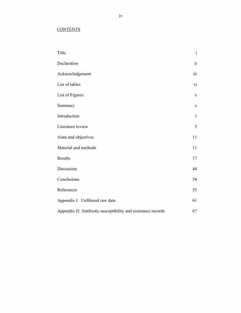
CONTENTS
Title i
Declaration ii
Acknowledgement iii
List of tables vi
List of Figures x
Summary x
Introduction 1
Literature review 5
Aims and objectives 11
Material and methods 11
Results 17
Discussion 44
Conclusions 54
References 55
Appendix I: Unfiltered raw data 61
Appendix II: Antibiotic susceptibility and resistance records 67
v
LIST OF TABLES
Table 1: Categorization of microbiology culture results from 301 patients with
acute complicated sinusitis. [Page 17]
Table 2: Age and gender statistics of the study population. [Page 19]
Table 3: Distribution of negative culture reports amongst 226 laboratory
culture reports by gender, age and presence of neurosurgical
complication. [Page 20]
Table 4: Age analysis of 163 patients in the positive culture population.
[Page 21]
Table 5: 233 bacteria and fungi isolated from 163 patients with acute
complicated sinusitis expressed as raw data and percentage of total
count. [Page 23]
Table 6: Comparative analysis of the frequency of occurrence, of aerobes,
facultative anaerobes, anaerobes and fungi in 163 patients with
positive culture specimen. [Page 24]
Table 7: Number of different microorganism species isolated per patient
within the positive culture population (n = 163). [Page24]
vi
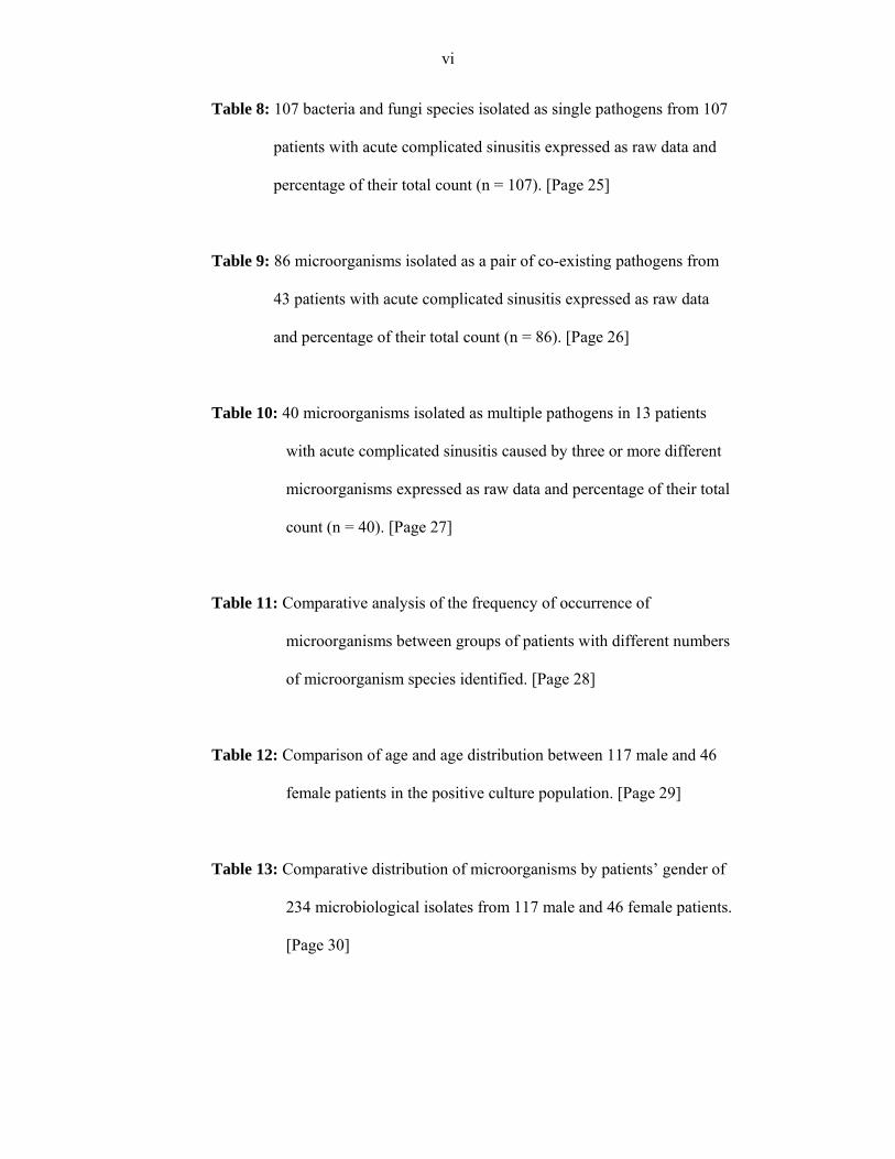
Table 8: 107 bacteria and fungi species isolated as single pathogens from 107
patients with acute complicated sinusitis expressed as raw data and
percentage of their total count (n = 107). [Page 25]
Table 9: 86 microorganisms isolated as a pair of co-existing pathogens from
43 patients with acute complicated sinusitis expressed as raw data
and percentage of their total count (n = 86). [Page 26]
Table 10: 40 microorganisms isolated as multiple pathogens in 13 patients
with acute complicated sinusitis caused by three or more different
microorganisms expressed as raw data and percentage of their total
count (n = 40). [Page 27]
Table 11: Comparative analysis of the frequency of occurrence of
microorganisms between groups of patients with different numbers
of microorganism species identified. [Page 28]
Table 12: Comparison of age and age distribution between 117 male and 46
female patients in the positive culture population. [Page 29]
Table 13: Comparative distribution of microorganisms by patients’ gender of
234 microbiological isolates from 117 male and 46 female patients.
[Page 30]
vii
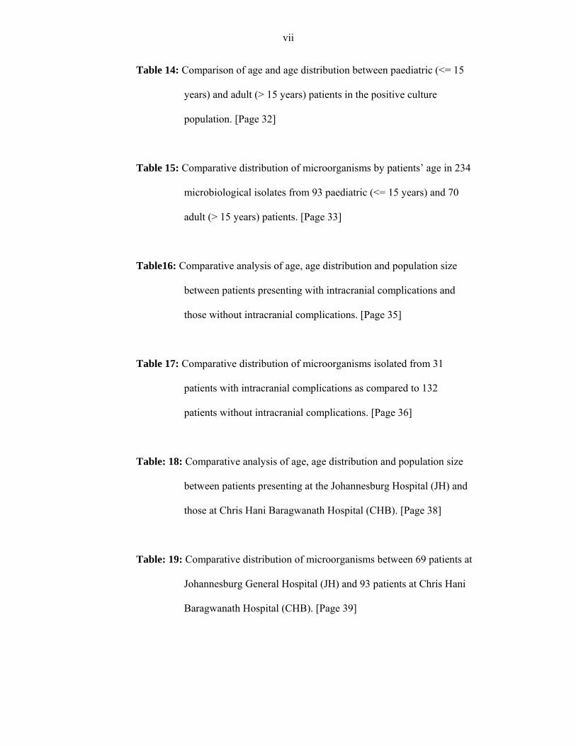
Table 14: Comparison of age and age distribution between paediatric (<= 15
years) and adult (> 15 years) patients in the positive culture
population. [Page 32]
Table 15: Comparative distribution of microorganisms by patients’ age in 234
microbiological isolates from 93 paediatric (<= 15 years) and 70
adult (> 15 years) patients. [Page 33]
Table16: Comparative analysis of age, age distribution and population size
between patients presenting with intracranial complications and
those without intracranial complications. [Page 35]
Table 17: Comparative distribution of microorganisms isolated from 31
patients with intracranial complications as compared to 132
patients without intracranial complications. [Page 36]
Table: 18: Comparative analysis of age, age distribution and population size
between patients presenting at the Johannesburg Hospital (JH) and
those at Chris Hani Baragwanath Hospital (CHB). [Page 38]
Table: 19: Comparative distribution of microorganisms between 69 patients at
Johannesburg General Hospital (JH) and 93 patients at Chris Hani
Baragwanath Hospital (CHB). [Page 39]
viii
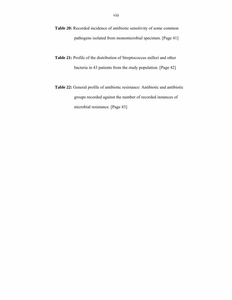
Table 20: Recorded incidence of antibiotic sensitivity of some common
pathogens isolated from monomicrobial specimen. [Page 41]
Table 21: Profile of the distribution of Streptococcus milleri and other
bacteria in 43 patients from the study population. [Page 42]
Table 22: General profile of antibiotic resistance: Antibiotic and antibiotic
groups recorded against the number of recorded instances of
microbial resistance. [Page 43]
ix
LIST OF FIGURES
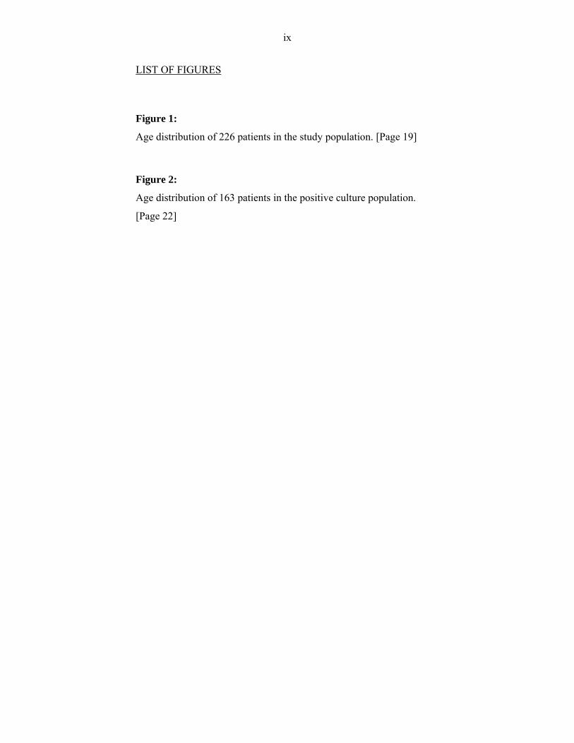
Figure 1:
Age distribution of 226 patients in the study population. [Page 19]
Figure 2:
Age distribution of 163 patients in the positive culture population.
[Page 22]
x
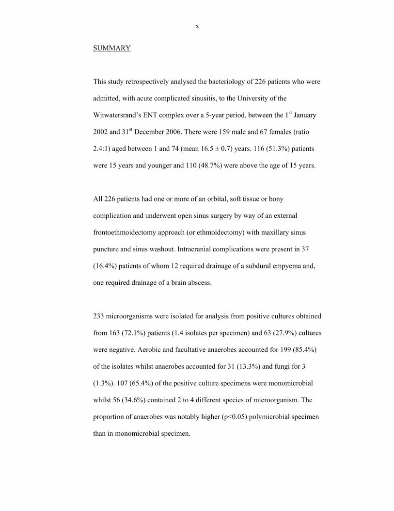
SUMMARY
This study retrospectively analysed the bacteriology of 226 patients who were
admitted, with acute complicated sinusitis, to the University of the
Witwatersrand’s ENT complex over a 5-year period, between the 1st January
2002 and 31st December 2006. There were 159 male and 67 females (ratio
2.4:1) aged between 1 and 74 (mean 16.5 ± 0.7) years. 116 (51.3%) patients
were 15 years and younger and 110 (48.7%) were above the age of 15 years.
All 226 patients had one or more of an orbital, soft tissue or bony
complication and underwent open sinus surgery by way of an external
frontoethmoidectomy approach (or ethmoidectomy) with maxillary sinus
puncture and sinus washout. Intracranial complications were present in 37
(16.4%) patients of whom 12 required drainage of a subdural empyema and,
one required drainage of a brain abscess.
233 microorganisms were isolated for analysis from positive cultures obtained
from 163 (72.1%) patients (1.4 isolates per specimen) and 63 (27.9%) cultures
were negative. Aerobic and facultative anaerobes accounted for 199 (85.4%)
of the isolates whilst anaerobes accounted for 31 (13.3%) and fungi for 3
(1.3%). 107 (65.4%) of the positive culture specimens were monomicrobial
whilst 56 (34.6%) contained 2 to 4 different species of microorganism. The
proportion of anaerobes was notably higher (p<0.05) polymicrobial specimen
than in monomicrobial specimen.
xi
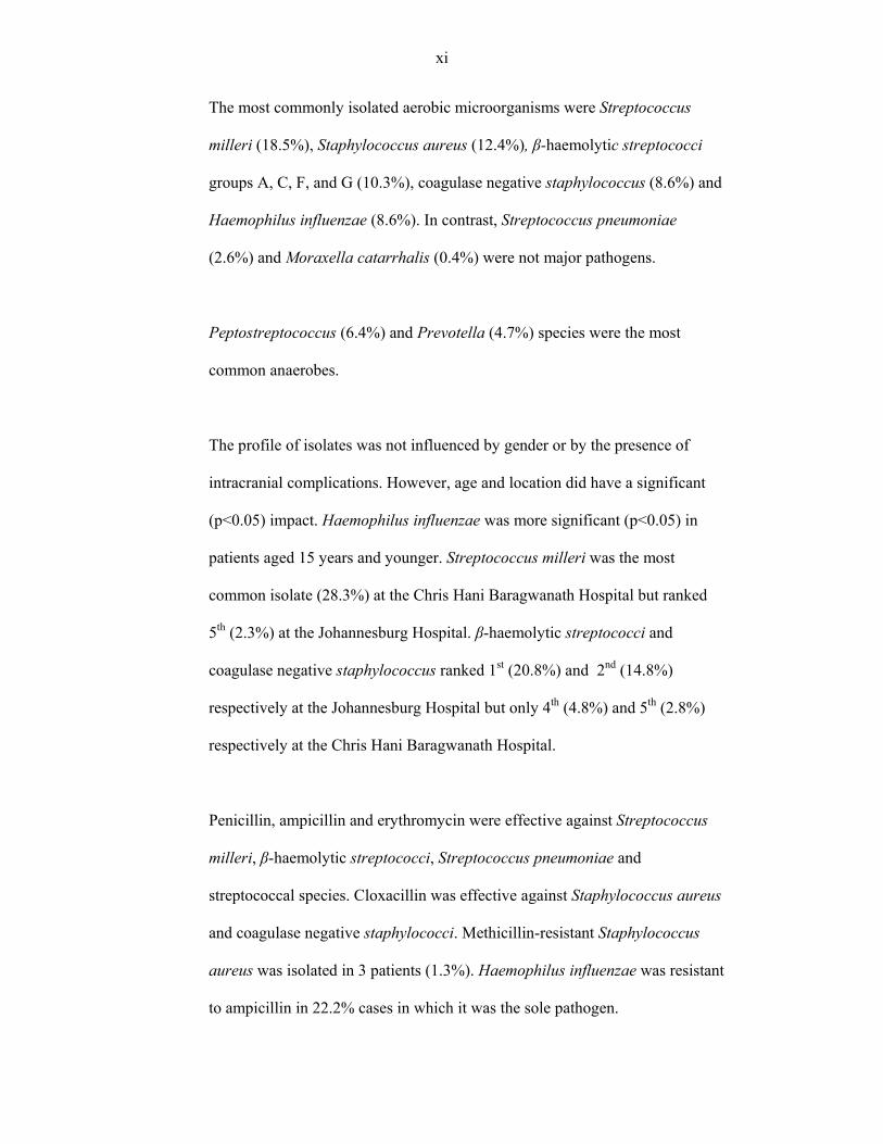
The most commonly isolated aerobic microorganisms were Streptococcus
milleri (18.5%), Staphylococcus aureus (12.4%), β-haemolytic streptococci
groups A, C, F, and G (10.3%), coagulase negative staphylococcus (8.6%) and
Haemophilus influenzae (8.6%). In contrast, Streptococcus pneumoniae
(2.6%) and Moraxella catarrhalis (0.4%) were not major pathogens.
Peptostreptococcus (6.4%) and Prevotella (4.7%) species were the most
common anaerobes.
The profile of isolates was not influenced by gender or by the presence of
intracranial complications. However, age and location did have a significant
(p<0.05) impact. Haemophilus influenzae was more significant (p<0.05) in
patients aged 15 years and younger. Streptococcus milleri was the most
common isolate (28.3%) at the Chris Hani Baragwanath Hospital but ranked
5th (2.3%) at the Johannesburg Hospital. β-haemolytic streptococci and
coagulase negative staphylococcus ranked 1st (20.8%) and 2nd (14.8%)
respectively at the Johannesburg Hospital but only 4th (4.8%) and 5th (2.8%)
respectively at the Chris Hani Baragwanath Hospital.
Penicillin, ampicillin and erythromycin were effective against Streptococcus
milleri, β-haemolytic streptococci, Streptococcus pneumoniae and
streptococcal species. Cloxacillin was effective against Staphylococcus aureus
and coagulase negative staphylococci. Methicillin-resistant Staphylococcus
aureus was isolated in 3 patients (1.3%). Haemophilus influenzae was resistant
to ampicillin in 22.2% cases in which it was the sole pathogen.
1
A 5-year review of the microbiology of acute
complicated bacterial sinusitis at the University of the
Witwatersrand
INTRODUCTION
Acute sinusitis defines a state in which there is rapid onset, progression and
persistence of signs and symptoms caused by inflammatory disease within the
paranasal sinuses. Viruses and bacteria are largely responsible for the
development of acute sinusitis. Often an initial viral inflammation of the
sinuses is superseded by invasion with a variety of bacterial pathogens and the
development of complications at adjacent and/or remote sites. Management of
acute complicated sinusitis requires an understanding of the anatomy of the
sinuses and the microbiology of the associated pathogens.
Embryology
The paranasal sinuses are airspaces within the bones of the face and skull that
are transposed as relations around the nasal cavity. There are four paired
sinuses namely the maxillary, ethmoid, frontal and sphenoid sinuses. The
paranasal sinuses develop as outpouches of the nasal cavity and remain in
communication with the nasal cavity through narrow openings (or ostia). The
maxillary and ethmoid sinuses are present at birth; the frontal and sphenoid
sinuses begin to develop after the ages of 2 and 7 years, respectively. The
2
sinuses attain adult size between the age of 12 and 18, with the sphenoid sinus
being the last to mature.
Physiology
The paranasal sinuses are lined with pseudostratified ciliated columnar
epithelium which is in continuity with the mucosa of the nasal cavity. The
epithelium also contains non-ciliated columnar cells, basal cells and mucus-
secreting goblet cells. The cilia in each sinus beat in a specific direction that
results in orderly movement of mucus and debris around the sinus and
ultimately through the sinus ostium and into the nasal cavity.
Normally the paranasal sinuses are relatively sterile with low titers of bacteria
of less than 1000 colony-forming units per milliliter of mucus. The sterility of
the cavities is maintained in part by the mucociliary clearance system, the
immune system of the body and nitric oxide production within the sinuses
(Palm J, Lidman C, Graf P et al., 2000). Factors that prevent or delay the
clearance of secretions from the paranasal sinuses result in the accumulation
of secretions and subsequent colonization of the sinuses by pathogenic
microorganisms by invasion from without and/or proliferation from within.
Definitions
Sinusitis describes a state in which pathogenic microorganisms colonize the
mucosal lining of the paranasal sinuses and cause an inflammatory disorder.
Viruses, bacteria, fungi and protozoa may cause the disease by infecting any
one, or combination, of the sinuses. The term rhinosinusitis, used
3
interchangeably with the term sinusitis, depicts the disease state as a
continuum of inflammation between the nasal and sinus mucosae. When 2 or
more paranasal sinuses are simultaneously infected, the term pansinusitis is
often used. Complications of paranasal sinus disease occur when the disease
extends to involve bone, the adjacent orbit and cranial cavities and their
respective contents.
Treatment
The mainstay of treatment of patients with sinusitis is medical and is directed
towards alleviating the symptoms and eliminating the invading organism
where possible. The treatment of sinusitis associated with complications
constitutes both a medical and, in certain cases, a surgical emergency.
At the University of the Witwatersrand’s ENT complex, patients with acute
complicated sinusitis are referred to the department of Otorhinolaryngology
usually when surgical intervention is anticipated. Prompt medical treatment
with or without surgery must be initiated. This includes the use of antibiotics
prior to obtaining laboratory identification of the offending pathogen(s).
Observed differences in the local prevalence of bacterial pathogens and the
continued emergence of antibiotic-resistant strains of bacteria influence the
choice of antibiotic used to initiate treatment (Ali A, Kurien M, Mathews SS,
Mathew J., 2005; Oxford LE, McClay J. 2005; Butler JC, Hofmann J, Cetron
MS, Elliot JA, Facklam RR, Breiman RF.1996).

4
The decision to conduct this study is prompted by the need to know the
prevalent bacteria associated with complicated sinusitis and their antibiotic
sensitivity patterns as seen at the University of the Witwatersrand’s ENT
complex.
5
LITERATURE REVIEW
Bacterial sinusitis is a common disease of worldwide distribution that affects
people of all ages. The course of the disease in patients with bacterial sinusitis
is varied, ranging from mild and insidious to debilitating and potentially life-
threatening (Ali A, Kurien M, Mathews SS, Mathew J. 2005; Oxford LE,
McClay J 2005). Bacterial sinusitis is usually preceded by an episode of viral
sinusitis caused by viruses responsible for the common cold. These include
such viruses as the adenovirus, rhinovirus, influenza virus, parainfluenza virus
and respiratory syncytial virus. The resultant sinus mucosal inflammation
causes oedema of the mucosa and obstruction of the sinus ostia and a
concomitant reduction in ciliary action.
It is classified into five clinical categories according to the duration of the
signs and symptoms of disease and the frequency with which they occur
(Piccirillo JF, 1998). These categories include:
Acute sinusitis in which symptoms and signs of the disease resolve completely
and do not exceed 4 weeks duration,
Recurrent acute sinusitis in which 2 to 4 episodes of acute sinusitis occur per
year with at least 8 weeks interval between each episode,
Subacute sinusitis in which symptoms of the disease persist for more than 4
weeks but not beyond 12 weeks,
Chronic sinusitis in which insidious symptoms persist for over 12 weeks,
6
Acute-on-chronic sinusitis in which a patient with chronic sinusitis
experiences and acute exacerbation of symptoms.
The presenting signs and symptoms in patients with sinusitis are those in
keeping with an inflammatory disease of the upper respiratory tract. The
findings include fever, headache, vomiting, nasal obstruction and discharge.
When complications occur other more ominous signs occur, the clinical
presentation being determined by the site and extent of the complications.
Involvement of the orbit and brain would result in the development of features
such as facial and periorbital swelling, proptosis, mood changes, seizures.
Infection of the paranasal sinus is associated with serious complications as a
result of the spread of the disease into adjacent and remote sites. Spread of the
disease from the sinuses occurs through areas of osteitic bone destruction,
congenital or acquired bony defects, or by way of septic thrombophlebitis of
communicating veins (Osguthorpe JD, Hochman M, 1993). The complications
of sinusitis fall into four categories namely orbital, intracranial, bone and soft
tissue. The highest occurrence of complications of sinusitis is recorded in
young male patients during the cold winter season (Oxford LE, McClay J,
2002).
Orbital complications are the most common complication of sinusitis (Ali A,
Kurien M, Mathews SS, Mathew J, 2005; Oxford LE, McClay J, 2005;
Schramm VL, Myers EN, Kennerdell JS, 1978), usually occurring as a result
of ethmoid sinusitis (Arjmand EM, Lusck RP, Muntz HR, 1993). The
7
Chandler Classification describes 5 types of orbital complications (Chandler
JR, Langenbrunner DJ, Stevens ER., 1970):
Preseptal cellulitis
Orbital cellulitis
Subperiosteal abscess
Orbital abscess
Cavernous sinus thrombosis
In the pre-antibiotic error, permanent sequelae including blindness occurred in
20% of patients with orbital complications (Gamble RC, 1933). Over the years
incidence of permanent orbital complications has reduced to around 6-10.5%
(Patt BS, Manning SC, 1991; Schramm VL, Myers EN, Kennerdell JS, 1978)
with some centres now reporting incidences as low as 1.8% (Oxford LE,
McClay J, 2005).
Intracranial complications are most commonly secondary to acute frontal
sinusitis and may include meningitis, cerebritis, epidural and subdural
empyemas, cerebral abscess and thrombosis of venous sinuses (Ali A, Kurien
M, Mathews SS, Mathew J. 2005; Clayman GL, Adams GL, Paugh DR et al
1991; Oxford LE, McClay J. 2005; Remmler D, Boles R. 1980). With
appropriate management the outcome of the disease will range from complete
resolution to transient or permanent neurological deficit, seizures or even
death (Younis RT, Lazar RH, 2002).
8
Bony complications include osteomyelitis of the frontal and maxillary sinus,
frontal subperiosteal abscess and frontal pyocoele (Ali A, Kurien M, Mathews
SS, Mathew J. 2005).
Periorbital and facial cellulitis are the predominant soft tissue complications.
The microbiology of bacterial sinusitis has been studied extensively in relation
to the causative organisms. The invading bacterial pathogens, in both acute
and chronic sinusitis, include a variety of aerobic, facultative and anaerobic
organisms. The most common pathogens found in patients with acute bacterial
sinusitis are aerobic and facultative anaerobes that include Streptococcus
pneumoniae, Haemophilus influenzae and Moraxella (Branhamella)
catarrhalis (Brook, 2002; Brook I, Frazier EH, 2004; Jousimies-Somer HR,
Savolainen S, Ylikoski JS, 1988; Tellez I, Alba LMD, Reyes MG, Patton E,
Hesles H dl G., 2006). Anaerobic organisms, such as Propionbacterium acnes
and Peptostreptococcus species, are found in as many as 31% of patients with
acute bacterial sinusitis (Brook I, 2005). Isolates from patients with chronic
sinusitis were found to have a mixture of both aerobic and anaerobic bacteria
with a preponderance of anaerobic bacteria such as Prevotella species,
Fusobacterium species, Peptostreptococcus species (Brook I, 1981). The
aerobic bacteria isolated from patients with chronic sinusitis included
Haemophilus influenzae, Staphylococcus aureus, Klebsiella pneumoniae and
Pseudomonas aeruginosa (Brook I, 2002; Frederick J, Braude AI, 1974;
Kamau JK, Macharia IM, Odhiambo PA, 2001).
9
Patients with bacterial pansinusitis are more likely to have a mixture of both
aerobic and anaerobic bacteria. Furthermore a study on 155 patients with
sinusitis demonstrated that the likelihood of anaerobic bacteria being present
was significantly high when multiple sinuses were concurrently diseased
(Brook I, 2004)
There appears to be a different frequency pattern of bacteria isolated from
patients with complicated sinusitis as compared to those presenting without
complications. The profile of offending bacteria also differs in reports from
different centres. Ali A et al (2005) isolated Staphylococcus aureus in 66.6%
of patients. Other organisms cultured in this study included Enterobacter
species, Streptococcus species, Protavella species and Bacteriodes. None of
the isolates included Haemophilus influenzae and Moraxella catarrhalis.
Special mention needs to be made of a group of streptococcal bacteria now
known as the Streptococcus milleri group of bacteria. This group is made up
of three species namely Streptococcus intermedius, Streptococcus consellatus
and Streptococcus anginosus. Their role as pathogens human pathogens was
not recognized until recently mainly because of difficulties in their laboratory
identification and because of confusion surrounding their taxonomy and
nomenclature (Ruoff KL, 1988). Oxford and McClay (2005) identified the
Streptococcus milleri group of bacteria as being the most frequent bacteria
isolated from children with complicated sinusitis. In their study ά-haemolytic
Streptococci species, Staphylococcus aureus and Streptococcus pneumoniae
followed in descending order of frequency. Haemophilus influenzae was
10
isolated from only one case, representing 0.9% of bacterial isolates. Moraxella
catarrhalis was not found in this study.
Acute sinusitis is extremely common and is the reason for which many
patients seek medical attention. Most often the illness is of viral origin and
about 2% of cases may become complicated by acute bacterial sinusitis (Lauer
J, 2003). In the absence of obvious bacterial complications it is not possible to
distinguish between viral and bacterial sinusitis on clinical grounds alone.
Despite the knowledge that viral infections are the predominant cause of
sinusitis many physicians continue to prescribe antibiotics as part of their first-
line of treatment (Gonzales R, Steiner JF, Sande MA., 1997). Over the years
the use and abuse of antibiotics has led to the emergence of antibiotic-resistant
strains of bacteria such a penicillin-resistant Streptococcus pneumoniae
(Butler JC, Hofmann J, Cetron MS, Elliot JA, Facklam RR, Breiman RF,
1996, Hofmann J, Cetron MS, Farley MM, Baughman WS, Facklam RR,
Elliot JA et al., 1995), Methicillin-resistant Staphylococcus aureus
and Pneumococci (Dowell SF, Schwartz B 1997; Kunin CM, 1993).
11
AIMS AND OBJECTIVE
The aim of the study is to:
Describe the frequency of the types of microorganisms isolated from patients
with acute complicated sinusitis at the teaching hospitals of the University of
the Witwatersrand
Document the antibiotic susceptibility and resistance of the isolated
microorganisms
Make recommendations on the choice of antibiotic to be used to initialize
treatment prior to obtaining laboratory bacterial culture and sensitivity results.
MATERIALS AND METHOD
Ethics Approval
This study was approved by the Standards and Ethics Committee of the
University of the Witwatersrand.
Study Location
The study was conducted at the four university teaching hospitals namely
Johannesburg General, Chris Hani Baragwanath, Helen Joseph and Coronation
Hospitals.
Study Period
The study extended over a period of 5 years; starting from the 1st January 2002
and ending on the 31st December 2006.
12
Study Population
The study includes all patients who underwent paranasal sinus surgery as part
of their management for acute complicated sinusitis and on whom sinus
specimen microbiology analysis was performed.
Data Collection
Patients were identified from three sources within each hospital, namely the:
ENT and neurosurgery emergency theatre operation registers
ENT and neurosurgery ward admission registers
Radiology CT scan registers.
The operating theatre emergency registers were used as the primary reference
to identify patients who were recorded as having had sinus surgery as an
emergency procedure. Both the ENT and neurosurgery registers were searched
so as to include those patients with acute sinusitis who additionally suffered
intracranial complications and may therefore have been registered as patients
outside the ENT department.
The ENT and neurosurgical ward admissions registers and the CT scan
register were used as secondary references to aid in the identification of those
patients whose information in the theatre register was unclear or lacking in
details (e.g. missing or partial hospital numbers, misspelled names, illegible
entry). These secondary references were also used as a screen to source for
patients whose information may not have been identified specifically in the
ENT and neurosurgery theatre registers. Those patients screened from this
13
secondary source included those who appeared in the ward register with a
diagnosis of sinusitis and those in-patients, from any hospital ward, who
appear in the CT scan register as having had a scan of either the paranasal
sinuses or the brain with the orbit.
All patients clearly identified from the theatre register were recorded as part of
the initial sample population. The patients’ information was then used to
search the database of the National Health Laboratory Service (NHLS) and all
findings were recorded placed into one of the following six categories:
Positive cultures in which pathogenic microorganisms were identified
Negative cultures in which the report states that there was “no bacterial
growth”
Histology reports in which a histological analysis but not a microbiological
analysis was performed on the specimen
Rejected specimen in which the specimen received by the laboratory was
unsuitable for processing
No results found in which no results were found in the database
Incomplete or incorrect information in which patients in the theatre register
did not have sufficient information to interrogate the NHLS database
Patients identified exclusively from the secondary references were cross-
checked against their recorded laboratory findings and were included in the
study population only if certain conditions were met. These conditions for
inclusion being that:
14
There was a microbiology or histology laboratory report for the identified
patient
The sample analysed was obtained from the paranasal sinuses
The report clearly stated that the sample was obtained from a patient with a
history of acute complicated sinusitis
Those patients selected from the secondary references who did not have
sufficient information to search the NHLS database were not included in the
study sample.
The following data was recorded for each patient:
Full name
Age
Sex
Hospital registration number
Date of operation (specimen collection)
Type of operation / complication
Type of isolated microorganisms
Antibiotic sensitivity and resistance
Laboratory results for microscopy culture and sensitivity were obtained at
each hospital from the computer register of the resident NHLS laboratory.
All data was recorded electronically using Microsoft Access database and
Microsoft Excel Spreadsheet.
15
For the purposes of this study, only those patients who were identified as
patients with acute complicated sinusitis, on whom sinus surgery was
performed and, for whom positive microbiological cultures were obtained
were included in the final study population and used for analysis.
Data Analysis and Presentation
Analysis of data was carried out by the MMed candidate (Dr IP Olwoch) at the
Department of Otorhinolaryngology Head and Neck Surgery. Standard
statistical methods are used. Student’s t test was used to analyse ordinal data
and the Chi Square (X2) test was used for dichotomous nominal data. A
probability (p) value of less than (or equal to) 0.05 is regarded as significant.
The following software applications were used for data manipulation,
statistical analysis and presentation:
Microsoft Access 2003 Database for data storage, retrieval and selection
Microsoft Excel 2003 Spreadsheet for descriptive data analysis, summary
statistics and comparison of sample means
Web Chi Square Calculator (Ball CN, Connor-Linton J, 2007) for comparison
of sample populations
Microsoft Word 2003 Word Processor for final documentation and
presentation
16
Evaluation of data from identified microorganisms with respect to prevalence,
age and gender of patients, and antibiotic sensitivity-resistance patterns was
done. Special attention is given to comparative analysis of:
The bacterial profile in male patients as compared to female patients.
The bacterial profile in patients under the age of 15-years as compared to
those over the age of 15 years.
The bacterial profile in those patients presenting with intracranial
complications as compared to those without.
The bacterial profile in patients presenting at the different hospitals within the
University ENT complex.
17
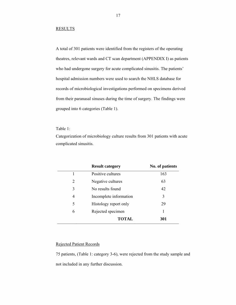
RESULTS
A total of 301 patients were identified from the registers of the operating
theatres, relevant wards and CT scan department (APPENDIX I) as patients
who had undergone surgery for acute complicated sinusitis. The patients’
hospital admission numbers were used to search the NHLS database for
records of microbiological investigations performed on specimens derived
from their paranasal sinuses during the time of surgery. The findings were
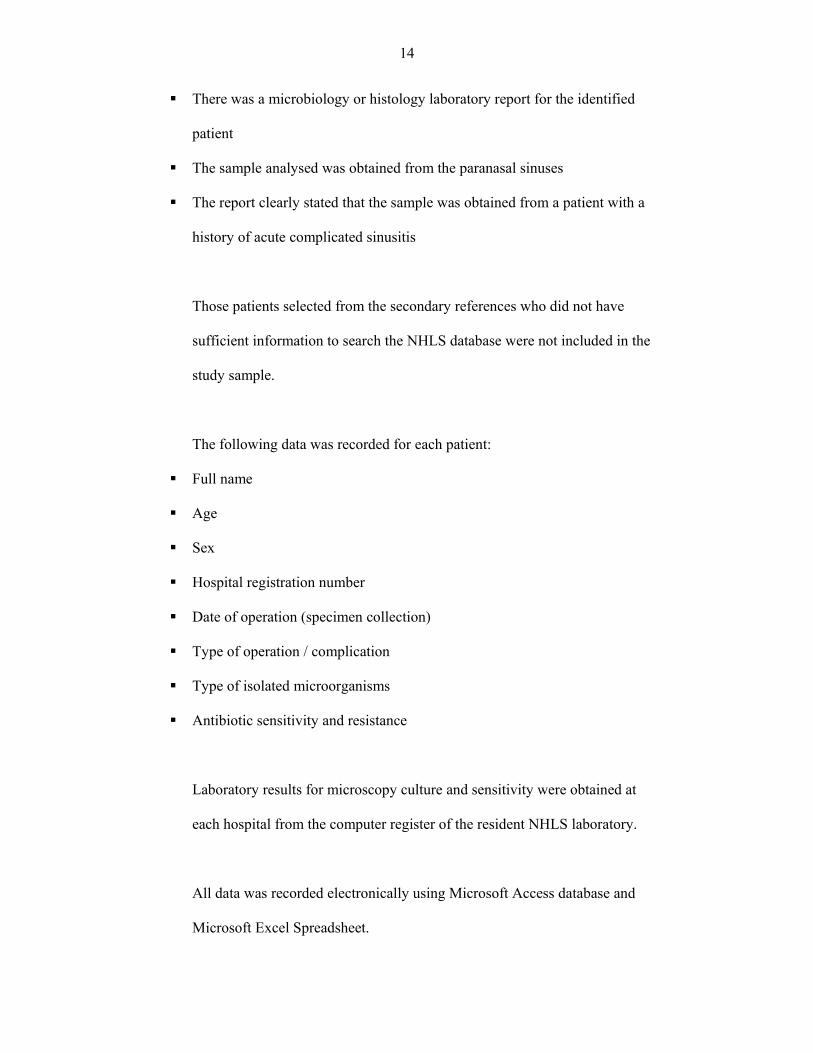
grouped into 6 categories (Table 1).
Table 1:
Categorization of microbiology culture results from 301 patients with acute
complicated sinusitis.
Result category No. of patients
1 Positive cultures 163
2 Negative cultures 63
3 No results found 42
4 Incomplete information 3
5 Histology report only 29
6 Rejected specimen 1
TOTAL 301
Rejected Patient Records
75 patients, (Table 1: category 3-6), were rejected from the study sample and
not included in any further discussion.
18
Study Population (n = 226)
All patients in the study population underwent open sinus surgery by way of
an external frontoethmoidectomy approach (or ethmoidectomy) with maxillary
sinus puncture and washout. In addition 37 (16.4%) patients of the study
population were diagnosed as having intracranial abscess formations. 13 of
these patients underwent concurrent neurosurgical craniotomy for drainage of
subdural empyema (n = 12) and brain abscess (n = 1).
All specimens were collected intra-operatively and therefore standard aseptic
conditions of collection are presumed to have been applied in all cases.
Specimens were analysed at the respective hospitals by the resident NHLS
laboratories in accordance with their Standard Operating Procedure Manual.
Specimen derived from the sinuses as sinus aspirates (or tissues) are cultured
aerobically at 35oC for 24 to 48 hours in:
5% horse blood agar
MacConkey agar
Chocolate agar
Thioglycolate broth
and, anaerobically at 35oC for 24 to 48 in 10% horse blood agar and Amikacin
blood agar. Direct gram staining and microscopy are performed after 24 hours.
Both positive and negative microbiology culture records were obtained from
226 patients (Table 1: category 1,2). Positive cultures were obtained in 163
(72.1%) cases and 63 (27.9%) records were reported as having “no bacterial
growth”.
19
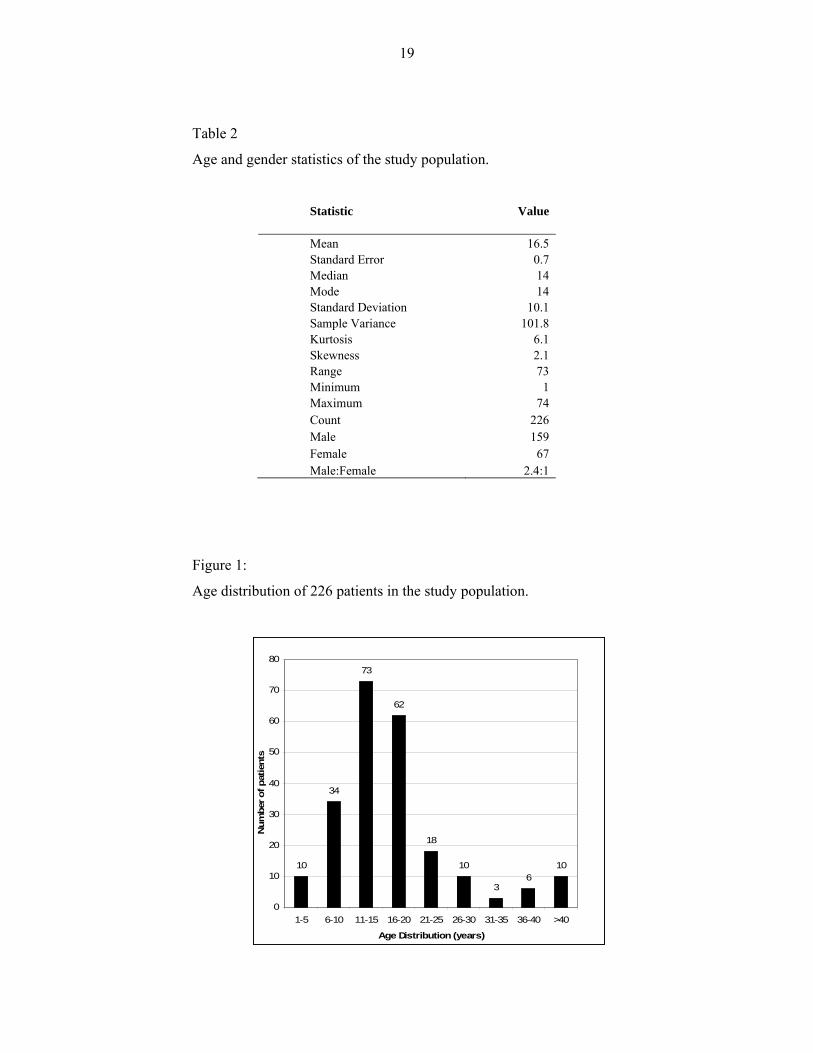
Table 2
Age and gender statistics of the study population.
Statistic Value Mean 16.5 Standard Error 0.7 Median 14 Mode 14 Standard Deviation 10.1 Sample Variance 101.8Kurtosis 6.1 Skewness 2.1Range 73 Minimum 1 Maximum 74 Count 226 Male 159 Female 67 Male:Female 2.4:1
Figure 1:
Age distribution of 226 patients in the study population.
10
34
73
62
18
10
36
10
0
10
20
30
40
50
60
70
80
1-5 6-10 11-15 16-20 21-25 26-30 31-35 36-40 >40
Age Distribution (years)
Num
ber o
f pat
ient
s
20
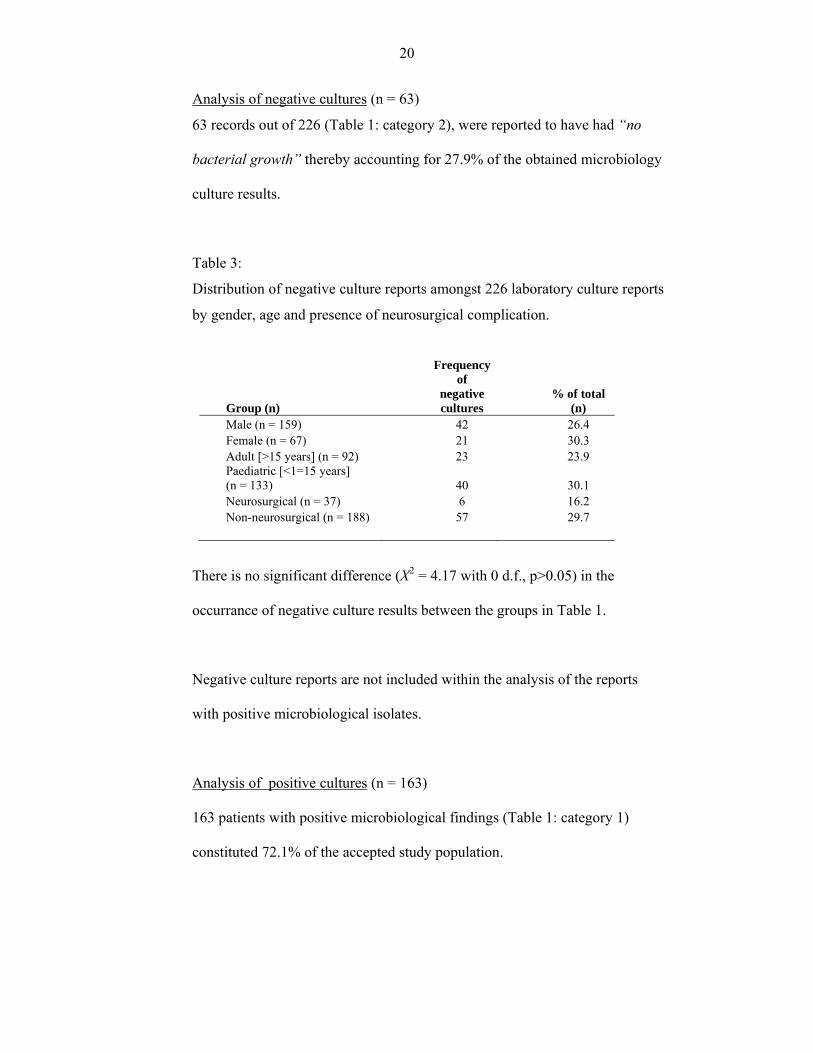
Analysis of negative cultures (n = 63)
63 records out of 226 (Table 1: category 2), were reported to have had “no
bacterial growth” thereby accounting for 27.9% of the obtained microbiology
culture results.
Table 3:
Distribution of negative culture reports amongst 226 laboratory culture reports
by gender, age and presence of neurosurgical complication.
Group (n)
Frequency of
negative cultures
% of total (n)
Male (n = 159) 42 26.4 Female (n = 67) 21 30.3 Adult [>15 years] (n = 92) 23 23.9 Paediatric [<1=15 years] (n = 133) 40 30.1 Neurosurgical (n = 37) 6 16.2 Non-neurosurgical (n = 188) 57 29.7
There is no significant difference (X2 = 4.17 with 0 d.f., p>0.05) in the
occurrance of negative culture results between the groups in Table 1.
Negative culture reports are not included within the analysis of the reports
with positive microbiological isolates.
Analysis of positive cultures (n = 163)
163 patients with positive microbiological findings (Table 1: category 1)
constituted 72.1% of the accepted study population.
21
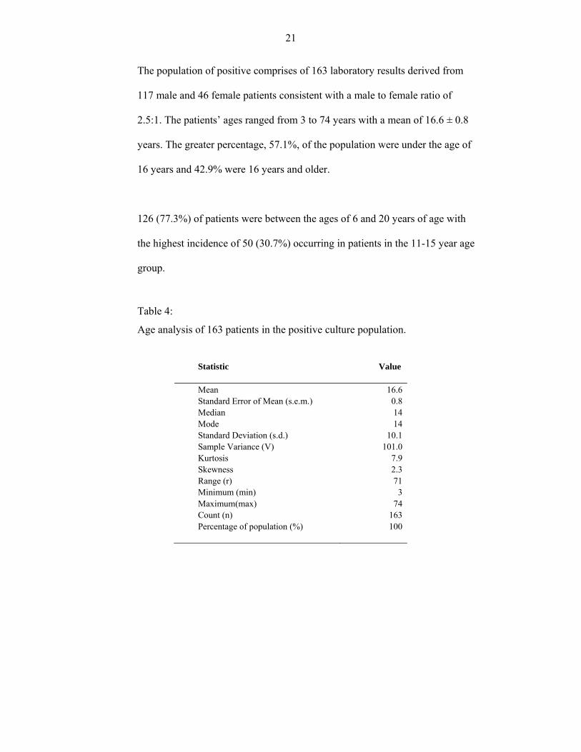
The population of positive comprises of 163 laboratory results derived from
117 male and 46 female patients consistent with a male to female ratio of
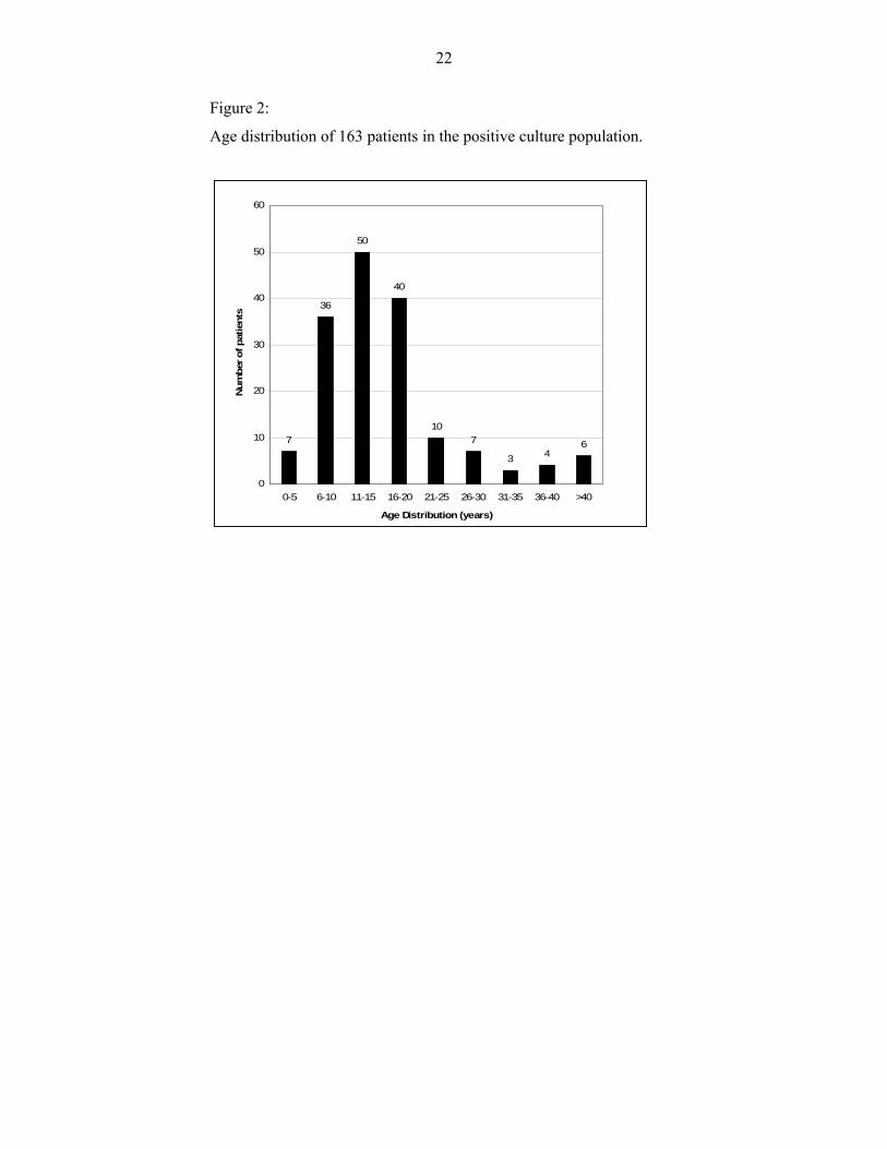
2.5:1. The patients’ ages ranged from 3 to 74 years with a mean of 16.6 ± 0.8
years. The greater percentage, 57.1%, of the population were under the age of
16 years and 42.9% were 16 years and older.
126 (77.3%) of patients were between the ages of 6 and 20 years of age with
the highest incidence of 50 (30.7%) occurring in patients in the 11-15 year age
group.
Table 4:
Age analysis of 163 patients in the positive culture population.
Statistic Value Mean 16.6Standard Error of Mean (s.e.m.) 0.8 Median 14 Mode 14 Standard Deviation (s.d.) 10.1 Sample Variance (V) 101.0 Kurtosis 7.9 Skewness 2.3 Range (r) 71 Minimum (min) 3 Maximum(max) 74 Count (n) 163 Percentage of population (%) 100
22
Figure 2:
Age distribution of 163 patients in the positive culture population.
7
36
50
40
107
3 46
0
10
20
30
40
50
60
0-5 6-10 11-15 16-20 21-25 26-30 31-35 36-40 >40
Age Distribution (years)
Num
ber o
f pat
ient
s
23
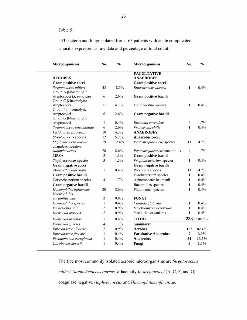
Table 5:
233 bacteria and fungi isolated from 163 patients with acute complicated
sinusitis expressed as raw data and percentage of total count.
Microorganisms No. % Microorganisms No. %
AEROBES FACULTATIVE ANAEROBES
Gram positive cocci Gram positive cocci Streptococcus milleri 43 18.5% Enterococcus durans 1 0.4% Group A β-haemolytic streptococci (S. pyogenes) 6 2.6% Gram positive bacilli Group C β-haemolytic streptococci 11 4.7% Lactobacillus species 1 0.4% Group F β-haemolytic streptococci 6 2.6% Gram negative bacilli Group G β-haemolytic streptococci 1 0.4% Eikenella corrodens 4 1.7% Streptococcus pneumoniae 6 2.6% Proteus mirabilis 1 0.4% Viridans streptococci 10 4.3% ANAEROBES Streptococcus species 12 5.2% Anaerobic cocci Staphylococcus aureus 29 12.4% Peptostreptococcus species 11 4.7% coagulase negative staphylococcus 20 8.6% Peptostreptococcus anaerobius 4 1.7% MRSA 3 1.3% Gram positive bacilli Staphylococcus species 3 1.3% Propionbacterium species 1 0.4% Gram negative cocci Gram negative bacilli Moraxella catarrhalis 1 0.4% Prevotella species 11 4.7% Gram positive bacilli Fusobacterium species 1 0.4% Corynebacterium species 4 1.7% Acinetobacter baumanii 1 0.4% Gram negative bacilli Bacteroides species 1 0.4% Haemophilus influenzae 20 8.6% Photobacter species 1 0.4% Haemophilus parainfluenzae 2 0.9% FUNGI Haemophilus species 1 0.4% Candida glabrata 1 0.4% Escherichia coli 2 0.9% Sacchromyces cerevisiae 1 0.4% Klebsiella oxytoca 2 0.9% Yeast-like organisms 1 0.4% Klebsiella ozaenae 1 0.4% TOTAL 233 100.0% Klebsiella species 4 1.7% Summary: Enterobacter cloacae 2 0.9% Aerobes 192 82.4% Enterobacter faecalis 1 0.4% Facultative Anaerobes 7 3.0% Pseudomonas aeruginosa 1 0.4% Anaerobes 31 13.3% Citrobacter koserii 1 0.4% Fungi 3 1.3%
The five most commonly isolated aerobic microorganisms are Streptococcus
milleri, Staphylococcus aureus, β-haemolytic streptococci (A, C, F, and G),
coagulase negative staphylococcus and Haemophilus influenzae.
24
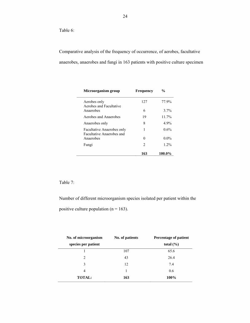
Table 6:
Comparative analysis of the frequency of occurrence, of aerobes, facultative
anaerobes, anaerobes and fungi in 163 patients with positive culture specimen
Microorganism group
Frequency
%
Aerobes only 127 77.9% Aerobes and Facultative Anaerobes 6 3.7%
Aerobes and Anaerobes 19 11.7%
Anaerobes only 8 4.9%
Facultative Anaerobes only 1 0.6% Facultative Anaerobes and Anaerobes 0 0.0%
Fungi 2 1.2%
163 100.0%
Table 7:
Number of different microorganism species isolated per patient within the
positive culture population (n = 163).
No. of microorganism
species per patient
No. of patients Percentage of patient
total (%)
1 107 65.6
2 43 26.4
3 12 7.4
4 1 0.6
TOTAL: 163 100%
25
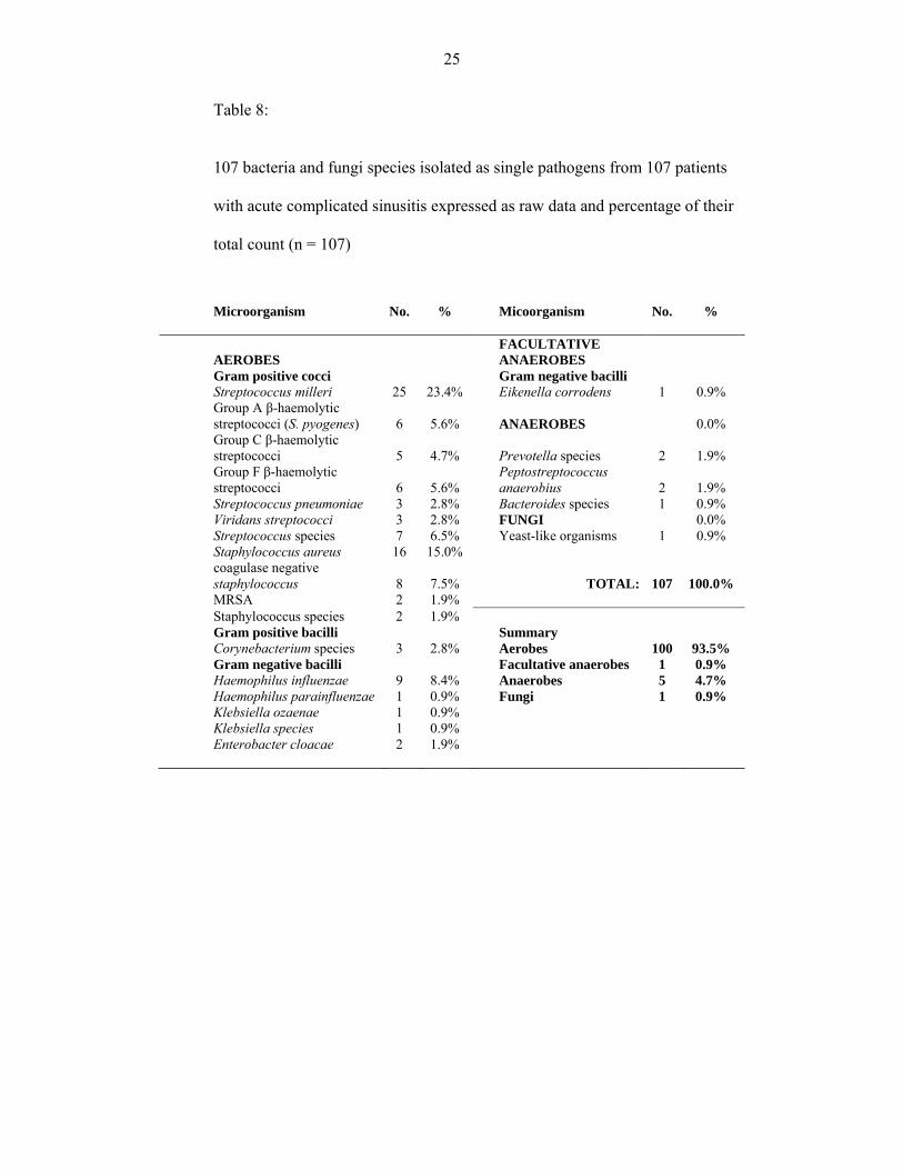
Table 8:
107 bacteria and fungi species isolated as single pathogens from 107 patients
with acute complicated sinusitis expressed as raw data and percentage of their
total count (n = 107)
Microorganism No. % Micoorganism No. %
AEROBES FACULTATIVE ANAEROBES
Gram positive cocci Gram negative bacilli Streptococcus milleri 25 23.4% Eikenella corrodens 1 0.9% Group A β-haemolytic streptococci (S. pyogenes) 6 5.6% ANAEROBES 0.0% Group C β-haemolytic streptococci 5 4.7% Prevotella species 2 1.9% Group F β-haemolytic streptococci 6 5.6%
Peptostreptococcus anaerobius 2 1.9%
Streptococcus pneumoniae 3 2.8% Bacteroides species 1 0.9% Viridans streptococci 3 2.8% FUNGI 0.0% Streptococcus species 7 6.5% Yeast-like organisms 1 0.9% Staphylococcus aureus 16 15.0% coagulase negative staphylococcus 8 7.5% TOTAL: 107 100.0% MRSA 2 1.9% Staphylococcus species 2 1.9% Gram positive bacilli Summary Corynebacterium species 3 2.8% Aerobes 100 93.5% Gram negative bacilli Facultative anaerobes 1 0.9% Haemophilus influenzae 9 8.4% Anaerobes 5 4.7% Haemophilus parainfluenzae 1 0.9% Fungi 1 0.9% Klebsiella ozaenae 1 0.9% Klebsiella species 1 0.9% Enterobacter cloacae 2 1.9%
26
Table 9:
86 microorganisms isolated as a pair of co-existing pathogens from 43 patients
with acute complicated sinusitis expressed as raw data and percentage of their
total count (n = 86)
Microorganisms No. % Microorganisms No. %
AEROBES FACULTATIVE ANAEROBES
Gram positive cocci Gram positive cocci Streptococcus milleri 13 15.1% Enterococcus durans 1 1.2% Group C β-haemolytic streptococci 6 7.0% Gram positive bacilli 0.0% Streptococcus pneumoniae 3 3.5% Lactobacillus species 1 1.2% Viridans streptococci 3 3.5% Gram negative bacilli 0.0%Streptococcus species 4 4.7% Eikenella corrodens 2 2.3% Staphylococcus aureus 8 9.3% Proteus mirabilis 1 1.2%coagulase negative staphylococcus 8 9.3% ANAEROBES 0.0% MRSA 1 1.2% Anaerobic cocci 0.0%
Staphylococcus species 1 1.2% Peptostreptococcus anaerobius 2 2.3%
Gram negative cocci 0.0% Peptostreptococcus species 9 10.5%
Moraxella catarrhalis 1 1.2% Gram positive bacilli 0.0%
Gram negative bacilli 0.0% Propionbacterium species 1 1.2%
Haemophilus influenzae 8 9.3% Gram negative bacilli 0.0% Haemophilus species 1 1.2% Prevotella species 3 3.5% Escherichia coli 1 1.2% Fusobacterium species 1 1.2%
Klebsiella oxytoca 1 1.2% Acinetobacter baumanii 1 1.2%
Klebsiella species 2 2.3% FUNGI 0.0% Pseudomonas aeruginosa 1 1.2% Candida glabrata 1 1.2%
Sacchromyces cerevisiae 1 1.2%
TOTAL: 86 100.0% Summary Aerobes 62 72.1%
Facultative anaerobes 5 5.8%
Anaerobes 17 19.8% Fungi 2 2.3%
27
Table 10:
40 microorganisms isolated as multiple pathogens in 13 patients with acute
complicated sinusitis caused by three or more different microorganisms
expressed as raw data and percentage of their total count (n = 40)
Microorganism No. % Microorganism No. %
AEROBES FACULTATIVE ANAEROBES
Gram positive cocci Gram negative bacilli
Streptococcus milleri 5 12.5% Eikenella corrodens 1 2.5%
Group G β-haemolytic streptococci 1 2.5% ANAEROBES Viridans streptococci 4 10.0% Anaerobic cocci
Streptococcus species 1 2.5%Peptostreptococcus species 2 5.0%
Staphylococcus aureus 5 12.5% Gram negative bacilli
coagulase negative staphylococcus 4 10.0% Prevotella species 6 15.0%
Gram positive bacilli Photobacter species 1 2.5%
Corynebacterium species 1 2.5% Gram negative bacilli Haemophilus influenzae 3 7.5% TOTAL: 40 100.0% Haemophilus parainfluenzae 1 2.5% Escherichia coli 1 2.5% Summary Klebsiella oxytoca 1 2.5% Aerobes 30 75.0%
Klebsiella species 1 2.5% Facultative Anaerobes 1 2.5%
Enterobacter faecalis 1 2.5% Anaerobes 9 22.5% Citrobacter koserii 1 2.5% Fungi 0 0.0%
12 patients had three pathogens isolated from their sinus specimen and 1
patient had 4 pathogens.
The single patient from who 4 pathogens were isolated was found to have
Streptococcus milleri, Viridans streptococci, Haemophillus influenzae and
Photobacter species.
28
Table 11:
Comparative analysis of the frequency of occurrence of microorganisms
between groups of patients with different numbers of microorganism species
identified.
No. of
microorganisms
per patient
Aerobes Facultative
Anaerobes
Anaerobes Fungi Total
1 100 1 5 1 107
2 62 5 17 2 86
3 27 1 8 0 36
4 3 0 1 0 4
192 7 31 3 233
Comparative analysis of the relative distribution of aerobes, facultative
anaerobes, anaerobes and fungi between the four groups of patients was made
using the Chi Squared Distribution. A highly significant difference (3 d.f. X2 =
18.5, p<0.05) was noted.
Distribution of aerobes and anerobes in monomicrobial verses polymicrobial
isolates
There is a significant difference (p<0.05) in the pattern of distribution of
aerobic and anaerobic microorganisms between the mono and polymicrobial
patient groups. This is most likely attributed to the high proportion of:
aerobic microorganisms in patients where a single species was isolated
anaerobic microorganisms in the polymicrobial isolates
29
Patients with monomicrobial isolates accounted for 107 of 163 (65.4%) of the
positive culture population whilst patients with polymicrobial isolates
accounted for the remaining 56 of 163 (34.6%).
Table 12:
Comparison of age and age distribution between 117 male and 46 female
patients in the positive culture population.
Statistic Male Female Mean 15.6 19.1 Standard Error 0.7 2.1 Median 14 15.5 Mode 14 13 Standard Deviation 7.7 14.2 Sample Variance 59.3 201.9 Kurtosis 3.8 4.5 Skewness 1.8 1.9 Range 44 71 Minimum 4 3 Maximum 48 74 Count 117 46 Percentage of total 71.8% 28.2%
Analysis of the mean age of the male and female patients was done with
Student’s t-test for comparison of the two sample means with unequal
variances. This showed no significant difference (t = 1.58, p>0.05) between
the mean ages of male and female patients.
30
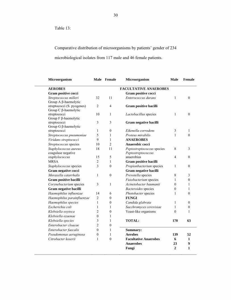
Table 13:
Comparative distribution of microorganisms by patients’ gender of 234
microbiological isolates from 117 male and 46 female patients.
Microorganism Male Female Microorganism Male Female AEROBES FACULTATIVE ANAEROBES Gram positive cocci Gram positive cocci Streptococcus milleri 32 11 Enterococcus durans 1 0 Group A β-haemolytic streptococci (S. pyogenes) 2 4 Gram positive bacilli Group C β-haemolytic streptococci 10 1 Lactobacillus species 1 0 Group F β-haemolytic streptococci 3 3 Gram negative bacilli Group G β-haemolytic streptococci 1 0 Eikenella corrodens 3 1 Streptococcus pneumoniae 5 1 Proteus mirabilis 1 0 Viridans streptococci 9 1 ANAEROBES Streptococcus species 10 2 Anaerobic cocci Staphylococcus aureus 18 11 Peptostreptococcus species 8 3 coagulase negative staphylococcus 15 5
Peptostreptococcus anaerobius 4 0
MRSA 2 1 Gram positive bacilli Staphylococcus species 3 0 Propionbacterium species 1 0 Gram negative cocci Gram negative bacilli Moraxella catarrhalis 1 0 Prevotella species 8 3 Gram positive bacilli Fusobacterium species 1 0Corynebacterium species 3 1 Acinetobacter baumanii 0 1 Gram negative bacilli Bacteroides species 0 1 Haemophilus influenzae 14 6 Photobacter species 1 0 Haemophilus parainfluenzae 2 0 FUNGI Haemophilus species 1 0 Candida glabrata 1 0 Escherichia coli 1 1 Sacchromyces cerevisiae 1 0 Klebsiella oxytoca 2 0 Yeast-like organisms 0 1 Klebsiella ozaenae 0 1 Klebsiella species 3 1 TOTAL: 170 63 Enterobacter cloacae 2 0 Enterobacter faecalis 0 1 Summary: Pseudomonas aeruginosa 0 1 Aerobes 139 52 Citrobacter koserii 1 0 Facultative Anaerobes 6 1 Anaerobes 23 9 Fungi 2 1
31
Comparison of the relative distribution of aerobes, facultative anaerobes,
anaerobes and fungi between male and female patients (Table 13) showed no
significant difference (X2 = 0.62, d.f. = 2, p>0.05) between the two groups.
The frequencies of the five most commonly isolated aerobic microorganisms
(refer to page 23) are compared between the male and female patients and are
shown to have no significant difference (X2 = 1.6, d.f = 4, p>0.05).
The frequencies of Peptostreptococcus and Prevotella species showed no
significant difference (X2 = 0.2, d.f = 1, p>0.05) between male and female
patients.
32
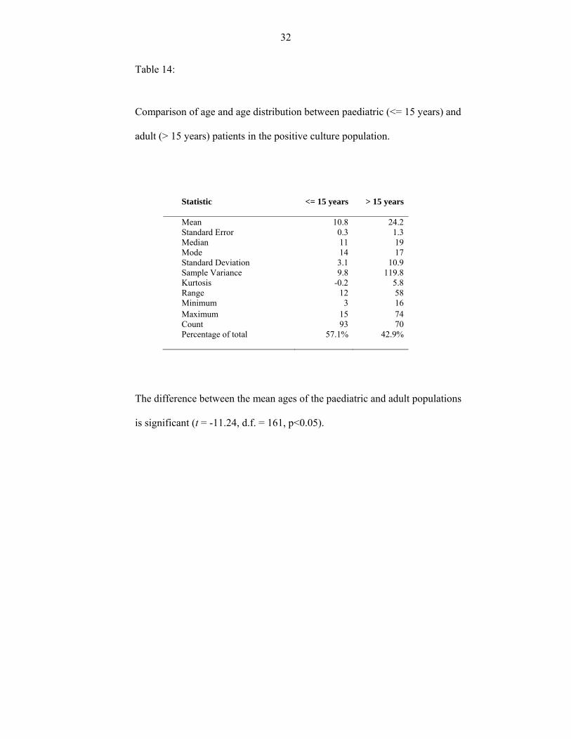
Table 14:
Comparison of age and age distribution between paediatric (<= 15 years) and
adult (> 15 years) patients in the positive culture population.
Statistic <= 15 years > 15 years Mean 10.8 24.2 Standard Error 0.3 1.3 Median 11 19 Mode 14 17 Standard Deviation 3.1 10.9 Sample Variance 9.8 119.8 Kurtosis -0.2 5.8 Range 12 58 Minimum 3 16 Maximum 15 74 Count 93 70 Percentage of total 57.1% 42.9%
The difference between the mean ages of the paediatric and adult populations
is significant (t = -11.24, d.f. = 161, p<0.05).
33
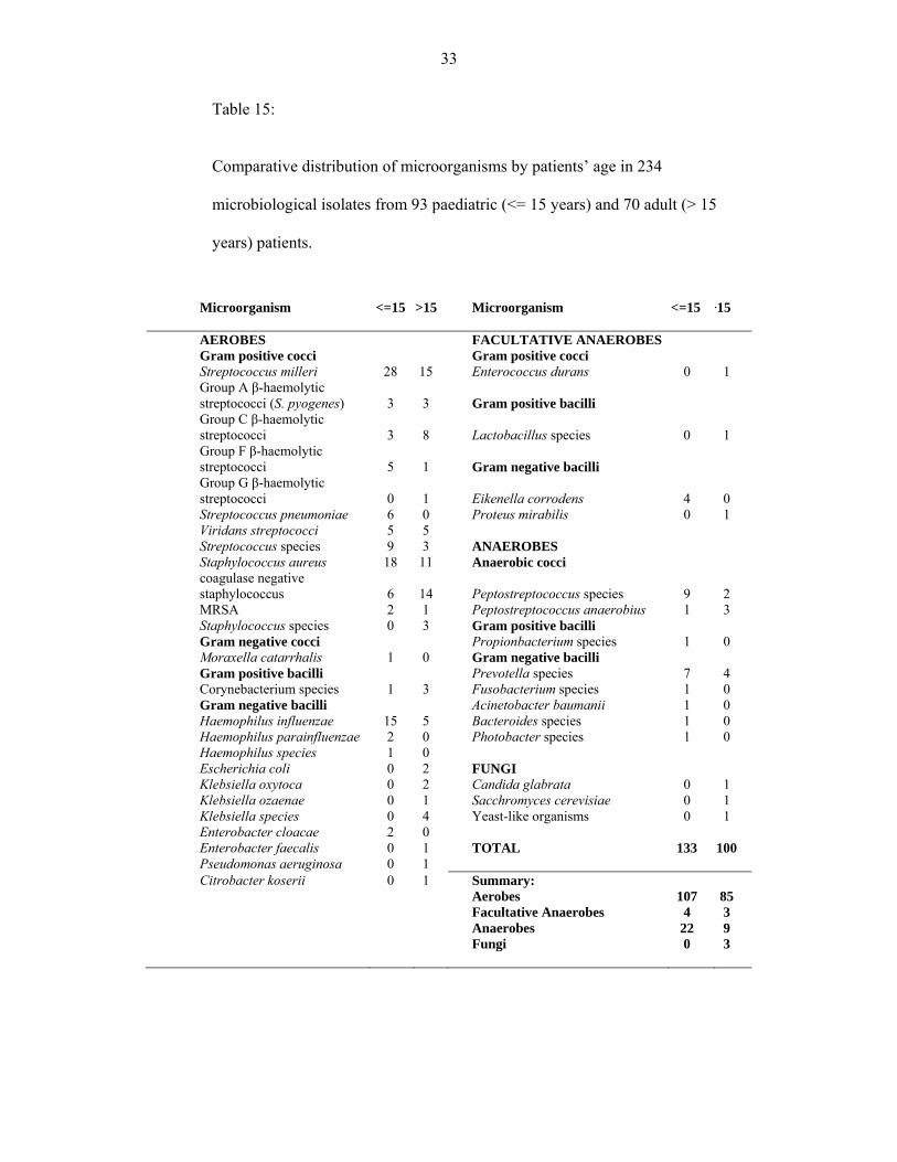
Table 15:
Comparative distribution of microorganisms by patients’ age in 234
microbiological isolates from 93 paediatric (<= 15 years) and 70 adult (> 15
years) patients.
Microorganism <=15 >15 Microorganism <=15 >15 AEROBES FACULTATIVE ANAEROBES Gram positive cocci Gram positive cocci Streptococcus milleri 28 15 Enterococcus durans 0 1 Group A β-haemolytic streptococci (S. pyogenes) 3 3 Gram positive bacilli Group C β-haemolytic streptococci 3 8 Lactobacillus species 0 1 Group F β-haemolytic streptococci 5 1 Gram negative bacilli Group G β-haemolytic streptococci 0 1 Eikenella corrodens 4 0 Streptococcus pneumoniae 6 0 Proteus mirabilis 0 1 Viridans streptococci 5 5 Streptococcus species 9 3 ANAEROBES Staphylococcus aureus 18 11 Anaerobic cocci coagulase negative staphylococcus 6 14 Peptostreptococcus species 9 2 MRSA 2 1 Peptostreptococcus anaerobius 1 3 Staphylococcus species 0 3 Gram positive bacilli Gram negative cocci Propionbacterium species 1 0 Moraxella catarrhalis 1 0 Gram negative bacilli Gram positive bacilli Prevotella species 7 4 Corynebacterium species 1 3 Fusobacterium species 1 0 Gram negative bacilli Acinetobacter baumanii 1 0 Haemophilus influenzae 15 5 Bacteroides species 1 0 Haemophilus parainfluenzae 2 0 Photobacter species 1 0 Haemophilus species 1 0 Escherichia coli 0 2 FUNGI Klebsiella oxytoca 0 2 Candida glabrata 0 1 Klebsiella ozaenae 0 1 Sacchromyces cerevisiae 0 1 Klebsiella species 0 4 Yeast-like organisms 0 1 Enterobacter cloacae 2 0 Enterobacter faecalis 0 1 TOTAL 133 100 Pseudomonas aeruginosa 0 1 Citrobacter koserii 0 1 Summary: Aerobes 107 85 Facultative Anaerobes 4 3
Anaerobes 22 9 Fungi 0 3
34
Comparison of the relative distribution of aerobes, facultative anaerobes,
anaerobes and fungi between paediatric and adult patients (Table 15) showed
no significant difference (X2 = 0.95, d.f. = 2, p>0.05) between the two groups.
The frequencies of the five most commonly isolated aerobic microorganisms
(refer to page 23) are compared between the paediatric and adult patients and
are shown to be significantly different (X2 = 11.3, d.f. = 4, p<0.05).
Haemophilus influenzae is significantly more common (p<0.05) amongst the
paediatric patients and coagulase negative staphylococcus is more common
(p<0.05) amongst the adult patients.
There is no significant difference in the frequencies of occurrance of
Streptococcus milleri, Staphylococcus aureus, β-haemolytic streptococci (A,
C, F, and G) between the adult and paediatric groups.
The frequencies of Peptostreptococcus and Prevotella species showed no
significant difference (X2 = 0.03, d.f = 1, p>0.05) between the two groups.
35
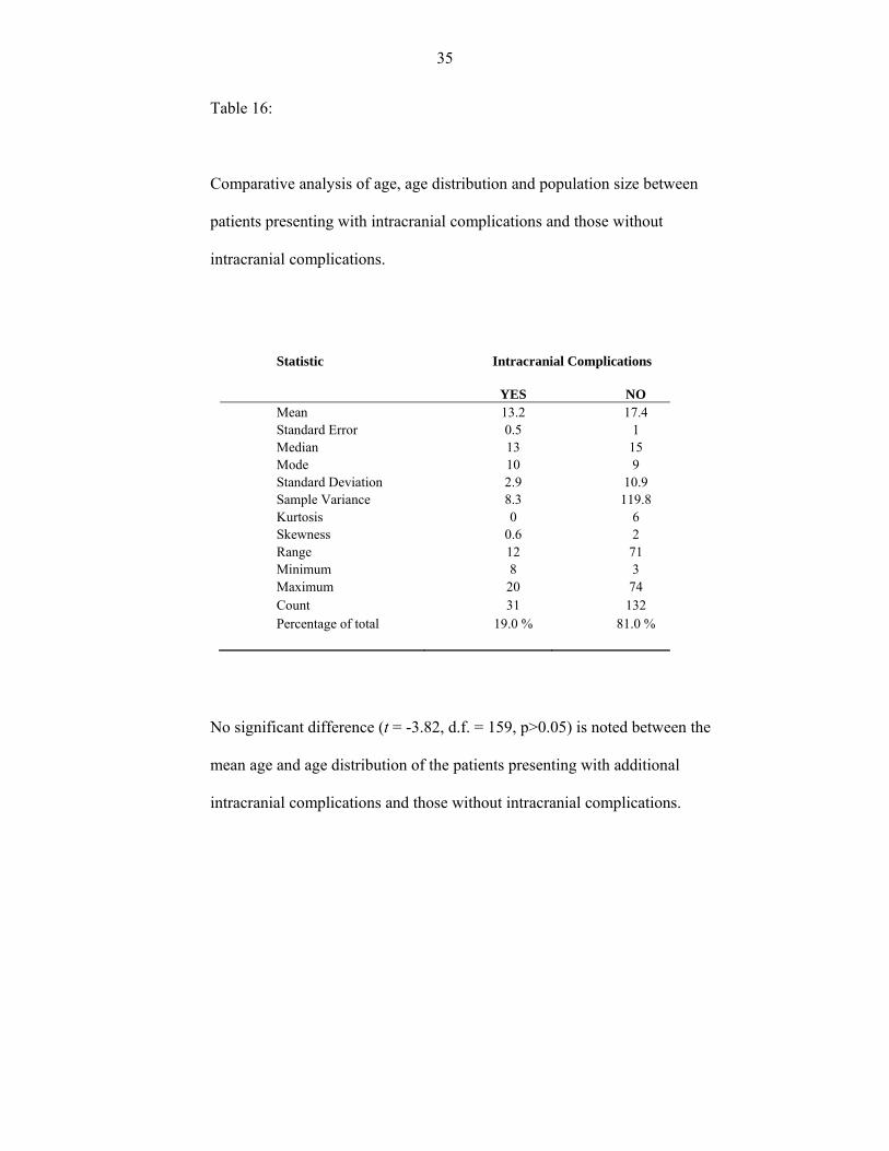
Table 16:
Comparative analysis of age, age distribution and population size between
patients presenting with intracranial complications and those without
intracranial complications.
Statistic
Intracranial Complications
YES NO Mean 13.2 17.4 Standard Error 0.5 1 Median 13 15 Mode 10 9 Standard Deviation 2.9 10.9 Sample Variance 8.3 119.8 Kurtosis 0 6 Skewness 0.6 2 Range 12 71 Minimum 8 3 Maximum 20 74 Count 31 132 Percentage of total 19.0 % 81.0 %
No significant difference (t = -3.82, d.f. = 159, p>0.05) is noted between the
mean age and age distribution of the patients presenting with additional
intracranial complications and those without intracranial complications.
36
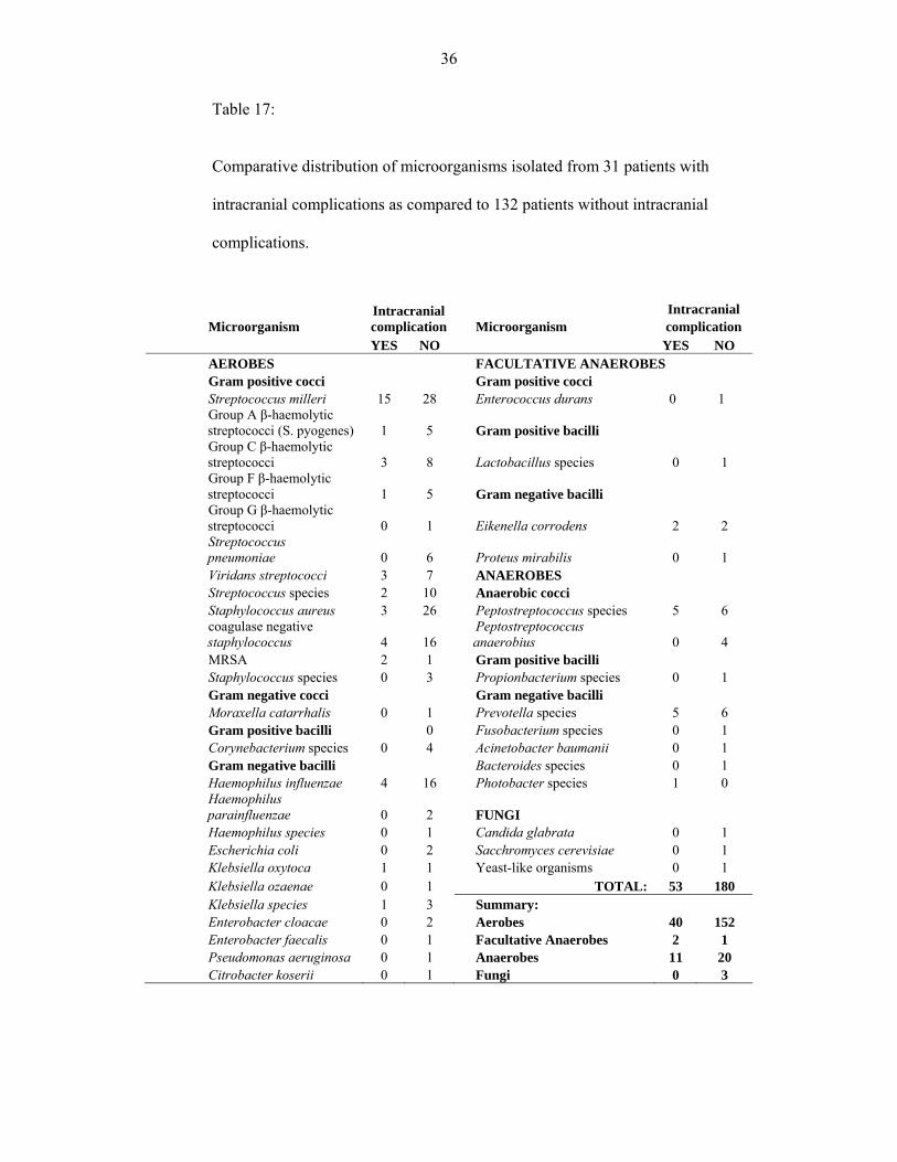
Table 17:
Comparative distribution of microorganisms isolated from 31 patients with
intracranial complications as compared to 132 patients without intracranial
complications.
Microorganism Intracranial complication Microorganism
Intracranial complication
YES NO YES NO AEROBES FACULTATIVE ANAEROBES Gram positive cocci Gram positive cocci Streptococcus milleri 15 28 Enterococcus durans 0 1 Group A β-haemolytic streptococci (S. pyogenes) 1 5 Gram positive bacilli Group C β-haemolytic streptococci 3 8 Lactobacillus species 0 1 Group F β-haemolytic streptococci 1 5 Gram negative bacilli Group G β-haemolytic streptococci 0 1 Eikenella corrodens 2 2 Streptococcus pneumoniae 0 6 Proteus mirabilis 0 1 Viridans streptococci 3 7 ANAEROBES Streptococcus species 2 10 Anaerobic cocci Staphylococcus aureus 3 26 Peptostreptococcus species 5 6 coagulase negative staphylococcus 4 16
Peptostreptococcus anaerobius 0 4
MRSA 2 1 Gram positive bacilli Staphylococcus species 0 3 Propionbacterium species 0 1 Gram negative cocci Gram negative bacilli Moraxella catarrhalis 0 1 Prevotella species 5 6 Gram positive bacilli 0 Fusobacterium species 0 1 Corynebacterium species 0 4 Acinetobacter baumanii 0 1 Gram negative bacilli Bacteroides species 0 1 Haemophilus influenzae 4 16 Photobacter species 1 0 Haemophilus parainfluenzae 0 2 FUNGI Haemophilus species 0 1 Candida glabrata 0 1 Escherichia coli 0 2 Sacchromyces cerevisiae 0 1Klebsiella oxytoca 1 1 Yeast-like organisms 0 1 Klebsiella ozaenae 0 1 TOTAL: 53 180 Klebsiella species 1 3 Summary: Enterobacter cloacae 0 2 Aerobes 40 152 Enterobacter faecalis 0 1 Facultative Anaerobes 2 1 Pseudomonas aeruginosa 0 1 Anaerobes 11 20 Citrobacter koserii 0 1 Fungi 0 3
37
Comparison of the relative distribution of aerobes, facultative anaerobes,
anaerobes and fungi isolated from those patients with intracranial
complications and those without (Table 17) showed no significant difference
(X2 = 5.39, d.f. = 2, p>0.05) between the two groups.
The frequencies of the five most commonly isolated aerobic microorganisms
(refer to page 23) are compared between the neurosurgical and non-
neurosurgical patients and are shown to have no significant difference (X2 =
6.4, d.f = 4, p>0.05).
The frequencies of Peptostreptococcus and Prevotella species showed no
significant difference (X2 = 0.4, d.f = 1, p>0.05) between the two groups.
38
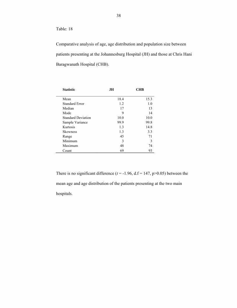
Table: 18
Comparative analysis of age, age distribution and population size between
patients presenting at the Johannesburg Hospital (JH) and those at Chris Hani
Baragwanath Hospital (CHB).
Statistic JH CHB
Mean 18.4 15.3 Standard Error 1.2 1.0 Median 17 13 Mode 9 14 Standard Deviation 10.0 10.0 Sample Variance 99.9 99.8 Kurtosis 1.3 14.8 Skewness 1.3 3.3 Range 45 71 Minimum 3 3 Maximum 48 74 Count 69 93
There is no significant difference (t = -1.96, d.f = 147, p>0.05) between the
mean age and age distribution of the patients presenting at the two main
hospitals.
39
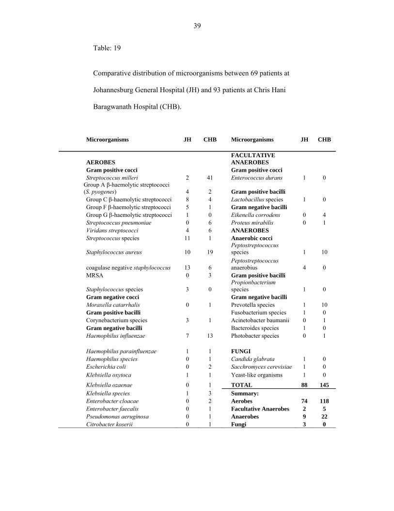
Table: 19
Comparative distribution of microorganisms between 69 patients at
Johannesburg General Hospital (JH) and 93 patients at Chris Hani
Baragwanath Hospital (CHB).
Microorganisms JH CHB Microorganisms JH CHB
AEROBES FACULTATIVE ANAEROBES
Gram positive cocci Gram positive cocci Streptococcus milleri 2 41 Enterococcus durans 1 0
Group A β-haemolytic streptococci (S. pyogenes) 4 2 Gram positive bacilli Group C β-haemolytic streptococci 8 4 Lactobacillus species 1 0 Group F β-haemolytic streptococci 5 1 Gram negative bacilli Group G β-haemolytic streptococci 1 0 Eikenella corrodens 0 4 Streptococcus pneumoniae 0 6 Proteus mirabilis 0 1 Viridans streptococci 4 6 ANAEROBES Streptococcus species 11 1 Anaerobic cocci
Staphylococcus aureus 10 19 Peptostreptococcus species 1 10
coagulase negative staphylococcus 13 6 Peptostreptococcus anaerobius 4 0
MRSA 0 3 Gram positive bacilli
Staphylococcus species 3 0Propionbacterium species 1 0
Gram negative cocci Gram negative bacilli Moraxella catarrhalis 0 1 Prevotella species 1 10Gram positive bacilli Fusobacterium species 1 0 Corynebacterium species 3 1 Acinetobacter baumanii 0 1 Gram negative bacilli Bacteroides species 1 0 Haemophilus influenzae 7 13 Photobacter species 0 1
Haemophilus parainfluenzae 1 1 FUNGI Haemophilus species 0 1 Candida glabrata 1 0 Escherichia coli 0 2 Sacchromyces cerevisiae 1 0 Klebsiella oxytoca 1 1 Yeast-like organisms 1 0
Klebsiella ozaenae 0 1 TOTAL 88 145 Klebsiella species 1 3 Summary: Enterobacter cloacae 0 2 Aerobes 74 118 Enterobacter faecalis 0 1 Facultative Anaerobes 2 5Pseudomonas aeruginosa 0 1 Anaerobes 9 22 Citrobacter koserii 0 1 Fungi 3 0
40
Comparison of the relative distribution of aerobes, facultative anaerobes,
anaerobes and fungi isolated from patients at Johannesburg General Hospital
with those from Chris Hani Baragwanath Hospital (Table 19) showed no
significant difference (X2 = 0.47, d.f. = 1, p>0.05) between the two groups.
The frequencies of the five most commonly isolated aerobic microorganisms
are compared between patients at the two hospitals (Table 19). The study
shows a significant difference (X2 = 40.7, d.f = 4, p<0.05) between common
organisms the Chris Hani Baragwanath Hospital and Johannesburg Hospital.
The frequencies of isolation of Streptococcus milleri and Haemophilus
influenzae are significantly higher (p<0.05) at the Chris Hani Baragwanath
Hospital (Table 19).
The frequencies of isolation of β-haemolytic streptococci (group A, C, G and
F) and coagulase negative staphylococcus are significantly higher (p<0.05) at
the Johannesburg Hospital. The frequency of isolation of Streptococcus milleri
at the Johannesburg Hospital is low (p<0.05).
There is no significant difference (p>0.05) in the frequencies of occurrance of
Staphylococcus aureus at the two main hospitals (Table 19).
The frequencies of Peptostreptococcus and Prevotella species at the two
hospitals (Table 19) showed no significant difference (X2 = 2.1, d.f. = 1,
p>0.05).
41
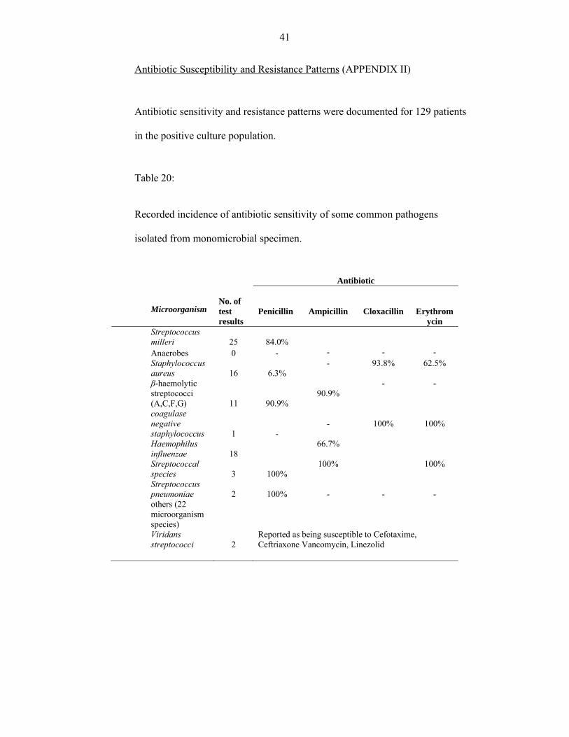
Antibiotic Susceptibility and Resistance Patterns (APPENDIX II)
Antibiotic sensitivity and resistance patterns were documented for 129 patients
in the positive culture population.
Table 20:
Recorded incidence of antibiotic sensitivity of some common pathogens
isolated from monomicrobial specimen.
Microorganism
No. of test results
Antibiotic
Penicillin
Ampicillin
Cloxacillin
Erythromycin
Streptococcus milleri 25 84.0%
Anaerobes 0 - - - - Staphylococcus aureus 16 6.3%
- 93.8% 62.5%
β-haemolytic streptococci (A,C,F,G) 11 90.9%
90.9%
- -
coagulase negative staphylococcus 1 -
-
100%
100%
Haemophilus influenzae 18
66.7%
Streptococcal species 3 100%
100% 100%
Streptococcus pneumoniae 2 100%
-
-
-
others (22 microorganism species) Viridans streptococci 2
Reported as being susceptible to Cefotaxime, Ceftriaxone Vancomycin, Linezolid
42
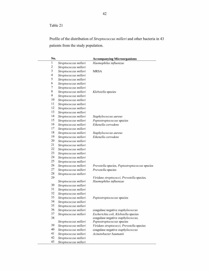
Table 21
Profile of the distribution of Streptococcus milleri and other bacteria in 43
patients from the study population.
No. Accompanying Microorganisms1 Streptococcus milleri Haemophilus influenzae2 Streptococcus milleri 3 Streptococcus milleri MRSA 4 Streptococcus milleri 5 Streptococcus milleri 6 Streptococcus milleri 7 Streptococcus milleri 8 Streptococcus milleri Klebsiella species 9 Streptococcus milleri 10 Streptococcus milleri 11 Streptococcus milleri 12 Streptococcus milleri 13 Streptococcus milleri14 Streptococcus milleri Staphylococcus aureus 15 Streptococcus milleri Peptostreptococcus species16 Streptococcus milleri Eikenella corrodens 17 Streptococcus milleri 18 Streptococcus milleri Staphylococcus aureus 19 Streptococcus milleri Eikenella corrodens 20 Streptococcus milleri 21 Streptococcus milleri 22 Streptococcus milleri 23 Streptococcus milleri 24 Streptococcus milleri 25 Streptococcus milleri 26 Streptococcus milleri Prevotella species, Peptostreptococcus species 27 Streptococcus milleri Prevotella species28 Streptococcus milleri 29
Streptococcus milleri Viridans streptococci, Prevotella species, Haemophilus influenzae
30 Streptococcus milleri 31 Streptococcus milleri 32 Streptococcus milleri33 Streptococcus milleri Peptostreptococcus species 34 Streptococcus milleri35 Streptococcus milleri 36 Streptococcus milleri coagulase negative staphylococcus 37 Streptococcus milleri Escherichia coli, Klebsiella species 38
Streptococcus milleri coagulase negative staphylococcus, Peptostreptococcus species
39 Streptococcus milleri Viridans streptococci, Prevotella species 40 Streptococcus milleri coagulase negative staphylococcus 41 Streptococcus milleri Acinetobacter baumanii 42 Streptococcus milleri 43 Streptococcus milleri
43
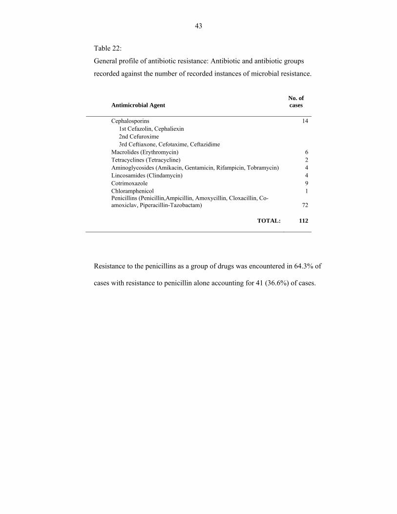
Table 22:
General profile of antibiotic resistance: Antibiotic and antibiotic groups
recorded against the number of recorded instances of microbial resistance.
Antimicrobial Agent No. of cases
Cephalosporins 14 1st Cefazolin, Cephaliexin 2nd Cefuroxime 3rd Ceftiaxone, Cefotaxime, Ceftazidime Macrolides (Erythromycin) 6 Tetracyclines (Tetracycline) 2 Aminoglycosides (Amikacin, Gentamicin, Rifampicin, Tobramycin) 4 Lincosamides (Clindamycin) 4 Cotrimoxazole 9 Chloramphenicol 1 Penicillins (Penicillin,Ampicillin, Amoxycillin, Cloxacillin, Co-amoxiclav, Piperacillin-Tazobactam) 72
TOTAL: 112
Resistance to the penicillins as a group of drugs was encountered in 64.3% of
cases with resistance to penicillin alone accounting for 41 (36.6%) of cases.
44
DISCUSSION
In this study we analysed the microbiology findings from 266 patients selected
from a pool of 301 who were diagnosed with acute complicated sinusitis
(Table 1). The mean age of the patient pool was 16.5 (± 0.7) years. The
patients were predominantly male with a male: female ratio of 2.4:1 (Table 2).
A peak incidence of 73 (32.3 %) occurred in paediatric patients aged 11-15
years with the next highest of 27.4% (62) being in adolescents and young
adults aged 16-20 years (Figure 1).
All patients in this study underwent open sinus surgery as part of their
management during which procedure sinus aspirates and tissues were derived
for microbiological analysis. We found positive cultures in 163 (72.1%) of
cases and negative cultures in 63 (27.9%) of cases which is within the range
(20-33.3%) noted other studies (Penttila M et al 1997; Mortimore et al 1998;
Ali et al 2005). No significant trends (p>0.05) in the occurrance of negative
culture results could be established between the patients on the basis of age,
gender or neurosurgical complication. However, those with neurosurgical
complications appeared to have a slightly lower percentage of negative
microbiology cultures at 16.2% (Table 3).
A total of 233 microorganisms were identified from 163 patients (an average
of 1.4 isolates per specimen) with positive microbiology culture results (Table
5). Aerobic and facultative anaerobes accounted for 199 (85.4%) of the
isolates whilst anaerobes accounted for 31 (13.3%). Two specimens were
45
found to have fungal growths and no bacteria and accounted for 3 (1.3%) of
the positive isolates.
When the data is viewed from the perspective of the patient, 134 (82.0%)
patients had aerobes and/or facultative anaerobes, 19 (11.7%) patients had
both aerobic and anaerobic isolates, 8 (4.9 %) had only anaerobic species and
2 (1.3%) had fungi isolated from their sinus specimen (Table 6). It may also
suffice to say that 27 (16.6%) of the patients with positive isolates had
anaerobes with or without the concurrent presence of aerobes.
The bacteriology of acute sinusitis has been extensively studied. Many reports
in medical literature mention the common aerobic microorganisms in acute
sinusitis as being Streptococcus pneumonia, Haemophilus influenzae and
Moraxella catarhalis (Wald et al 1981; Snyman et al 1988; Wald 1992;
Penttilla et al 1997; Jousimies-Somer et al 1988; Brook 2002; Brook, Frazier
2004; Brook 2005(b); Tellez et al 2006). Stapylococcus aureus and anerobic
bacteria are given greater recognition as pathogens in chronic sinusitis
(Ramadan H 1995, Brook I, 2002; Frederick J, Braude AI, 1974; Kamau at al
2001; Wald 1992; Brook 2005(b)). However, there are also reports that
highlight Staphylococcus aureus as an important pathogen in acute sinusitis
(Hnatuk LAP et al 1994). It is worth noting that all these studies differ from
this study in that they were conducted on patients who did not have severe
disease associated with complications.
46
In this study Streptococcus milleri was the most frequently isolated
microorganism accounting for 43 (18.5 %) isolates. Only recently has
Streptococcus milleri been recognized as a human pathogen accounting for its
absence in earlier reports (Ruoff KL, 1988). Next in descending order of
frequency were Staphylococcus aureus 29 (12.4%), β-haemolytic streptococci
24 (10.8%), coagulase negative staphylococcus 20 (8.6%) and Haemphilus
influenzae 20 (8.6%). Streptococcus pneumoniae accounted for only 6 (2.6%)
of the isolates and only one isolate containing Moraxella catarrhalis (0.4%)
was identified.
Four retrospective studies on the bacteriology of acute complicated sinusitis,
were conducted on a study population with a similar profile to this study. Two
studies (Mortimore S, Wormald PJ, Oliver S. 1998; Oxford LE, McClay J.
2005) found Streptococcus milleri to be the most frequently isolated
microorganism and concurred with the findings of this study. In their patients
with orbital and intracranial complications, Mortimore et al (1998) similarly
observed that Staphylococcus aureus (25%) was the next most frequent
isolate, that there was a low frequency of Streptococcus pneumoniae (4%) and
Moraxella catarrhalis (0.0%) was notably absent. Oxford and McClay (2005)
ranked Staphylococcus aureus and coagulase negative staphylococci third and
fourth respectively. The two other studies (Rosenfeld EA 1993, Ali et al 2005)
both identified Staphylococcus aureus and anaerobes as the predominant
isolates.
47
Anaerobic bacteria are generally associated with chronic sinusitis.
Peptostreptococci, Prevotella, Fusobacterium, Propionbacterium and
Bacteroides are the anaerobes most frequently encountered and reported in
many articles (Ramadan H 1995, Brook 1981, 2002, 2004). There does not
appear to be any consistent pattern in the order of frequency with which they
appear. Anaerobes were isolated 31 times (13.3%) in this study, and the most
frequently isolated anaerobes were Peptostreptococci 15 (6.4%) and
Prevotella species 11 (4.7%). Similarly Brook (2005) isolated
Peptostreptococci in 4 (8.5%) of patients with acute sinusitis. Oxford and
McClay (2004) reported an 18.6% occurrance of anaerobes and identified
Prevotella 4 (8.3%) and Bacteroides 2 (4.2%) species as the most common.
Similarly Ali et al (2005) isolated Prevotella 1 (8.3%) and Bacteroides
(8.3%). A study on children with intracranial abscesses (Rosenfeld E, Rowley
1993) isolated Bacteroides species in 3 out of 4 positive culture specimens.
Mortimore et al (1998) reported a much lower frequency of 1.6% of anaerobes
and made no mention of the identity of the species. The pattern of anaerobes
encountered in this study closely resembles that seen in patients with chronic
sinusitis.
Monomicrobial and Polymicrobial Isolates
Brook 2004 demonstrated the distribution of microorganisms between the
various sinuses was not uniform with the possibility of some sinuses being
free of pathogens. The number of isolates within each positive culture
specimen varied (Table 7). Single microbial species were isolated from 107
(65.6%) specimen whilst polymicrobial isolates, numbering 2 to 4 species per
48
specimen, were found in 56 (34.4%) of cases. Those patients with
monomicrobial isolates had a significantly (p<0.05) higher proportion of
aerobic isolates than those with polymicrobial isolates. Polymicrobial isolates
were found to be associated with a significantly higher (p<0.05) occurrance of
anaerobic microorganisms (Tables 8, 9, 10, 11). Polymicrobial specimen and
the prominence of anaerobes suggests the presence of chronic disease
(Ramadan H 1995, Brook 1981, 2002, 2004).
Male and Female Patients
There was no significant difference (p>0.05) between male and female
patients in the relative distribution of aerobes, facultative anaerobes, anaerobes
and fungi (Table 13). There are no studies which suggest there is any
difference in the microbiology of sinusitis between male and female patients.
Paediatric and Adult Populations
93 (57.1%) of the study population were 15 years and younger (Table 15). No
statistically significant difference (p>0.05) was shown between paediatric and
adult patients in the relative distribution of aerobes, facultative anaerobes,
anaerobes and fungi.
There was a difference in the priority order of the frequency of occurrence of
aerobic bacteria in the paediatric patients. Streptococcus milleri (21.1%, n =
28) and Staphylococcus aureus (13.5%, n = 18) remained in the top two
positions with the third and fourth positions being occupied by Haemophilus
influenzae (11.3%, n = 15) and β-haemolytic streptococci (8.3%, n =11). In
49
the adult patients the frequent aerobes were the same as those common to the
whole patient population.
Haemophilus influenzae occurred more frequently (p<0.05) in the paediatric
group (11.3%, n = 15) as compared to the adult group (5.0%, n = 5). Two
studies within the same region (Snyman et al 1988, Mortimore et al 1998) find
a higher frequencies of Haemophilus influenzae, in 6 (22.2%) and 9 (16.0%)
of isolates, as compared either age group in this study.
On the other hand coagulase negative staphylococcus occurred more
frequently (p<0.05) in the adult patients (14.0%, n = 14) than in the paediatric
patients (4.5%, n = 6). Moraxella catarrhalis was identified in one (0.8%)
isolate in the paediatric group in contrast to other studies (Penttilla et al 1997,
Wald et al 1981, Brook et al 2005b, Tellez et al 2006) that show Moraxella
catarrhalis to be an important pathogen in children and adults.
Neurosurgical and Non-neurosurgical Cases
There was no significant difference (p>0.05) between in the relative
distribution of aerobes, facultative anaerobes, anaerobes and fungi in patients
with intracranial complications as compared to those without such
complications (Table 17).
There were three observations worth noting from the comparative analysis of
patients with intracranial complications. This study finds Streptococcus milleri
to be the predominant pathogen in patients with intracranial complications.
This finding is also reported by Mortimore et al (1998) and Oxford and
50
McClay (2005). The second is that anaerobes account for 20.8% (n = 11) of
the isolates from patients with intracranial complications as compared to
11.1% (n = 20) in those without intracranial complications. Several reports
(Brook 2005c; Rosenfeld 1994, Mortimore et al 1998, Oxford, McClay 2005,
Ali et el 2005) agree that anaerobes are commonly isolated from patents with
intracranial complications. The third observation is that Streptococcus
pneumoniae was not isolated from patients with intracranial complications.
Johannesburg and Chris Hani Baragwanath Hospitals
The Johannesburg Hospital and Chris Hani Baragwanath Hospitals are located
to the north and south respectively, of the Johannesburg city centre and are
separated by distance of about 30 kilometres. Most of the data collected in this
study was derived from these two hospitals. The frequencies of the five most
commonly isolated aerobic microorganisms (refer to page 23) occurring in
patients at these two hospitals (Table 19) are found to be significantly different
(p<0.05). This finding emphasizes the importance of having to know the local
prevalence of microorganisms in ones area of practice (Lauer J 2003).
The order of selected common microorganisms (refer to page 23) isolated at
Johannesburg hospital occur in the order listed below:
β-haemolytic streptococci, 18 (20.5%)
coagulase negative staphylococcus, 13 (14.8%)
Staphylococcus aureus, 10 (11.4%)
Haemophilus influenzae, 7 (8.0%)
Streptococcus milleri, 2 (2.3%)
51
In contrast, the top order of the same selected common (refer to page 23)
isolated microorganisms at the Chris Hani Baragwanath Hospital are:
Streptococcus milleri, 41 (28.3%)
Staphylococcus aureus, 19 (13.1%)
Haemophilus influenzae, 13 (9.0%)
β-haemolytic streptococci, 7 (4.8%)
coagulase negative staphylococcus, 4 (2.8%)
The frequencies of isolation of Streptococcus milleri 41 (28.3%) and
Haemophilus influenzae 13 (9.0%) were noted to be significantly higher
(p<0.05) at the Chris Hani Baragwanath Hospital. The frequencies of isolation
of β-haemolytic streptococci (group A, C, G and F) 18 (20.5%) and coagulase
negative staphylococcus 13 (14.8%) are significantly higher (p<0.05) at the
Johannesburg Hospital. The frequency of isolation of Streptococcus milleri 2
(2.3%) at the Johannesburg Hospital is low (p<0.05). The frequencies of
Staphylococcus aureus, Peptostreptococcus and Prevotella species at the two
hospitals showed no significant difference (p>0.05).
Ruoff (1988) pointed out that Streptococcus milleri has similar characteristics
to the β-haemolytic streptococci (A, C, F and G) with which it is often
mistaken to be. It is possible that this may partially explain the high frequency
of β-haemolytic streptococci 18 (20.5%) coupled with the low frequency of
Streptococcus milleri 2 (2.3%) seen at the Johannesburg Hospital (Table 19).
52
Children under the age of 16 years accounted for 40.6 % of the patients at the
Johannesburg Hospital and 68.8% at the Chris Hani Baragwanath Hospital
making the child to adult ratio 2:3 and 2:1 in the two hospitals respectively.
The proportionally larger adult population of the Johannesburg Hospital would
account for the higher occurrance of coagulase negative staphylococci. In a
similar fashion, the higher proportion of children at Chris Hani Baragwanath
Hospital would account for the significantly higher (p<0.05) occurrance of
Haemophilus influenzae at this hospital.
Antibiotic Susceptibility and Resistance
Antibiotic susceptibility and resistance records were obtained for 129 patients.
Susceptibility tests were not consistently reported for all common antibiotics
and therefore significance levels of microorganism susceptibility and
resistance to antibiotics were not tested in this study. With the results available
certain trends were observed from which reasonable inferences could be made
from specimen that contained single organisms.
Streptococcus milleri was found to be highly susceptible to penicillin,
ampicillin and erythromycin. Streptococcus milleri is recorded as being
sensitive to penicillin in 84% of tested cases (Table 20) with the remaining
16% being tested against cephalosporins. No penicillin resistance to
Streptococcus milleri is reported in this study. Unfortunately this apparent
benefit is offset by the fact that 18 of the 43 specimens that contained
Streptococcus milleri were polymicrobial and contained penicillin resistant
microorganisms in all cases (Table 21).
53
Penicillin, ampicillin and erythromycin were highly effective against β-
haemolytic streptococci, Streptococcus pneumoniae and streptococcal species.
Cloxacillin was a highly effective agent against Staphylococcus aureus and
coagulase negative staphylococci. Methicillin-resistant Staphylococcus was
isolated in specimens from 2 children and 1 adult and was found to be
susceptible to vancomycin.
Haemophilus influenzae was resistant to ampicillin in 2 of 9 cases in which it
was the sole pathogen.
A simple count of the number of reported instances of antibiotic resistance
was tabulated (Table 22). The penicillins, cotrimoxazole and the
cephalosporins had the highest frequency. Snyman et al (1988) reported a
similar trend and additionally pointed out that 100% of anaerobes were
susceptible to metronidazole. In this study some anaerobes were found to be
susceptible to clindamycin and no susceptibility testing to metronidazole was
recorded.
54
CONCLUSION
Streptococcus milleri, Staphylococcus aureus, β-haemolytic streptococci,
coagulase negative staphylococci, Haemophilus influenzae and the anaerobes,
Peptostreptococci and Prevotella species, are important causative pathogens
found in patients presenting with acute complicated bacterial sinusitis to the
Witwatersrand ENT complex.
The frequent isolation of Staphylococcus aureus, coagulase negative
staphylococci and anaerobes suggests acute exacerbation of chronic sinusitis
to be a common presenting form of the disease.
Haemophilus influenzae appears to be a more important pathogen in children
than in adults.
Streptococcus pneumoniae and Moraxella catarrhalis are not major pathogens
in patients with acute complicated sinusitis at the University of the
Witwatersrand ENT complex.
Initial antibiotic therapy, prior to obtaining culture and sensitivity results,
should include antibiotics that cover:
Streptococcus milleri, β-haemolytic streptococci, Staphylococcus aureus,
coagulase negative staphylococci and anaerobes in children and adults
Haemophilus influenzae especially in children
55
REFERENCES
Ali A, Kurien M, Mathews SS, Mathew J. Complications of infective
rhinosinusitis: experience from a developing country. Singapore Med J 2005;
46:540-544
Arjmand EM, Lusck RP, Muntz HR. Pediatric sinusitis and subperiosteal
abscess formation: diagnosis and treatment. Otolaryngol Head Neck Surg
1993; 109:886-894
Ball CN, Connor-Linton J. Web chi square calculator.
www.georgetown.edu/faculty/ballc/webtools/web_chi.html Department of
Linguistics, University of Georgetown 2007
Brook I. Bacteriological features of chronic sinusitis in children. JAMA 1981;
246:967-969
Brook I. Bacteriology of acute and chronic frontal sinusitis. Arch Otolaryngol
Head Neck Surg 2002; 128:583-585
Brook I. Discrepancies in the recovery of bacteria from multiple sinuses in
acute and chronic sinusitis. J Med Microbiol 2004; 53:879-885
Brook I, Frazier EH. Microbiology of recurrent acute rhinosinusitis.
Laryngoscope 2004, 114: 129-131 .
56
Brook I. Bacteriology of acute and chronic ethmoid sinusitis. J Clin Microbiol
2005; 43 (7):3479-3480
Brook I. Microbiology of acute exacerbation of chronic sinusitis. Ann Otol
Rhinol Laryngol 2005b; 114:573-576
Brook I. Microbiology of intracranial abcesses and their associated sinusitis.
Arch Otolaryngol Head Neck 2005c; 131:1017-1019
Butler JC, Hofmann J, Cetron MS, Elliot JA, Facklam RR, Breiman RF. The
continued emergence of drug-resistant Streptococcus pneumoniae in the
United States: An update from the Centers for Disease Control and
Prevention’s Pneumococal Sentinel Surveillance System. J Infect Dis 1996;
174:986-993
Chandler JR, Langenbrunner DJ, Stevens ER. The pathogenesis of orbital
complications of sinusitis. Laryngoscope 1970; 80:1414-1428
Clayman GL, Adams GL, Paugh DR et al. Intracranial complications of
paranasal sinusitis: a combined institutional review. Laryngoscope 1991;
101:234-239
Dowell SF, Schwartz B. Resistant pneumococci: protecting patients through
judicious use of antibiotics. Am Fam Physician 1997; 55:1647-1654
57
Frederick J, Braude AI. Anaerobic infection of the paranasal sinuses. N Engl J
Med 1974; 290:135-137
Gamble RC. Acute inflammation of the orbit in children. Arch Ophthalmol
1933; 10:483-497
Gonzale R, Bartlett JG, Besser RE, Cooper RJ, Hickner JM, Hoffman JR,
Sande MA. Princiles of appropriate antibiotic use for treatment of acute
respiratory tract infections in adults: background, specific aims and methods.
Ann Intern Med 2001; 134:479-486
Gonzales R, Steiner JF, Sande MA. Antibiotic prescribing for adults with
colds, upper respiratory tract infections and bronchitis by ambulatory care
physicians. JAMA 1997; 278:901-904
Hofmann J, Cetron MS, Farley MM, Baughman WS, Facklam RR, Elliot JA et
al. The prevalence of drug resistant Streptococcus pneumoniae in Atlanta. N
Engl J Med 1995; 333:481-486
Hnatuk LAP. Macdonald RE, Papsin BC. Isolated sphenoid sinusitis: The
Toronto Hospital for Sick Children experience and literature review. J
Otolaryngol 1994; 23:36-41
58
Jousimies-Somer HR, Savolainen S, Ylikoski JS. Bacteriological findings of
acute maxillary sinusitis in young adults. J Clin Microbiol 1988; 26:1919-
1925
Kamau JK, Macharia IM, Odhiambo PA. Bacteriology of chronic maxillary
sinusitis at Kenyatta National Hospital, Nairobi. East Afr Med J 2001;
78(7):343-5
Kunin CM. Resistance to antimicrobial drugs – a worldwide calamity. Ann
Intern Med 1993; 118:557-561).
Lauer J. Acute community-acquired sinusitis: A review of epidemiology and
management. Infect Med 2003; 20(1):44-48
Mortimore S, Wormald PJ, Oliver S. Antibiotic choice in acute and
complicated sinusitis. J Laryngol Otol 1998; 112:264-268
Osguthorpe JD, Hochman M. Inflammatory sinus diseases affecting the orbit.
Otolaryngol Clin North Am 1993; 26:657-671
Oxford LE, McClay J. Complications of acute sinusitis in children.
Otolaryngol head neck Surg 2005; 133:32-37
Palm J, Lidman C, Graf P et al. Nitric oxide is reduced in patients with HIV.
Acta Otolaryngol 2000; 120:420-423
59
Penttilla M, Savolainen S, Kiukaannieme H, Forsblom B, Jousimies-Somer.
Bacterial findings in acute maxillary sinusitis: European study. Acta Otol
(Stockh) 1997; supppl 529:165-168
Patt BS, Manning SC. Blindness resulting from the orbital complications of
sinusitis. Otolaryngol Head Neck Surg 1991; 104:789-795
Piccirillo JF, Acute rhinosinusitis: Staging, diagnosis/outcomes assessment.
Infect Med 1998; 15:16-25
Ramadan H. What is the bacteriology of chronic sinusitis in adults? Am J
Otolaryngol 1995; 16(5):303-306
Remmler D, Boles R. Intracranial complications of frontal sinusitis.
Laryngoscope 1980; 90:1814-1824
Rosenfeld EA, Rowley AH. Infectious intracranial complications of sinusitis,
other than meningitis, in children: 12 year review. Clin Infect Dis 1994;
18:750-4
Ruoff KL. Streptococcus anginosus (“Streptococcus milleri”): The
unrecognized pathogen. Clin Microbiol Rev 1988; 1:102-108
60
Schramm VL, Myers EN, Kennerdell JS. Orbital complications of acute
sinusitis: evaluation, management and outcome. Otolaryngol 1978; 86:221-
230
Tellez I, Alba LMD, Reyes MG, Patton E, Hesles H dl G. Microbiology of
acute sinusitis in Mexican patients. Arch Med Res 2006; 37:395-398
Younis RT, Lazar RH, Anand VK. Intracranial complications of sinusitis: a
15-year review of 39 cases. ENT J 2002; 81:636-644
Wald ER, Milmoe GJ, Bowen A’D, Ledesma-Medina J, Salamon N,
Bluestone CD. Acute maxillary sinusitis in children. N Eng J Med 1981;
304(13):749-754
Wald ER. Microbiology of acute and chronic sinusitis in children. J Allery
Clin Immunol 1992; 90:452-460
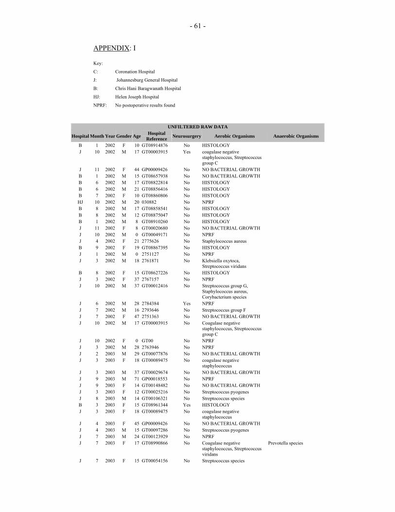
- 61 -
APPENDIX: I
Key:
C: Coronation Hospital
J: Johannesburg General Hospital
B: Chris Hani Baragwanath Hospital
HJ: Helen Joseph Hospital
NPRF: No postoperative results found
UNFILTERED RAW DATA
Hospital Month Year Gender Age Hospital Reference Neurosurgery Aerobic Organisms Anaerobic Organisms
B 1 2002 F 10 GT08914876 No HISTOLOGY J 10 2002 M 17 GT00003915 Yes coagulase negative
staphylococcus, Streptococcus group C
J 11 2002 F 44 GP00009426 No NO BACTERIAL GROWTH B 1 2002 M 15 GT08657938 No NO BACTERIAL GROWTH B 6 2002 M 17 GT08822814 No HISTOLOGY B 6 2002 M 21 GT08856416 No HISTOLOGY B 7 2002 F 10 GT08860806 No HISTOLOGY HJ 10 2002 M 20 030882 No NPRF B 8 2002 M 17 GT08858541 No HISTOLOGY B 8 2002 M 12 GT08875047 No HISTOLOGY B 1 2002 M 8 GT08910260 No HISTOLOGY J 11 2002 F 8 GT00020680 No NO BACTERIAL GROWTH J 10 2002 M 0 GT00049171 No NPRF J 4 2002 F 21 2775626 No Staphylococcus aureus B 9 2002 F 19 GT08867395 No HISTOLOGY J 1 2002 M 0 2751127 No NPRF J 3 2002 M 18 2761871 No Klebsiella oxytoca,
Streptococcus viridans
B 8 2002 F 15 GT08627226 No HISTOLOGY J 3 2002 F 37 2767157 No NPRF J 10 2002 M 37 GT00012416 No Streptococcus group G,
Staphylococcus aureus, Corybacterium species
J 6 2002 M 28 2784384 Yes NPRF J 7 2002 M 16 2793646 No Streptococcus group F J 7 2002 F 47 2751363 No NO BACTERIAL GROWTH J 10 2002 M 17 GT00003915 No Coagulase negative
staphylococcus, Streptococcus group C
J 10 2002 F 0 GT00 No NPRF J 3 2002 M 28 2763946 No NPRF J 2 2003 M 29 GT00077876 No NO BACTERIAL GROWTH J 3 2003 F 18 GT00089475 No coagulase negative
staphylococcus
J 3 2003 M 37 GT00029674 No NO BACTERIAL GROWTH J 9 2003 M 71 GP00018553 No NPRF J 9 2003 F 14 GT00148482 No NO BACTERIAL GROWTH J 3 2003 F 12 GT00025216 No Streptococcus pyogenes J 8 2003 M 14 GT00106321 No Streptococcus species B 3 2003 F 15 GT08961344 Yes HISTOLOGY J 3 2003 F 18 GT00089475 No coagulase negative
staphylococcus
J 4 2003 F 45 GP00009426 No NO BACTERIAL GROWTH J 4 2003 M 15 GT00097286 No Streptococcus pyogenes J 7 2003 M 24 GT00123929 No NPRF J 7 2003 F 17 GT08990866 No Coagulase negative
staphylococcus, Streptococcus viridans
Prevotella species
J 7 2003 F 15 GT00054156 No Streptococcus species
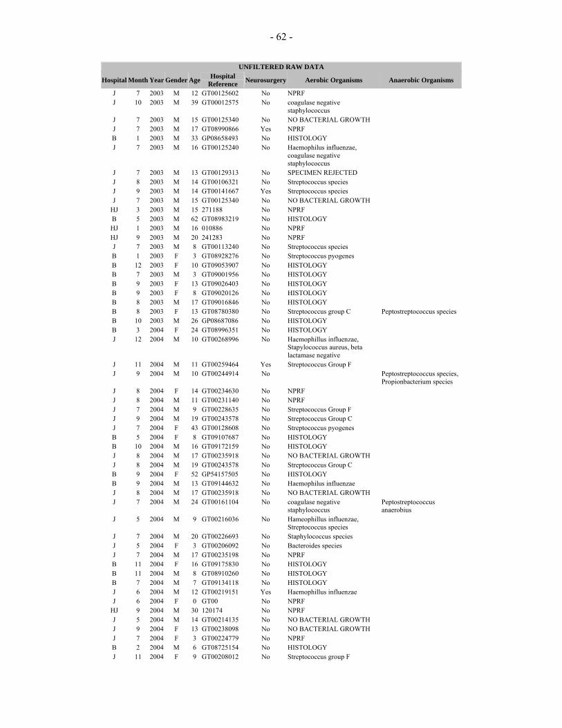
- 62 -
UNFILTERED RAW DATA
Hospital Month Year Gender Age Hospital Reference Neurosurgery Aerobic Organisms Anaerobic Organisms
J 7 2003 M 12 GT00125602 No NPRF J 10 2003 M 39 GT00012575 No coagulase negative
staphylococcus
J 7 2003 M 15 GT00125340 No NO BACTERIAL GROWTH J 7 2003 M 17 GT08990866 Yes NPRF B 1 2003 M 33 GP08658493 No HISTOLOGY J 7 2003 M 16 GT00125240 No Haemophilus influenzae,
coagulase negative staphylococcus
J 7 2003 M 13 GT00129313 No SPECIMEN REJECTED J 8 2003 M 14 GT00106321 No Streptococcus species J 9 2003 M 14 GT00141667 Yes Streptococcus species J 7 2003 M 15 GT00125340 No NO BACTERIAL GROWTH
HJ 3 2003 M 15 271188 No NPRF B 5 2003 M 62 GT08983219 No HISTOLOGY HJ 1 2003 M 16 010886 No NPRF HJ 9 2003 M 20 241283 No NPRF J 7 2003 M 8 GT00113240 No Streptococcus species B 1 2003 F 3 GT08928276 No Streptococcus pyogenes B 12 2003 F 10 GT09053907 No HISTOLOGY B 7 2003 M 3 GT09001956 No HISTOLOGY B 9 2003 F 13 GT09026403 No HISTOLOGY B 9 2003 F 8 GT09020126 No HISTOLOGY B 8 2003 M 17 GT09016846 No HISTOLOGY B 8 2003 F 13 GT08780380 No Streptococcus group C Peptostreptococcus species B 10 2003 M 26 GP08687086 No HISTOLOGY B 3 2004 F 24 GT08996351 No HISTOLOGY J 12 2004 M 10 GT00268996 No Haemophillus influenzae,
Stapylococcus aureus, beta lactamase negative
J 11 2004 M 11 GT00259464 Yes Streptococcus Group F J 9 2004 M 10 GT00244914 No Peptostreptococcus species,
Propionbacterium species J 8 2004 F 14 GT00234630 No NPRF J 8 2004 M 11 GT00231140 No NPRF J 7 2004 M 9 GT00228635 No Streptococcus Group F J 9 2004 M 19 GT00243578 No Streptococcus Group C J 7 2004 F 43 GT00128608 No Streptococcus pyogenes B 5 2004 F 8 GT09107687 No HISTOLOGY B 10 2004 M 16 GT09172159 No HISTOLOGY J 8 2004 M 17 GT00235918 No NO BACTERIAL GROWTH J 8 2004 M 19 GT00243578 No Streptococcus Group C B 9 2004 F 52 GP54157505 No HISTOLOGY B 9 2004 M 13 GT09144632 No Haemophilus influenzae J 8 2004 M 17 GT00235918 No NO BACTERIAL GROWTH J 7 2004 M 24 GT00161104 No coagulase negative
staphylococcus Peptostreptococcus anaerobius
J 5 2004 M 9 GT00216036 No Hameophillus influenzae, Streptococcus species
J 7 2004 M 20 GT00226693 No Staphylococcus species J 5 2004 F 3 GT00206092 No Bacteroides species J 7 2004 M 17 GT00235198 No NPRF B 11 2004 F 16 GT09175830 No HISTOLOGY B 11 2004 M 8 GT08910260 No HISTOLOGY B 7 2004 M 7 GT09134118 No HISTOLOGY J 6 2004 M 12 GT00219151 Yes Haemophillus influenzae J 6 2004 F 0 GT00 No NPRF
HJ 9 2004 M 30 120174 No NPRF J 5 2004 M 14 GT00214135 No NO BACTERIAL GROWTH J 9 2004 F 13 GT00238098 No NO BACTERIAL GROWTH J 7 2004 F 3 GT00224779 No NPRF B 2 2004 M 6 GT08725154 No HISTOLOGY J 11 2004 F 9 GT00208012 No Streptococcus group F
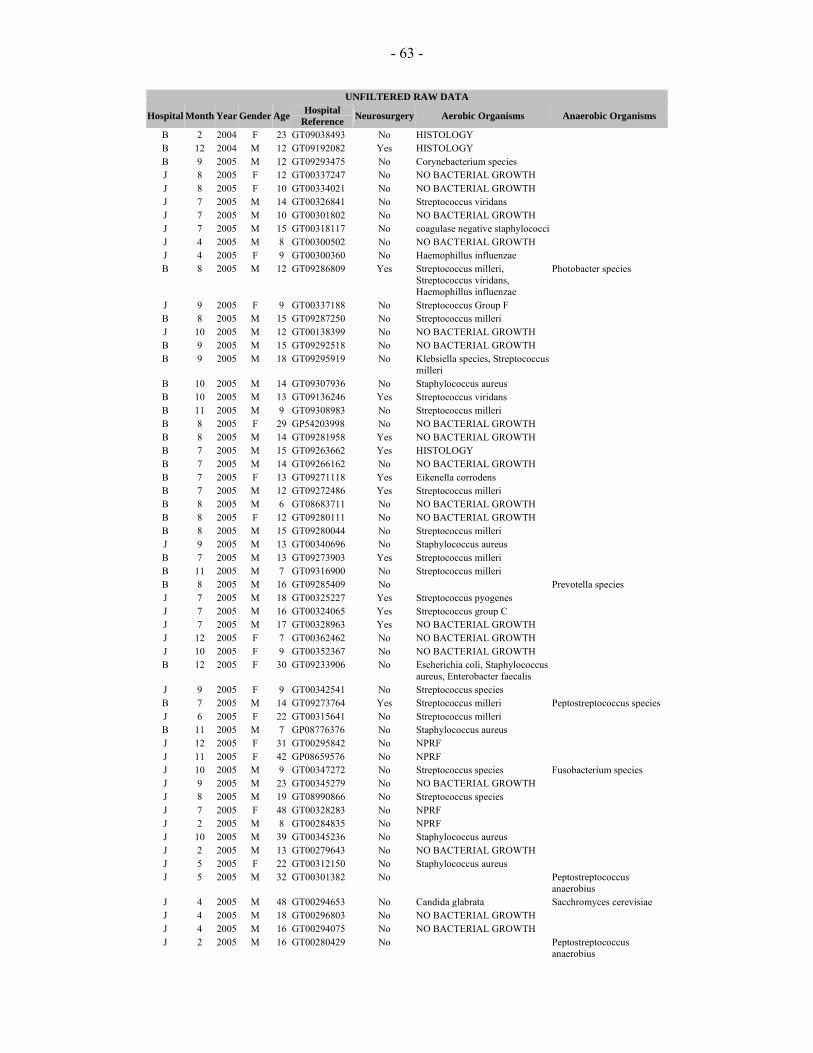
- 63 -
UNFILTERED RAW DATA
Hospital Month Year Gender Age Hospital Reference Neurosurgery Aerobic Organisms Anaerobic Organisms
B 2 2004 F 23 GT09038493 No HISTOLOGY B 12 2004 M 12 GT09192082 Yes HISTOLOGY B 9 2005 M 12 GT09293475 No Corynebacterium species J 8 2005 F 12 GT00337247 No NO BACTERIAL GROWTH J 8 2005 F 10 GT00334021 No NO BACTERIAL GROWTH J 7 2005 M 14 GT00326841 No Streptococcus viridans J 7 2005 M 10 GT00301802 No NO BACTERIAL GROWTH J 7 2005 M 15 GT00318117 No coagulase negative staphylococci J 4 2005 M 8 GT00300502 No NO BACTERIAL GROWTH J 4 2005 F 9 GT00300360 No Haemophillus influenzae B 8 2005 M 12 GT09286809 Yes Streptococcus milleri,
Streptococcus viridans, Haemophillus influenzae
Photobacter species
J 9 2005 F 9 GT00337188 No Streptococcus Group F B 8 2005 M 15 GT09287250 No Streptococcus milleri J 10 2005 M 12 GT00138399 No NO BACTERIAL GROWTH B 9 2005 M 15 GT09292518 No NO BACTERIAL GROWTH B 9 2005 M 18 GT09295919 No Klebsiella species, Streptococcus
milleri
B 10 2005 M 14 GT09307936 No Staphylococcus aureus B 10 2005 M 13 GT09136246 Yes Streptococcus viridans B 11 2005 M 9 GT09308983 No Streptococcus milleri B 8 2005 F 29 GP54203998 No NO BACTERIAL GROWTH B 8 2005 M 14 GT09281958 Yes NO BACTERIAL GROWTH B 7 2005 M 15 GT09263662 Yes HISTOLOGY B 7 2005 M 14 GT09266162 No NO BACTERIAL GROWTH B 7 2005 F 13 GT09271118 Yes Eikenella corrodens B 7 2005 M 12 GT09272486 Yes Streptococcus milleri B 8 2005 M 6 GT08683711 No NO BACTERIAL GROWTH B 8 2005 F 12 GT09280111 No NO BACTERIAL GROWTH B 8 2005 M 15 GT09280044 No Streptococcus milleri J 9 2005 M 13 GT00340696 No Staphylococcus aureus B 7 2005 M 13 GT09273903 Yes Streptococcus milleri B 11 2005 M 7 GT09316900 No Streptococcus milleri B 8 2005 M 16 GT09285409 No Prevotella species J 7 2005 M 18 GT00325227 Yes Streptococcus pyogenes J 7 2005 M 16 GT00324065 Yes Streptococcus group C J 7 2005 M 17 GT00328963 Yes NO BACTERIAL GROWTH J 12 2005 F 7 GT00362462 No NO BACTERIAL GROWTH J 10 2005 F 9 GT00352367 No NO BACTERIAL GROWTH B 12 2005 F 30 GT09233906 No Escherichia coli, Staphylococcus
aureus, Enterobacter faecalis
J 9 2005 F 9 GT00342541 No Streptococcus species B 7 2005 M 14 GT09273764 Yes Streptococcus milleri Peptostreptococcus species J 6 2005 F 22 GT00315641 No Streptococcus milleri B 11 2005 M 7 GP08776376 No Staphylococcus aureus J 12 2005 F 31 GT00295842 No NPRF J 11 2005 F 42 GP08659576 No NPRF J 10 2005 M 9 GT00347272 No Streptococcus species Fusobacterium species J 9 2005 M 23 GT00345279 No NO BACTERIAL GROWTH J 8 2005 M 19 GT08990866 No Streptococcus species J 7 2005 F 48 GT00328283 No NPRF J 2 2005 M 8 GT00284835 No NPRF J 10 2005 M 39 GT00345236 No Staphylococcus aureus J 2 2005 M 13 GT00279643 No NO BACTERIAL GROWTH J 5 2005 F 22 GT00312150 No Staphylococcus aureus J 5 2005 M 32 GT00301382 No Peptostreptococcus
anaerobius J 4 2005 M 48 GT00294653 No Candida glabrata Sacchromyces cerevisiae J 4 2005 M 18 GT00296803 No NO BACTERIAL GROWTH J 4 2005 M 16 GT00294075 No NO BACTERIAL GROWTH J 2 2005 M 16 GT00280429 No Peptostreptococcus
anaerobius
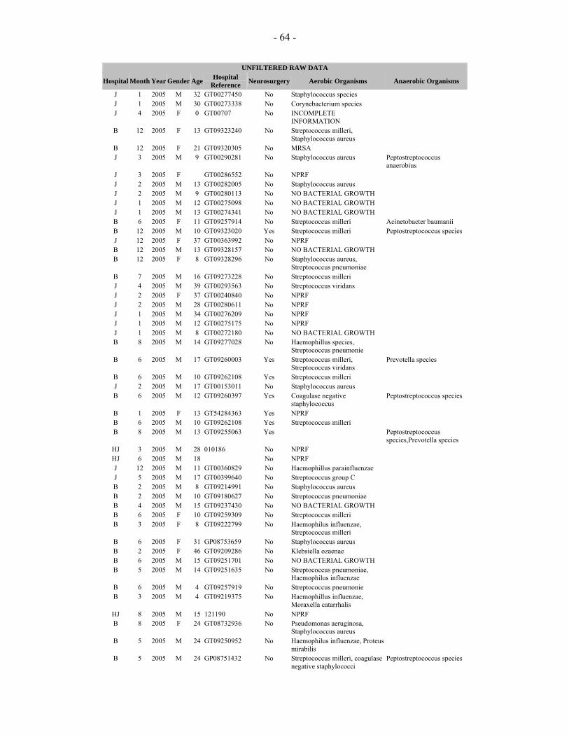
- 64 -
UNFILTERED RAW DATA
Hospital Month Year Gender Age Hospital Reference Neurosurgery Aerobic Organisms Anaerobic Organisms
J 1 2005 M 32 GT00277450 No Staphylococcus species J 1 2005 M 30 GT00273338 No Corynebacterium species J 4 2005 F 0 GT00707 No INCOMPLETE
INFORMATION
B 12 2005 F 13 GT09323240 No Streptococcus milleri, Staphylococcus aureus
B 12 2005 F 21 GT09320305 No MRSA J 3 2005 M 9 GT00290281 No Staphylococcus aureus Peptostreptococcus
anaerobius J 3 2005 F GT00286552 No NPRF J 2 2005 M 13 GT00282005 No Staphylococcus aureus J 2 2005 M 9 GT00280113 No NO BACTERIAL GROWTH J 1 2005 M 12 GT00275098 No NO BACTERIAL GROWTH J 1 2005 M 13 GT00274341 No NO BACTERIAL GROWTH B 6 2005 F 11 GT09257914 No Streptococcus milleri Acinetobacter baumanii B 12 2005 M 10 GT09323020 Yes Streptococcus milleri Peptostreptococcus species J 12 2005 F 37 GT00363992 No NPRF B 12 2005 M 13 GT09328157 No NO BACTERIAL GROWTH B 12 2005 F 8 GT09328296 No Staphylococcus aureus,
Streptococcus pneumoniae
B 7 2005 M 16 GT09273228 No Streptococcus milleri J 4 2005 M 39 GT00293563 No Streptococcus viridans J 2 2005 F 37 GT00240840 No NPRF J 2 2005 M 28 GT00280611 No NPRF J 1 2005 M 34 GT00276209 No NPRF J 1 2005 M 12 GT00275175 No NPRF J 1 2005 M 8 GT00272180 No NO BACTERIAL GROWTH B 8 2005 M 14 GT09277028 No Haemophillus species,
Streptococcus pneumonie
B 6 2005 M 17 GT09260003 Yes Streptococcus milleri, Streptococcus viridans
Prevotella species
B 6 2005 M 10 GT09262108 Yes Streptococcus milleri J 2 2005 M 17 GT00153011 No Staphylococcus aureus B 6 2005 M 12 GT09260397 Yes Coagulase negative
staphylococcus Peptostreptococcus species
B 1 2005 F 13 GT54284363 Yes NPRF B 6 2005 M 10 GT09262108 Yes Streptococcus milleri B 8 2005 M 13 GT09255063 Yes Peptostreptococcus
species,Prevotella species HJ 3 2005 M 28 010186 No NPRF HJ 6 2005 M 18 No NPRF J 12 2005 M 11 GT00360829 No Haemophillus parainfluenzae J 5 2005 M 17 GT00399640 No Streptococcus group C B 2 2005 M 8 GT09214991 No Staphylococcus aureus B 2 2005 M 10 GT09180627 No Streptococcus pneumoniae B 4 2005 M 15 GT09237430 No NO BACTERIAL GROWTH B 6 2005 F 10 GT09259309 No Streptococcus milleri B 3 2005 F 8 GT09222799 No Haemophilus influenzae,
Streptococcus milleri
B 6 2005 F 31 GP08753659 No Staphylococcus aureus B 2 2005 F 46 GT09209286 No Klebsiella ozaenae B 6 2005 M 15 GT09251701 No NO BACTERIAL GROWTH B 5 2005 M 14 GT09251635 No Streptococcus pneumoniae,
Haemophilus influenzae
B 6 2005 M 4 GT09257919 No Streptococcus pneumonie B 3 2005 M 4 GT09219375 No Haemophillus influenzae,
Moraxella catarrhalis
HJ 8 2005 M 15 121190 No NPRF B 8 2005 F 24 GT08732936 No Pseudomonas aeruginosa,
Staphylococcus aureus
B 5 2005 M 24 GT09250952 No Haemophilus influenzae, Proteus mirabilis
B 5 2005 M 24 GP08751432 No Streptococcus milleri, coagulase negative staphylococci
Peptostreptococcus species
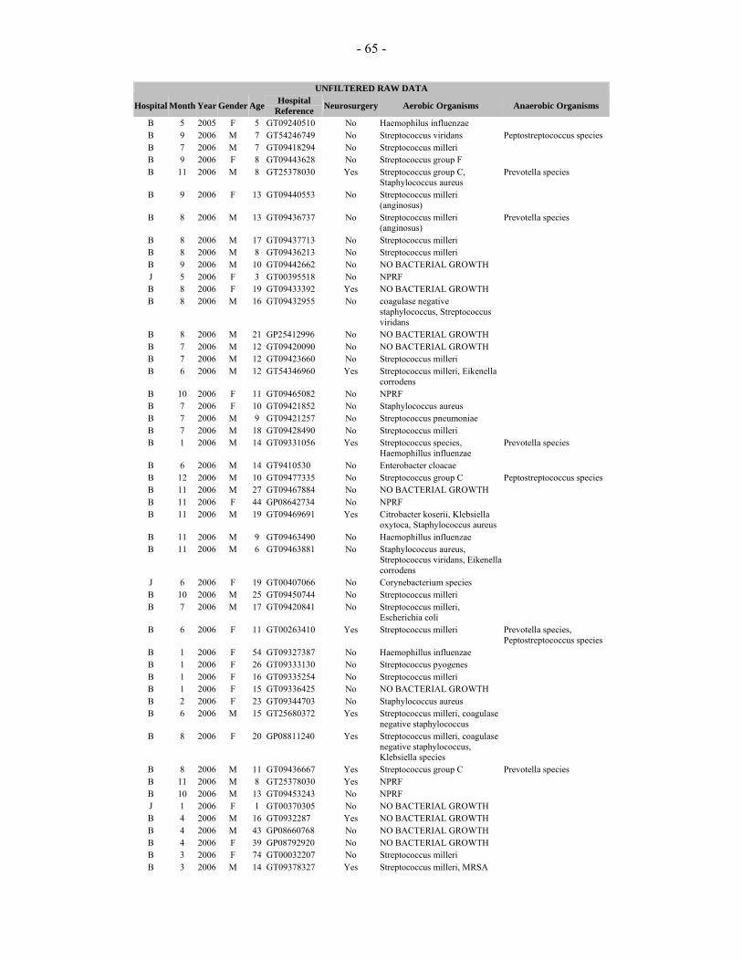
- 65 -
UNFILTERED RAW DATA
Hospital Month Year Gender Age Hospital Reference Neurosurgery Aerobic Organisms Anaerobic Organisms
B 5 2005 F 5 GT09240510 No Haemophilus influenzae B 9 2006 M 7 GT54246749 No Streptococcus viridans Peptostreptococcus species B 7 2006 M 7 GT09418294 No Streptococcus milleri B 9 2006 F 8 GT09443628 No Streptococcus group F B 11 2006 M 8 GT25378030 Yes Streptococcus group C,
Staphylococcus aureus Prevotella species
B 9 2006 F 13 GT09440553 No Streptococcus milleri (anginosus)
B 8 2006 M 13 GT09436737 No Streptococcus milleri (anginosus)
Prevotella species
B 8 2006 M 17 GT09437713 No Streptococcus milleri B 8 2006 M 8 GT09436213 No Streptococcus milleri B 9 2006 M 10 GT09442662 No NO BACTERIAL GROWTH J 5 2006 F 3 GT00395518 No NPRF B 8 2006 F 19 GT09433392 Yes NO BACTERIAL GROWTH B 8 2006 M 16 GT09432955 No coagulase negative
staphylococcus, Streptococcus viridans
B 8 2006 M 21 GP25412996 No NO BACTERIAL GROWTH B 7 2006 M 12 GT09420090 No NO BACTERIAL GROWTH B 7 2006 M 12 GT09423660 No Streptococcus milleri B 6 2006 M 12 GT54346960 Yes Streptococcus milleri, Eikenella
corrodens
B 10 2006 F 11 GT09465082 No NPRF B 7 2006 F 10 GT09421852 No Staphylococcus aureus B 7 2006 M 9 GT09421257 No Streptococcus pneumoniae B 7 2006 M 18 GT09428490 No Streptococcus milleri B 1 2006 M 14 GT09331056 Yes Streptococcus species,
Haemophillus influenzae Prevotella species
B 6 2006 M 14 GT9410530 No Enterobacter cloacae B 12 2006 M 10 GT09477335 No Streptococcus group C Peptostreptococcus species B 11 2006 M 27 GT09467884 No NO BACTERIAL GROWTH B 11 2006 F 44 GP08642734 No NPRF B 11 2006 M 19 GT09469691 Yes Citrobacter koserii, Klebsiella
oxytoca, Staphylococcus aureus
B 11 2006 M 9 GT09463490 No Haemophillus influenzae B 11 2006 M 6 GT09463881 No Staphylococcus aureus,
Streptococcus viridans, Eikenella corrodens
J 6 2006 F 19 GT00407066 No Corynebacterium species B 10 2006 M 25 GT09450744 No Streptococcus milleri B 7 2006 M 17 GT09420841 No Streptococcus milleri,
Escherichia coli
B 6 2006 F 11 GT00263410 Yes Streptococcus milleri Prevotella species, Peptostreptococcus species
B 1 2006 F 54 GT09327387 No Haemophillus influenzae B 1 2006 F 26 GT09333130 No Streptococcus pyogenes B 1 2006 F 16 GT09335254 No Streptococcus milleri B 1 2006 F 15 GT09336425 No NO BACTERIAL GROWTH B 2 2006 F 23 GT09344703 No Staphylococcus aureus B 6 2006 M 15 GT25680372 Yes Streptococcus milleri, coagulase
negative staphylococcus
B 8 2006 F 20 GP08811240 Yes Streptococcus milleri, coagulase negative staphylococcus, Klebsiella species
B 8 2006 M 11 GT09436667 Yes Streptococcus group C Prevotella species B 11 2006 M 8 GT25378030 Yes NPRF B 10 2006 M 13 GT09453243 No NPRF J 1 2006 F 1 GT00370305 No NO BACTERIAL GROWTH B 4 2006 M 16 GT0932287 Yes NO BACTERIAL GROWTH B 4 2006 M 43 GP08660768 No NO BACTERIAL GROWTH B 4 2006 F 39 GP08792920 No NO BACTERIAL GROWTH B 3 2006 F 74 GT00032207 No Streptococcus milleri B 3 2006 M 14 GT09378327 Yes Streptococcus milleri, MRSA
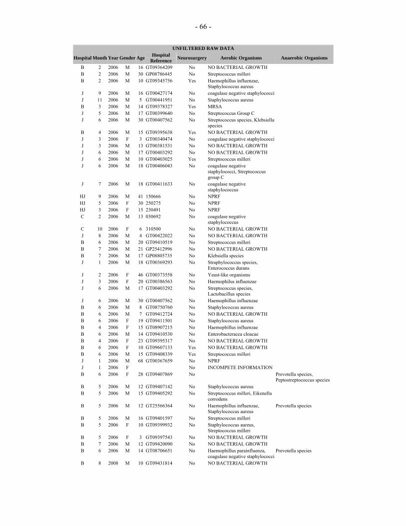
- 66 -
UNFILTERED RAW DATA
Hospital Month Year Gender Age Hospital Reference Neurosurgery Aerobic Organisms Anaerobic Organisms
B 2 2006 M 16 GT09364209 No NO BACTERIAL GROWTH B 2 2006 M 30 GP08786445 No Streptococcus milleri B 2 2006 M 10 GT09345756 Yes Haemophillus influenzae,
Staphylococcus aureus
J 9 2006 M 16 GT00427174 No coagulase negative staphylococci J 11 2006 M 5 GT00441951 No Staphylococcus aureus B 3 2006 M 14 GT09378327 Yes MRSA J 5 2006 M 17 GT00399640 No Streptococcus Group C J 6 2006 M 30 GT00407562 No Streptococcus species, Klebsiella
species
B 4 2006 M 15 GT09395638 Yes NO BACTERIAL GROWTH J 3 2006 F 3 GT00340474 No coagulase negative staphylococci J 3 2006 M 13 GT00381531 No NO BACTERIAL GROWTH J 6 2006 M 17 GT00403292 No NO BACTERIAL GROWTH J 6 2006 M 10 GT00403025 Yes Streptococcus milleri J 6 2006 M 18 GT00406043 No coagulase negative
staphylococci, Streptococcus group C
J 7 2006 M 18 GT00411633 No coagulase negative staphylococcus
HJ 9 2006 M 41 150666 No NPRF HJ 5 2006 F 30 250275 No NPRF HJ 3 2006 F 15 230491 No NPRF C 2 2006 M 13 030692 No coagulase negative
staphylococcus
C 10 2006 F 6 310500 No NO BACTERIAL GROWTH J 8 2006 M 4 GT00422022 No NO BACTERIAL GROWTH B 6 2006 M 20 GT09410519 No Streptococcus milleri B 7 2006 M 21 GP25412996 No NO BACTERIAL GROWTH B 7 2006 M 17 GP08805735 No Klebsiella species J 1 2006 M 18 GT00369293 No Straphylococcus species,
Enterococcus durans
J 2 2006 F 46 GT00373558 No Yeast-like organisms J 3 2006 F 20 GT00386563 No Haemophilus influenzae J 6 2006 M 17 GT00403292 No Streptococcus species,
Lactobacillus species
J 6 2006 M 30 GT00407562 No Haemophillus influenzae B 6 2006 M 8 GT08750760 No Staphylococcus aureus B 6 2006 M 7 GT09412724 No NO BACTERIAL GROWTH B 6 2006 F 19 GT09411501 No Staphylococcus aureus B 4 2006 F 15 GT08907215 No Haemophillus influenzae B 6 2006 M 14 GT09410530 No Enterobacteracea cloacae B 4 2006 F 23 GT09395317 No NO BACTERIAL GROWTH B 6 2006 F 10 GT09607133 Yes NO BACTERIAL GROWTH B 6 2006 M 15 GT09408339 Yes Streptococcus milleri J 1 2006 M 68 GT00367659 No NPRF J 1 2006 F No INCOMPETE INFORMATION B 6 2006 F 28 GT09407869 No Prevotella species,
Peptostreptococcus species B 5 2006 M 12 GT09407142 No Staphylococcus aureus B 5 2006 M 15 GT09405292 No Streptococcus milleri, Eikenella
corrodens
B 5 2006 M 12 GT25566364 No Haemophillus influenzae, Staphylococcus aureus
Prevotella species
B 5 2006 M 16 GT09401597 No Streptococcus milleri B 5 2006 F 10 GT09399932 No Staphylococcus aureus,
Streptococcus milleri
B 5 2006 F 3 GT09397543 No NO BACTERIAL GROWTH B 7 2006 M 12 GT09420090 No NO BACTERIAL GROWTH B 6 2006 M 14 GT08706651 No Haemophillus parainfluenza,
coagulase negative staphylococciPrevotella species
B 8 2008 M 10 GT09431814 No NO BACTERIAL GROWTH
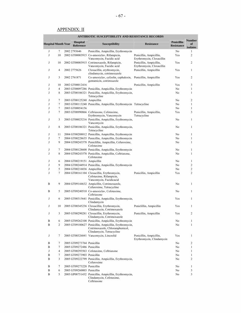
- 67 -
APPENDIX: II ANTIBIOTIC SUSCEPTIBILITY AND RESISTANCE RECORDS
Hospital Month Year Hospital Reference Susceptibility Resistance Penicillin
Resistance
Number of
isolates J 7 2002 2793646 Penicillin, Ampicillin, Erythromycin No 1 J 10 2002 GT00003915 Co-amoxiclav, Rifampicin,
Vancomycin, Fucidic acid Penicillin, Ampicillin, Erythromycin, Cloxacillin
Yes 2
J 10 2002 GT00003915 Cotrimoxazole, Rifampicin, Vancomycin, Fucidic acid
Penicillin, Ampicillin, Erythromycin, Cloxacillin
Yes 2
J 4 2002 2775626 Cloxacillin, erythromycin, clindamycin, cotrimoxazole
Penicillin, Ampicillin Yes 1
J 3 2002 2761871 Co-amoxiclav, cefzolin, cephalexin, gentamicin, cotrimxazole
Penicillin, Ampicillin Yes 2
J 10 2002 GT00012416 Penicillin, Ampicillin Yes 3 J 4 2003 GT00097286 Penicillin, Ampicillin, Erythromycin No 1 J 8 2003 GT00106321 Penicillin, Ampicillin, Erythromycin,
Tetracycline No 1
J 7 2003 GT00125240 Ampicillin No 2 J 7 2003 GT00113240 Penicillin, Ampicillin, Erythromycin Tetracycline No 1 J 7 2003 GT00054156 No 1 J 7 2003 GT08990866 Ceftriaxone, Cefotaxime,
Erythromycin, Vancomycin Penicillin, Ampicillin, Tetracycline
Yes 3
J 3 2003 GT00025216 Penicillin, Ampicillin, Erythromycin, Vancomycin
No 1
J 8 2003 GT00106321 Penicillin, Ampicillin, Erythromycin, Tetracycline
No 1
J 11 2004 GT00208012 Penicillin, Ampicillin, Erythromycin No 1 J 7 2004 GT00228635 Penicillin, Ampicillin, Erythromycin No 1 J 9 2004 GT00243578 Peniclillin, Ampicillin, Cefuroxime,
Cefotaxime No 1
J 7 2004 GT00128608 Penicillin, Ampicillin, Erythromycin No 1 J 8 2004 GT00243578 Penicillin, Ampicillin, Ceftriaxone,
Cefotaxime No 1
J 6 2004 GT00219151 Ampicillin No 1 J 9 2004 GT00244914 Penicillin, Ampicillin, Erythromycin No 2 J 5 2004 GT00216036 Ampicillin No 2 J 7 2004 GT00161104 Cloxacillin, Erythromycin,
Cefotaxime, Rifampicin, Vancomycin, Fucidicacid
Penicillin, Ampicillin Yes 2
B 9 2004 GT09144632 Ampicillin, Cotrimoxazole, Cefuroxime, Tetracycline
No 1
B 5 2005 GT09240510 Co-amoxiclav, Cefotaxime, Ceftriaxone
No 1
J 6 2005 GT00315641 Penicillin, Ampicillin, Erythromycin, Clindamycin
Yes 1
J 10 2005 GT00345236 Cloxacillin, Erythromycin, Clindamycin, Cotrimoxazole
Peniclillin, Ampicillin Yes 1
J 3 2005 GT00290281 Cloxacillin, Erythromycin, Clindamycin, Cotrimoxazole
Penicillin, Ampicillin Yes 2
B 6 2005 GT09262108 Penicillin, Ampicillin, Erythromycin No 1 B 2 2005
GT09180627 Penicillin, Ampicillin, Erythromycin, Cotrimoxazole, Chloramphenicol, Clindamycin, Tetracycline
No 1
J 7 2005 GT00326841 Vancomycin, Linezolid Penicillin, Ampicillin, Erythromycin, Clindamycin
Yes 1
B 7 2005 GT09273764 Penicillin No 2 B 7 2005 GT09272486 Penicillin No 1 J 4 2005 GT00293563 Cefotaxime, Ceftriaxone No 1 B 7 2005 GT09273903 Penicillin No 1 B 3 2005 GT09222799 Penicillin, Ampicillin, Erythromycin,
Cefuroxime No 2
B 7 2005 GT09273228 Penicillin No 1 B 6 2005 GT09260003 Penicillin No 3 B 5 2005 GP08751432 Penicillin, Ampicillin, Erythromycin,
Clindamycin, Cefotaxime, Ceftriaxone
No 3
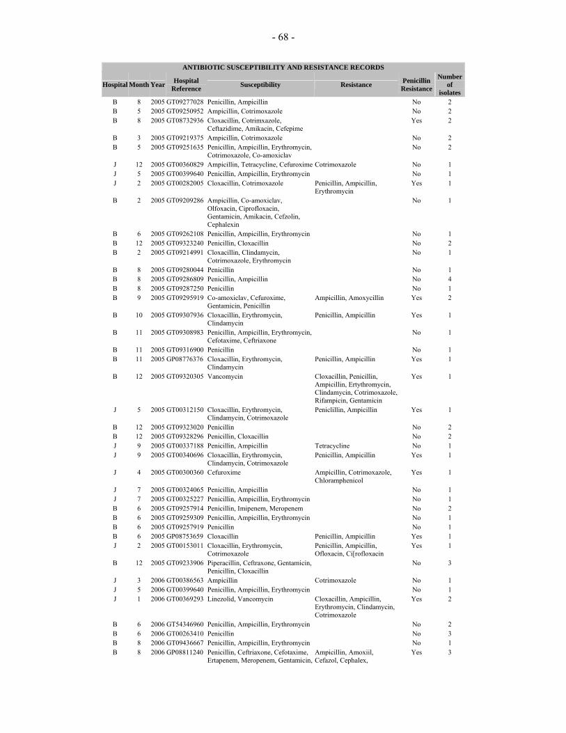
- 68 -
ANTIBIOTIC SUSCEPTIBILITY AND RESISTANCE RECORDS
Hospital Month Year Hospital Reference Susceptibility Resistance Penicillin
Resistance
Number of
isolates B 8 2005 GT09277028 Penicillin, Ampicillin No 2 B 5 2005 GT09250952 Ampicillin, Cotrimoxazole No 2 B 8 2005 GT08732936 Cloxacillin, Cotrimxazole,
Ceftazidime, Amikacin, Cefepime Yes 2
B 3 2005 GT09219375 Ampicillin, Cotrimoxazole No 2 B 5 2005 GT09251635 Penicillin, Ampicillin, Erythromycin,
Cotrimoxazole, Co-amoxiclav No 2
J 12 2005 GT00360829 Ampicillin, Tetracycline, Cefuroxime Cotrimoxazole No 1 J 5 2005 GT00399640 Penicillin, Ampicillin, Erythromycin No 1 J 2 2005 GT00282005 Cloxacillin, Cotrimoxazole Penicillin, Ampicillin,
Erythromycin Yes 1
B 2 2005 GT09209286 Ampicillin, Co-amoxiclav, Olfoxacin, Ciprofloxacin, Gentamicin, Amikacin, Cefzolin, Cephalexin
No 1
B 6 2005 GT09262108 Penicillin, Ampicillin, Erythromycin No 1 B 12 2005 GT09323240 Penicillin, Cloxacillin No 2 B 2 2005 GT09214991 Cloxacillin, Clindamycin,
Cotrimoxazole, Erythromycin No 1
B 8 2005 GT09280044 Penicillin No 1 B 8 2005 GT09286809 Penicillin, Ampicillin No 4 B 8 2005 GT09287250 Penicillin No 1 B 9 2005 GT09295919 Co-amoxiclav, Cefuroxime,
Gentamicin, Penicillin Ampicillin, Amoxycillin Yes 2
B 10 2005 GT09307936 Cloxacillin, Erythromycin, Clindamycin
Penicillin, Ampicillin Yes 1
B 11 2005 GT09308983 Penicillin, Ampicillin, Erythromycin, Cefotaxime, Ceftriaxone
No 1
B 11 2005 GT09316900 Penicillin No 1 B 11 2005 GP08776376 Cloxacillin, Erythromycin,
Clindamycin Penicillin, Ampicillin Yes 1
B 12 2005 GT09320305 Vancomycin Cloxacillin, Penicillin, Ampicillin, Ertythromycin, Clindamycin, Cotrimoxazole, Rifampicin, Gentamicin
Yes 1
J 5 2005 GT00312150 Cloxacillin, Erythromycin, Clindamycin, Cotrimoxazole
Peniclillin, Ampicillin Yes 1
B 12 2005 GT09323020 Penicillin No 2 B 12 2005 GT09328296 Penicillin, Cloxacillin No 2 J 9 2005 GT00337188 Penicillin, Ampicillin Tetracycline No 1 J 9 2005 GT00340696 Cloxacillin, Erythromycin,
Clindamycin, Cotrimoxazole Penicillin, Ampicillin Yes 1
J 4 2005 GT00300360 Cefuroxime Ampicillin, Cotrimoxazole, Chloramphenicol
Yes 1
J 7 2005 GT00324065 Penicillin, Ampicillin No 1 J 7 2005 GT00325227 Penicillin, Ampicillin, Erythromycin No 1 B 6 2005 GT09257914 Penicillin, Imipenem, Meropenem No 2 B 6 2005 GT09259309 Penicillin, Ampicillin, Erythromycin No 1 B 6 2005 GT09257919 Penicillin No 1 B 6 2005 GP08753659 Cloxacillin Penicillin, Ampicillin Yes 1 J 2 2005 GT00153011 Cloxacillin, Erythromycin,
Cotrimoxazole Penicillin, Ampicillin, Ofloxacin, Ci[rofloxacin
Yes 1
B 12 2005 GT09233906 Piperacillin, Ceftraxone, Gentamicin, Penicillin, Cloxacillin
No 3
J 3 2006 GT00386563 Ampicillin Cotrimoxazole No 1 J 5 2006 GT00399640 Penicillin, Ampicillin, Erythromycin No 1 J 1 2006 GT00369293 Linezolid, Vancomycin Cloxacillin, Ampicillin,
Erythromycin, Clindamycin, Cotrimoxazole
Yes 2
B 6 2006 GT54346960 Penicillin, Ampicillin, Erythromycin No 2 B 6 2006 GT00263410 Penicillin No 3 B 8 2006 GT09436667 Penicillin, Ampicillin, Erythromycin No 1 B 8 2006 GP08811240 Penicillin, Ceftriaxone, Cefotaxime,
Ertapenem, Meropenem, Gentamicin, Ampicillin, Amoxiil, Cefazol, Cephalex,
Yes 3
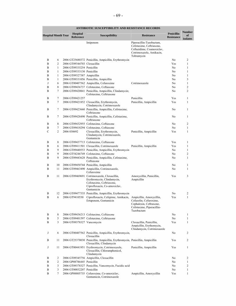
- 69 -
ANTIBIOTIC SUSCEPTIBILITY AND RESISTANCE RECORDS
Hospital Month Year Hospital Reference Susceptibility Resistance Penicillin
Resistance
Number of
isolates Imipenem Piperacillin-Tazobactam,
Cefotaxime, Ceftriaxone, Ceftazidime, Coamoxiclav, Cotrimoxazole, Amikacin, Tobramycin
B 6 2006 GT25680372 Penicillin, Ampicillin, Erythromycin No 2 B 2 2006 GT09344703 Cloxacillin Yes 1 B 1 2006 GT09335254 Penicillin No 1 B 1 2006 GT09333130 Penicillin No 1 B 1 2006 GT09327387 Ampicillin No 1 B 1 2006 GT09331056 Penicillin, Ampicillin No 3 J 6 2006 GT00407562 Ampicillin, Cefuroxime Cotrimoxazole No 1 B 8 2006 GT09436737 Cefotaxime, Ceftiaxone No 2 B 7 2006 GT09420841 Penicillin, Ampicillin, Clindamycin;
Cefotaxime, Ceftriaxone No 2
B 7 2006 GT09421257 Penicillin Yes 1 B 7 2006 GT09421852 Cloxacillin, Erythromycin,
Clindamycin, Cotrimoxazole Penicillin, Ampicillin Yes 1
B 7 2006 GT09423660 Penicillin, Ampicillin, Cefotaxime, Ceftriaxone
No 1
B 7 2006 GT09428490 Penicillin, Ampicillin, Cefotaxime, Ceftriaxone
No 1
B 8 2006 GT09432955 Cefotaxime, Ceftiaxone No 2 B 7 2006 GT09418294 Cefotaxime, Ceftiaxone No 1 C 2 2006 030692 Cloxacillin, Erythromycin,
Clindamycin, Cotrimoxazole, Gentamicin
Penicillin, Ampicillin Yes 1
B 8 2006 GT09437713 Cefotaxime, Ceftiaxone Yes 1 B 6 2006 GT09411501 Cloxacillin, Cotrimoxazole Penicillin, Ampicillin Yes 1 B 9 2006 GT09440553 Penicillin, Ampicillin, Erythromycin No 1 B 9 2006 GT54246749 Cefotaxime, Ceftiaxone No 2 B 9 2006 GT09443628 Penicillin, Ampicillin, Cefotaxime,
Ceftiaxone No 1
B 10 2006 GT09450744 Penicillin, Ampicillin No 1 B 11 2006 GT09463490 Ampicillin, Cotrimoxazole,
Cefuroxime No 1
B 11 2006 GT09469691 Cotrimoxazole, Cloxacillin, Erythromycin, Clindamycin, Cefotaxime, Ceftriaxone, Ciprofloxacin, Co-amoxiclav, Gentamicin
Amoxycillin, Penicillin, Ampicillin
Yes 3
B 12 2006 GT09477335 Penicillin, Ampicillin, Erythromycin No 2 B 6 2006 GT9410530 Ciprofloxacin, Cefepime, Amikacin,
Ertapenem, Gentamicin Ampicillin, Amoxycillin, Cefazolin, Cefuroxime, Cephalexin, Ceftraxone, Cefotaxime, Piperacillin-Tazobactam
Yes 1
B 8 2006 GT09436213 Cefotaxime, Ceftiaxone No 1 B 5 2006 GT09401597 Cefotaxime, Ceftriaxone No 1 B 3 2006 GT09378327 Vancomycin Cloxacillin, Penicillin,
Ampicillin, Erythomycin, Clindamycin, Cotrimoxazole
Yes 1
J 6 2006 GT00407562 Penicillin, Ampicillin, Erythromycin, Cloxacillin
No 2
B 11 2006 GT25378030 Penicillin, Ampicillin, Erythromycin, Cloxacillin, Clindamycin
Penicillin, Ampicillin Yes 2
J 11 2006 GT00441951 Erythromycin, Cotrimoxazole, Cloxacillin, Chloramphenicol, Clindamycin
Penicillin, Ampicillin Yes 1
B 2 2006 GT09345756 Ampicillin, Cloxacillin No 2 B 2 2006 GP08786445 Penicillin No 1 B 3 2006 GT09378327 Penicillin, Vancomycin, Fucidic acid No 2 B 3 2006 GT00032207 Penicillin No 1 B 7 2006 GP08805735 Cefuroxime, Co-amoxiclav,
Gentamicin, Cotrimoxazole Ampicillin, Amoxycillin Yes 1
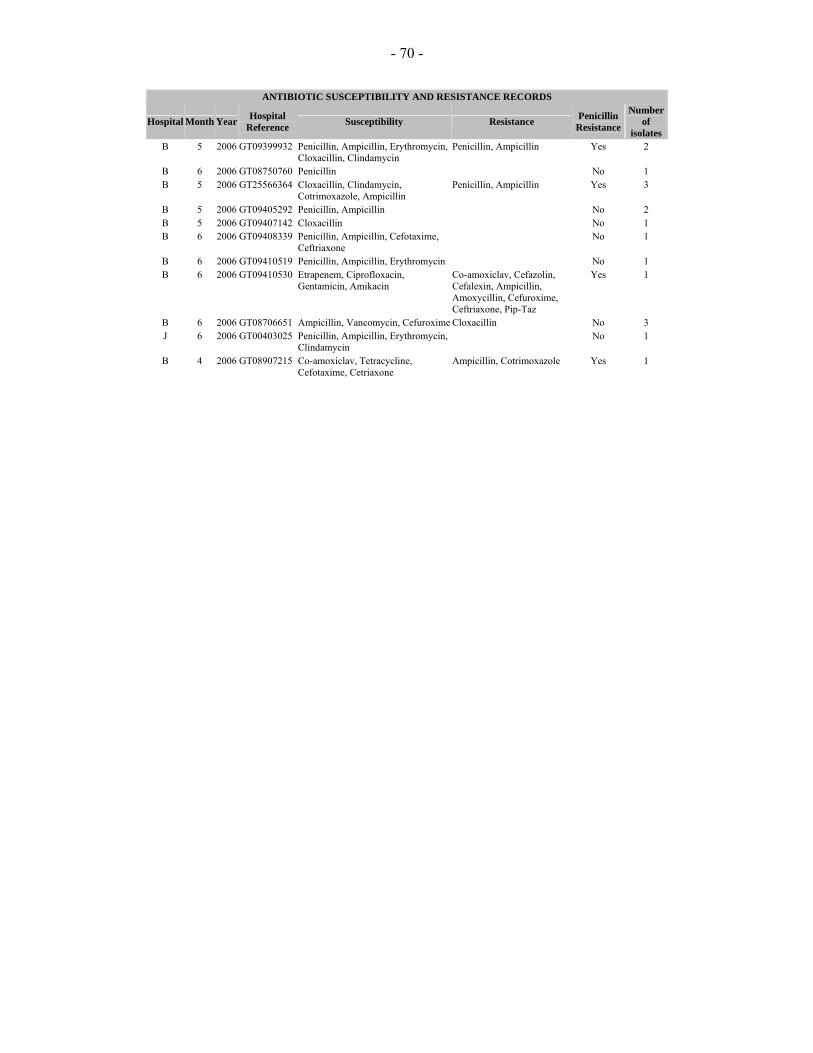
- 70 -
ANTIBIOTIC SUSCEPTIBILITY AND RESISTANCE RECORDS
Hospital Month Year Hospital Reference Susceptibility Resistance Penicillin
Resistance
Number of
isolates B 5 2006 GT09399932 Penicillin, Ampicillin, Erythromycin,
Cloxacillin, Clindamycin Penicillin, Ampicillin Yes 2
B 6 2006 GT08750760 Penicillin No 1 B 5 2006 GT25566364 Cloxacillin, Clindamycin,
Cotrimoxazole, Ampicillin Penicillin, Ampicillin Yes 3
B 5 2006 GT09405292 Penicillin, Ampicillin No 2 B 5 2006 GT09407142 Cloxacillin No 1 B 6 2006 GT09408339 Penicillin, Ampicillin, Cefotaxime,
Ceftriaxone No 1
B 6 2006 GT09410519 Penicillin, Ampicillin, Erythromycin No 1 B 6 2006 GT09410530 Etrapenem, Ciprofloxacin,
Gentamicin, Amikacin Co-amoxiclav, Cefazolin, Cefalexin, Ampicillin, Amoxycillin, Cefuroxime, Ceftriaxone, Pip-Taz
Yes 1
B 6 2006 GT08706651 Ampicillin, Vancomycin, Cefuroxime Cloxacillin No 3 J 6 2006 GT00403025 Penicillin, Ampicillin, Erythromycin,
Clindamycin No 1
B 4 2006 GT08907215 Co-amoxiclav, Tetracycline, Cefotaxime, Cetriaxone
Ampicillin, Cotrimoxazole Yes 1