Embed Size (px)
Citation preview
The Role of Ultrasound in the Diagnosis of Breast Disease 325
(a) (b) (c)
(d) (e)
Figure 9.14 86-year-old female could not access the stereotactic biopsy table,and the breast compressed thickness measured less than 2 cm. (a) Craniocaudalleft mammogram shows clustered calcifications in the medial left breast associatedwith a palpable triangular marker. (b) Grayscale high-resolution ultrasound showsthe clustered microcalcifications (arrow). (c) Ultrasound-guided vacuum-assistedcore needle biopsy using a Suros Celero 12-gauge biopsy device showsthe grouped calcifications within the needle trough. (d) Specimen radiographydemonstrates targeted calcifications in at least two cores. (e) Postbiopsy leftmammogram shows clip marker placement at site of targeted calcifications, whichhave been largely removed during biopsy.
9.9 Breast Cancer Imaging
Breast ultrasound is an important, cost-effective, and quick imagingtool for staging patients with breast cancer in our institution at theM.D. Anderson Cancer Center. Disease is assessed for unifocalityversus multifocality versus multicentricity (Fig. 9.15); and histologicalconfirmation of additional sites of disease via ultrasound-guided biopsy atthe time of ultrasound evaluation is possible.49–52 Regional lymph nodesare assessed to include the ipsilateral axillary, infraclavicular, internalmammary, and supraclavicular nodal basins (Fig. 9.16). Sonographicfeatures that suggest lymph node abnormality include the absence of acentral echogenic “fatty hilus,” and subtle eccentric cortical hypertrophythat can be hypoechoic relative to the rest of the nodal cortex53 (Fig. 9.16).Because afferent lymphatic channels enter a node through the peripheryof the cortex, abnormalities of the cortex can indicate early metastaticinvolvement. It is therefore critical to direct the needle tip to thehypoechoic abnormal cortex (Fig. 9.16) and not the central hilus or the
326 Chapter 9
(d) (e)
(a) (b) (c)
Figure 9.15 56-year-old female presents for staging mammogram and ultrasoundstatus after outside diagnosis of unifocal invasive lobular carcinoma in inferior rightbreast. (a) Craniocaudal and lateromedial right mammograms show postbiopsy clip(arrow) and associated architectural distortion in right 6-o’clock position, markingsite of known invasive lobular cancer. No further mammographic abnormality isseen. (b) Grayscale ultrasound shows known invasive lobular cancer in right 7-o’clock position, presenting as an area of heterogeneous breast tissue (arrow)with marked posterior acoustic shadowing. (c) Grayscale ultrasound shows secondarea of irregular heterogeneous breast tissue in right upper, outer quadrant 10-o’clock position (arrow). Core needle biopsy showed invasive lobular cancer athistopathology, consistent with multicentric disease. (d) Extended-field-of-viewgrayscale ultrasound shows two separate masses in right 7- and 10-o’clockpositions, 5 cm apart. (e) Postprocedural mammograms after placement of secondmarker clip in 10-o’clock position confirms multicentric disease.
normal cortical regions during biopsy to ensure the highest possible yieldof metastases.54–57 Any suspicious lymph nodes identified at the timeof staging ultrasound are subjected to ultrasound-guided needle biopsywith immediate on-site evaluation by a dedicated breast cytopathologist,allowing for accurate nodal staging [American Joint Committee on Cancer(AJCC) criteria] and comprehensive care for the breast cancer patient.54–57
The highest-order suspicious node detected is subjected to biopsy, asthe N stage impacts overall staging, determines eligibility for variouschemotherapy protocols, and also contributes to adjuvant radiation therapyplanning.58 Additionally, these sites of disease can also be used to assessresponse in patients undergoing neoadjuvant chemotherapy.
9.10 Tumor Response
Ultrasound is effective in the assessment of tumor response to neoadjuvantchemotherapy.59 Volume measurements of a mass by ultrasound are
The Role of Ultrasound in the Diagnosis of Breast Disease 327
(a) (b) (c)
(e)(d)
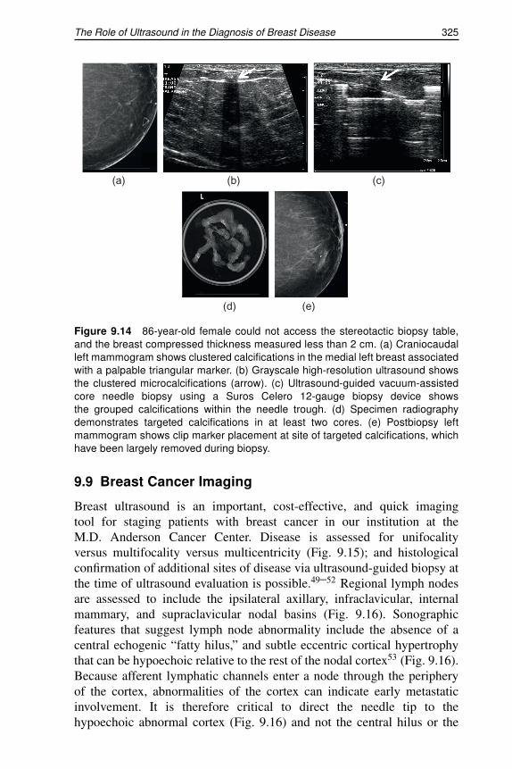
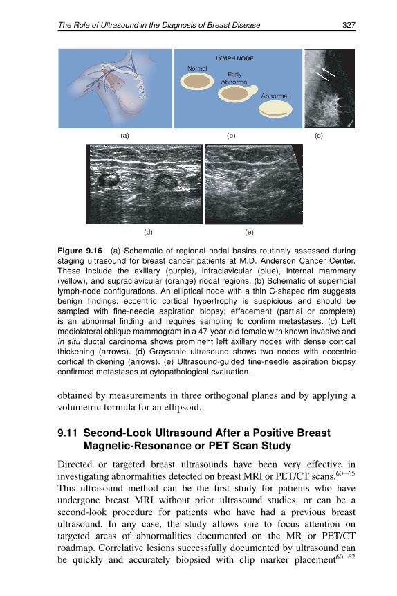
Figure 9.16 (a) Schematic of regional nodal basins routinely assessed duringstaging ultrasound for breast cancer patients at M.D. Anderson Cancer Center.These include the axillary (purple), infraclavicular (blue), internal mammary(yellow), and supraclavicular (orange) nodal regions. (b) Schematic of superficiallymph-node configurations. An elliptical node with a thin C-shaped rim suggestsbenign findings; eccentric cortical hypertrophy is suspicious and should besampled with fine-needle aspiration biopsy; effacement (partial or complete)is an abnormal finding and requires sampling to confirm metastases. (c) Leftmediolateral oblique mammogram in a 47-year-old female with known invasive andin situ ductal carcinoma shows prominent left axillary nodes with dense corticalthickening (arrows). (d) Grayscale ultrasound shows two nodes with eccentriccortical thickening (arrows). (e) Ultrasound-guided fine-needle aspiration biopsyconfirmed metastases at cytopathological evaluation.
obtained by measurements in three orthogonal planes and by applying avolumetric formula for an ellipsoid.
9.11 Second-Look Ultrasound After a Positive BreastMagnetic-Resonance or PET Scan Study
Directed or targeted breast ultrasounds have been very effective ininvestigating abnormalities detected on breast MRI or PET/CT scans.60–65
This ultrasound method can be the first study for patients who haveundergone breast MRI without prior ultrasound studies, or can be asecond-look procedure for patients who have had a previous breastultrasound. In any case, the study allows one to focus attention ontargeted areas of abnormalities documented on the MR or PET/CTroadmap. Correlative lesions successfully documented by ultrasound canbe quickly and accurately biopsied with clip marker placement60–62
328 Chapter 9
(a)
(c)
(b)
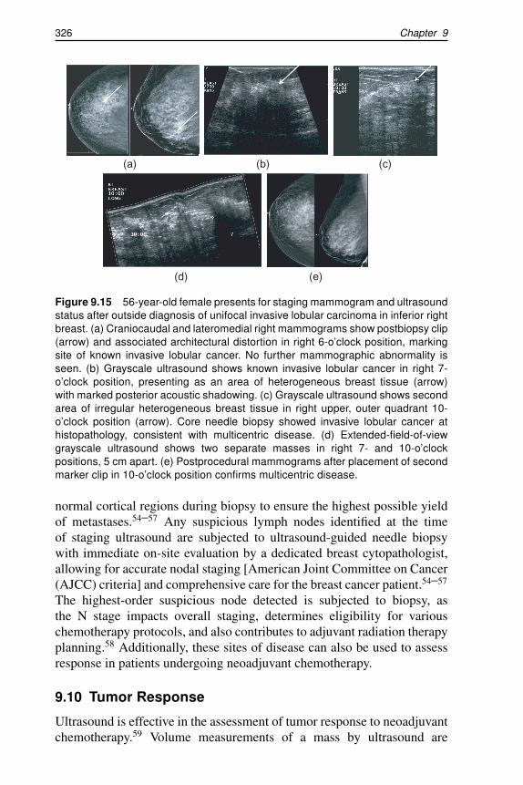
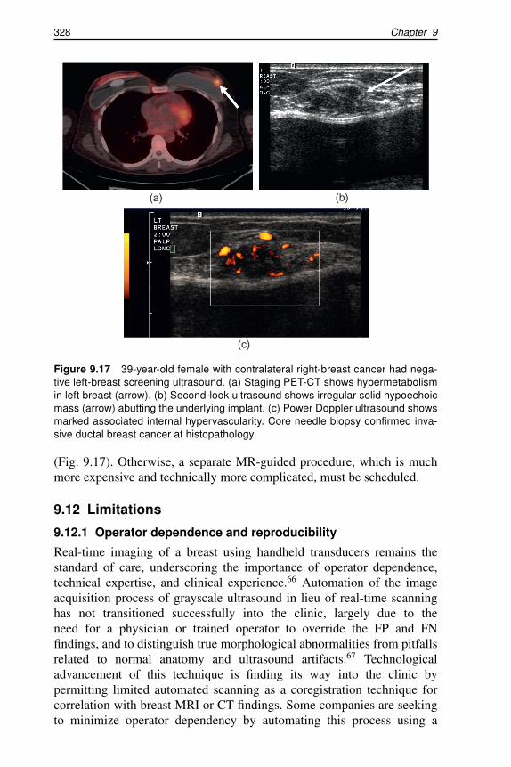
Figure 9.17 39-year-old female with contralateral right-breast cancer had nega-tive left-breast screening ultrasound. (a) Staging PET-CT shows hypermetabolismin left breast (arrow). (b) Second-look ultrasound shows irregular solid hypoechoicmass (arrow) abutting the underlying implant. (c) Power Doppler ultrasound showsmarked associated internal hypervascularity. Core needle biopsy confirmed inva-sive ductal breast cancer at histopathology.
(Fig. 9.17). Otherwise, a separate MR-guided procedure, which is muchmore expensive and technically more complicated, must be scheduled.
9.12 Limitations
9.12.1 Operator dependence and reproducibility
Real-time imaging of a breast using handheld transducers remains thestandard of care, underscoring the importance of operator dependence,technical expertise, and clinical experience.66 Automation of the imageacquisition process of grayscale ultrasound in lieu of real-time scanninghas not transitioned successfully into the clinic, largely due to theneed for a physician or trained operator to override the FP and FNfindings, and to distinguish true morphological abnormalities from pitfallsrelated to normal anatomy and ultrasound artifacts.67 Technologicaladvancement of this technique is finding its way into the clinic bypermitting limited automated scanning as a coregistration technique forcorrelation with breast MRI or CT findings. Some companies are seekingto minimize operator dependency by automating this process using a