Embed Size (px)
Citation preview

APW Dental Services, PC
IN THIS ISSUE:APW: New Features, New Services, New Update
New technology. We are pleased to announce that APW will soon install a new 3G (third generation) cone beam volumetric tomography (CBVT) scannerthat will revolutionize our imaging. This new technology utilizes a 12-bit imageintensifier that displays 4096 shades of gray as opposed to the current 256shades captured by our 8-bit. You will immediately see the difference in greaterclarity and greater accuracy. Patients benefit from scan time being reduced from70 to 30 seconds, and the already low-dose radiation will be reduced by 4 to 5times. We expect the new machine to be installed by year’s end.
Strategic Partners. APW has partnered with Biomedical Modeling, Inc.,Boston, MA, who is the leader in creating 3D anatomically accurate stereolithic biomedical models. These models have a wide range ofapplications and are used for pre-surgical treatment planning for conjoinedSiamese twins; enable accurate anaplastic prostheses to be manufactured for trauma and cancer victims; to repair congenital defects; and now, forsurgical guides for dental implant placement based on APW CBVT dental scans. BMI has partnered with the Marotta Dental Studio to fabricate surgical guides from these BioDental™ Models.
Services. Our cone beam studies are used for many purposes that include:dental implants, TMJ (both open and closed), impacted teeth, craniofacialabnormalities, pathology, mixed-dentition analysis, mid-palate implants, andmore. Since so much information can be learned from these scans whencompared to dental X-rays or panorexes, we encourage cone beam tomographsfor interim diagnoses during mid-orthodontic treatment, for chronic endodonticlesions, or after a patient has already had a diagnostic scan in the same jaw atAPW and post-treatment sites, such as healing sockets, ridge augmentations,and sinus grafts need to be evaluated. To utilize this mid-treatment service, weoffer a special fee of $125 with the understanding that a report will only beprovided for a single prescribed site. Call for more information.
SimPlant® Master Site.APW is the only U.S. dentalradiological lab that is also a SimPlant® Master Site.In addition to NewTom®
8x11” films or glossy prints,we provide CT scans inSimPlant® Versions 7, 8, andsoon-to-be-released Version 9 that renders sagittal slices through 3D images.
3D CONE BEAM
VOLUMETRIC
TOMOGRAPHY
DENTAL CT SCANSAPW Dental Services, PC • 34 East 62nd Street, New York, NY 10021 Vol. 2, October 2004
Surgical Guides
BioDental™ Models
Craniofrontal Dysplasia
APW Dental Services, PC | phone: 212.838.8302 | toll free: 1.888.APW.XRAY | fax: 212.838.8462 | web: www.apwdentalservices.com
97260_APW 09/24/2004 05:56 AM Page 1
APW Dental Services, PC | phone: 212.838.8302 | toll free: 1.888.APW.XRAY | fax: 212.838.8462 | web: www.apwdentalservices.com
2
97260_APW 09/24/2004 05:56 AM Page 2
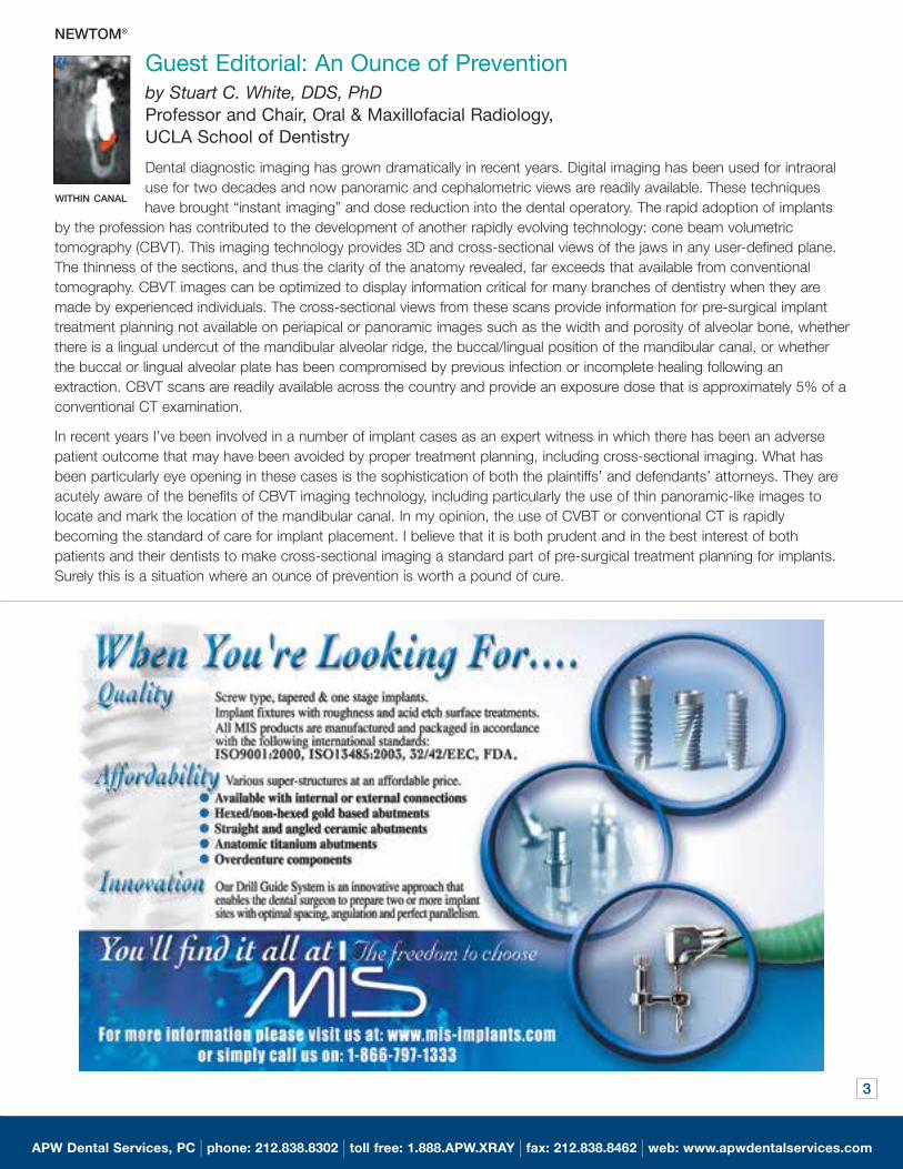
Guest Editorial: An Ounce of Prevention by Stuart C. White, DDS, PhDProfessor and Chair, Oral & Maxillofacial Radiology, UCLA School of Dentistry
Dental diagnostic imaging has grown dramatically in recent years. Digital imaging has been used for intraoral
use for two decades and now panoramic and cephalometric views are readily available. These techniques
have brought “instant imaging” and dose reduction into the dental operatory. The rapid adoption of implants
by the profession has contributed to the development of another rapidly evolving technology: cone beam volumetric
tomography (CBVT). This imaging technology provides 3D and cross-sectional views of the jaws in any user-defined plane.
The thinness of the sections, and thus the clarity of the anatomy revealed, far exceeds that available from conventional
tomography. CBVT images can be optimized to display information critical for many branches of dentistry when they are
made by experienced individuals. The cross-sectional views from these scans provide information for pre-surgical implant
treatment planning not available on periapical or panoramic images such as the width and porosity of alveolar bone, whether
there is a lingual undercut of the mandibular alveolar ridge, the buccal/lingual position of the mandibular canal, or whether
the buccal or lingual alveolar plate has been compromised by previous infection or incomplete healing following an
extraction. CBVT scans are readily available across the country and provide an exposure dose that is approximately 5% of a
conventional CT examination.
In recent years I’ve been involved in a number of implant cases as an expert witness in which there has been an adverse
patient outcome that may have been avoided by proper treatment planning, including cross-sectional imaging. What has
been particularly eye opening in these cases is the sophistication of both the plaintiffs’ and defendants’ attorneys. They are
acutely aware of the benefits of CBVT imaging technology, including particularly the use of thin panoramic-like images to
locate and mark the location of the mandibular canal. In my opinion, the use of CVBT or conventional CT is rapidly
becoming the standard of care for implant placement. I believe that it is both prudent and in the best interest of both
patients and their dentists to make cross-sectional imaging a standard part of pre-surgical treatment planning for implants.
Surely this is a situation where an ounce of prevention is worth a pound of cure.
WITHIN CANAL
NEWTOM®
APW Dental Services, PC | phone: 212.838.8302 | toll free: 1.888.APW.XRAY | fax: 212.838.8462 | web: www.apwdentalservices.com
3
97260_APW_2 09/29/2004 04:03 AM Page 3
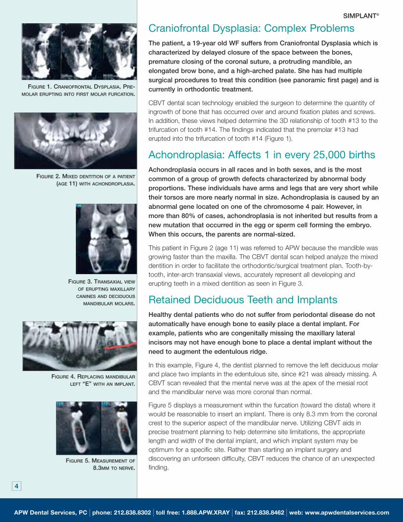
Craniofrontal Dysplasia: Complex ProblemsThe patient, a 19-year old WF suffers from Craniofrontal Dysplasia which ischaracterized by delayed closure of the space between the bones,premature closing of the coronal suture, a protruding mandible, anelongated brow bone, and a high-arched palate. She has had multiplesurgical procedures to treat this condition (see panoramic first page) and iscurrently in orthodontic treatment.
CBVT dental scan technology enabled the surgeon to determine the quantity of
ingrowth of bone that has occurred over and around fixation plates and screws.
In addition, these views helped determine the 3D relationship of tooth #13 to the
trifurcation of tooth #14. The findings indicated that the premolar #13 had
erupted into the trifurcation of tooth #14 (Figure 1).
Achondroplasia: Affects 1 in every 25,000 birthsAchondroplasia occurs in all races and in both sexes, and is the mostcommon of a group of growth defects characterized by abnormal bodyproportions. These individuals have arms and legs that are very short whiletheir torsos are more nearly normal in size. Achondroplasia is caused by anabnormal gene located on one of the chromosome 4 pair. However, inmore than 80% of cases, achondroplasia is not inherited but results from anew mutation that occurred in the egg or sperm cell forming the embryo.When this occurs, the parents are normal-sized.
This patient in Figure 2 (age 11) was referred to APW because the mandible was
growing faster than the maxilla. The CBVT dental scan helped analyze the mixed
dentition in order to facilitate the orthodontic/surgical treatment plan. Tooth-by-
tooth, inter-arch transaxial views, accurately represent all developing and
erupting teeth in a mixed dentition as seen in Figure 3.
Retained Deciduous Teeth and ImplantsHealthy dental patients who do not suffer from periodontal disease do notautomatically have enough bone to easily place a dental implant. Forexample, patients who are congenitally missing the maxillary lateralincisors may not have enough bone to place a dental implant without theneed to augment the edentulous ridge.
In this example, Figure 4, the dentist planned to remove the left deciduous molar
and place two implants in the edentulous site, since #21 was already missing. A
CBVT scan revealed that the mental nerve was at the apex of the mesial root
and the mandibular nerve was more coronal than normal.
Figure 5 displays a measurement within the furcation (toward the distal) where it
would be reasonable to insert an implant. There is only 8.3 mm from the coronal
crest to the superior aspect of the mandibular nerve. Utilizing CBVT aids in
precise treatment planning to help determine site limitations, the appropriate
length and width of the dental implant, and which implant system may be
optimum for a specific site. Rather than starting an implant surgery and
discovering an unforseen difficulty, CBVT reduces the chance of an unexpected
finding.
FIGURE 3. TRANSAXIAL VIEW
OF ERUPTING MAXILLARY
CANINES AND DECIDUOUS
MANDIBULAR MOLARS.
FIGURE 2. MIXED DENTITION OF A PATIENT
(AGE 11) WITH ACHONDROPLASIA.
FIGURE 4. REPLACING MANDIBULAR
LEFT “E” WITH AN IMPLANT.
FIGURE 5. MEASUREMENT OF
8.3MM TO NERVE.
SIMPLANT®
FIGURE 1. CRANIOFRONTAL DYSPLASIA. PRE-MOLAR ERUPTING INTO FIRST MOLAR FURCATION.
APW Dental Services, PC | phone: 212.838.8302 | toll free: 1.888.APW.XRAY | fax: 212.838.8462 | web: www.apwdentalservices.com
4
97260_APW_1 09/28/2004 08:45 AM Page 4
5
APW Dental Services, PC | phone: 212.838.8302 | toll free: 1.888.APW.XRAY | fax: 212.838.8462 | web: www.apwdentalservices.com
97260_APW 09/24/2004 05:56 AM Page 5
6
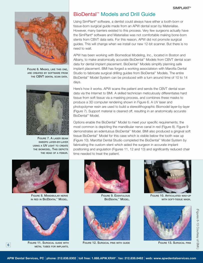
BioDental™ Models and Drill GuideUsing SimPlant® software, a dentist could always have either a tooth-born or
tissue-born surgical guide made from an APW dental scan by Materialise.
However, many barriers existed to this process. Very few surgeons actually have
the SimPlant® software and Materialise was not comfortable making bone-born
stents from CBVT data sets. For this reason, APW did not promote surgical
guides. This will change when we install our new 12-bit scanner. But there is no
need to wait.
APW has been working with Biomedical Modeling, Inc., located in Boston and
Albany, to make anatomically accurate BioDental™ Models from CBVT dental scan
data for dental implant placement. BioDental™ Models simplify planning safe
implant placement. BMI has forged a working associateion with Marotta Dental
Studio to fabricate surgical drilling guides from BioDental™ Models. The entire
BioDental™ Model System can be produced with a turn around time of 10 to 14
days.
Here’s how it works. APW scans the patient and sends the CBVT dental scan
data via the Internet to BMI. A skilled technician meticulously differentiates hard
tissue from soft tissue via a masking process, and combines these masks to
produce a 3D computer rendering shown in Figure 6. A UV laser and
photopolymer resin are used to build a stereolithographic Biomodel layer-by-layer
(Figure 7). Support material is cleaned off, resulting in an anatomically accurate
BioDental™ Model.
Options enable the BioDental™ Model to meet your specific requirements; the
most common is depicting the mandibular nerve canal in red (Figure 8). Figure 9
demonstrates an edentulous BioDental™ Model. BMI also produced a gingival soft
tissue BioDental™ Model for this case which is visible below the tooth wax up
(Figure 10). Marottal Dental Studio completed the BioDental™ Model System by
fabricating the custom stent which aided the surgeon in accurate implant
positioning and angulation (Figures 11, 12 and 13) and significantly reduced chair
time needed to treat the patient.
FIGURE 6. MASKS, LIKE THIS ONE,ARE CREATED BY SOFTWARE FROM
THE CBVT DENTAL SCAN DATA.
FIGURE 7. A LASER BEAM
SWEEPS LAYER-BY-LAYER
USING A UV LIGHT TO CREATE
THE BIOMODEL. THIS DEPICTS
THE HEAD OF A FEMUR.
FIGURE 8. MANDIBULAR NERVE
IN RED IN BIODENTAL™ MODEL.FIGURE 9. EDENTULOUS
BIODENTAL™ MODEL. FIGURE 10. ARTICULATED WAX-UP
WITH SOFT-TISSUE MASK.
FIGURE 11. SURGICAL GUIDE WITH
METAL TUBES FOR IMPLANTS.FIGURE 12. SURGICAL PINS WITH GUIDE FIGURE 13. SURGICAL PINS
APW Dental Services, PC | phone: 212.838.8302 | toll free: 1.888.APW.XRAY | fax: 212.838.8462 | web: www.apwdentalservices.com
SIMPLANT®(Fig
ures 6-1
3 C
ourtesy o
f BM
I.)
97260_APW_2 09/29/2004 12:21 PM Page 6
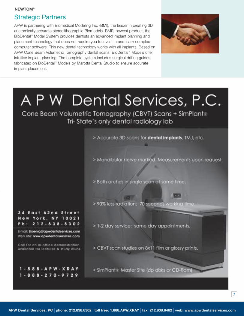
APW is partnering with Biomedical Modeling Inc. (BMI), the leader in creating 3D
anatomically accurate stereolithographic Biomodels. BMI’s newest product, the
BioDental™ Model System provides dentists an advanced implant planning and
placement technology that does not require you to invest in and learn complex
computer software. This new dental technology works with all implants. Based on
APW Cone Beam Volumetric Tomography dental scans, BioDental™ Models offer
intuitive implant planning. The complete system includes surgical drilling guides
fabricated on BioDental™ Models by Marotta Dental Studio to ensure accurate
implant placement.
Strategic Partners
7
NEWTOM®
APW Dental Services, PC | phone: 212.838.8302 | toll free: 1.888.APW.XRAY | fax: 212.838.8462 | web: www.apwdentalservices.com
7
97260_APW 09/24/2004 05:56 AM Page 7
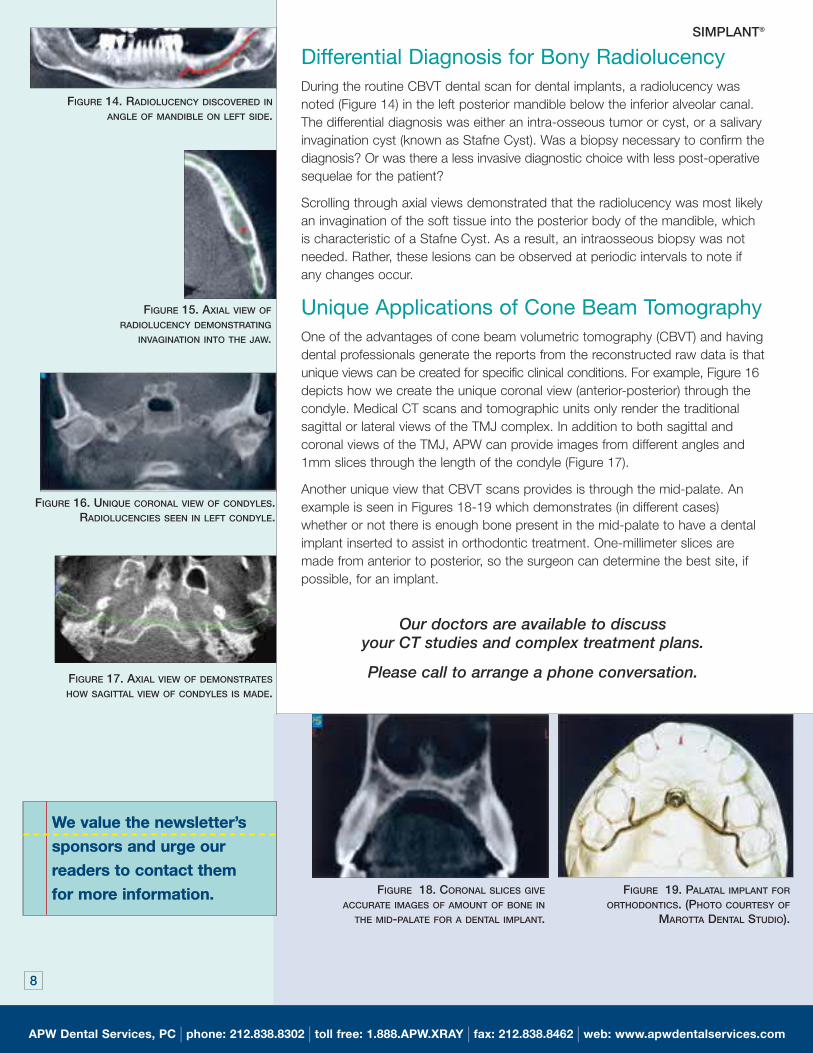
Differential Diagnosis for Bony RadiolucencyDuring the routine CBVT dental scan for dental implants, a radiolucency was
noted (Figure 14) in the left posterior mandible below the inferior alveolar canal.
The differential diagnosis was either an intra-osseous tumor or cyst, or a salivary
invagination cyst (known as Stafne Cyst). Was a biopsy necessary to confirm the
diagnosis? Or was there a less invasive diagnostic choice with less post-operative
sequelae for the patient?
Scrolling through axial views demonstrated that the radiolucency was most likely
an invagination of the soft tissue into the posterior body of the mandible, which
is characteristic of a Stafne Cyst. As a result, an intraosseous biopsy was not
needed. Rather, these lesions can be observed at periodic intervals to note if
any changes occur.
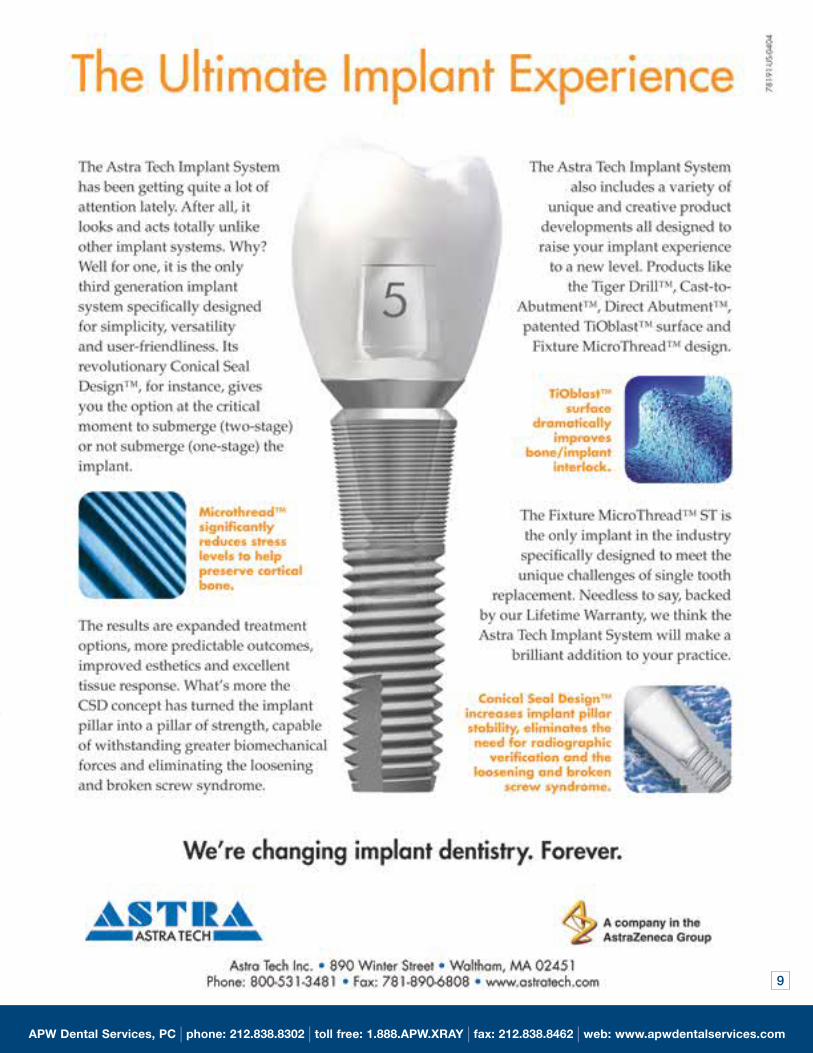
Unique Applications of Cone Beam TomographyOne of the advantages of cone beam volumetric tomography (CBVT) and having
dental professionals generate the reports from the reconstructed raw data is that
unique views can be created for specific clinical conditions. For example, Figure 16
depicts how we create the unique coronal view (anterior-posterior) through the
condyle. Medical CT scans and tomographic units only render the traditional
sagittal or lateral views of the TMJ complex. In addition to both sagittal and
coronal views of the TMJ, APW can provide images from different angles and
1mm slices through the length of the condyle (Figure 17).
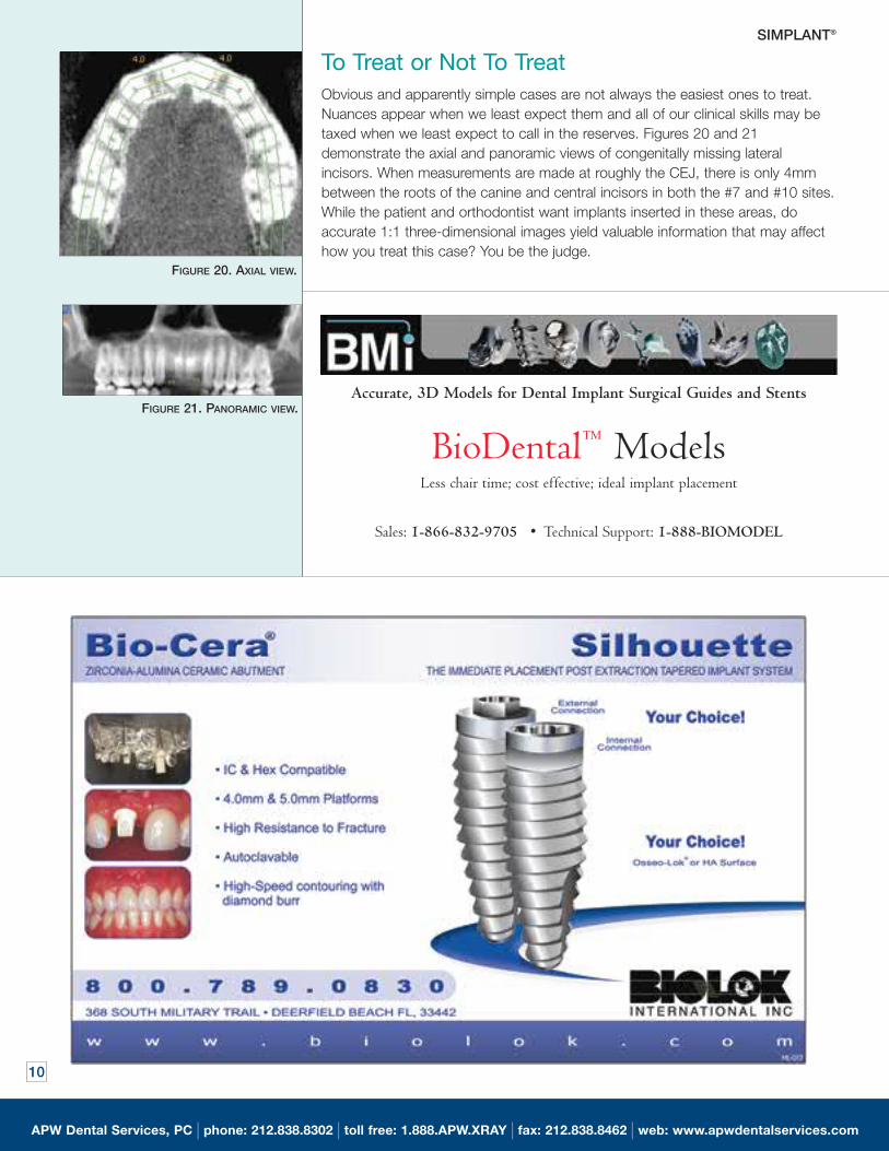
Another unique view that CBVT scans provides is through the mid-palate. An
example is seen in Figures 18-19 which demonstrates (in different cases)
whether or not there is enough bone present in the mid-palate to have a dental
implant inserted to assist in orthodontic treatment. One-millimeter slices are
made from anterior to posterior, so the surgeon can determine the best site, if
possible, for an implant.
Our doctors are available to discuss your CT studies and complex treatment plans.
Please call to arrange a phone conversation.
FIGURE 14. RADIOLUCENCY DISCOVERED IN
ANGLE OF MANDIBLE ON LEFT SIDE.
FIGURE 15. AXIAL VIEW OF
RADIOLUCENCY DEMONSTRATING
INVAGINATION INTO THE JAW.
FIGURE 16. UNIQUE CORONAL VIEW OF CONDYLES.RADIOLUCENCIES SEEN IN LEFT CONDYLE.
FIGURE 17. AXIAL VIEW OF DEMONSTRATES
HOW SAGITTAL VIEW OF CONDYLES IS MADE.
FIGURE 19. PALATAL IMPLANT FOR
ORTHODONTICS. (PHOTO COURTESY OF
MAROTTA DENTAL STUDIO).
FIGURE 18. CORONAL SLICES GIVE
ACCURATE IMAGES OF AMOUNT OF BONE IN
THE MID-PALATE FOR A DENTAL IMPLANT.
APW Dental Services, PC | phone: 212.838.8302 | toll free: 1.888.APW.XRAY | fax: 212.838.8462 | web: www.apwdentalservices.com
8
SIMPLANT®
We value the newsletter’s
sponsors and urge our
readers to contact them
for more information.
97260_APW 09/24/2004 05:56 AM Page 8
9
APW Dental Services, PC | phone: 212.838.8302 | toll free: 1.888.APW.XRAY | fax: 212.838.8462 | web: www.apwdentalservices.com
97260_APW 09/24/2004 05:56 AM Page 9
APW Dental Services, PC | phone: 212.838.8302 | toll free: 1.888.APW.XRAY | fax: 212.838.8462 | web: www.apwdentalservices.com
10
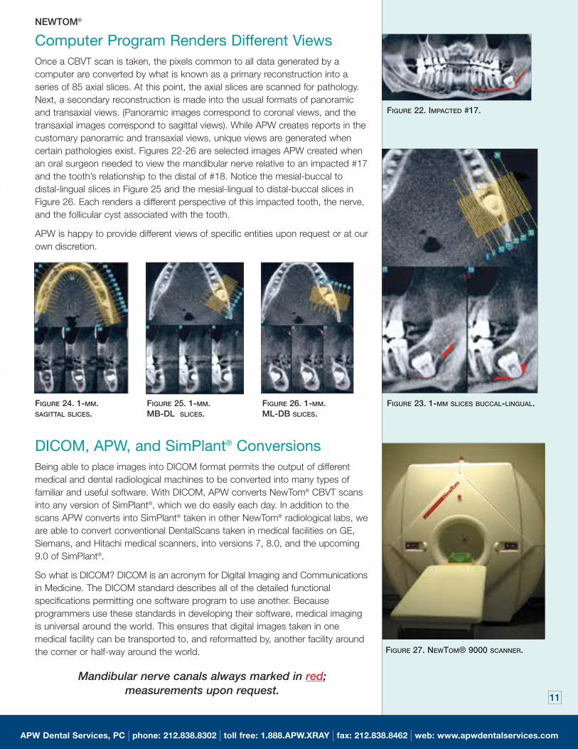
FIGURE 20. AXIAL VIEW.
FIGURE 21. PANORAMIC VIEW.
SIMPLANT®
To Treat or Not To Treat Obvious and apparently simple cases are not always the easiest ones to treat.
Nuances appear when we least expect them and all of our clinical skills may be
taxed when we least expect to call in the reserves. Figures 20 and 21
demonstrate the axial and panoramic views of congenitally missing lateral
incisors. When measurements are made at roughly the CEJ, there is only 4mm
between the roots of the canine and central incisors in both the #7 and #10 sites.
While the patient and orthodontist want implants inserted in these areas, do
accurate 1:1 three-dimensional images yield valuable information that may affect
how you treat this case? You be the judge.
Accurate, 3D Models for Dental Implant Surgical Guides and Stents
BioDental™ ModelsLess chair time; cost effective; ideal implant placement
Sales: 1-866-832-9705 • Technical Support: 1-888-BIOMODEL
97260_APW 09/24/2004 05:56 AM Page 10
Computer Program Renders Different ViewsOnce a CBVT scan is taken, the pixels common to all data generated by a
computer are converted by what is known as a primary reconstruction into a
series of 85 axial slices. At this point, the axial slices are scanned for pathology.
Next, a secondary reconstruction is made into the usual formats of panoramic
and transaxial views. (Panoramic images correspond to coronal views, and the
transaxial images correspond to sagittal views). While APW creates reports in the
customary panoramic and transaxial views, unique views are generated when
certain pathologies exist. Figures 22-26 are selected images APW created when
an oral surgeon needed to view the mandibular nerve relative to an impacted #17
and the tooth’s relationship to the distal of #18. Notice the mesial-buccal to
distal-lingual slices in Figure 25 and the mesial-lingual to distal-buccal slices in
Figure 26. Each renders a different perspective of this impacted tooth, the nerve,
and the follicular cyst associated with the tooth.
APW is happy to provide different views of specific entities upon request or at our
own discretion.
DICOM, APW, and SimPlant® ConversionsBeing able to place images into DICOM format permits the output of different
medical and dental radiological machines to be converted into many types of
familiar and useful software. With DICOM, APW converts NewTom® CBVT scans
into any version of SimPlant®, which we do easily each day. In addition to the
scans APW converts into SimPlant® taken in other NewTom® radiological labs, we
are able to convert conventional DentalScans taken in medical facilities on GE,
Siemans, and Hitachi medical scanners, into versions 7, 8.0, and the upcoming
9.0 of SimPlant®.
So what is DICOM? DICOM is an acronym for Digital Imaging and Communications
in Medicine. The DICOM standard describes all of the detailed functional
specifications permitting one software program to use another. Because
programmers use these standards in developing their software, medical imaging
is universal around the world. This ensures that digital images taken in one
medical facility can be transported to, and reformatted by, another facility around
the corner or half-way around the world.
Mandibular nerve canals always marked in red; measurements upon request.
11
APW Dental Services, PC | phone: 212.838.8302 | toll free: 1.888.APW.XRAY | fax: 212.838.8462 | web: www.apwdentalservices.com
FIGURE 22. IMPACTED #17.
FIGURE 23. 1-MM SLICES BUCCAL-LINGUAL.FIGURE 24. 1-MM.SAGITTAL SLICES.
FIGURE 25. 1-MM. MB-DL SLICES.
FIGURE 26. 1-MM. ML-DB SLICES.
FIGURE 27. NEWTOM® 9000 SCANNER.
NEWTOM®
97260_APW_2 09/29/2004 04:05 AM Page 11
APW Dental Services, PC
34 East 62nd Street
New York, NY 10021
Demonstration in your office
Please contact APW’s executive
administrator, Lisa Koenig, to
arrange an
in-office demonstration of our services.
We will demonstrate how SimPlant and
NewTom tomography will help you give
the best care possible for your patients.
UPCOMING EVENTS:
October 6, 2004
“CT Scans for Esthetic
Implant Placement”
Dr. Alan Winter
North East Periodontal
Study Group
Sheraton at Woodbridge
Iselin, NJ
October 13, 2004
“The ABCs of CT Scans
in Dentistry”
Drs. Winter and Pollack
NY County Dental Society
6 East 43rd Street
New York, NY
October 22, 2004
Exhibitor
NESP Fall Meeting
NY Marriott Marquis Hotel
1535 Broadway
New York, NY
April 14, 2005
“Newest Advances in
CT Scans.”
Dr. Alan A. Winter
2nd Dist Dent Society
Ft. Hamilton C.C.
Brooklyn, NY
MEET DR. HERBERT H. FROMMER
Generations of NYU dental students have learned radiology from Dr. Herbert H. Frommer.What many do not know is that after attending Columbia University, Herb received his DDS
from Columbia in 1957. Herb was in the Naval Reserve and before settling into NYU, taught
at what was then Seton Hall College of Dentistry, and is now UMDNJ. Herb joined the
faculty of NYUCD in 1969 and became a full tenured professor in 1992. He is currently
Professor and Director of Radiology, and Diplomate of the Board of the American Academy
of Oral & Maxillofacial Radiology. APW is proud of Herb’s accomplishments and thankful he
is part of our team.
SIMPLANT® NEWTOM®
STUDY CLUBS AND DENTAL ORGANIZATIONS
Call APW Dental Services to arrange for a lecture to your study club or dental organization.
Learn how easy it is to incorporate the rapidly changing world of computed tomography into
your restorative/cosmetic or specialty practice. Understand how axial and transaxial 3D
views take the guesswork out of complex diagnostic problems. Discover how
12-bit technology, BioDental™ models, and surgical guides will take the worry out of surgical
planning for dental implants.
Contact Lisa Koenig at 212-838-8302 or toll free at: 1-888-APW-XRAY.
CONVERT MEDICAL CTAND NEWTOM CBVTSCANS TO SIMPLANT®
APW can convert medical
CT scans, DentaScans,
NewTom 9000 and all other
cone beam volumetric
tomographic scans to
SimPlant® in versions 7,
8.0, and upcoming 9.0,
with NEXT DAY Service.
Please call our toll free
number for details.
1-888-APW-XRAY
1-888-270-9729
97260_APW_1 09/28/2004 07:37 AM Page 12