Embed Size (px)
Citation preview
Bull. Org. mond. Sante 1958, 18, 945-961Bull. Wid Hith Org. 1981,9
THE INTRADERMAL TEST IN THE DIAGNOSISOF BILHARZIASIS
J. PELLEGRINOInsttuto Nacional de Endemias Rurais (Centro de Pesquisas de Belo Horizonte)
and Instituto de Biologia, Faculdade de Filosofia daUniversidade de Minas Gerais, Belo Horizonte, Brazil
SYNOPSIS
The diagnosis of bilharziasis by detection of schistosome eggsin the urine or faeces of patients is often not possible in the laterstages of the disease, when elimination of the eggs is renderedincreasingly difficult. To overcome this difficulty, an immunologicaltest has been developed, based on the cutaneous response to aninjection of an antigen. prepared from the cercariae, adult worms,eggs or miracidia of Schistosoma.
In this paper, the author reviews the various procedures forobtaining the basic material for antigen preparation, describesbriefly the method of preparation, outlines the nature and proper-ties of the specific antigens, and discusses the skin-testing procedure,with special reference to the criteria for evaluating the results.He points out that the intradermal test may prove useful in epi-demiological surveys as well as in the diagnosis of individual cases.
The diagnosis of bilharziasis during the initial stage of the disease (frankhaematuria, in the vesical type, and toxaemic manifestations accompaniedby gastro-intestinal disorders, in the intestinal types) may be easily made bydetecting schistosome eggs in the patient's urine (S. haematobium) or faeces(S. mansoni and S. japonicum). However, at later stages, the number ofworms may decrease and the elimination of eggs becomes more and moredifficult, owing not only to the gradually increasing cellular reactions thattend to wall them in, but also to the growing connective tissue whichprevents them from escaping. Therefore, in endemic areas, the prevalenceof bilharziasis, when evaluated by examining stool or urine samples, orboth, from unselected people, always gives figures lower than expected,since most patients are in the chronic stage of the infection.
Attempts to overcome these inherent difficulties in diagnosis led to theuse of immunological methods, which have proved to be very helpful forconfirming the diagnosis of individual cases of schistosome infections.
- 945663
J. PELLEGRINO
When used in epidemiological surveys, such methods provide a fairly goodapproximation of the true prevalence of the disease.
In this report the writer will review the procedures for procuring thebasic material for antigen preparation and the main aspects regarding theuse of the intradermal test for the diagnosis of schistosome infections.
Procurement of Basic MaterialCercariae
The following methods for concentrating schistosome cercariae forantigen preparation have been devised:
(a) Centrifugation of living cercariae (Oliver-Gonzailez & Pratt, 1944;Katzin & Most, 1946) or ofdead cercariae killed by heat (Lurie & De Meillon,1951). As to the first method, Bozicevich & Hoyem (1947) and Standen(1950) have pointed out that the cercariae, even when spun at 4000 revolu-tions per minute (r.p.m.) for 5 minutes are able to swim back actively to thetop immediately after centrifugation.
(b) Immobilization of cercariae by chemical or physical agents prior tosedimentation. Bozicevich & Hoyem (1947) used ethanol and Martins (1949)a small quantity of ether to narcotize the cercariae, which then sink to thebottom. The sedimentation may also be accomplished by putting thecercarial suspension into the refrigerator (4-10°C). The latter method wasemployed by Bozicevich & Hoyem (1947), Mayer & Pifano (1949) andOliver-Gonzailez, Bauman & Benenson (1954).
(c) Deposition of cercariae on filter-paper. This technique, used byAlves & Blair (1946), has also been employed by Blair & Ross (1948),Rodrigues da Silva & Costa (1949), Standen (1950) and Pereira (1951).Antigens may be prepared with the filter-paper still wet or after it has beendried.
(d) Accelerated vertical migration of cercariae. Standen (1950, 1952)claims to have easily concentrated cercariae of S. mansoni by putting thecercarial suspension in a separating-funnel, cooling its lower part withice-cold water and illuminating its top from the sides. After a few minutesthe cercariae will have gathered together in the bright upper layer of theliquid. The contaminants are then removed by drawing off the lower layer.
(e) Concentration of cercariae in crucibles and funnels with fritted-glassbottoms. The porosity of the plates should be such as to let only water andsmall contaminants pass through, so that the cercariae are easily concen-trated (Pellegrino & Macedo, 1955).
Appreciable amounts of S. mansoni cercariae practically free fromundesirable matter may be obtained by the following method (Pellegrino &Nunes, 1956):
946
INTRADERMAL TEST IN DIAGNOSIS OF BILHARZIASIS
1. Infected snails, previously washed and cleaned, are dispensed inbeakers containing about 300 ml of water, in groups of 20-50. They arethen exposed to the sun or to electric light for 1-3 hours, in order to inducethe shedding of cercariae.
2. The cercarial suspension is poured through 4 layers of surgicalgauze to remove snail faeces and other debris.
3. The cercariae are concentrated in Biichner funnels, with a capacity of350 ml or greater, fitted with plates of medium porosity (maximum poresize, about 201u).
4. The heavy cercarial suspension is poured into conical flasks andplaced in the refrigerator for sedimentation.
5. The supernatant is discarded and the cercariae in the remaining smallvolume of liquid are transferred to a beaker so that a thin layer is formed.
6. The material in the beaker is frozen under dry ice and then desiccatedin vacuum in the presence of calcium chloride. The dried cercariae are keptin glass ampoules and sealed in vacuum.
About 100-150 mg of dried cercariae may be obtained in a day, which isa highly satisfactory yield for antigen preparation (Pellegrino-unpub-lished data, 1957).
Adult worms
The recovery of adult schistosomes from the mesenteric-portal systemmay be easily accomplished by means of simplified perfusion techniques(Brandt & Finch, 1946; Yolles et al., 1947; Pan & Hunter, 1951; Ruiz, 1952,1953b; Pellegrino & Siqueira, 1956). The perfusion is generally performedin two stages:
1. Perfusion of the liver, in which the physiological salt solution runsunder pressure through the hepatic veins into the portal system, reaching theexit at the portal vein.
2. Perfusion of the mesenteric vessels, in which the solution is allowedto flow into the aorta and run off by the portal vein.
A previous injection of heparin, which prevents blood from clotting,renders the perfusion and the collection of the worms easier.
Most schistosomes lodged in the mesenteric-portal system are carriedaway with the perfusion fluid. When a large number of worms is required,as for antigen preparation, the guinea-pig has proved to be a suitableanimal despite being considered a poor host of S. mansoni (Moore, Yolles& Meleney, 1949; Stirewalt, Kuntz & Evans, 1951). Pellegrino & Siqueira(1956) were able to recover 51 702 worms after liver and mesentericperfusions carried out on 38 highly infected guinea-pigs.
947
J. PELLEGRINO
Eggs
Interest in egg antigens was aroused when Oliver-Gonzalez, Bauman& Benenson (1954, 1955) reported that skin-tests with such antigens,negative in most patients before treatment, became positive after therapyin about 80% of cases. These workers used the liver and intestines ofinfected mice and hamsters as sources of eggs. The liver and intestineswere cut up into small pieces, homogenized in a Waring blender and thenstrained through a finely woven sieve into conical flasks for the sedimenta-tion of eggs. Most extraneous matter was removed after several sedimen-tations in saline. Griffiths & Beesley (1955) concentrated S. mansonieggs from infected mouse livers homogenized in a Waring blender and thenstrained through several sieves of gradually decreasing meshes. Theresulting suspension was poured through organdie and silk to removesmall contaminants. Recently Coker & Lichtenberg (1956) outlined asimple procedure for isolating S. mansoni eggs from the liver of experi-mentally infected hamsters by a combination of comminution, filtrationand sedimentation through a viscous medium (sucrose).
As the number of S. mansoni eggs in the small intestine of mice isremarkable 8 weeks after infection and the tendency to granuloma formationis still little pronounced at that stage (Brener, 1956), the following techniquefor egg concentration has been devised (Pellegrino-unpublished data.1957):
1. Mice infected with about 150 cercariae (S. mansoni) are killed,preferably between 8 and 11 weeks after exposure, and their intestinesremoved.
2. The small intestine is cut off and thoroughly opened and washed.It is then placed in a Petri dish containing physiological salt solutionand left at 37°C for 2 hours.
3. The intestinal mucosa is scraped with the aid of a microscope slidein order to remove most of the available material.
4. The suspension is homogenized in a Waring blender for 1 minuteand, after being filtered through 4 layers of surgical gauze, is finally strainedthrough a sieve (US Standard No. 100).
5. The eggs are allowed to settle in a crystallizing-dish, 6 cm high,for 15 minutes.
6. The supernatant is removed, by means of a pipette connected toa vacuum pump, so that a thin layer (about 0.5 cm) is left.
7. Physiological salt solution is poured into the crystallizing-dish upto a height of 3 cm and the sedimentation of eggs is observed througha stereoscopic microscope. When sedimentation is complete, the super-natant is pipetted off as in step 6.
948

INTRADERMAL TEST IN DIAGNOSIS OF BILHARZIASIS
8. This washing in saline is repeated as many times as are necessaryto remove most of the intestinal debris. As the eggs settle rapidly, only4-5 minutes are required for each washing.
9. The egg suspension is poured into a centrifuge tube and spun at1500 r.p.m. for 5 minutes.
10. The sediment is frozen under dry ice and desiccated in the presenceof calcium chloride.
The amount of eggs obtained by this technique is quite satisfactoryfor antigen preparation.Miracidia
Blair & Ross (1948) make reference to the use of an extract preparedwith S. haematobium miracidia, stating that this antigen, even after havingbeen stored at room temperature for several years, provided results similarto those obtained with cercarial antigen of the same species of Schistosoma.However, they were forced to stop using it, owing to difficulties in securinga satisfactory yield of miracidia. Recently, Sherif (1956) described a methodfor collecting and washing schistosome eggs (mainly of S. haematobium)by means of which a plentiful supply of miracidia may be obtained. Themiracidia are killed by adding 96% ethanol to the original suspensionand are then centrifuged and dried under vacuum.
Simplified techniques for concentration, based on the positive photo-tropism and negative geotropism of miracidia, have been devised (Stunkard,1946; Ingalls et al., 1949; Kagan, Short & Nez, 1954; Chaia, 1956).
When appreciable amounts of S. mansoni miracidia are required,the following procedure can give fairly good results (Pellegrino-unpub-lished data, 1957):
1. S. mansoni eggs from the intestines of infected mice are concentratedas previously described.
2. The saline egg suspension is poured into a glass crucible fitted witha plate of medium porosity.
3. Dechlorinated water is added to the suspension in the crucibleso that any excess of sodium chloride is carried away by percolation.
4. The egg suspension, now in dechlorinated water, is exposed to thelight of a 60-watt electric lamp. The ova will soon hatch, liberating free-swimming miracidia.
5. With a pipette, the miracidial suspension is transferred to anothercrucible. Immature and degenerate eggs, as well as empty shells, willremain at the bottom of the first crucible. The heavy miracidial suspensionis then transferred from the second crucible to a watch-glass and, finally,is frozen and desiccated for antigen preparation.
949

J. PELLEGRINO
Preparation of Antigens
As has been shown, desiccated schistosome cercariae, adult worms,eggs and miracidia can be easily obtained for antigen preparation. Therefore,the use of extracts from the digestive glands of infected snails (Fairley& Williams, 1927; Manson-Bahr, 1929; Taliaferro & Taliaferro, 1931;Vogel, 1932; Ramsay, 1934; Martins, 1949; Davies & Eliakim, 1954)or of heterologous antigens (Hassan & Betashe, 1934; Culbertson & Rose,1942; Mayer & Pifano, 1945b, 1946; Guerra, Mayer & Di Prisco, 1945;Peres & Pena, 1945; Culbertson, Rose & Oliver-Gonzalez, 1947; Martins,1949; Lurie & De Meillon, 1951) should not be encouraged. Cercarialand adult-worm extracts have been widely used as skin-testing material.Egg antigens have been employed by Oliver-Gonzalez, Bauman & Benenson(1954, 1955) and Pellegrino (unpublished data, 1957). Miracidial antigenshave been used by Blair & Ross (1948) and, more recently, by Sherif(1956).
As a rule, antigens are prepared with desiccated and finely groundmaterial. Extracts of fresh cercariae or adult worms have occasionallybeen employed as antigens (Bozicevich & Hoyem, 1947; Pellegrino-unpublished data, 1957). Physiological and Coca solution have been usedas the extracting medium, phenol or merthiolate sometimes being added.Extraction is generally carried out at room temperature (2-3 days) or inthe refrigerator at 4-8°C (2-8 days). It has also been performed at 36-37°C during short periods (Pesigan et al., 1951, 1954; Oliver-Gonzilez,1953; Oliver-Gonzailez & Pratt, 1944; Davies & Eliakim, 1954). Wrightet al. (1947) effect the extraction of allergenic substances from adult wormsin a relatively short time: 12 hours in the refrigerator. Extracts have beenpreserved with phenol (0.3-0.5 %) or merthiolate (1: 5000 to 1: 10 000).In some cases Seitz filters (Mayer & Pifano, 1945a, 1946; Rodrigues daSilva & Costa, 1949; Pesigan et al., 1951, 1954; Pessoa & Barros, 1953;Sherif, 1956) or porcelain ones (Coutinho, 1948, 1949, 1951, 1952a) havebeen employed.
Purified antigens (removal of lipoids, isolation of polysaccharides)have been prepared by some authors (Martins, 1949; Coutinho, 1952b;Ruiz, 1953a; Horstman, Chaffee & Bauman, 1954; Pellegrino et al., 1956).
Nature and Properties of Antigens
According to Martins (1949) and Ruiz (1953a), the active fraction ofantigens that is capable of eliciting cutaneous responses in patients withbilharziasis must be a polysaccharide. In fact, substances acting chemicallyas polysaccharides have been extracted from cercariae and adult worms(S. mansoni) and successfully used for skin-testing. However, Pellegrino
950
INTRADERMAL TEST IN DIAGNOSIS OF BILHARZIASIS
et al. (1956) believe that the polysaccharides-at least those extracted fromS. mansoni cercariae by Fuller's technique-may not be the only activefraction.
Data concerning the storage of antigens seem to indicate that they arehighly stable. Pratt & Oliver-Gonzailez (1947) did not detect any loss ofpotency in cercarial antigens which had been stored in the refrigerator forone year. According to Mayer & Pifano (1945a), Blair & Ross (1948),Martins (1949), Coutinho (1951), Lurie & De Meillon (1951) and Pellegrino(unpublished data, 1957), antigens commonly used for skin-tests in bil-harziasis do not undergo any appreciable changes during storage. However,Pesigan et al. (1951) observed that an adult-worm antigen (S. japonicum)prepared three months previously was slightlv less potent than a fresh one.
Technique of Intradermal Test
The intradermal test is generally performed on the volar surface of theforearm and, less frequently, on the dorsal surface. It must be mentioned,however, that skin-tests performed on the scapular region are significantlymore intense (Mayer & Pifano, 1945a; Martins, 1949; Pellegrino-un-published data, 1957).
The amounts of antigen injected vary from 0.01 ml to 0.1 ml. Recently,the smaller volumes (0.01-0.05 ml) have been chosen, to prevent false-positive reactions due to tissue injury. Antigen dilutions range from 1: 1000to 1: 10 000, though, occasionally, stronger or weaker concentrations havebeen used.
Positive reactions are normally immediate and reach their maximum15 minutes after injection of the antigen. However, since Pessoa & Barros(1953) have noticed that patients in the later stages of the disease, showingspleen enlargement and anaemia, may react slowly (30 minutes or more),observation for a longer period is sometimes required. Reading is done byobserving the wheal and recording its characteristics, diameter length, etc.,or by taking a tracing of it on transparent paper (Oliver-Gonzalez & Pratt,1944), or even by drawing its outline in ink and then pressing it againstslightly wet absorbent paper (Martins, 1949; Pellegrino & Macedo, 1956;Pellegrino, Memoria & Macedo, 1957).
Delayed reactions have occasionally been recorded (Khalil & Hassan,1932; Boza, 1942; Martins, 1949; Pessoa & Barros, 1953).
Criteria for Evaluation of Results
A critical review of the diagnosis of bilharziasis by the intradermal testhas shown that a number of criteria have been used for the interpretationof results. Such criteria may be grouped as follows:
'P
951
J. PELLEGRINO
Inaccurate or subjective criteria
Regrettably, some authors who have carried out intradermal testsusing antigens prepared from S. bovis or other helminths-Khalil & Hassan,1932 (S. bovis antigen); Hassan & Betashe, 1934 (Fasciola gigantica antigen);Culbertson, Rose & Oliver-Gonz'alez, 1946 (Planaria maculata antigen);Oliver-Gonzalez et al., 1953 (Fasciola hepatica and Pneumoneces medio-plexus antigens, simultaneously with that of S. mansoni cercariae)-haveused inaccurate criteria for the evaluation of the results. Lurie & DeMeillon (1951), for instance, just state that " a definite extension in the sizeof the wheal was taken as a positive result ".
Criteria based on enlargement of initial wheal
Blair & Ross (1948) and Coutinho (1951) consider an intradermal testpositive when 15 minutes after the antigen injection the wheal diameteris twice its initial size.
Criteria based on increase of antigen wheal in relation to control wheal
To several authors, a reaction in which the diameter of the antigenwheal exceeds that of the control wheal by 3-4 mm or more is consideredpositive (Katzin & Most, 1946; Bozicevich & Hoyem, 1947; Wright et al.,1957; Pesigan et al., 1951, 1954; Davies & Eliakim, 1954; Horstman,Chaffee & Bauman, 1954; Oliver-Gonzalez, Bauman & Benenson, 1954).
Criteria based on diameter of antigen wheal
The size of the wheal diameter at the 15 minutes' reading is used as abasis of interpretation by several authors who, however, do not agree asto the diameter size beyond which a reaction can be considered positive:20 mm (Fairley & Williams, 1927); 23 mm (Ramsay, 1934); 12 mm (Peres &Pena, 1945); 10 mm (Davies & Eliakim, 1955; Sherif, 1956).
Criteria based on degree of intensity ofpositive reactions
Most South American workers classify positive reactions into severalgroups according to their intensity (Mayer & Pifano, 1945a, 1949; Lopes,1945; Coutinho, 1948, 1949, 1952a, 1952b; Martins, 1949; Rodrigues daSilva & Costa, 1949; Pereira, 1951; Pessoa & Barros, 1953; Ruiz, 1953a).Wheal enlargement, pseudopodia formation and, in some instances, thepresence of erythema and pruritus are usually taken into consideration.
Criterion based on size of wheal area
Aiming at a more accurate and objective evaluation of results, Pellegrino& Macedo (1956) suggested the following procedure: 15 minutes after the
952

INTRADERMAL TEST IN DIAGNOSIS OF BILHARZJASIS
antigen injection (0.05 ml on the volar surface of the forearm) the outlineof the wheal is drawn in ink and transferred, by pressure, to slightly wetabsorbent paper. The wheal area is then determined with a planimeter orin the following way: a series of circles, outlining areas ranging from0.5 to 1.2 cm2, is drawn on thick transparent paper or celluloid; besidethese circles, 3 rectangles, with areas of 1.0, 2.0 and 4.0 cm2 and surroundedby a chequered area of which the small squares measure 0.05 cm2, are alsodrawn. It is very easy to obtain a rapid and fairly good estimate of thewheal area by placing the transparent paper over the wheal transfer. Thefollowing criteria were established after preliminary tests on patients withbilharziasis as well as on healthy individuals:
(a) negative reactions: wheals with areas up to 0.9 cm2;(b) doubtful reactions: wheals with areas of 1.0 or 1.1 cm2;(c) positive reactions: wheals with areas of 1.2 cm2 or more.
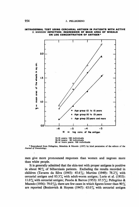
On estimating the wheal areas obtained by intradermal tests using cer-carial antigen in concentrations ranging from 10-3 to 10-6, in three age-groups of patients with bilharziasis, Pellegrino, Memoria & Macedo(1957) demonstrated the existence of a linear relationship between themean area responses and the log concentrations of the antigen (see figurebelow).
Sensitivity and Specificity of Test
In the course of schistosome infection the skin becomes sensitized tospecific antigens subsequent to the development of circulating antibodies.Positive intradermal tests are usually observed from the fourth week afterexposure (WHO Expert Committee on Bilharziasis, 1953). Wright et al.(1947), using S. mansoni antigens diluted 1: 1000, obtained negative intra-dermal tests in American soldiers newly infected with S. japonicum duringthe Leyte campaign; on the other hand, tests performed with the sameantigen in local patients were positive. Mayer & Pifano (1945a, 1946)observed positive reactions 35 days after S. mansoni exposure and Lurie,De Meillon & Stoffberg (1952) observed them during the seventh week ofdisease in one case (S. haematobium). The duration of the infection probablycontributes materially to sensitization and, early in the course of the disease,such sensitization is not at a high level (Wright et al., 1947).
The data at present available on the sensitivity of the test cannot beadequately compared, either because of the diversity of antigens and criteriaof interpretation or because of the lack of uniformity in the groups studied.It is known that the cutaneous reaction is stronger in adults than in children(Mayer & Pifano, 1946; Martins, 1949; Lurie et al., 1953; Pessoa & Barros,1953; Pellegrino, Memoria & Macedo, 1957). According to Martins (1949),
953
J. PELLEGRINO
INTRADERMAL TEST USING CERCARIAL ANTIGEN IN PATIENTS WITH ACTIVES. MANSONI INFECTION: REGRESSION OF MEAN AREA OF WHEALS
ON LOG CONCENTRATION OF ANTIGEN *
2.01-.
10-15 years: 100 individuals16-19 years: 50 individuals20 or more years: 100 individuals
* Reproduced from Pellegrino, Mem6ria & Macedo (1957) by kind permission of the editors of theJournal of Parasitology.
men give more pronounced responses than women and negroes morethan white people.
It is generally admitted that the skin-test with proper antigens is positivein about 90% of bilharziasis patients. Excluding the results recorded inchildren (Tavares da Silva (1945): 45.6%; Martins (1949): 78.2% withcercarial antigen and 83.3% with adult-worm antigen; Lurie et al. (1953):11.0% with cercarial antigen; Pessoa & Barros (1953): 87.5%; Pellegrino &Macedo (1956): 79.0 %), there are few cases in which figures lower than 90%are reported (Bozicevich & Hoyem (1947): 63.0% with cercarial antigen
954
E
c._
0C0
0
IC
0
0
00
921
1.5 I-
1.0 I-
-6 -5 -4 -3
X = log conc. of the antigen
* Age group 10 to 15 yearsA Age group 16 to 19 yearso Age group 20 years and moreF
I I
LLZ) I
I I II
INTRADERMAL TEST IN DIAGNOSIS OF BILHARZIASIS
and 80.0% with adult-worm antigen; Rodrigues da Silva & Costa (1949):84.0%; Pereira (1951): 88.6%; Pesigan et al. (1951): 76.5%; Pesigan etal. (1954): 84.1 %.A higher degree of sensitivity has been attained by performing the test
with cercarial, adult-worm and miracidial antigens instead of with extractsderived from infected snails or heterologous antigens (Martins, 1949;Mayer & Di Prisco, 1949; Prata, 1953; Prata et al., 1956k Sherif, 1956).However, the diversity of criteria adopted for reading the test has rendereddifficult any comparative study. According to Martins (1949) the proportionof false-positive reactions in non-exposed individuals is about 5 %. Thisproportion may be reduced with the use of purified antigens. Positiveintradermal tests were occasionally observed by Hsii & Ameel (1955) withantigens of S. mansoni (cercariae) and S. japonicum (adult worms) in cases ofcercarial dermatitis. Chung et al. (1955) observed positive skin-tests in 6out of 18 cases of paragonimiasis using S. japonicum antigen. No cross-reactions were observed in 8 patients with clonorchiasis (Chung, Weng &Hou, 1955).
Standardization of Antigens
The antigens used in the intradermal test for the diagnosis of bilharziasishave not generally been properly standardized before use. Thus, arbitrarydilutions, commonly ranging from 1: 1000 to 1: 10 000, have been madefrom desiccated material. In some cases, preliminary tests have beenperformed on bilharziasis patients as well as on healthy individuals to checkthe effectiveness of the antigen (Mayer & Pifano, 1946; Bozicevich & Hoyem,1947; Coutinho, 1951, 1952b).
Some workers standardize antigens by estimating the approximatenumber of cercariae or adult worms in a given volume of extract (Mayer &Pifano (1945a): 500 worms per each 3-4 ml of extracting medium; Alves &Blair (1946) and Blair & Ross (1948): 1000 cercariae per ml; Rodrigues daSilva & Costa (1949): 2000 cercariae per ml; Pesigan et al. (1951, 1954):500-1000 worms per ml; Pessoa & Barros (1953): 500 worms per each4-5 ml of extracting medium).
Quantitative determination of nitrogen (0.001 mg ofN per ml) was usedby Lopes (1945) to standardize cercarial (S. mansoni) antigens.
Several workers have tried to standardize antigens biologically, withthe aim of determining suitable doses for the test, by injecting differentdilutions and volumes into patients with bilharziasis (Bozicevich & Hoyem,1947; Katzin & Most, 1946; Martins, 1949; Most et al., 1950).
Recently, attempts have been made to base biological standardization(Pellegrino-unpublished data, 1957) on the linear relationship between thelog concentration of antigen and the mean areas of the wheals (Pellegrino,Memoria & Macedo, 1957).
955
J. PELLEGRINO
The Intradermal Test in Epidemiological Surveys
The prevalence of bilharziasis in endemic areas is generally assessed byexamination of the stools (S. mansoni and S. japonicum) and urine (S. hae-matobium)-usually a single sample of each-of unselected people. Labor-atory proof of the infection, however, is often not attained, especially inlight or advanced cases of S. mansoni or S. japonicum infections when feweggs are eliminated, either because they are scarce or because eliminationis rendered difficult by the growing intestinal fibrosis. It is generally ac-cepted that, even when the most efficient methods of concentration are used,stool examination does not reveal more than 70% of the patients withbilharziasis mansoni. Pesigan (quoted by Davies & Eliakim, 1955) believesthat, in epidemiological surveys, the examination of a single faecal samplecannot show more than 30% of patients infected with S. japonicum. Theuse of immunological methods for the diagnosis of bilharziasis in epide-miological surveys, then, presents many advantages: saving of time, equip-ment and personnel; quick results; increased accuracy; etc. In masstreatment campaigns, the intradermal test provides a reliable means ofscreening out infected persons; stool or urine samples of the positive reactorscan then be examined for schistosome eggs.
The use of intradermal tests as a means of diagnosis in epidemiologicalsurveys has not been resorted to as often as it should have been. So far asthe writer is aware, only a few authors have reported on the use of skin-testing for such a purpose (Ramsay, 1934; Pesigan et al., 1954; Horstman,Chaffee & Bauman, 1954; Davies & Eliakim, 1955).
The screening of infected individuals and the estimation of the prevalenceof the disease, in a given region, are rendered easier when the wheal area istaken as the basis for evaluating the results. The data obtained in surveysmay be plotted on graphs where the ordinates represent the frequency of agiven wheal area and the abscissae the size of the areas. Two differentfrequency curves, which overlap slightly, are usually obtained: the firstcurve gives the proportion of individuals presenting negative reactions; thesecond one gives the proportion of positive reactors. It must be taken intoconsideration that age, sex and race may influence sensitivity to thetest.
The Intradermal Test as a Criterion of Cure
The reactions to intradermal tests before and after specific treatment arestill open to investigation. Authors do not agree as to the value of suchtests as a criterion of cure.
956
INTRADERMAL TEST IN DIAGNOSIS OF BILHARZIASIS
Since Fairley & Williams' report in 1927, it has been shown that positivereactions may persist after treatment. This finding has been confirmed bymany investigators, although some cases of negative reactions in curedpatients have been observed (Manson-Bahr, 1929; Taliaferro & Taliaferro,1931; Khalil & Hassan, 1932; Oliver-Gonzalez & Pratt, 1944; Lopes, 1945;Mayer & Pifano, 1945a, 1946; Rodrigues da Silva & Costa, 1949; Oliver-Gonz'alez, 1953; Davies & Eliakim, 1954; Oliver-Gonzalez, Bauman &Benenson, 1954; Pesigan et al., 1951).
Martins (1949) carried out intradermal tests, before and after treatment,on 214 individuals passing eggs of S. mansoni and did not obtain a singlenegative reaction after treatment, merely observing a decrease of reactionintensity in 65 patients (42 cured; 23 failures). On the contrary, he pointedout that most patients showed more intense reactions after completionof therapy. Similar observations have been reported by Mayer & Pifano(1946). According to Martins (1949) and Pellegrino (unpublished data, 1957),no value should be attached to the intradermal test in the assessment of cure.
Alves & Blair (1946), Katzin & Most (1946) and Most et al. (1950)claim that, where patients have been skin-tested with a reliable antigenand found to give a positive reaction, a reversion to a negative reactionafter treatment is supporting evidence that a cure has been effected. Alves& Blair (1946) suggest that further treatment is needed in cases showingpositive skin reactions six months after the end of a course of therapy.Rodrigues da Silva (1948) thinks that the intradermal test is valuablefor checking active cases of bilharziasis.
Recent reports from Oliver-Gonz'alez and his co-workers (Oliver-Gonzalez, Bauman & Benenson, 1954, 1955; Oliver-Gonzalez, Ramos& Coker, 1955) have shown that only a small percentage of patients withactive S. mansoni infection (8.3 %) give positive cutaneous reactions whenthe test is performed with egg antigen. However, after treatment, thepercentage of positive reactors gradually increases, reaching about 80%after one year. According to Oliver-Gonz'alez, Bauman & Benenson(1954), negative results before therapy are due to the neutralization ofsensitizing antibodies by the schistosome eggs continuously laid in thetissues, and therefore cutaneous sensitization to schistosome eggs canbe demonstrated by the intradermal test only after the spontaneous deathof the worms or successful therapy. These findings have not been confirmedby Pellegrino (unpublished data, 1957). Oliver-Gonz'alez and his colleaguessuggest that the intradermal reaction with egg antigen, together with thecircumoval precipitation test, would provide a good criterion for evaluatingthe efficacy of drug therapy. On the other hand, Sherif (1956) claimedthat bilharziasis patients (S. haematobium and S. mansoni) who hadcompleted a full course of specific treatment all gave negative reactionswhen tested with miracidial antigen 3-6 months after the completion oftreatment.
957

J. PELLEGRINO
Passive Transfer of Cutaneous Allergy
Taliaferro & Taliaferro (1931) were the first to show that the sera ofbilharziasis patients contain a circulating antibody capable of sensitizingthe skin of normal individuals. Guerra, Mayer & Di Prisco (1945) confirmedthis finding and demonstrated that it is possible, by passive transfer, todifferentiate antigens prepared with S. mansoni adult worms from thoseprepared with Fasciola hepatica.
Recently, several passive-transfer tests have been performed with thesera of patients infected with S. mansoni. In most cases the Prausnitz-Kiistner test was positive. By heating the sera at 56°C for 2 hours, theantibody responsible for the skin sensitization is destroyed. The reversePrausnitz-Kiustner test always gave negative results (Pellegrino-unpub-lished data, 1957).
Conclusions
The intradermal test is a useful method for the diagnosis of bilharziasis,especially in the chronic stage of the disease, when it is often not possibleto detect schistosome eggs in the urine or faeces of the patient.
From the data at present available on this test, it would appear thatfurther studies are required on the following aspects:
(a) Standardization of the technique of the test on an objective basis.(b) Use of the test in epidemiological surveys.(c) Preparation of purified antigens.(d) Biological standardization of antigens, based on the linear relation-
ship between the log concentration of antigen and the mean area response.(e) Behaviour of the intradermal and passive-transfer tests before and
after treatment.
RJISUMIt
Le diagnostic de la bilharziose par mise en evidence des ceufs de schistosomes dansl'urine et les feces est souvent impossible aux stades avances de la maladie, alors que lafibrose intestinale 6tendue rend leur 6limination difficile. Pour obvier a cet inconve-nient, on a mis au point un test immunologique fond6 sur la r6ponse cutan6e a l'injectiond'un antigene pr6pare a partir de cercaires, de vers adultes, d'aeufs ou de miracidia. Cetest a donn6 de bons resultats dans le diagnostic individuel. Dans les enquetes epid6mio-logiques, il indique avec une assez bonne approximation la prevalence de la maladie.
L'auteur d6crit les methodes d'extraction des substances antig6niques. L'antig6nedesseche et finement broy6 est reconstitu6 au moment de l'injection. La fraction activesemble etre un polysaccharide; certains auteurs estiment que cette fraction n'est pas seuleen jeu.
958
INTRADERMAL TEST IN DIAGNOSIS OF BILHARZIASIS 959
Le test intradermique est g6n6ralement effectu6 dans le bras; la quantit6 inject6evarie de 0,01 'a 0,1 ml. Les reactions sont immediates, le plus souvent, et atteignent leurmaximum en 15 minutes. Des reactions differees ont cependant 6t6 observ6es. On apropose divers criteres d'estimation des reactions. L'auteur et ses collaborateurs ontsugg6re une methode de relev6 et de calcul de la surface de la zone reagissante et declassement des r6actions selon cette surface. Ils ont demontr6 qu'il existait un rapportlineaire entre la moyenne des reponses et le logarithme de la concentration de l'antigene,et ont propose une m6thode de standardisation de l'antigene sur cette base. Ce sont desanticorps specifiques qui determinent la sensibilisation de la peau. Le test devient positifenviron 4 semaines apres infection. Au debut, la sensibilisation peut etre faible, et il estprobable que la dur6e de l'infection contribue a la sensibilisation cutan6e. On admetg6n6ralement que le test cutane pratiqu6 avec des antigenes convenables est positif chez90% des malades. La proportion des reactions faussement positives (evaluee a 5% danscertains travaux) diminuera 'a mesure que l'on purifiera l'antigene. Dans les enquetesepidemiologiques, il est notoire que 1'examen des feces ne permet pas de deceler plusde 70% des malades (s'il s'agit d'un examen unique, cette proportion peut s'abaissera 30 %, ainsi que l'ont montre des experiences avec S. japonicum). Le test cutane presentede nombreux avantages: 6conomie de temps, d'appareils et de personnel, resultats rapides,plus grande precision. On n'a pas encore tire de ce test tout le parti possible, dans lesenqu8tes epidemiologiques. La valeur du test comme critere de guerison est encorediscut6e. Les anticorps produits par l'infection bilharzienne peuvent etre transmis parinjection, d'un individu malade a un individu sain, et provoquer la reaction cutanee.L'anticorps responsable de la sensibilit6 cutan6e est detruit par chauffage de 2 heuresa 560C.
REFERENCES
Alves, W. & Blair, D. M. (1946) Lancet, 2, 556Blair, D. M. & Ross, W. F. (1948) Ann. trop. Med. Parasit., 42, 46Boza, F. V. (1942) Contribucion al estudio de las reacciones cutaneas alergicas en la
schistosomiasis mansoni, Caracas (Thesis)Bozicevich, J. & Hoyem, H. M. (1947) Nat. Inst. Hlth Bull., 189, 199Brandt, J. L. & Finch, E. P. (1946) Proc. Soc. exp. Biol. (N.Y.), 61, 22Brener, Z. (1956) Rev. bras. Malar., 8, 565Chaia, G. (1956) Rev. bras. Malar., 8, 355Chung, H., Weng, H. & Hou, T. (1955) Chin. med. J., 73, 1Chung, H. et al. (1955) Chin. med. J., 73, 368Coker, C. M. & Lichtenberg, F. (1956) Proc. Soc. exp. Biol. (N.Y.), 92, 780Coutinho, J. 0. (1948) Rev. paul. Med., 33, 15Coutinho, J. 0. (1949) Rev. clin. S. Paulo, 25, 7Coutinho, J. 0. (1951) Arch. Hig. (S. Paulo), 15, 3Coutinho, J. 0. (1952a) An. Fac. Med. S. Paulo, 26, 145Coutinho, J. 0. (1952b) Folia clin. biol. (S. Paulo), 18, 121Culbertson, J. T. & Rose, H. M. (1942) Amer. J. Hyg., 36, 311Culbertson, J. T., Rose, H. M. & Oliver-Gonzalez, J. (1946) J. Parasit., 32, 20Culbertson, J. T., Rose, H. M. & Oliver-Gonzalez, J. (1947) J. Infect. Dis., 80, 218Davies, M. A. & Eliakim, M. (1954) Amer. J. trop. Med. Hyg., 3, 728Davies, M. A. & Eliakim, M. (1955) Ann. trop. Med. Parasit., 49, 9Fairley, N. H. & Williams, F. E. (1927) Med. J. Aust., 14, 811Griffiths, R. B. & Beesley, W. N. (1955) Trans. roy. Soc. trop. Med. Hyg., 49, 301Guerra, P., Mayer, M. & Di Prisco, J. (1945) Rev. Sanid. Asist. soc., 10, 51Hassan, A. & Betashe, M. (1934) J. roy. Egypt. med. Ass., 17, 991
960 J. PELLEGRINO
Horstman, H. A., Chaffee, E F. & Bauman, P. M. (1954) Amer. J. trop. Med. Hyg., 3,914Hsu, H. F. & Ameel, D. J. (1955) J. Parasit., 41, No. 6, Section 2 (Suppl.), p. 23Ingalls, J. W. et al. (1949) J. Parasit., 35, 147Kagan, I. G., Short, R. B. & Nez, M. M. (1954) J. Parasit., 40, 424Katzin, H. & Most, H. (1946) Bull. U.S. Army med. Dep., 6, 613Khalil, M, & Hassan, A. (1932) J. roy. Egypt. med. Ass., 15, 129Lopes, D. M. (1945) Minas med., 12, 58Lurie, H. I. & De Meillon, B. (1951) S. Afr. med. J., 25, 321Lurie, H. I., De Meillon, B. & Stoffberg, N. (1952) S. Afr. med. J., 26, 1005Lurie, H. I. et al. (1953) S. Afr. med. J., 27, 295Manson-Bahr, P. (1929) J. Helminth., 7, 99Martins, A. V. (1949) Diagno'stico de laboratorio de esquistossomose mansoni, Belo
Horizonte (Thesis, University of Minas Gerais), 265 pp.Mayer, M. & Di Prisco, J. R. (1949) Arch. Venez. Pat. trop., 1, 36Mayer, M. & Pifano, F. (1945a) Rev. Sanid. Asist. soc., 10, 3Mayer, M. & Pifano, F. (1945b) Rev. Sanid. Asist. soc., 10, 45Mayer, M. & Pifano, F. (1946) El diagnostico de rutina de la schistosomiasis mansoni
por la intradermorreaccion y la reacci6n de Fairley en la campana sanitdria anti-bilharziana, Caracas (XII Conferencia Sanitaria Panamericana. Cuadernos Amarillos,No. 18, 22 pp.)
Mayer, M. & Pifano, F. (1949) Arch. venez. Pat. trop., 1, 1Moore, D. V., Yolles, T. K. & Meleney, H. E. (1949) J. Parasit., 35, 156Most, H. et al. (1950) Amer. J. trop. Med., 30, 239Oliver-Gonzdlez, J. (1953) Amer. J. trop. Med. Hyg., 2, 79Oliver-GonzAlez, J., Bauman, P. M. & Benenson, A. S. (1954) Proc. Soc. exp. Bio.
(N.Y.), 87, 186Oliver-GonzAlez, J., Bauman, P. M. & Benenson, A. S. (1955) Amer. J. trop. Med. Hyg.,
4, 443Oliver-GonzAlez, J. & Pratt, C. K. (1944) Puerto Rico J. publ. Hlth, 20, 242Oliver-GonzAlez, J., Ramos, F. L. & Coker, C. M. (1955) Amer. J. trop. Med. Hyg., 4,908Pan, C. & Hunter, G. W. (1951) J. Lab. clin. Med., 37, 815Pellegrino, J. & Macedo, D. G. (1955) J. Parasit., 41, 329Pellegrino, J. & Macedo, D. G. (1956) Rev. bras. Malar., 8, 499Pellegrino, J., Memoria, J. M. P. & Macedo, D. G. (1957) J. Parasit., 43, 304Pellegrino, J. & Nunes, R. (1956) Rev. bras. Malar., 8, 397Pellegrino, J. & Siqueira, A. F. (1956) Rev. bras. Malar., 8, 589Pellegrino, J. et al. (1956) Rev. bras. Malar., 8, 527Pereira, 0. A. (1951) Resen. clin-cient., 20, 331Peres, J. N. & Pena, 0. (1945) Rev. bras. Biol., 5, 413Pesigan, T. P. (1953) Further studies on intradernal test in schistosomiasis japonica.
In: Atti del VI Congresso internazionale di Microbiologia, Roma, 6-12 settembre 1953,Roma, vol. 3, p. 40
Pesigan, T. P. et al. (1951) J. Philipp. med. Ass., 27, 212Pesigan, T. P. et al. (1954) J. Philipp. med. Ass., 30, 14Pess6a, S. & Barros, P. R. (1953) Hospital (Rio de J.), 43, 19Prata, A. R. (1953) Valor da intradermo-rea!do no diagnostico da esquistossomose mansoni.
In: Anais do X Congresso Brasiliero de Higiene, Belo Horizonte, p. 360Prata, A. R. et al. (1956) Bol. Fund. GonCalo Moniz, No. 8, 13 pp.Pratt, C. K. & Oliver-GonzAlez, J. (1947) Puerto Rico J. publ. Hlth, 22, 254Ramsay, G. W. (1934) W. Afr. med. J., 8, 2Rodrigues da Silva, J. (1948) Rev. bras. Med., 5, 794Rodrigues da Silva, J. & Costa, P. D. (1949) Med. Cirurg. Farm., 161, 497Ruiz, J. M. (1952) Mem. Inst. Butantan, 24, 101Ruiz, J. M. (1953a) Mem. Inst. Butantan, 25, 5
INTRADERMAL TEST IN DIAGNOSIS OF BILHARZIASIS 961
Ruiz, J. M. (1953b) Mem. Inst. Butantan, 25, 29Sherif, A. F. (1956) Ann. trop. Med. Parasit., 50, 105Standen, 0. D. (1950) Trans. roy. Soc. trop. Med. Hyg., 43, 527Standen, 0. D. (1952) J. Helminth., 26, 25Stirewalt, M. A., Kuntz, R. E. & Evans, A. S. (1951) Amer. J. trop. Med., 31, 57Stunkard, H. W. (1946) J. Parasit., 32, 539Taliaferro, W. H. & Taliaferro, L. G. (1931) Puerto Rico J. pubi. Hlth, 7, 23Tavares da Silva, L. C. (1945) Estudo mJdico-ciruirgico da esquistossomiase de Manson,
Recife (Thesis)Vogel, H. (1932) Arch. Schiffs- u. Tropenhyg., 36, 384World Health Organization, Expert Committee on Bilharziasis (1953) Wld Hlth Org.
techn. Rep. Ser., 65Wright, W. H. et al. (1947) Amer. J. Hyg., 45, 150Yolles, T. K. et al. (1947) J. Parasit., 33, 419