Embed Size (px)
Citation preview

1
The effects of CO2 laser with or without nano-hydroxyapatite paste in the occlusion of dentinal tubules.
Mohammed Abbood Al-maliky1*, Ali Shukur Mahmood2, Tamara Sardar Al-
Karadaghi2, Christoph Kurzmann1, Markus Laky3, Alexander Franz4 , Andreas Moritz1,3
1Division of Conservative Dentistry and Periodontology, Bernhard Gottlieb University Clinic of Dentistry,
Medical University of Vienna, Sensengasse 2a, A-1090, Vienna, Austria.
2Department of Biomedical Applications, Institute of Laser for Postgraduate Studies, University of Baghdad, Aljadriya Campus; 216+, Baghdad, Iraq. 3Division of Dental Student Training and Patient Care, Bernhard Gottlieb University Clinic of Dentistry,
Medical University of Vienna, Sensengasse 2a, A-1090, Vienna, Austria.
4Central Research Unit and Division of Conservative Dentistry, Bernhard Gottlieb University Clinic of Dentistry, Medical University of Vienna, Sensengasse 2a, A-1090, Vienna, Austria.
*Correspondence to: Mohammed Abbood Al-maliky. Division of Conservative Dentistry and Periodontology, Bernhard Gottlieb University Clinic of Dentistry, Medical University of Vienna, Sensengasse 2a, A-1090, Vienna,
Austria. Email: [email protected]
1*Mohammed Abbood Al-maliky, MSc, B.D.S. E-mail: [email protected]
2Ali Shukur Mahmood1, CABS, MBChB, HDIPLM. E-mail: [email protected]
2Tamara Sardar Al-Karadaghi, PhD, MSc, B.D.S. E-mail: [email protected]
1Christoph Kurzmann, DMD E-mail: [email protected]
3Markus Laky, MD, DMD. E-mail: [email protected]
4Alexander Franz, PhD. E-mail: [email protected]
1,3Andreas Moritz, MD, DMD. E-mail: [email protected]

2
Abstract
The aim of this study was to evaluate a new treatment modality for the occlusion of dentinal tubules (DTs)
via the combination of 10.6 µm carbon dioxide (CO2) laser and nanoparticle hydroxyapatite paste (n-
HAp). Forty-six sound human molars were used in the current experiment. Ten of the molars were used to
assess the temperature elevation during lasing. Thirty were evaluated for dentinal permeability test,
subdivided into 3 groups: The control group (C), laser only (L-) and laser plus n-HAp (L+). Six samples,
two per group, were used for surface and cross section morphology, evaluated through scanning electron
microscope (SEM). The temperature measurement results showed that the maximum temperature increase
was 3.2 ºC. Morphologically groups (L-) and (L+) presented narrower DTs, and almost a complete
occlusion of the dentinal tubules for group (L+) was found. The Kruskal-Wallis non-parametric test for
permeability test data showed statistical differences between the groups (p<0.05). For intergroup
comparison all groups were statistically different from each other, with group (L+) showing significant
less dye penetration than the control group. We concluded that CO2 laser in moderate power density
combined with n-HAp seems to be a good treatment modality for reducing the permeability of dentin.
1. Introduction
Dentin hypersensitivity (DH) arises from exposed dentin in response to tactile, thermal, osmotic,
evaporative and chemical stimuli, which cannot be attributed to any other form of dental defect or
pathology. DH is characterized by short, sharp pain and is often faced as a bothering symptom in dental
clinics [1]. Basically, exposure of the dentin results from one of two processes, either removal of the
enamel covering the crown of the tooth, or denudation of the root surface by loss of cement and overlying
periodontal tissues [2, 3].
Hypersensitive teeth have a larger number and wider diameter of exposed DTs than normal teeth.
That is the reason why treatment modalities often focus on decreasing the radius of the open dentinal
tubules (DTs) [4]. Scanning electron microscopic (SEM) examinations of human DTs showed a number of
approximately 20,000/mm2 at the surface of peripheral Dentin [5]. Isik et al. [6] stated that on untreated
Dentin, the diameter of DTs ranges from 1.76–2.12 µm.
In 2006, Bartold [7] postulated that 14.3% of all patients have some degree of sensitivity. Incidence
peaks around the third decade of life with no gender preference [8, 9]. Maxillary premolars are the most
commonly affected teeth and cold drinks are most often the triggering factor [10, 11]. DH prevalence
seems to range from 60% to 98% in patient with periodontitis [12]. So dentin hypersensitivity is a
widespread and common problem.
In spite of extensive research, DH mechanism and management are still only partially understood.
One mechanism in the treatment of DH is hydrodynamic, here the treatment focus in on desensitizing
3
agents and dentifrices mainly containing fluorides that have the ability to seal or occlude the DTs through
calcium fluoride crystal precipitation. Another one is neural, by decreasing the activity of the dentinal
sensory nerve. Potassium nitrate is mainly used for this mechanism. Until now, no treatment certainly
eliminates DH [10, 13].
In 1935, Grossman [14] listed the basic requirements that an ideal dentifrice or desensitizer should
have, which are still valid now: nontoxic material, not irritating the pulp, easy to apply and spread,
consistently effective, permanently active, rapidly performing and it should not cause tooth discoloration.
Considering possible materials to use for this purpose, nano-Hydroxyapatite (n-HAp) could be a good
option because of its similar composition to tooth and bone. It is a widely accepted material in dentistry
and in medicine, because it has a very high level of biocompatibility and bioactivity [15, 16]. n-HAp
particle diameter is in the nanometer scale, which is much smaller than that of the DTs. In general, surface
area and chemical reactivity of the material increases with decreasing particle size. Assuming equal
masses of nano- and micrometer particle diameter of the same material, the surface area and chemical
reactivity are approximately 1000-fold greater [17].
Since the invention of the laser by Maiman [18] in 1960, researchers have investigated laser
applications in dentistry, and since that time lasers have added additional revolutionary treatment options
for both hard and soft tissue applications in dentistry.
The mechanisms by which lasers act on tissue depend on factors of the tissue itself and the laser
parameters. Lasers have also been used in the treatment of DH. High output power laser systems such as
neodymium: yttrium–aluminum–garnet (Nd:YAG), erbium:yttrium–aluminum–garnet (Er:YAG) and
carbon dioxide (CO2) can decrease or even eliminate dentinal pain due to their ability to occlude DTs [19,
20]. Diode laser achieved good results in DH treatment through causing stenosis of the DTs and reduced
dye penetration across the dentin [21]. Pashley et al. [22] reported that CO2 laser irradiation is able to
occlude DTs and decrease dentinal permeability by reducing hydraulic conductance.
The selective absorption of 10.6 µm CO2 laser by hydroxyapatite paste makes this wavelength the
appropriate candidate for treatment of DH. To our knowledge, there are no studies addressing the
combination of a CO2 laser and a nanoparticle hydroxyapatite paste with this extremely small particle size.
This small particle size possesses the ability to enter inside the DTs to increase [23] its surface area and to
promote its absorption onto tooth surfaces.
This study, investigates in vitro the effects of CO2 laser with or without n-HAp on dentin
permeability, temperature elevation and morphology.

4
2. Materials and methods
2.1 Sample preparation. In this study 46 sound extracted human molars were selected (due to periodontal
indications) after approval of the ethics review committee of the Medical University of Vienna on research
(EK Nr: 980/2009). Thirty molars were used for the permeability test, six samples for the SEM
examination, and the last ten teeth were used for temperature measurement. After teeth apices were
mounted in acrylic resin, two horizontal sections were made using a diamond saw blade (915 DC,
Meisinger, Germany), which is mounted on a low speed hand piece (W&H A 25 RM, Dabi Atlante,
Austria) under running distilled water. One section was done at the cement-enamel junction and the
second 3 mm apical to the first one. The cementum was removed by a periodontal curette (4L-4R, GC-
AMERICAN, USA) before the second sectioning was done. Samples of 3x4 mm area were obtained from
each molar. The teeth were immersed in 1% citric acid solution for 5 minutes [24] for smear layer
removal, then washed with distilled water in an ultrasonic bath for 15 minutes and dried with gauze
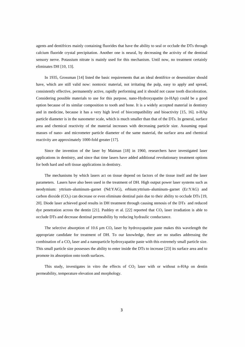
(FIGURE 1).
The surface and cross section morphology of all samples was evaluated using SEM (Tabletop
Microscope TM-1000, Hitachi high technologies corp., Tokyo, Japan). Six samples were used; three of
them were utilized for assessment of surface morphology and the other three samples were used to
examine the cross section morphology after different treatment modalities. For the cross section
examination, the three samples were fractured with a dental chisel after making a groove at the pulpal side
of the sample opposite to the lasing side, with the aid of a diamond bur mounted on a high speed air motor
with water spray.
A whole tooth was used for the temperature measurement experiment. The lasing was done after
cementum removal and marking an area of 3x4 mm for lasing. A hole was made with a diamond fissure
bur in the root surface opposite to the lasing area, until reaching the pulp cavity. The teeth were mounted
in acrylic resin at the crown portion (FIGURE 2).
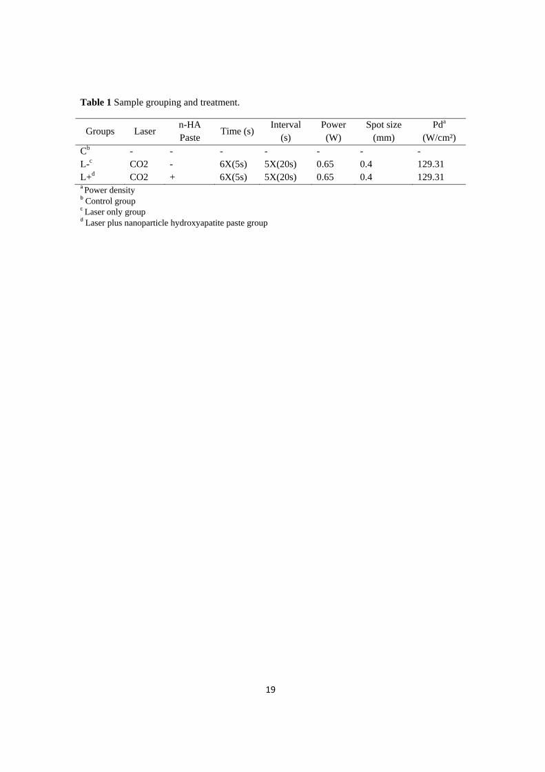
2.2 Samples grouping and treatment. Thirty six exposed DTs samples were divided into three groups
(n=12) (TABLE 1), the control (C), laser (L-) and laser plus n -HAp (L+), two per group for the SEM and
ten for each group for the permeability test. The (C) group samples received no treatment after surface
conditioning with 1% citric acid. For the group (L-) the samples were irradiated with a CO2 laser only, and
for the (L+) group the CO2 laser irradiation was done after applying the n-HAp Paste (M K Impex Corp.,
Ontario, Canada). With an average particle size of 60 nm, the paste was prepared by mixing the n-HAp
powder with distilled water, then it was added to the dentin surface by a micro-brush 10 times over a
period of 10 minutes with hand pressure. After that, excess paste was brushed away. The samples were
irradiated with a CO2 laser (OpusDuo ECTM, Lumenis Germany GmbH), 10.6 µm wavelength, 0.65 W, in
a continuous mode perpendicular to dentin surface, with 5 mm defocus distance and a power density of

5
(129.33 W/cm2). The lasing was done according to Moritz et al. [20], 6 times for 5s with 20s interval for
cooling in between. After treatment the samples were rinsed with a distilled water spray for 15 seconds.
2.3 Dye penetration test. The specimens were coated with three layers of nail varnish except in the marked
area, then were immersed in an aqueous solution of 2 % methylene blue dye for 1 hour at room
temperature. The samples were then washed under tap water, dried, cut longitudinally and the cross-
section of each sample was examined with a stereomicroscope (Hamilton, Altay Scientific, Rome, Italy).
The permeability test was evaluated by using the measure pictures V 1.0 software (CAD-KAS
Kassler Computer software GbR, Germany). A stereomicroscope under the magnification of x50 was used
to measure the length of dye penetration in the DTs from the outer surface of the root toward the pulp
chamber. For the standardization of the samples measurements, the length of dye penetration inside the
dentin was divided by the whole thickness of the sample. Then the results multiples by 100%.
2.4 Temperature measurement. The measurements were done opposite to the lasing area under the
following conditions. Part of the root was immersed in water, which was heated by an accurate digital
hotplate (Cemaric*, Thermo Scientific Inc., MA, USA) for teeth temperature stabilization at 37 ± 0.5˚C
while being immersed in water. A K type thermocouple was used, which was connected to a digital
multilogger thermometer (AMPROBE TMD-56, Everett, WA, USA), with basic accuracy of ± 0.05%,
through a universal serial bus controller connected to a computer software (AMPROPE multi-line V3.0).
The temperature recording was done every second. A thermal compound of 5.6 W/MK thermal
conductivity (Arctic MX-2, China) was injected inside the pulp chamber, to confirm a contact between the
thermocouple and the dentin surface during temperature measurements. Then a horizontal tooth sectioning
through the pre-marked lasing area was done to measure the thickness of all samples by a vernier caliper
(TOPEX Sp. z o.o. S.K., Warsaw, Poland), from the peripheral dentin surface to the pulpal one.
3. Results
3.1 Dye penetration test. The data obtained from dye penetration from all the experimental groups were
statistically analyzed using SPSS Statistics 20 (IBM® Corp., NY, USA). Descriptive statistics were done
to obtain the means and standard deviations (TABLE 2).
Group (C) showed an obvious dye penetration approaching the pulpal side of the dentin with a mean
of 86.52%, while (L+) exhibited only a slight penetration of the dye beyond the dentin surface with a
mean of 16.22%. In group (L-) the mean was 49.39% which seemed to have dye penetration in the DTs.
To check if the obtained data distribution is normal, the Shapiro-Wilk test was implemented, and the
test statistics showed that data were not normally distributed (P < 0.05). To examine if the groups were
statistically different, the Kruskal-Wallis non-parametric test was used and the obtained descriptive level

6
was 0.002, which revealed that the groups were significantly different. For inter-groups comparisons the
post-hoc Dunnett multiple comparison test was performed. The group (L+) possessed highly significant
less dye penetration compared to the control group (C) (P < 0.001) (FIGURE 3); (L+) and (L-) groups
were also statistically different (P < 0.05). Finally the laser group was significantly different compared to
the control group (C) (P < 0.05).
3.2 SEM evaluation. The SEM showed the morphological characteristics of all different groups of the
experiment. In group (C), the DTs were widely open and the whole area was free from smear layer
(FIGURE 4A and 5A). The specimens of group (L-) (FIGURE 4B) showed a smaller diameter of DTs
which is also noted in the cross section micrograph (FIGURE 5B). A large number of DTs were occluded
due to the melting effect of the CO2 laser. In the combined group (L+) (FIGURE 4C) most tubules were
occluded by n-HAp, and there were signs of melting of the n-HAp. The cross section micrograph of the
combined group (L+) (FIGURE 5C) showed melting of the n-HAp, penetration into the tubules and
formation of plug over it.
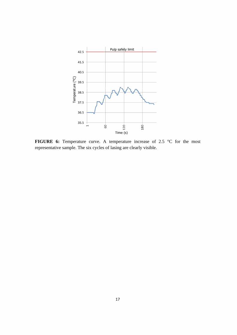
3.3 Temperature measurements. Laser irradiation showed a mean temperature rise of 2.33 ºC ± 0.56 ºC,
and the maximum temperature rise was 3.2 ºC. The corresponding average dentin thickness was 2.19 mm
± 0.24 mm. The curve of the temperature elevation course (FIGURE 6), showed a temperature increase of
2.5 ºC, revealing a favorable temperature drop in the 20 seconds interval between each 5 second lasing
cycle.
4. Discussion
Various studies have been conducted to evaluate the effect of n-HAp on enamel and dentin
remineralization [25, 26]. In this study we are trying to assess the effectiveness of this material in the
treatment of DH. In this work, the ability of a CO2 laser to melt the nanoparticle hydroxyapatite paste and
fusion the open DTs was examined regarding the morphology and permeability of dentin. Additionally,
temperature measurements were performed.
The results of the three groups in this study showed that (L+) group has the greater decrease in dentin
permeability. This outcome was supported by SEM as most of DTs were sealed by n-HAp plugs.
The (L+) group reduced the permeability more than (L-). This may indicate that the addition of n-
HAp was effective in reducing dentinal permeability. In the (L-) group dye penetration inside the dentin
was 49%. These results can be compared to Matsui et al. [21] who recorded a 41% dye extension inside
the tooth dentin thickness. Bonin et al. [27] reported a reduction in the dentinal permeability with 1 W
using CO2 laser alone. In the present study the n-HAp paste and the CO2 laser with 0.65 W proved to
reduce the dentinal permeability to an acceptable level at a lower power setting.

7
In this study, 1% citric acid was used to remove the smear layer leaving a clean surface free of
tubules plugs and without surface damage (FIGURE 4A). Citric acid is one of the agents used to remove
the smear layer from the dentin surface [28, 29] which may simulate DH in vivo conditions due to its
presence in juices, vegetables and fruits [30]. Samples were stored without adding any antiseptics, as they
may affect the dentin permeability due to mineral trapping inside the DTs [31].
On SEM micrographs, for group (L+) most of the DTs were occluded. The n-HAp particles appeared
to be melted, recrystallized and trapped inside the tubules forming a plug inside their orifices (FIGURE
5C). These plugs resisted the 15s air-water spray that was done after the lasing, remained bonded to the
DTs (FIGURE 4C). This may prove the ability of the CO2 laser in melting and bonding the n-HAp
particles to the DTs. In pilot experiments of this study SEM Shows that adding n-HAp paste only not
resisted the 15s air-water spray. Leaving open DTs like the SEM of the control group (C) with some
remnant of the n-HAp paste remaining on the dentin surface (FIGURE 7). The control group was chosen
to be representative of open DTs. We may say that the combined treatment or the indirect method as first
stated by Moritz et al. [20, 32] is a step forward to reduce the shortcoming of either treatment alone [23,
24, 33].
As shown in the (L-) micrograph (FIGURE 4B), dentin surface melting and narrowing of DTs
diameter had been also noted. Similar results were presented by Caker et al. [24] demonstrated a
reduction of DTs diameter after exposure to CO2 laser. In the (L+) group micrograph, the melting of n-
HAp may be due to the nanometer particles size of the n-HAp which reveals a large surface area
promoting absorption on the dentin surface [23].
We measured the temperature to ensure that our parameter is within the pulp safety limit of a 5.5 °C
temperature increase as reported by Zach and Cohen [34]. The maximum recorded value was a
temperature increase of 3.2 °C inside the pulp chamber, with an average dentin thickness of 2.19 mm.
This result is in agreement with Moritz et al. [20] who recorded a 2.5 °C as maximum temperate increase.
As the heat dissipated to the surrounding medium, the air or the water [35], we made a hole opposite the
lasing area, so that the thermocouple inserted inside the pulp chamber was isolated by the tooth structure,
and may not detect the heat dissipated to that medium. We used a whole tooth instead of sectioned dentin
samples for temperature measurement, such as we had done in the permeability test. According to the
basic law of thermodynamics: dQ = m c dT, where dQ is the heat content, m represents tooth mass, c is
the heat capacity and dT is the linear change in the temperature. The sample with a smaller mass may
exhibit a higher temperature after the lasing procedure. A thermal compound was used inside the pulp
chamber to prevent a gap formation between the thermocouple and the dentin.
According to the manufacturer specifications, the n-HAp used in this study is water insoluble. This
may give favorable expectations, while Romano et al. [33] used the calcium hydroxide paste in
combination with CO2 laser, they stated that the stability of this paste is still a point for open discussion
due to the solubility of this material.
8
Due to its high absorption in both Hydroxyapatite and water, the CO2 laser with a wavelength of 10.6
µm, has high absorption coefficient in dentin (800 cm-1), presenting satisfactory superficial interaction.
This kind of interaction is required for sealing DTs to reduce fluid passage across the dentin and
consequently DH pain relieve [35, 36].
Due to the presence of the dental pulp in vivo, this work needs to be confirmed clinically. The
presence of pulp circulation in vivo may be of advantage in reducing the temperature elevation due to
lasing as it acts as a heat sink that dissipates the generated heat [37]. Also, odontoblast cells in the dental
pulp of primates and dogs could be indirectly activated by low power CO2 laser leading to reactional
dentinogenesis [38, 39].
DH treatment by laser seems to be simple, quick and effective [21]. He et al. [40] concluded through
his systematic review that laser treatment with correct and controlled parameters will not lead to adverse
effects. Our experiment with 0.65 W of moderate power density showed that there was no damage,
carbonization or cracks on the dentin surface, which is in accordance with previous studies employed for
DH treatment [32, 35, 41].
This experiment showed that n-HAp melted and plugged over most of the DTs, reduced dentinal
permeability with an acceptable temperature increase using a CO2 laser of 0.65 W with a moderate power
density.
5. Conclusions
Based on the results of the present study, the combination of nano-hydroxyapatite paste and a CO2
laser of moderate power density occluded the dentinal tubules and reduced the permeability of exposed
dentin. This preliminary experiment gives primary evidence of a new treatment modality for dentin
hypersensitivity. Further comprehensive clinical studies are needed to assess the clinical potential of this
combined treatment.
Author Disclosure Statement
No conflicting financial interests exist.
References
1. G. R. Holland, M. N. Narhi, M. Addy, L. Gangarosa and R. Orchardson, "Guidelines for the design and conduct of clinical trials on dentine hypersensitivity," J Clin Periodontol, vol. 24, no. 11, pp. 808‐813, 1997. 2. M. Addy, "Dentine hypersensitivity: definition, prevalence distribution and aetiology. In: Addy, M., Embery, G., Edgar, W.M., and Orchardson, R., editors. Tooth wear and sensitivity: Clinical advances in restorative dentistry," London: Martin Dunitz, pp. 239‐248, 2000. 3. M. Addy and M. L. Hunter, "Can tooth brushing damage your health? Effects on oral and dental tissues," Int Dent J, vol. 53 Suppl 3, pp. 177‐186, 2003. 4. L. Rimondini, C. Baroni and A. Carrassi, "Ultrastructure of hypersensitive and non‐sensitive dentine. A study on replica models," J Clin Periodontol, vol. 22, no. 12, pp. 899‐902, 1995.

9
5. R. Garberoglio and M. Brannstrom, "Scanning electron microscopic investigation of human dentinal tubules," Arch Oral Biol, vol. 21, no. 6, pp. 355‐362, 1976. 6. A. G. Isik, B. Tarim, A. A. Hafez, F. S. Yalcin, U. Onan and C. F. Cox, "A comparative scanning electron microscopic study on the characteristics of demineralized dentin root surface using different tetracycline HCl concentrations and application times," J Periodontol, vol. 71, no. 2, pp. 219‐225, 2000. 7. P. M. Bartold, "Dentinal hypersensitivity: a review," Aust Dent J, vol. 51, no. 3, pp. 212‐218; quiz 276, 2006. 8. J. A. Gerschman, J. Ruben and J. Gebart‐Eaglemont, "Low level laser therapy for dentinal tooth hypersensitivity," Aust Dent J, vol. 39, no. 6, pp. 353‐357, 1994. 9. P. Dowell and M. Addy, "Dentine hypersensitivity‐‐a review. Aetiology, symptoms and theories of pain production," J Clin Periodontol, vol. 10, no. 4, pp. 341‐350, 1983. 10. R. Orchardson and D. G. Gillam, "Managing dentin hypersensitivity," J Am Dent Assoc, vol. 137, no. 7, pp. 990‐998; quiz 1028‐1029, 2006. 11. L. P. Gangarosa, Sr., "Current strategies for dentist‐applied treatment in the management of hypersensitive dentine," Arch Oral Biol, vol. 39 Suppl, pp. 101s‐106s, 1994. 12. M. B. Chabanski, D. G. Gillam, J. S. Bulman and H. N. Newman, "Prevalence of cervical dentine sensitivity in a population of patients referred to a specialist Periodontology Department," J Clin Periodontol, vol. 23, no. 11, pp. 989‐992, 1996. 13. K. Markowitz and S. Kim, "Hypersensitive teeth. Experimental studies of dentinal desensitizing agents," Dent Clin North Am, vol. 34, no. 3, pp. 491‐501, 1990. 14. L. I. Grossman, "A systematic method for the treatment of hypersensitive dentin," J. Am. Dent Assoc, vol. 22, pp. 592‐602, 1935. 15. M. Hannig and C. Hannig, "Nanomaterials in preventive dentistry," Nat Nanotechnol, vol. 5, no. 8, pp. 565‐569, 2010. 16. J. Vandiver, D. Dean, N. Patel, W. Bonfield and C. Ortiz, "Nanoscale variation in surface charge of synthetic hydroxyapatite detected by chemically and spatially specific high‐resolution force spectroscopy," Biomaterials, vol. 26, no. 3, pp. 271‐283, 2005. 17. C. Buzea, Pacheco, II and K. Robbie, "Nanomaterials and nanoparticles: sources and toxicity," Biointerphases, vol. 2, no. 4, pp. Mr17‐71, 2007. 18. T. H. Maiman, "Stimulated Optical Radiation in Ruby," Nature, vol. 187, no. 4736, pp. 493‐494, 1960. 19. R. Birang, J. Poursamimi, N. Gutknecht, F. Lampert and M. Mir, "Comparative evaluation of the effects of Nd:YAG and Er:YAG laser in dentin hypersensitivity treatment," Lasers Med Sci, vol. 22, no. 1, pp. 21‐24, 2007. 20. A. Moritz, N. Gutknecht, U. Schoop, K. Goharkhay, D. Ebrahim, J. Wernisch and W. Sperr, "The advantage of CO2‐treated dental necks, in comparison with a standard method: results of an in vivo study," J Clin Laser Med Surg, vol. 14, no. 1, pp. 27‐32, 1996. 21. S. Matsui, M. Kozuka, J. Takayama, K. Ueda, H. Nakamura, K. Ito, M. Kimura, H. Miura, Y. Tsujimoto, T. Kondoh, T. Ikemi and K. Matsushima, "Stimulatory Effects of CO(2) Laser, Er:YAG Laser and Ga‐Al‐As Laser on Exposed Dentinal Tubule Orifices," J Clin Biochem Nutr, vol. 42, no. 2, pp. 138‐143, 2008. 22. E. L. Pashley, J. A. Horner, M. Liu, S. Kim and D. H. Pashley, "Effects of CO2 laser energy on dentin permeability," J Endod, vol. 18, no. 6, pp. 257‐262, 1992. 23. J. S. Kim, S. Y. Han, H. K. Kwon and B. I. Kim, "Synergistic effect of dentinal tubule occlusion by nano‐carbonate apatite and CO2 laser in vitro," Photomed Laser Surg, vol. 31, no. 8, pp. 392‐397, 2013.

10
24. G. Cakar, B. Kuru, S. D. Ipci, Z. M. Aksoy, I. Okar and S. Yilmaz, "Effect of Er:YAG and CO2 lasers with and without sodium fluoride gel on dentinal tubules: a scanning electron microscope examination," Photomed Laser Surg, vol. 26, no. 6, pp. 565‐571, 2008. 25. P. Tschoppe, D. L. Zandim, P. Martus and A. M. Kielbassa, "Enamel and dentine remineralization by nano‐hydroxyapatite toothpastes," J Dent, vol. 39, no. 6, pp. 430‐437, 2011. 26. K. Najibfard, K. Ramalingam, I. Chedjieu and B. T. Amaechi, "Remineralization of early caries by a nano‐hydroxyapatite dentifrice," J Clin Dent, vol. 22, no. 5, pp. 139‐143, 2011. 27. P. Bonin, R. Boivin and J. Poulard, "Dentinal permeability of the dog canine after exposure of a cervical cavity to the beam of a CO2 laser," J Endod, vol. 17, no. 3, pp. 116‐118, 1991. 28. C. Reis, G. De‐Deus, F. Leal, E. Azevedo, T. Coutinho‐Filho and S. Paciornik, "Strong effect on dentin after the use of high concentrations of citric acid: an assessment with co‐site optical microscopy and ESEM," Dent Mater, vol. 24, no. 12, pp. 1608‐1615, 2008. 29. N. G. Kumar and D. S. Mehta, "Short‐term assessment of the Nd:YAG laser with and without sodium fluoride varnish in the treatment of dentin hypersensitivity‐‐a clinical and scanning electron microscopy study," J Periodontol, vol. 76, no. 7, pp. 1140‐1147, 2005. 30. N. X. West, J. A. Hughes and M. Addy, "Erosion of dentine and enamel in vitro by dietary acids: the effect of temperature, acid character, concentration and exposure time," J Oral Rehabil, vol. 27, no. 10, pp. 875‐880, 2000. 31. R. S. McGuckin and D. H. Pashley, "Effect of disinfection/sterilization treatments on Gluma‐mediated dentin shear bond strengths," Am J Dent, vol. 3, no. 6, pp. 278‐282, 1990. 32. A. Moritz, U. Schoop, K. Goharkhay, M. Aoid, P. Reichenbach, M. A. Lothaller, J. Wernisch and W. Sperr, "Long‐term effects of CO2 laser irradiation on treatment of hypersensitive dental necks: results of an in Vivo study," J Clin Laser Med Surg, vol. 16, no. 4, pp. 211‐215, 1998. 33. G. A. Gholami, R. Fekrazad, A. Esmaiel‐Nejad and K. A. Kalhori, "An evaluation of the occluding effects of Er;Cr:YSGG, Nd:YAG, CO(2) and diode lasers on dentinal tubules: a scanning electron microscope in vitro study," Photomed Laser Surg, vol. 29, no. 2, pp. 115‐121, 2011. 34. L. Zach and G. Cohen, "PULP RESPONSE TO EXTERNALLY APPLIED HEAT," Oral Surg Oral Med Oral Pathol, vol. 19, pp. 515‐530, 1965. 35. A. C. Romano, A. C. Aranha, B. L. da Silveira, S. L. Baldochi and P. Eduardo Cde, "Evaluation of carbon dioxide laser irradiation associated with calcium hydroxide in the treatment of dentinal hypersensitivity. A preliminary study," Lasers Med Sci, vol. 26, no. 1, pp. 35‐42, 2011. 36. D. Fried, M. J. Zuerlein, C. Q. Le and J. D. Featherstone, "Thermal and chemical modification of dentin by 9‐11‐microm CO2 laser pulses of 5‐100‐micros duration," Lasers Surg Med, vol. 31, no. 4, pp. 275‐282, 2002. 37. M. Lin, F. Xu, T. J. Lu and B. F. Bai, "A review of heat transfer in human tooth‐‐experimental characterization and mathematical modeling," Dent Mater, vol. 26, no. 6, pp. 501‐513, 2010. 38. J. Melcer, M. T. Chaumette, S. Zeboulon, F. Melcer, R. Hasson, R. Merard, Y. Pinaudeau, J. Dejardin and R. Weill, "Preliminary report on the effect of the CO2 laser beam on the dental pulp of the Macaca mulatta primate and the beagle dog," J Endod, vol. 11, no. 1, pp. 1‐5, 1985. 39. J. Melcer, M. T. Chaumette and F. Melcer, "Dental pulp exposed to the CO2 laser beam," Lasers Surg Med, vol. 7, no. 4, pp. 347‐352, 1987. 40. S. He, Y. Wang, X. Li and D. Hu, "Effectiveness of laser therapy and topical desensitising agents in treating dentine hypersensitivity: a systematic review," J Oral Rehabil, vol. 38, no. 5, pp. 348‐358, 2011. 41. A. Barone, U. Covani, R. Crespi and G. E. Romanos, "Root surface morphological changes after focused versus defocused CO2 laser irradiation: a scanning electron microscopy analysis," J Periodontol, vol. 73, no. 4, pp. 370‐373, 2002.

11
FIGURE 1: Permeability test setup and method. 1. Tooth sectioning, 2. Samples collection and cementum removal, 3. Immersion in 1% citric acid for 5 min., 4. Marking of the lasing area and nail varnish coating, 5. Mounting in acrylic resin mold, 6. Lasing, 7. Immersion in 2% methylene blue for 1 hr., 8. Longitudinal sectioning, 9. Stereomicroscopic examination. FIGURE 2: Experimental setup for temperature measurement. FIGURE 3: Dye penetration test. D: dentin surface side, P: pulpal side. (a) Control group (C), (b) laser only (L-) and (c) the laser plus nano-hydroxyapatite paste (L+). The (C) group shows a full length dye penetration while for the (L+) group the dye is confined mainly to the surface with very slight dye penetration.
FIGURE 4: Dentine surface scanning electron microscope (SEM) micrographs. (a) Control group (C), (b) laser only (L-) and (c) the laser plus nano-hydroxyapatite paste (L+). The original magnification was x1200.
FIGURE 5: Cross-sectional scanning electron microscope (SEM) micrographs. (a) Control group (C), (b) laser only (L-) and (c) the laser plus nano-hydroxyapatite paste (L+). The original magnification was x5000.
FIGURE 6: Temperature curve. A temperature increase of 2.5 °C for the most representative sample. The six cycles of lasing are clearly visible.
FIGURE 7: SEM micrograph of a pilot sample in which n-HAp only is applied on DTs. Showing open DTs like the SEM of the control group (C) with some remnant of the n-HAp paste remaining on the dentin surface. The original magnification was x1200.
FIGcollof th7. Ster
GURE 1: Plection and he lasing arImmersion reomicrosco
Permeabilitcementum rrea and nail
in 2% mopic examin
ty test seturemoval, 3. l varnish comethylene nation.
12
up and meImmersionating, 5. Mblue for 1
ethod. 1. Tn in 1% citriounting in a1 hr., 8.
Tooth sectiic acid for 5acrylic resinLongitudin
oning, 2.Sa5 min., 4. Mn mold, 6. L
nal sectioni
amples Marking Lasing, ing, 9.

FIG
GURE 2: Exxperimentall setup for t
13
temperature measuremeent

FIGgrou(C) con
GURE 3: Dup (C), (b) group sho
fined mainl
Dye penetralaser only (
ows a full lly to the sur
ation test. D(L-) and (c)length dye rface with v
14
D: dentin su the laser ppenetration
very slight d
urface sidelus nano-hyn while for
dye penetrat
, P: pulpal ydroxyapatir the (L+) tion.
side. (a) Cte paste (L+group the
Control +). The dye is

FIGCon(L+
GURE 4: Dntrol group +). The origi
Dentine sur(C), (b) las
inal magnifi
rface scannser only (L
fication was
15
ning electro-) and (c) tx1200.
on microscothe laser plu
ope (SEM) us nano-hyd
micrograpdroxyapatite
hs. (a) e paste

FIGCon(L+
GURE 5: Cntrol group +). The origi
Cross-sectio(C), (b) las
inal magnifi
onal scanninser only (L
fication was
16
ng electron-) and (c) tx5000.
n microscopthe laser plu
pe (SEM) mus nano-hyd
micrographdroxyapatite
hs. (a) e paste
FIGrepr
GURE 6: Tresentative s
Temperatursample. The
re curve. Ae six cycles
17
A temperat of lasing ar
ture increasre clearly vi
se of 2.5 isible.
°C for thee most

FIGShoHAp
GURE 7: SEowing open p paste rem
EM microgrDTs like th
maining on th
raph of a pihe SEM of thhe dentin su
18
ilot sample ihe control gurface. The
in which n-group (C) woriginal ma
HAp only iwith some reagnification
is applied onemnant of thn was x1200
n DTs. he n-0.
19
Table 1 Sample grouping and treatment.
Groups Laser n-HA Paste
Time (s) Interval
(s) Power (W)
Spot size (mm)
Pda (W/cm²)
Cb - - - - - - - L-c CO2 - 6X(5s) 5X(20s) 0.65 0.4 129.31 L+d CO2 + 6X(5s) 5X(20s) 0.65 0.4 129.31 a Power density b Control group c Laser only group d Laser plus nanoparticle hydroxyapatite paste group

20
Table 2 Mean of percentage of dye penetration and standard deviation in each group.
a Control group b Laser only group c Laser plus nanoparticle hydroxyapatite paste group
Groups Mean Std. Deviation Ca 86.52% 20.10 L-b 49.39% 43.29 L+c 16.22% 23.47