Embed Size (px)
Citation preview

04/19/23 1
Cranial Nerves AssessmentCranial Nerves Assessment20092009
Sheeba Jacob R.N., B.S.N.,Sheeba Jacob R.N., B.S.N.,
Victoria Kim RN B.S.N.Victoria Kim RN B.S.N.

04/19/23 2
GoalsGoals
Goal:Goal:1 .Students will be able to identify 1 .Students will be able to identify the 12 cranial nerves by name and the 12 cranial nerves by name and assess the function of each assess the function of each (knowledge).(knowledge).
2. The student will be able to 2. The student will be able to comprehend the anatomical comprehend the anatomical significance of the assessment. significance of the assessment. (Comprehension)(Comprehension)

04/19/23 3
ObjectivesObjectives
1.1. Objectives, Upon completion of this learning Objectives, Upon completion of this learning experience the student should be able to:experience the student should be able to:1.1. Identify the 12 cranial nerves.Identify the 12 cranial nerves.2.2. Demonstrate the ability to interpret the test of visual Demonstrate the ability to interpret the test of visual
acuity using a snellen chart.acuity using a snellen chart.3.3. Assess the visual fields by recognizing correct Assess the visual fields by recognizing correct
technique for confrontation.technique for confrontation.4.4. Perform the cover test.Perform the cover test.5.5. Correctly assess PERRLA (Pupils equal, round, reactive Correctly assess PERRLA (Pupils equal, round, reactive
to light and accomodation).to light and accomodation).6.6. Comprehend the correct technique for performing the Comprehend the correct technique for performing the
whisper test, Weber and Rhinne test.whisper test, Weber and Rhinne test.7.7. Perform assessment of neck including the lymph nodes.Perform assessment of neck including the lymph nodes.8.8. Use the technique of inspecting and palpating the head Use the technique of inspecting and palpating the head
and scalp, anterior posterior chest, and sensorimotor and scalp, anterior posterior chest, and sensorimotor functions.functions.

04/19/23 4
Identify the 12 cranial Identify the 12 cranial nerves.nerves.

04/19/23 5
The Cranial NervesThe Cranial Nerves. 1. . 1. Video Cranial Nerves
University of Utah (University of Utah (Hyperlinkhttp://www.youtube.com/watch?Hyperlinkhttp://www.youtube.com/watch?v=CL1KNziYmzo)v=CL1KNziYmzo)
Watch the video and Watch the video and reviewing the reviewing the different anatomical different anatomical sections of the brainsections of the brain
The nurse, in The nurse, in assessing the 12 assessing the 12 cranial nerves, is cranial nerves, is testing the testing the functions of various functions of various parts of the brain parts of the brain 2. . Anatomy of Brain
-http://www.righthealth.com/search/Picture_Of_Labeled_Brain/overview/google_imagesearch?img=3

04/19/23 6
Cranial Nerve AnatomyCranial Nerve Anatomy
• There are 12 pairs of There are 12 pairs of cranial nervescranial nerves
• CN I SmellCN I Smell• CN II VisionCN II Vision• CN III, IV, VI OculomotorCN III, IV, VI Oculomotor• CN V Trigeminal CN V Trigeminal
Sensorimotor muscles of Sensorimotor muscles of the Jawthe Jaw
• CN VII Sensorimotor of the CN VII Sensorimotor of the faceface
• CN VIII Hearing CN VIII Hearing • CN IX, X, XII Mouth, CN IX, X, XII Mouth,
esophagus, oropharynxesophagus, oropharynx• CN XI Cervical Spine and CN XI Cervical Spine and
shouldershoulder
3. Cranial Nerve http://www.becomehealthynow.com/images/organs/nervous/cranial_nerves_bh.jpg

04/19/23 7
Knowledge Test 1Knowledge Test 1
• How many Cranial How many Cranial Nerves are there?Nerves are there?
1212
66
33
1010

04/19/23 8
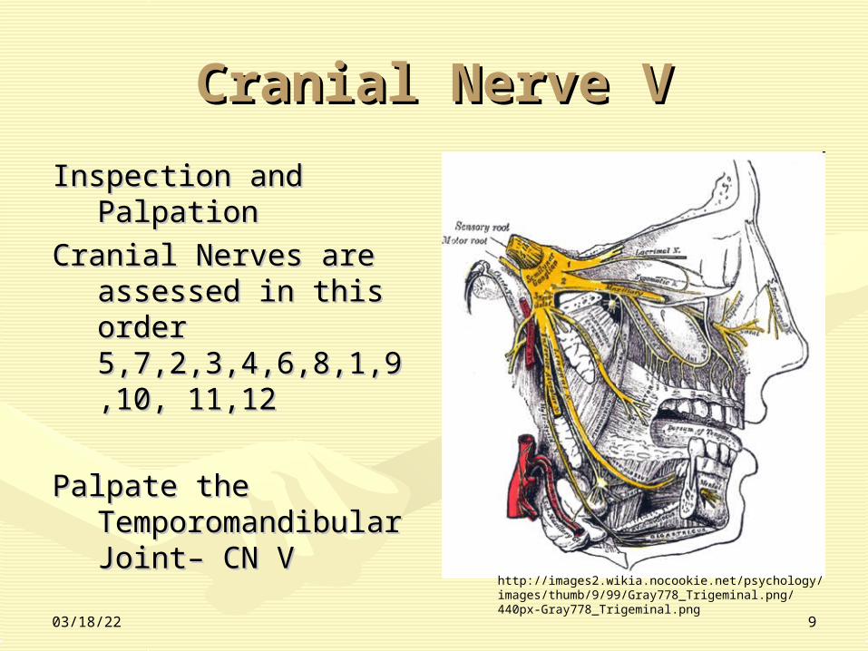
Cranial Nerve VCranial Nerve V • Cranial Nerve VCranial Nerve V
• CN V Trigeminal CN V Trigeminal Sensorimotor muscles of Sensorimotor muscles of the Jawthe Jaw
• The temperomandibular The temperomandibular joint is palpated while the joint is palpated while the patient clenches jaw, patient clenches jaw, opens and closes mouth, opens and closes mouth, moves jaw side to side and moves jaw side to side and forward against pressure forward against pressure CN V, trigeminal NerveCN V, trigeminal Nerve
• CN V is further tested CN V is further tested with light touch a with light touch a wisp of cotton in three wisp of cotton in three areas .areas .
Temporomandibular Temporomandibular joint and Massater joint and Massater MuscleMuscle
Cranial Nerve V -http://www.fotosearch.com/LIF155/mm103010/
04/19/23 9
Cranial Nerve VCranial Nerve V
Inspection and Inspection and PalpationPalpation
Cranial Nerves are Cranial Nerves are assessed in this assessed in this order order 5,7,2,3,4,6,8,1,9,10, 5,7,2,3,4,6,8,1,9,10, 11,1211,12
Palpate the Palpate the Temporomandibular Temporomandibular Joint– CN VJoint– CN V http://images2.wikia.nocookie.net/psychology/images/
thumb/9/99/Gray778_Trigeminal.png/440px-Gray778_Trigeminal.png

04/19/23 10
Knowledge Test 1Knowledge Test 1
Q. Which cranial Q. Which cranial nerve nerve is further is further tested with tested with light touchlight touch a a whisp of cotton whisp of cotton in three areas .in three areas .
VIVI
VV
VIIVII
IIIIII

04/19/23 11
Cranial Nerve VII Cranial Nerve VII FunctionFunction
Cranial Nerve VIICranial Nerve VII• Definition cranial nerves sevenDefinition cranial nerves seven• The facial nerve VII (nThe facial nerve VII (nervus facialiservus facialis) )
comes from the pons in the hindbrain. comes from the pons in the hindbrain. It is a strong motor, sensory and It is a strong motor, sensory and parasympathetic nerve. Its numerous parasympathetic nerve. Its numerous branches connect to the face muscles, branches connect to the face muscles, the skin around the earlobes and the skin around the earlobes and various exocrine glands in the head..various exocrine glands in the head..

04/19/23 12
Cranial Nerve VII, Facial Cranial Nerve VII, Facial NerveNerve
1.1. Cranial Nerve VII Cranial Nerve VII Anatomy and Anatomy and Physiology Physiology http://www.megasystemsusa.com/app/home/home.aspx

04/19/23 13
Cranial Nerve VII Cranial Nerve VII AnatomyAnatomy
• Facial Nerve (nervus Facial Nerve (nervus facialis)facialis)
• Comes from the Pons Comes from the Pons in the Midbrainin the Midbrain
• SensorySensory• MotorMotor• Branches connect to Branches connect to
the face muscles, the the face muscles, the skin around the skin around the earlobes and various earlobes and various exocrine glands in the exocrine glands in the head..head..

04/19/23 14
Assessment Cranial Assessment Cranial Nerve VIINerve VII
• Assessment - is tested by observing Assessment - is tested by observing the presence and symmetry of the the presence and symmetry of the patients facial muscle movements patients facial muscle movements – SmilingSmiling– Frowning Frowning – Showing teethShowing teeth– Puffing out cheeks Puffing out cheeks – Raising eyebrows Raising eyebrows – Resisting examiners attempt to open Resisting examiners attempt to open
the eyesthe eyes

04/19/23 15
Knowledge Test 1Knowledge Test 1
Q. A test of Cranial Q. A test of Cranial VII is? VII is?
Protruding the Protruding the tonguetongue
BlinkingBlinking
Flapping handsFlapping hands
SmilingSmiling

04/19/23 16
Cranial Nerve II, Optic Cranial Nerve II, Optic Nerve Function of SightNerve Function of Sight
Definition Cranial Nerve Definition Cranial Nerve the optic the optic nerve II (vnerve II (vervus opticuservus opticus), which ), which facilitates sight,facilitates sight,
http://www.megasystemsusa.com/app/home/home.aspx

04/19/23 17
Cranial Nerve II, Optic Cranial Nerve II, Optic Nerve AnatomyNerve Anatomy
1.1. Cranial Nerve II Cranial Nerve II Anatomy and Anatomy and Physiology Physiology http://www.megasystemsusa.com/app/home/home.aspx
1.Cranial Nerve II Anatomy and Physiology

04/19/23 18
• Assessment Cranial nerve IIAssessment Cranial nerve II– Visual AcuityVisual Acuity – part of cranial nerve to – part of cranial nerve to
is tested .is tested .• Near visual acuity, must be done having Near visual acuity, must be done having
patient read a small snellen equivalent patient read a small snellen equivalent • Visual fieldsVisual fields, part of cranial nerves are , part of cranial nerves are
tested by confrontationtested by confrontation

04/19/23 19
Cranial Nerve II, Optic Cranial Nerve II, Optic Nerve Nerve
Visual AcuityVisual Acuity• Using a Using a Snellen eye chart
• Using the E Snellen Chart hold it out in front of Using the E Snellen Chart hold it out in front of them 14 inchesthem 14 inches– The chart is usually read while standing at a distance of The chart is usually read while standing at a distance of
20 feet. Acuity is represented as a fraction, with the 20 feet. Acuity is represented as a fraction, with the distance at which you are standing being the numerator distance at which you are standing being the numerator (top part of fraction), and the normal maximum legible (top part of fraction), and the normal maximum legible viewing distance ("Distance" on the chart above) as the viewing distance ("Distance" on the chart above) as the denominator (bottom of fraction). So if, at 20 feet, you denominator (bottom of fraction). So if, at 20 feet, you can read the letters on the row marked "40", this means can read the letters on the row marked "40", this means you have visual acuity of 20/40 or better: 1/2 normal. you have visual acuity of 20/40 or better: 1/2 normal. From 10 feet, if the smallest letters you could read were From 10 feet, if the smallest letters you could read were on the "40" line, this would give you an acuity of 10/40: on the "40" line, this would give you an acuity of 10/40: 1/4 normal. If you are nearsighted, your vision will 1/4 normal. If you are nearsighted, your vision will become more normal the closer you stand to the chart. become more normal the closer you stand to the chart. http://www.i-see.org/eyecharts.htmlhttp://www.i-see.org/eyecharts.html
• Patients wearing corrective lenses should be tested Patients wearing corrective lenses should be tested with lenses in place .with lenses in place .
• Each eye is tested separatelyEach eye is tested separately

04/19/23 20
Cranial Nerve II, Optic Cranial Nerve II, Optic NerveNerve
Confrontation TestConfrontation Test• Visual fieldsVisual fields, part of cranial nerves are , part of cranial nerves are
tested by confrontation tested by confrontation • Examination of the patient's left eye Examination of the patient's left eye
visual fields by confrontation. The visual fields by confrontation. The patient is asked to identify the number of patient is asked to identify the number of fingers, which the examiner raises in fingers, which the examiner raises in each quadrant while centering his gaze each quadrant while centering his gaze on the examiner's right eye on the examiner's right eye ttp://books.google.com/books?ttp://books.google.com/books?id=B4V85KlNZfYC&pg=PA17&lpg=PA17&dq=Cranial+Nerve+II+confrontation&source=bl&ots=uYsPFaid=B4V85KlNZfYC&pg=PA17&lpg=PA17&dq=Cranial+Nerve+II+confrontation&source=bl&ots=uYsPFa_qDC&sig=vqSKzT8LIgYjRoAEiyBzM_JB6No&hl=en&ei=wiKYSqDBCIigsgOd7-_qDC&sig=vqSKzT8LIgYjRoAEiyBzM_JB6No&hl=en&ei=wiKYSqDBCIigsgOd7-WzAg&sa=X&oi=book_result&ct=result&resnum=1#v=onepage&q=Cranial%20Nerve%20IIWzAg&sa=X&oi=book_result&ct=result&resnum=1#v=onepage&q=Cranial%20Nerve%20II%20confrontation&f=false%20confrontation&f=false
• Want to see a Video of Confrontation? Want to see a Video of Confrontation? http://www.youtube.com/watch?http://www.youtube.com/watch?v=XiEw7v7OyBwv=XiEw7v7OyBw

04/19/23 21
Cranial Nerve III, IV, VICranial Nerve III, IV, VIPupillary FunctionPupillary Function
• Part Nine –Cranial Nerve III, IV, VI Pupillary Part Nine –Cranial Nerve III, IV, VI Pupillary FunctionFunction
• Definition Definition – The oculomotor nerve III (nThe oculomotor nerve III (n. .
oculomotorius) oculomotorius) stretches from the stretches from the front edge of the pons to the eye front edge of the pons to the eye socket. There it connects to four socket. There it connects to four external eye muscles (mexternal eye muscles (musculi recti usculi recti superiorsuperior, , inferior et medialisinferior et medialis and and mmusculus obliquus inferiorusculus obliquus inferior). It ). It also carries parasympathetic also carries parasympathetic nerves for closing the pupil nerves for closing the pupil ((usculus sphincter pupillaeusculus sphincter pupillae) and ) and the accommodation (mthe accommodation (musculus usculus ciliarisciliaris).).
– The trochlear nerve IV (nThe trochlear nerve IV (nervus ervus trochlearistrochlearis) runs from the brain to ) runs from the brain to the tendons on the eye muscles in the tendons on the eye muscles in the orbit. It connects an external the orbit. It connects an external motor eye muscle (mmotor eye muscle (musculus usculus obliquus superiorobliquus superior).).
– The abducens nerve VI (nThe abducens nerve VI (nervus ervus abducensabducens) is a motor nerve which ) is a motor nerve which connects to the external optic connects to the external optic muscles (mmuscles (musculus rectus usculus rectus lateralislateralis). If this nerve fails, the ). If this nerve fails, the eyes can become cross-eyed eyes can become cross-eyed (s(strabismus convergenstrabismus convergens).).
1.1. Assessment – CN III, IV, VIAssessment – CN III, IV, VI2.2. The cranial nerves of each The cranial nerves of each
eye is assessed separately eye is assessed separately for pupillary reactions to for pupillary reactions to lightlight
3.3. cranial nerves III, IV and or cranial nerves III, IV and or VI a further tested by VI a further tested by evaluating the extra ocular evaluating the extra ocular movements through the movements through the six six cardinal fields of gazecardinal fields of gaze. This . This examination allows examination allows assessment of each muscle assessment of each muscle in its primary field of action in its primary field of action Video link belowVideo link below
4.4. cranial nerves III, IV and VI cranial nerves III, IV and VI are further tested by are further tested by performing the performing the cover- cover- uncover testuncover test

04/19/23 22
Part 9--Cranial Nerve III, Part 9--Cranial Nerve III, IV, VI IV, VI
– The cranial nerves of each eye The cranial nerves of each eye is assessed separately for is assessed separately for pupillary reactions to light,pupillary reactions to light,
– Testing is performed twice on Testing is performed twice on each eyeeach eye
– Each pupil is evaluated for its Each pupil is evaluated for its direct, and consensual direct, and consensual reaction to lightreaction to light
– Pupils are also examined for Pupils are also examined for accommodation, PERRLA accommodation, PERRLA Links to video belowLinks to video below• http://www.youtube.com/http://www.youtube.com/
watch?v=iTncbhfbl6Awatch?v=iTncbhfbl6A• http://www.youtube.com/http://www.youtube.com/
watch?v=E2XzBaOOX8gwatch?v=E2XzBaOOX8g

04/19/23 23
Cranial Nerve III, IV, VI Cranial Nerve III, IV, VI • 2. cranial nerves III, IV and 2. cranial nerves III, IV and
or VI a further tested by or VI a further tested by evaluating the extra ocular evaluating the extra ocular movements through the six movements through the six cardinal fields of gaze. This cardinal fields of gaze. This examination allows examination allows assessment of each muscle assessment of each muscle in its primary field of action in its primary field of action Video link belowVideo link below
– http://http://www.youtube.com/www.youtube.com/watch?watch?
v=UDR7Bv=UDR7B__2sQM__2sQM
– right and up right superior right and up right superior rectus and left inferior rectus and left inferior obliqueoblique
– right lateral rectus and right lateral rectus and left medial rectusleft medial rectus
– right and down right right and down right inferior rectus and left inferior rectus and left superior obliquesuperior oblique
– . left and up left superior . left and up left superior rectus and right inferior rectus and right inferior obliqueoblique
– left lateral rectus and left lateral rectus and right medial rectusright medial rectus
– left and down left inferior left and down left inferior rectus and right superior rectus and right superior obliqueoblique

04/19/23 24
Cranial Nerve III, IV, VI Cranial Nerve III, IV, VI
• cranial nerves III, cranial nerves III, IV and VI are IV and VI are further tested by further tested by performing the performing the cover- uncover testcover- uncover test– Link Link
http://www.youtubehttp://www.youtube.com/watch?.com/watch?v=PRa7mPx2XVsv=PRa7mPx2XVs

04/19/23 25
Part 10-Cranial Nerve VIII Part 10-Cranial Nerve VIII HearingHearing
1.1. Part TEN –Cranial Part TEN –Cranial Nerve VIII, HearingNerve VIII, Hearing
2.2. Definition Definition The The vestibulocochlear vestibulocochlear nerve VIII (nnerve VIII (n. . vestibulocochlearisvestibulocochlearis) ) is a sensory nerve is a sensory nerve branch which comes branch which comes from the pons in the from the pons in the brain. It reaches to brain. It reaches to the inner ear and the inner ear and serves to carry serves to carry hearing and balance hearing and balance senses.senses.
• Assessment - Assessment - cranial nerve eight cranial nerve eight is tested using this is tested using this screening hearing screening hearing test or the whisper test or the whisper teststests
• Cranial nerve eight Cranial nerve eight is also tested by is also tested by performing the performing the Weber and Rinne Weber and Rinne teststests

04/19/23 26
Cranial Nerve VIII, Cranial Nerve VIII, Vestibulocochlear nerve Vestibulocochlear nerve VIII VIII
1.1. Cranial Nerve VII Cranial Nerve VII Anatomy and Physiology Anatomy and Physiology http://www.megasystemshttp://www.megasystemsusa.com/app/home/home.usa.com/app/home/home.aspx aspx
2. External Ear --2. External Ear --Both Both outer ears are outer ears are deliberately and deliberately and thoroughly inspected and thoroughly inspected and palpated palpated
http://medicalimages.http://medicalimages.allrefer.com/large/meallrefer.com/large/medical-findings-based-dical-findings-based-on-ear-anatomy.jpg on-ear-anatomy.jpg

04/19/23 27
Cranial Nerve VIII, Hearing Cranial Nerve VIII, Hearing AnatomyAnatomy

04/19/23 28
Performing the Whisper Performing the Whisper TestTest
• Check the patients response Check the patients response to your whispered voice one to your whispered voice one ear at a timeear at a time
• Mask the hearing in the Mask the hearing in the other ear by having the other ear by having the patient place a finger in the patient place a finger in the ear canal and gently move it ear canal and gently move it rapidly up-and-down.rapidly up-and-down.
• Stand to the side of the Stand to the side of the patient at a consistent patient at a consistent distance best for you, about distance best for you, about 1 to 2 feet away from the ear 1 to 2 feet away from the ear being tested, and out of the being tested, and out of the patients line of visionpatients line of vision
• Whisper a combination of Whisper a combination of three letters and numbers three letters and numbers very softly and ask the very softly and ask the patient to repeat the words patient to repeat the words heardheard
• Normal findings .Normal findings .– The patient should hear The patient should hear
softly whispered words in softly whispered words in each ear at that distance each ear at that distance of about 1 to 2 feet, of about 1 to 2 feet, responding correctly more responding correctly more than 50% of the timethan 50% of the time

04/19/23 29
Weber Test of HearingWeber Test of Hearing
• Weber and Rinne test is Weber and Rinne test is used to compare used to compare hearing by bone hearing by bone conduction with that of conduction with that of air conductionair conduction
• Hold the base of the Hold the base of the tuning fork with one tuning fork with one hand without touching hand without touching the tines, and stroke or the tines, and stroke or tap the tines gently . tap the tines gently . With your other hand, With your other hand, setting the tuning fork setting the tuning fork in vibrationin vibration

04/19/23 30
Performing Weber testPerforming Weber test
• Perform the Weber test Perform the Weber test by placing the base of by placing the base of the vibrating tuning fork the vibrating tuning fork on the midline vertex of on the midline vertex of the patients headthe patients head
• Ask the patient if the Ask the patient if the sound is heard equally in sound is heard equally in both ears or is better in both ears or is better in one earone ear
• Normal finding Normal finding lateralization of sound. Is lateralization of sound. Is their lateralization of their lateralization of sound?sound?
• To test the reliability To test the reliability of the patient’s of the patient’s response, repeat the response, repeat the procedure while procedure while occluding one ear, occluding one ear, asking the patient in asking the patient in which hear the which hear the sound is best heard. sound is best heard. It should be heard It should be heard best in the occluded best in the occluded ear.ear.

04/19/23 31
Rinne Test of HearingRinne Test of Hearing
• The Rinne test is The Rinne test is performed by placing performed by placing the base of the vibrating the base of the vibrating tuning fork against the tuning fork against the patient’s mastoid bonepatient’s mastoid bone
• Begin counting or Begin counting or timing the interval with timing the interval with your watch .your watch .
• Ask the patient to tell Ask the patient to tell you when the sound is you when the sound is no longer heard, noting no longer heard, noting the number of secondsthe number of seconds
• Quickly position the Quickly position the still vibrating tines 1cm still vibrating tines 1cm to 2 cm from the to 2 cm from the auditory canal, and auditory canal, and again ask the patient to again ask the patient to tell you when the sound tell you when the sound is no longer heardis no longer heard
• Continue counting or Continue counting or timing the interval to timing the interval to determine the length of determine the length of time, the sound is time, the sound is heard by air conductionheard by air conduction

04/19/23 32
Normal Findings Rinne Normal Findings Rinne TestTest
• Compare the number of seconds sound is Compare the number of seconds sound is heard by bone conduction versus air heard by bone conduction versus air conduction. conduction.
• Normal Findings: The air conducted Normal Findings: The air conducted sound should be heard twice as long as sound should be heard twice as long as bone conducted sound, after bone bone conducted sound, after bone conduction stops. For example, if bone conduction stops. For example, if bone conducted sound is heard for 15 seconds, conducted sound is heard for 15 seconds, the air conducted sound should be heard the air conducted sound should be heard for an additional 15 seconds.for an additional 15 seconds.

04/19/23 33
Part 11-Cranial Nerve I, Part 11-Cranial Nerve I, SmellSmell
1.1. The sense of smell is The sense of smell is developed by the mucous developed by the mucous membranes in the nose membranes in the nose ((nasus). nasus). Here a small Here a small area has a layer of area has a layer of sensory sells, the sensory sells, the olfactory epithelium. olfactory epithelium. This picks up smells and This picks up smells and sends them to the brain sends them to the brain ((cerebrum)cerebrum). All . All information received by information received by the brain from the main the brain from the main sensory organs is called sensory organs is called sensory signals.sensory signals.
1.1. AssessmentAssessment1.1. The nose is inspected and The nose is inspected and
palpated externally .palpated externally .
2.2. Inspected internally with Inspected internally with the light and speculum of the light and speculum of an otoscopean otoscope
3.3. The patency of the nose is The patency of the nose is also assessed be also assessed be occluding each nostril.occluding each nostril.
4.4. Test cranial nerve 1 with Test cranial nerve 1 with odor differentiation of odor differentiation of each nostrileach nostril

04/19/23 34
Cranial Nerve 1, Cranial Nerve 1, Olfactory NerveOlfactory Nerve
Cranial Nerve 1Olfactory

04/19/23 35
Cranial Nerve IX and X, Cranial Nerve IX and X, SwallowingSwallowing
1.1. The glossopharyngeal nerve IX The glossopharyngeal nerve IX (n(n. glossopharyngeus. glossopharyngeus) is a ) is a motor, sensory and motor, sensory and parasympathetic nerve branch parasympathetic nerve branch which comes from the extended which comes from the extended spinal cord (mspinal cord (medulla oblongataedulla oblongata). ). Its branches connect to the Its branches connect to the tongue, the pharyngeal muscles, tongue, the pharyngeal muscles, the ear drum, the lower the ear drum, the lower thorangeal skin and the ear wax thorangeal skin and the ear wax glands.glands.
2.2. The vagus nerve X (The vagus nerve X (n. vagusn. vagus) ) comes from the extended spinal comes from the extended spinal cord. It has motor and sensory cord. It has motor and sensory threads which reachs from the threads which reachs from the neck to the stomach-intestinal neck to the stomach-intestinal tract. On its path, it connects to tract. On its path, it connects to numerous muscles in the larynx, numerous muscles in the larynx, thorax, gullet and intestinal thorax, gullet and intestinal tract, but also the glands, tract, but also the glands, glandular organs and the ear glandular organs and the ear canal.canal.
•
1.1. Assessment SwallowingAssessment Swallowing1.1. The entire oral cavity is The entire oral cavity is
inspected inspected 2.2. CN IX and X are tested by CN IX and X are tested by
1.1. Having the patient swallowHaving the patient swallow2.2. This can be observed during This can be observed during
the thyroid assessmentthe thyroid assessment3.3. Observing movement of the Observing movement of the
palate during phonation.palate during phonation.4.4. A comment about quality of A comment about quality of
the patients voice should be the patients voice should be noted .noted .
Testing the gag reflex, does Testing the gag reflex, does not need to be assessed not need to be assessed and is usually only and is usually only tested if neurological tested if neurological impairment is suspected impairment is suspected . .
The sense of taste on the The sense of taste on the posterior third of the posterior third of the tongue does not need tongue does not need these testedthese tested

04/19/23 36
Cranial Nerve IX and X, Cranial Nerve IX and X, SwallowingSwallowing

04/19/23 37
Cranial Nerve IX, XCranial Nerve IX, X
Cranial Nerve 1Olfactory

04/19/23 38
The thyroid glandThe thyroid gland• The thyroid gland (glandula The thyroid gland (glandula
thyroidea)thyroidea)• The two oval side lobes The two oval side lobes
(lobus dexter, lobus sinister) (lobus dexter, lobus sinister) of the thyroid gland are of the thyroid gland are located at the front of the located at the front of the neck to the right and left of neck to the right and left of the windpipe (trachea) the windpipe (trachea) underneath the larynx underneath the larynx (larynx). The two lobes are (larynx). The two lobes are joined by a narrow bridge of joined by a narrow bridge of tissue, the isthumus, on a tissue, the isthumus, on a level with the 2nd to 4th level with the 2nd to 4th tracheal cartilages.tracheal cartilages.
• It has a plentiful supply of It has a plentiful supply of blood. Mutations in the form blood. Mutations in the form of hard patches and of hard patches and considerable enlargements considerable enlargements (goiter, struma) are not rare (goiter, struma) are not rare and indicate malfunctions of and indicate malfunctions of the gland.the gland.

04/19/23 39
The thyroid glandThe thyroid gland
The thyroid gland is The thyroid gland is inspected and inspected and palpated before palpated before and during and during swallowing .swallowing .
The patient is The patient is provided a couple provided a couple water to facilitate water to facilitate swallowing during swallowing during this part of the this part of the assessmentassessment

04/19/23 40
Cranial Nerve XIICranial Nerve XII
• The hypoglossal The hypoglossal nerve XII (nnerve XII (nervus ervus hypoglossushypoglossus) ) comes from the comes from the extended spinal extended spinal cord. This motor cord. This motor nerve connects to nerve connects to the muscles of the the muscles of the tongue.tongue.
•

04/19/23 41
Cranial Nerve XIICranial Nerve XII
• Cranial nerve XII Cranial nerve XII assessed by assessed by observing observing – The movement of The movement of
the tongue laterally the tongue laterally mediallymedially
– Including a Including a statement about statement about patients ability to patients ability to articulatearticulate
•

04/19/23 42
Part 13-Range of Motion of Part 13-Range of Motion of the Cervical Spinethe Cervical Spine
• The Cervical Vertebrae The Cervical Vertebrae ((Vertebrae cervicalesVertebrae cervicales))
• • The cervical vertebral column is The cervical vertebral column is
the one most capable of the one most capable of movement. The first cervical movement. The first cervical vertebrae (vertebrae (atlasatlas) and the second ) and the second cervical vertebrae (cervical vertebrae (axisaxis) deviate ) deviate significantly from the basic form significantly from the basic form of the cervical vertebrae. For of the cervical vertebrae. For instance, the atlas does not have instance, the atlas does not have the body of the vertebrae or spine the body of the vertebrae or spine of the vertebrae. Instead of that, of the vertebrae. Instead of that, it has two arches with joint it has two arches with joint surfaces for the axis and the surfaces for the axis and the occipital bone (occipital bone (os occipitaleos occipitale). ).
• The second cervical vertebra has The second cervical vertebra has a tooth-like process (a tooth-like process (dens axisdens axis), ), which connects the front surface which connects the front surface of the joint of the atlas and axis. of the joint of the atlas and axis. The cervical vertebrae 3-6 are The cervical vertebrae 3-6 are very similar to one another. very similar to one another.
• The range of motion The range of motion of the cervical spine is of the cervical spine is evaluated by having evaluated by having the patient .the patient .- put his or her chin - put his or her chin on the chest .on the chest .- lift chin to ceiling- lift chin to ceiling-turn chin toward -turn chin toward each shouldereach shoulder- touch each ear - touch each ear toward corresponding toward corresponding shouldershoulder

04/19/23 43
Cervical Vertebrae Cervical Vertebrae AnatomyAnatomy

04/19/23 44
Cranial Nerve XICranial Nerve XI
• Cranial nerve 11 is Cranial nerve 11 is tested by having the tested by having the patients .patients .– Shrugged the their Shrugged the their
shoulders against shoulders against resistance .resistance .
– By turning their head By turning their head against the examiner hand against the examiner hand bilaterallybilaterally
– Observe the Observe the Sternocleidomastoid and Sternocleidomastoid and trapeze muscles for equal trapeze muscles for equal sizesize
– General examination of General examination of the patients anterior neckthe patients anterior neck

04/19/23 45
Assessment of NeckAssessment of NeckObserve the Sternocleidomastoid and trapeze used Observe the Sternocleidomastoid and trapeze used
muscles for equal sizemuscles for equal size
• The sternomastoid muscle (M. The sternomastoid muscle (M. sternocleidomastoideus) goes sternocleidomastoideus) goes at an incline on the neck from at an incline on the neck from the upper edge of the the upper edge of the breastbone (sternum) and breastbone (sternum) and collarbone (clavicula) to the collarbone (clavicula) to the mastoid process of the mastoid process of the temporal bone (os temporale). temporal bone (os temporale). It can be used one-sided and It can be used one-sided and two-sided. On one side, it two-sided. On one side, it bends the cervical vertebral bends the cervical vertebral column to the same side and column to the same side and rotates the head to the rotates the head to the opposite side while raising the opposite side while raising the chin. It raises the chin on both chin. It raises the chin on both sides, bends the cervical sides, bends the cervical vertebral column forwards and vertebral column forwards and it raises and supports the it raises and supports the thorax. thorax.

04/19/23 46
NeckNeck
• All lymph nodes in All lymph nodes in the head, posterior the head, posterior and anterior neck, and anterior neck, and supraclavicular and supraclavicular regions are regions are palpatedpalpated
• The specific lymph The specific lymph nodes are named nodes are named while examining while examining the specific areasthe specific areas

04/19/23 47
Lymph Nodes (must be Lymph Nodes (must be named)named)
1.1. OccipitalOccipital
2.2. PostauricularPostauricular
3.3. PreauricularPreauricular
4.4. Posterior CervicalPosterior Cervical
5.5. SupraclavicularSupraclavicular
6.6. SubmandibularSubmandibular
7.7. SubmentalSubmental
8.8. Superficial Anterior Superficial Anterior CervicalCervical
9.9. Inferior anterior Inferior anterior cervicalcervical

04/19/23 48
TracheaTrachea
• The position of the The position of the trachea is palpatedtrachea is palpated

04/19/23 49
Auscultate the CarotidsAuscultate the Carotids• The carotids are also The carotids are also
auscultated bilaterally auscultated bilaterally using the belle of the using the belle of the stethescope while the stethescope while the patient is holding his patient is holding his breathbreath
• Note – the thyroid is also Note – the thyroid is also auscultated only if it is auscultated only if it is enlargedenlarged
• The carotid arteries, which The carotid arteries, which each extend towards the head each extend towards the head on either side, branch off in on either side, branch off in the so-called the so-called carotid carotid bifurcation bifurcation on the level of the on the level of the upper edge of the larynx into upper edge of the larynx into an inner and outer carotid an inner and outer carotid artery (artery (arteria carotis externa arteria carotis externa et internaet interna). The outer carotid ). The outer carotid artery supplies blood to the artery supplies blood to the thyroid gland (thyroid gland (glandula glandula thyreoideathyreoidea), larynx (), larynx (larynxlarynx), ), oral cavity (oral cavity (cavum oriscavum oris), ), masticatory muscles and the masticatory muscles and the face; the inner carotid artery face; the inner carotid artery supplies blood to the eye supplies blood to the eye ((oculusoculus) and to most of the ) and to most of the brain (brain (cerebrumcerebrum).).

04/19/23 50
Carotids AnatomyCarotids Anatomy

04/19/23 51
Summary Part 1Summary Part 1
• The cranial nervesThe cranial nerves• The appearance of The appearance of
the the sternoclidomastoid sternoclidomastoid muscle and the muscle and the trapezius muscle trapezius muscle
• Lymph Nodes of Lymph Nodes of the head and neckthe head and neck
• Position of the Position of the tracheatrachea
• Start Part 2 Start Part 2

04/19/23 52
ReferencesReferences
1.1. Video Cranial Nerves , The University of Utah, Video Cranial Nerves , The University of Utah, Videohttp://www.youtube.com/watch?Videohttp://www.youtube.com/watch?v=CL1KNziYmzov=CL1KNziYmzo
2.2. Brain Anatomy Brain Anatomy http://craig.f12network.com/microbiology/Ch26_BL/http://craig.f12network.com/microbiology/Ch26_BL/homepage_BL.htmhomepage_BL.htm
3.3. Cranial NerveCranial Nerve http://www.becomehealthynow.com/images/organs/nervous/cranial_nerves_bh.jpg
4.4. Foto search Stock Photography and Stock FootageFoto search Stock Photography and Stock FootageRoyalty Free Images, Publitek, Inc. dba FotosearchRoyalty Free Images, Publitek, Inc. dba Fotosearch21155 Watertown Road, Waukesha, WI 53186-1898 21155 Watertown Road, Waukesha, WI 53186-1898 USA USA http://www.fotosearch.com/illustration/brain_5.html http://www.fotosearch.com/illustration/brain_5.html

04/19/23 53