Embed Size (px)
DESCRIPTION
8/16/12 Squirrel of Organization pg 19. Sponge: P. 56: Explain this statement. 1. Cells are the basic unit of structure and function in all living things. All living things are made up of cells. P. 67: 2. What are two advantages of being multi-cellular? - PowerPoint PPT Presentation
Citation preview
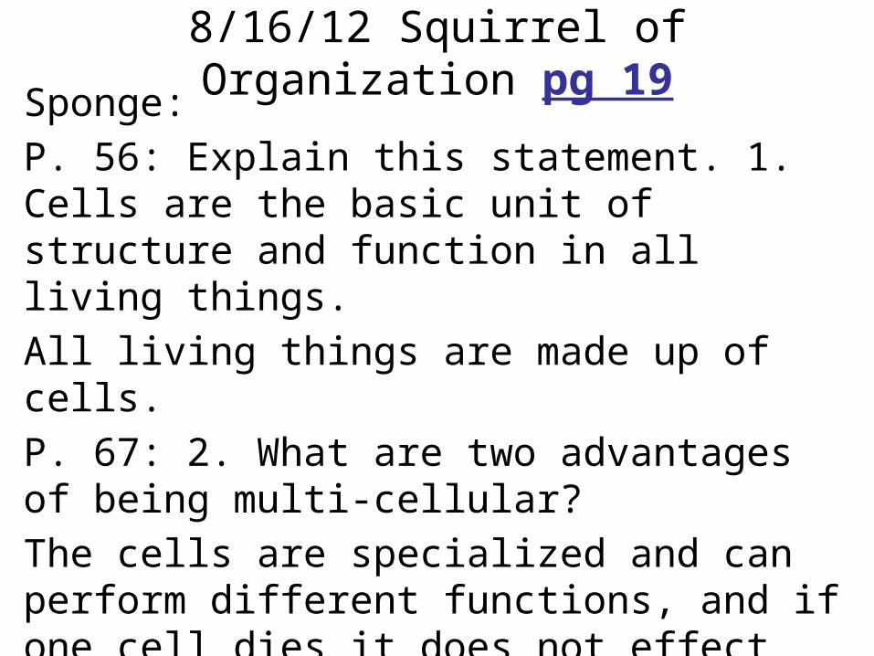
Sponge: P. 56: Explain this statement. 1. Cells are the basic unit of structure and function in all living things.All living things are made up of cells.P. 67: 2. What are two advantages of being multi-cellular? The cells are specialized and can perform different functions, and if one cell dies it does not effect the organism.
8/16/12 Squirrel of Organization pg 19
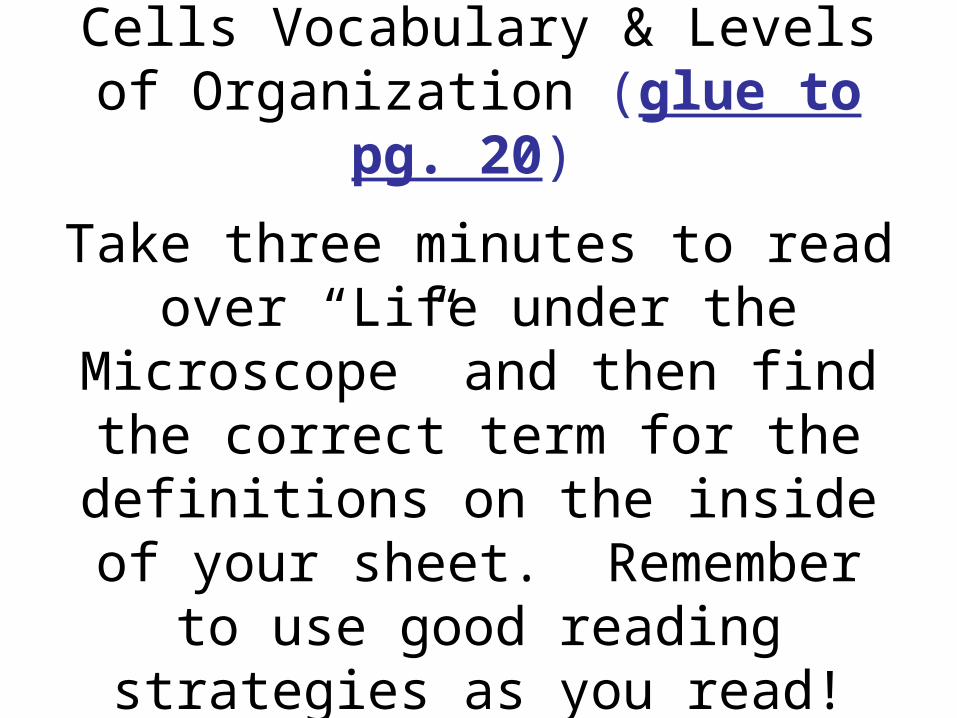
Cells Vocabulary & Levels of Organization (glue to pg. 20)
Take three minutes to read over “Life under the Microscope” and then find the correct term for the definitions on the inside of your sheet. Remember to use good reading strategies as you read!
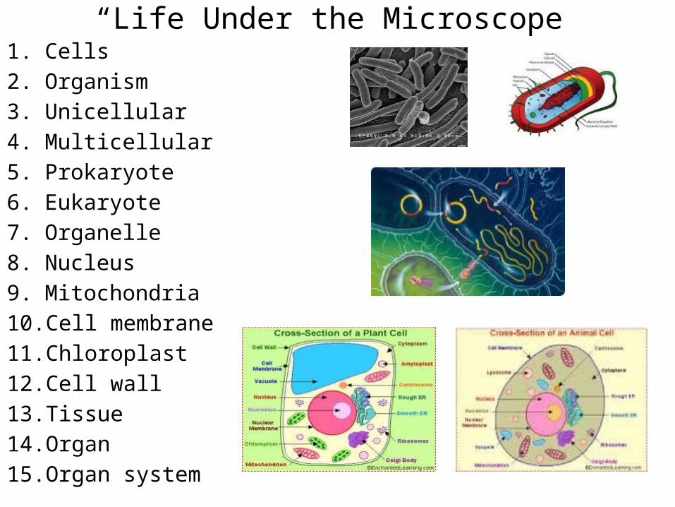
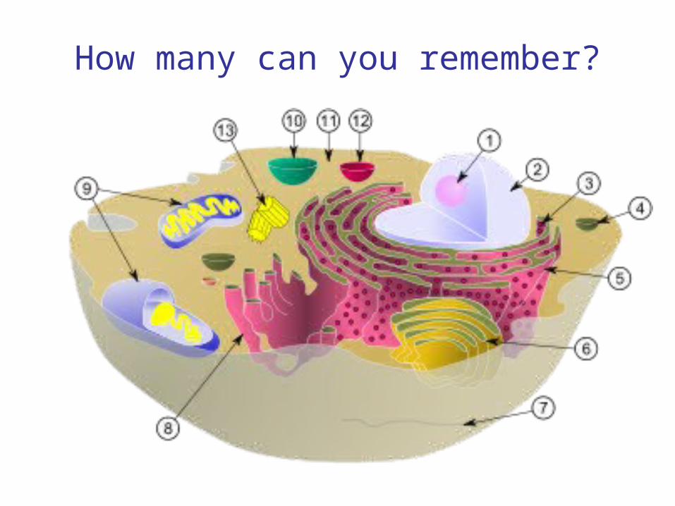
“Life Under the Microscope”1. Cells2. Organism3. Unicellular4. Multicellular5. Prokaryote6. Eukaryote7. Organelle8. Nucleus9. Mitochondria10. Cell membrane11. Chloroplast12. Cell wall13. Tissue14. Organ15. Organ system
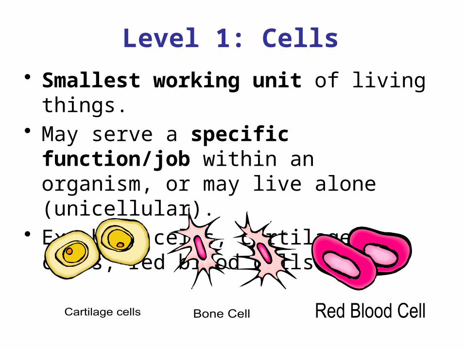
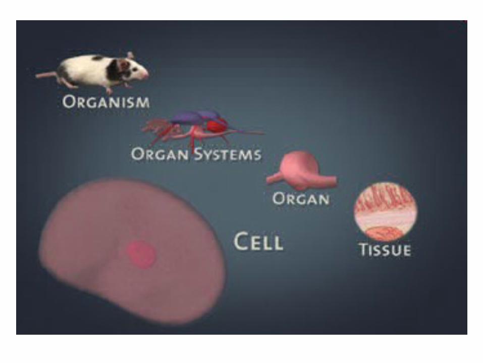
Level 1: Cells• Smallest working unit of living things.• May serve a specific function/job within
an organism, or may live alone (unicellular).
• Ex: bone cells, cartilage cells, red blood cells.
Level 2: Tissues(No, not THAT kind of tissue!)
• Made up of cells that are similar in structure/function that work together to
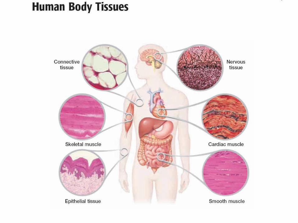
perform a specific function or job.• Ex: Humans have FOUR basic tissues:
connective (fat, cartilage, bone, blood); epithelial (skin), nervous and muscular
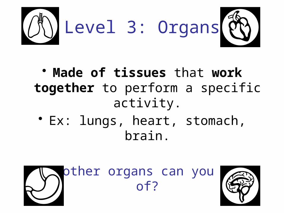
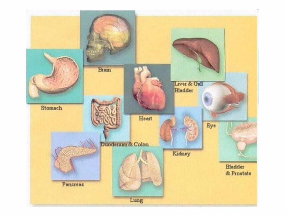
Level 3: Organs
• Made of tissues that work together to perform a specific activity.
• Ex: lungs, heart, stomach, brain.
What other organs can you think of?
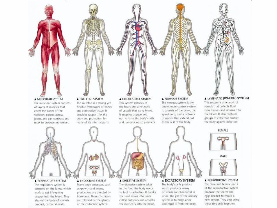
Level 4: Organ Systems
Groups of one or more organs working together to perform
specific functions for the organism.
Our human body has 11 organ systems. Can you name them?
Level 5: Organism
• Entire living things that carry out all basic life functions.
Meaning… they are made of cells, share similar chemicals, can take in and use
energy, grow and develop, reproduce, and sense and respond to changes in their
surroundings. They’re ALIVE!
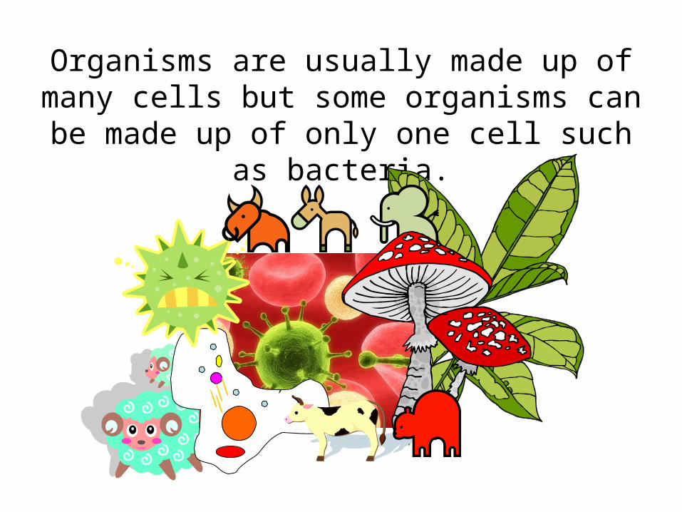
Organisms are usually made up of many cells but some organisms can be made up of only
one cell such as bacteria.
“Squirrel of Organization” In each circle, draw a colorful
representative picture to match that specific level. For
example, in the circle labeled “cell”, draw a picture of a
specific type of cell you might find in a squirrel.
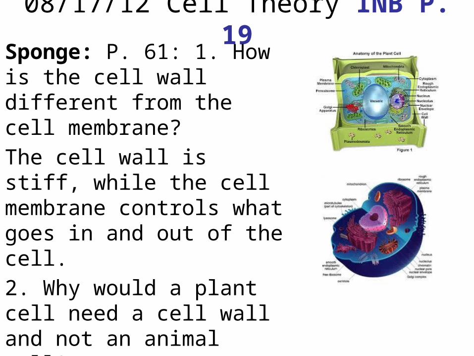
08/17/12 Cell Theory INB P. 19Sponge: P. 61: 1. How is the cell wall different from the cell membrane?The cell wall is stiff, while the cell membrane controls what goes in and out of the cell.2. Why would a plant cell need a cell wall and not an animal cell?Plants can’t move so the cell wall protects and supports the cell.
“Cell Theory” Review Q’s1. WAFLS, water, air, food, living space, and shelter2. Solid-rock, liquid-water, gas-oxygen3. 2 or more cells combined together, ex. Water4. Solid-bone, liquid-blood, gas-oxygen5. OGRRs, organized structure, growth &
development, reproduction, and response to surroundings
6. Ask a question7. Form a hypothesis8. Using your 5 senses9. Maintain balance of a stable internal environment10.shelter
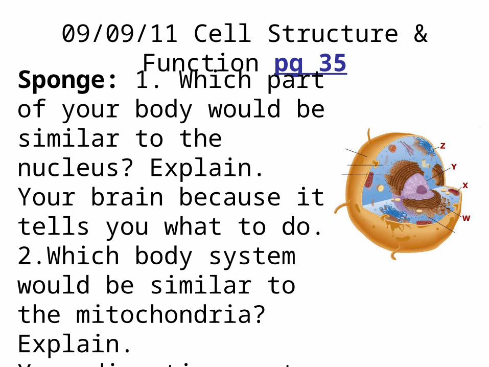
09/09/11 Cell Structure & Function pg 35Sponge: 1. Which part of your body would be similar to the nucleus? Explain.Your brain because it tells you what to do.2.Which body system would be similar to the mitochondria? Explain.Your digestive system because it turns food into energy.
Cell Organelle Graphic Organizer glue to pg 20
The cell theory tells us that…
1. All living things are made up of cells2. Cells are the smallest working units of all
living things3. All cells come from pre-existing cells
through cell division
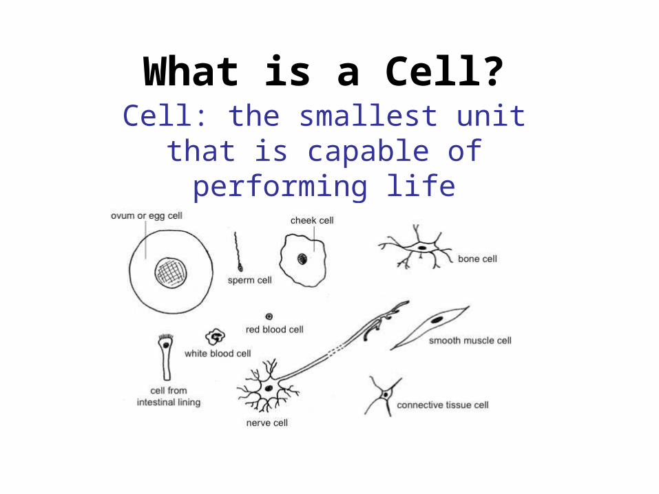
What is a Cell?Cell: the smallest unit that is
capable of performing life functions.
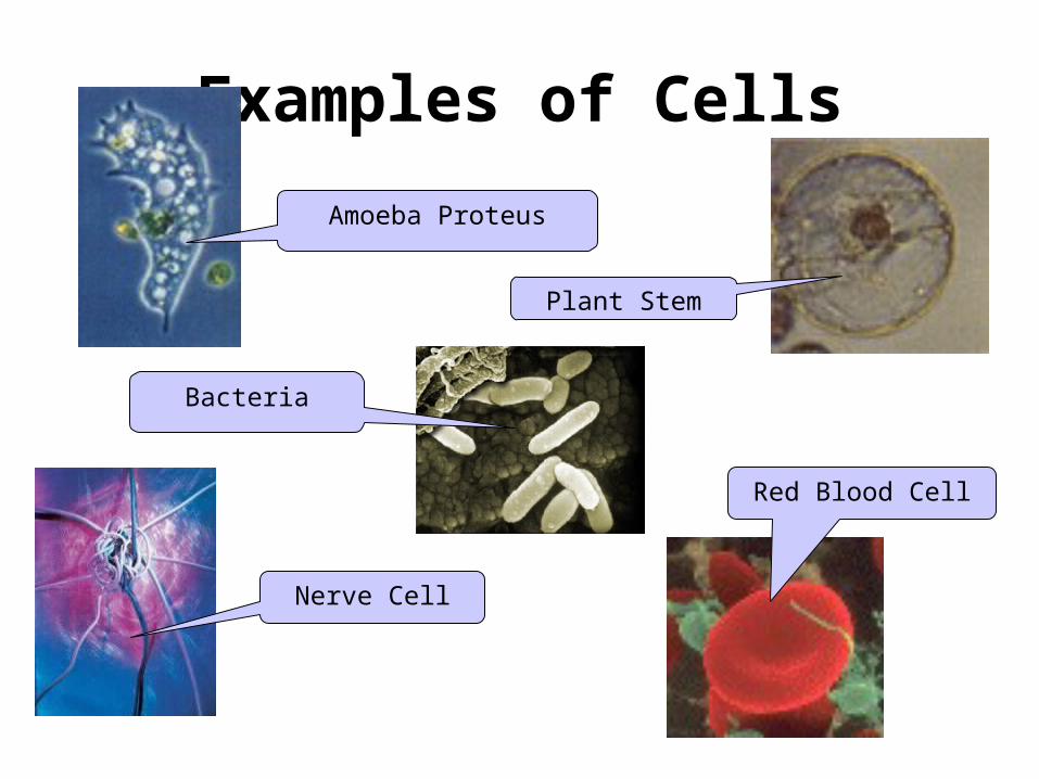
Examples of CellsAmoeba Proteus
Plant Stem
Red Blood Cell
Nerve Cell
Bacteria
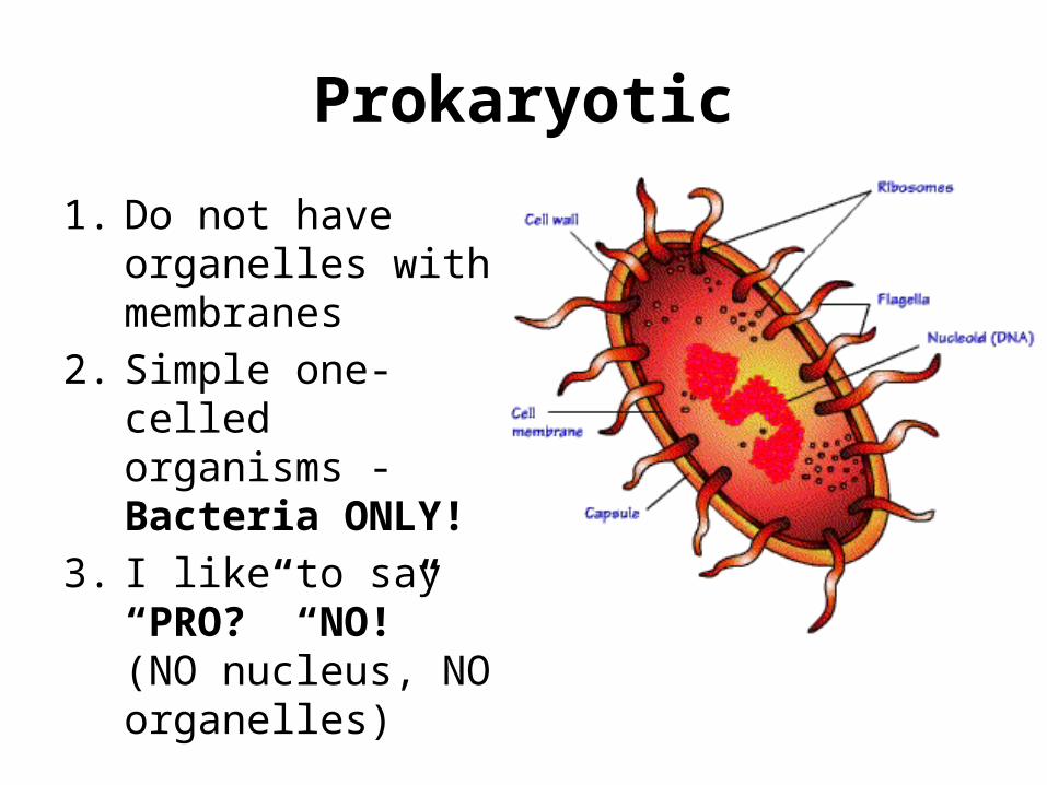
Prokaryotic1. Do not have
organelles with membranes
2. Simple one-celled organisms - Bacteria ONLY!
3. I like to say “PRO?” “NO!” (NO nucleus, NO organelles)
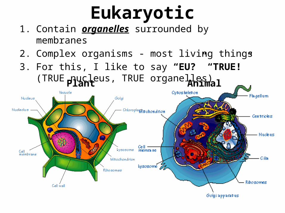
Eukaryotic1. Contain organelles surrounded by membranes2. Complex organisms - most living things3. For this, I like to say “EU?” “TRUE!” (TRUE nucleus,
TRUE organelles)
Plant Animal
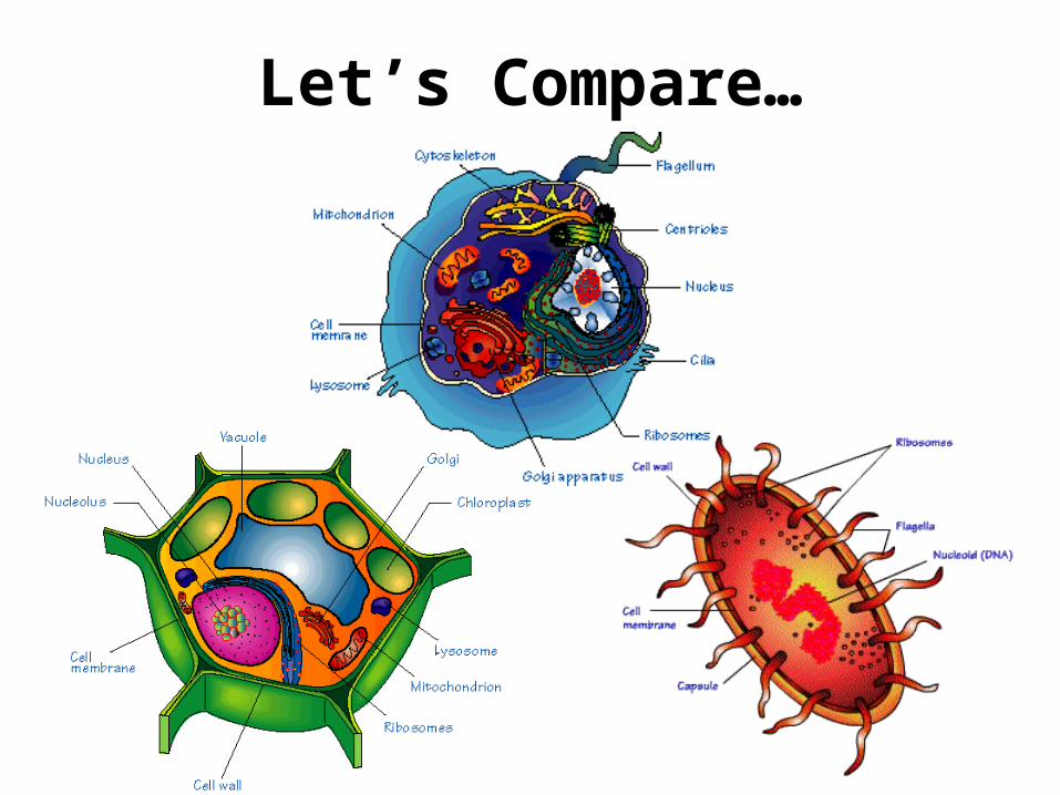
Let’s Compare…
Make your own analogy!
For each organelle on the following slides, think of a picture analogy that will help you
to remember the organelle and what it does! You will draw this in the last column
of your organizer.
The first one is already done for you!
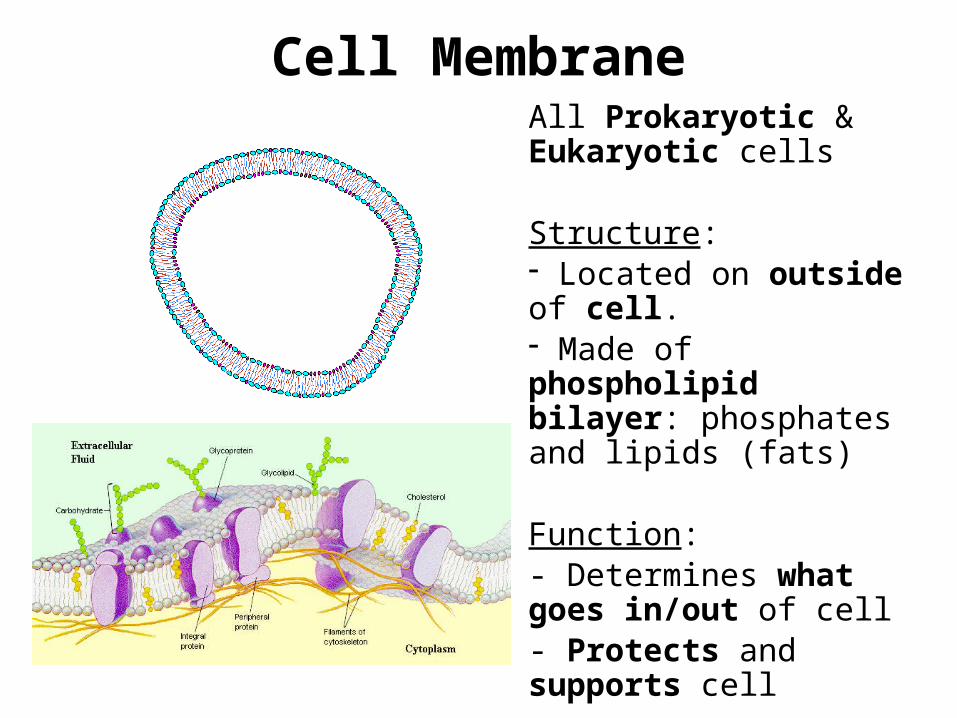
Cell MembraneAll Prokaryotic & Eukaryotic cells
Structure: - Located on outside of cell. - Made of phospholipid bilayer: phosphates and lipids (fats)
Function: - Determines what goes in/out of cell- Protects and supports cell
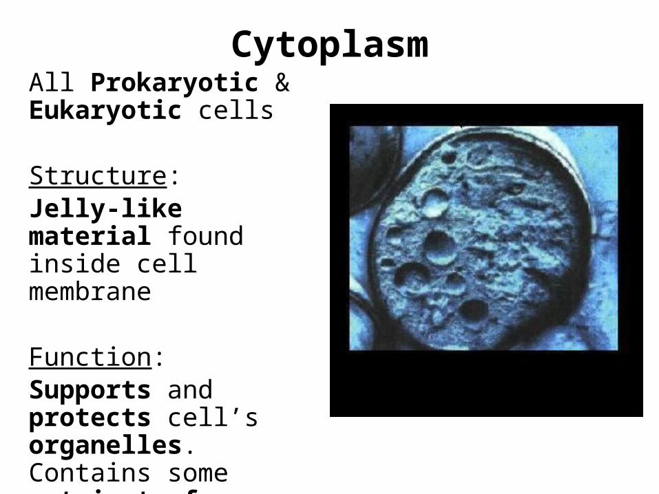
CytoplasmAll Prokaryotic & Eukaryotic cells
Structure: Jelly-like material found inside cell membrane
Function: Supports and protects cell’s organelles. Contains some nutrients for cell.
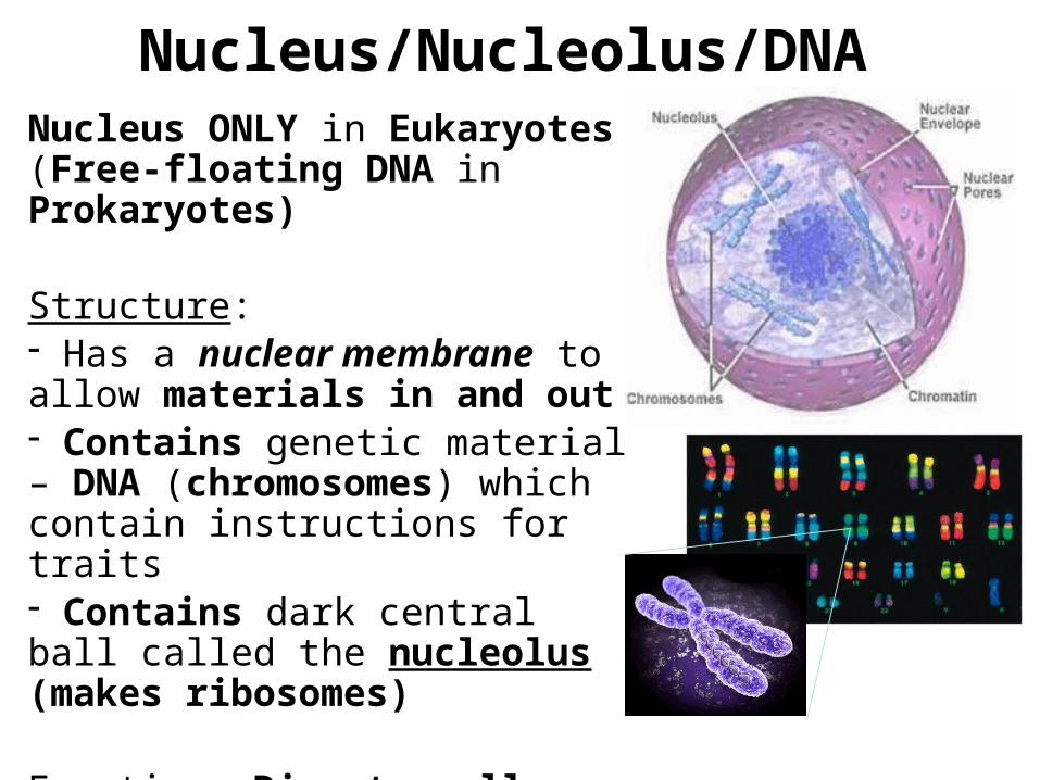
Nucleus/Nucleolus/DNA Nucleus ONLY in Eukaryotes (Free-floating DNA in Prokaryotes)
Structure: - Has a nuclear membrane to allow materials in and out- Contains genetic material – DNA (chromosomes) which contain instructions for traits- Contains dark central ball called the nucleolus (makes ribosomes)
Function: Directs cell activities.
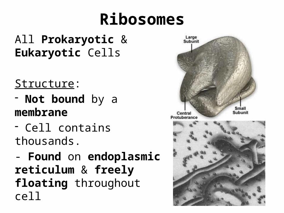
RibosomesAll Prokaryotic & Eukaryotic Cells
Structure: - Not bound by a membrane- Cell contains thousands.- Found on endoplasmic reticulum & freely floating throughout cell
Function: Make protein
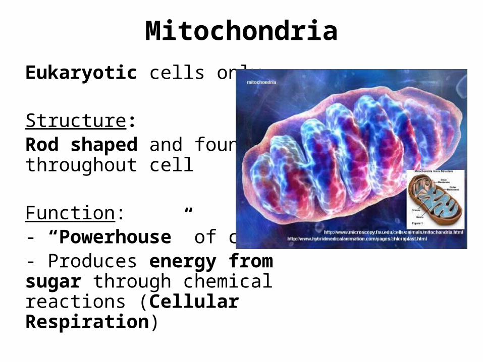
MitochondriaEukaryotic cells only
Structure: Rod shaped and found throughout cell
Function:- “Powerhouse” of cell- Produces energy from sugar through chemical reactions (Cellular Respiration)
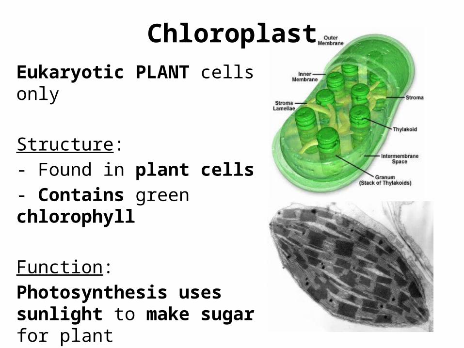
ChloroplastEukaryotic PLANT cells only
Structure: - Found in plant cells- Contains green chlorophyll
Function:Photosynthesis uses sunlight to make sugar for plant
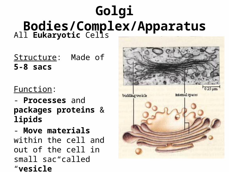
Golgi Bodies/Complex/ApparatusAll Eukaryotic Cells
Structure: Made of 5-8 sacs
Function:- Processes and packages proteins & lipids- Move materials within the cell and out of the cell in small sac called “vesicle”
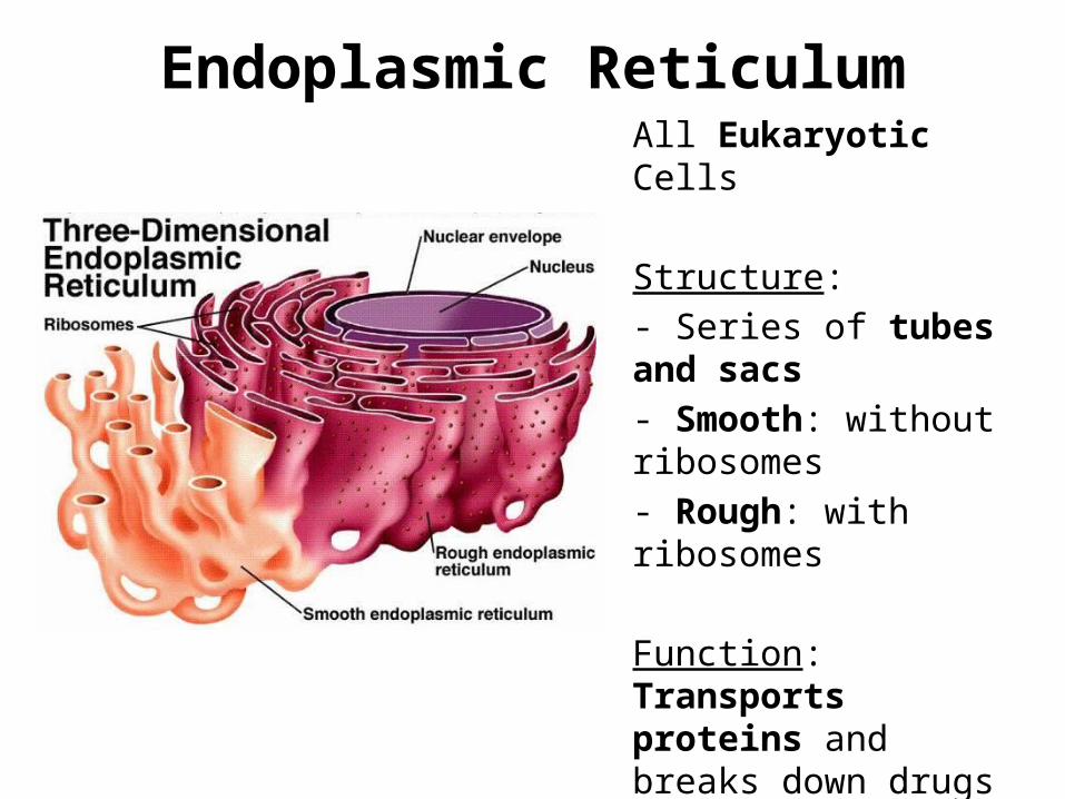
Endoplasmic ReticulumAll Eukaryotic Cells
Structure: - Series of tubes and sacs- Smooth: without ribosomes- Rough: with ribosomes
Function: Transports proteins and breaks down drugs in the cell
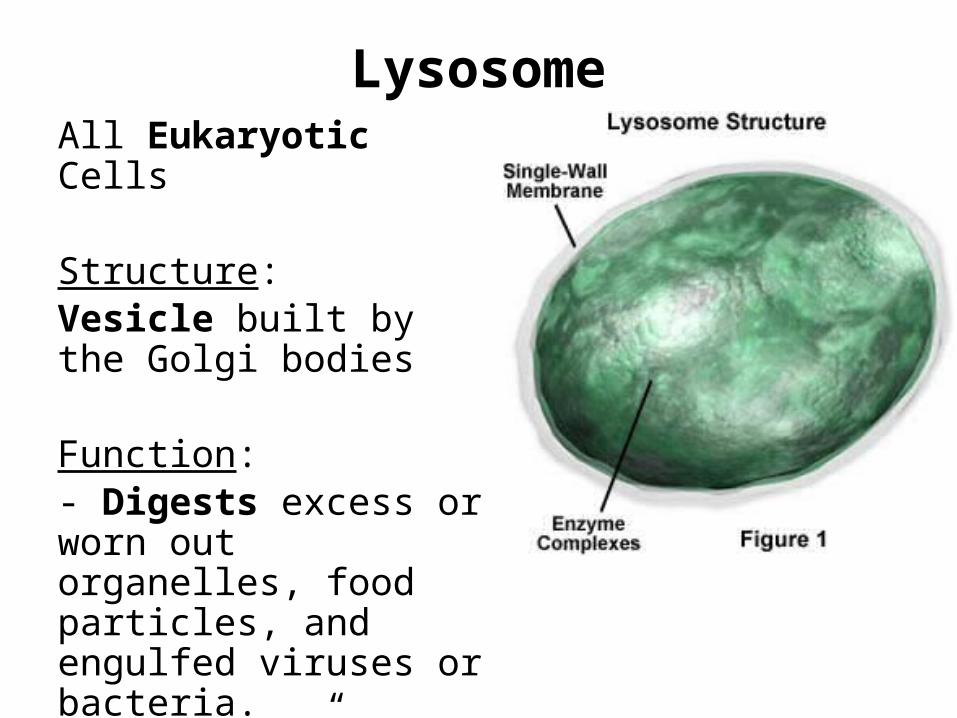
LysosomeAll Eukaryotic Cells
Structure: Vesicle built by the Golgi bodies
Function:- Digests excess or worn out organelles, food particles, and engulfed viruses or bacteria. - “Disposal” of the cell
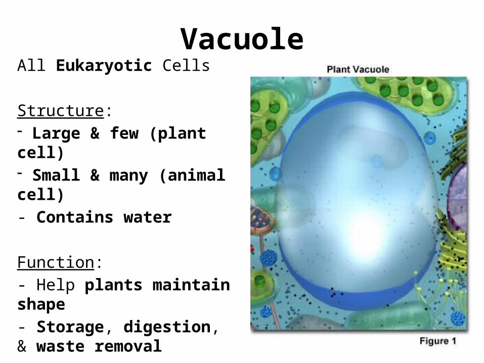
VacuoleAll Eukaryotic Cells
Structure:- Large & few (plant cell)- Small & many (animal cell)- Contains water
Function:- Help plants maintain shape- Storage, digestion, & waste removal
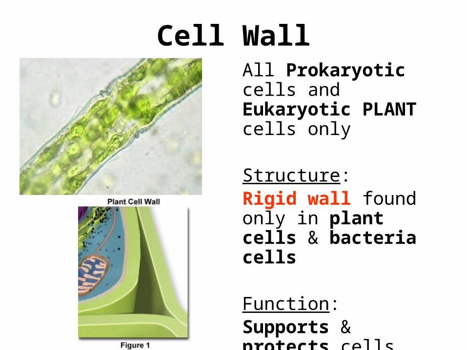
Cell WallAll Prokaryotic cells and Eukaryotic PLANT cells only
Structure:Rigid wall found only in plant cells & bacteria cells
Function:Supports & protects cells
Left Side HomeworkWrite the following directions in your own
words:
Pick your favorite organelle from our lesson today, and draw that organelle as a
superhero comic book character performing it’s job.
Creativity, coloring, and the job it performs will all be factors in your grade!
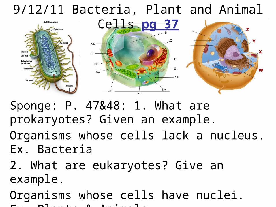
Sponge: P. 47&48: 1. What are prokaryotes? Given an example. Organisms whose cells lack a nucleus. Ex. Bacteria2. What are eukaryotes? Give an example.Organisms whose cells have nuclei. Ex. Plants & Animals
9/12/11 Bacteria, Plant and Animal Cells pg 37
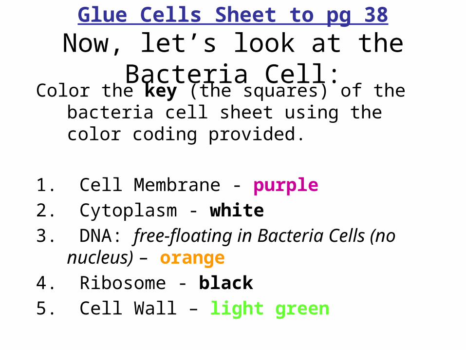
Glue Cells Sheet to pg 38Now, let’s look at the Bacteria Cell:Color the key (the squares) of the bacteria cell
sheet using the color coding provided.
1. Cell Membrane - purple2. Cytoplasm - white3. DNA: free-floating in Bacteria Cells (no
nucleus) – orange4. Ribosome - black5. Cell Wall – light green
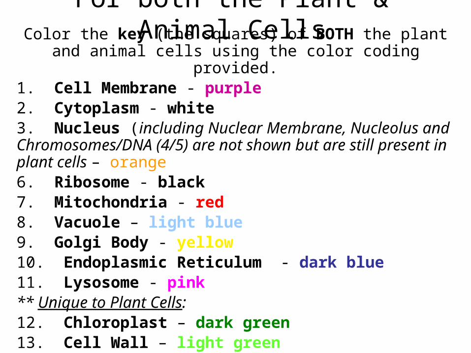
For both the Plant & Animal CellsColor the key (the squares) of BOTH the plant and animal
cells using the color coding provided.1. Cell Membrane - purple2. Cytoplasm - white3. Nucleus (including Nuclear Membrane, Nucleolus and Chromosomes/DNA (4/5) are not shown but are still present in plant cells – orange6. Ribosome - black7. Mitochondria - red 8. Vacuole – light blue 9. Golgi Body - yellow10. Endoplasmic Reticulum - dark blue11. Lysosome - pink** Unique to Plant Cells: 12. Chloroplast – dark green13. Cell Wall – light green
Homework for TonightColor the cells according to the
keys provided. You MAY NOT use colors other than those you were given today. Make sure to review
your organelles.
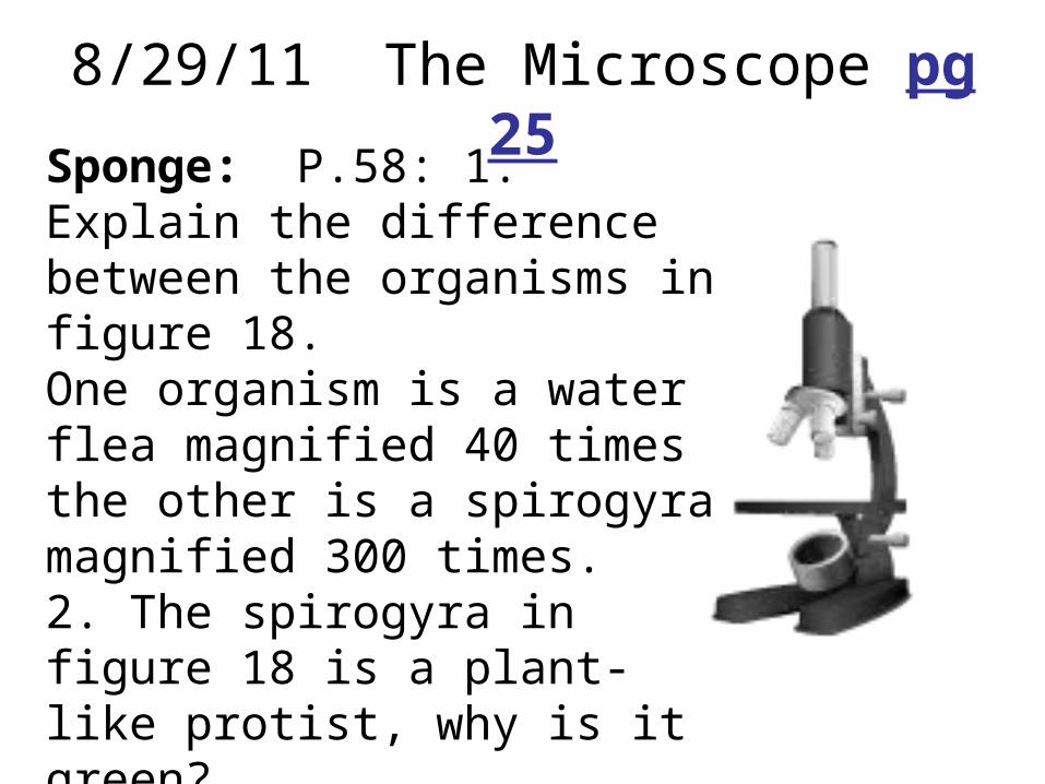
8/29/11 The Microscope pg 25Sponge: P.58: 1. Explain the difference between the organisms in figure 18.One organism is a water flea magnified 40 times the other is a spirogyra magnified 300 times.2. The spirogyra in figure 18 is a plant-like protist, why is it green?It is green because it has chlorophyll in chloroplast to capture sunlight to make food.
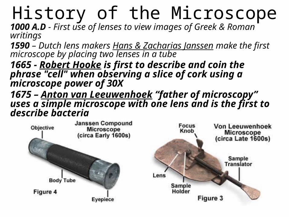
History of the Microscope1000 A.D - First use of lenses to view images of Greek & Roman writings1590 – Dutch lens makers Hans & Zacharias Janssen make the first microscope by placing two lenses in a tube1665 - Robert Hooke is first to describe and coin the phrase "cell" when observing a slice of cork using a microscope power of 30X 1675 – Anton van Leeuwenhoek “father of microscopy” uses a simple microscope with one lens and is the first to describe bacteria
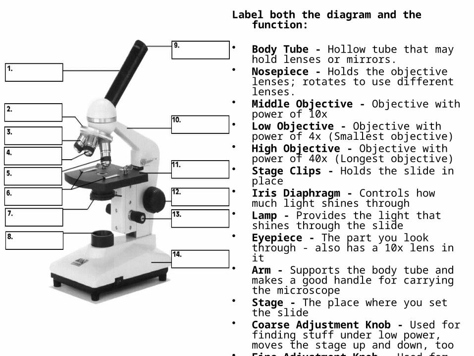
Label both the diagram and the function:
• Body Tube - Hollow tube that may hold lenses or mirrors.
• Nosepiece - Holds the objective lenses; rotates to use different lenses.
• Middle Objective - Objective with power of 10x
• Low Objective - Objective with power of 4x (Smallest objective)
• High Objective - Objective with power of 40x (Longest objective)
• Stage Clips - Holds the slide in place • Iris Diaphragm - Controls how much light
shines through• Lamp - Provides the light that shines through
the slide• Eyepiece - The part you look through - also
has a 10x lens in it• Arm - Supports the body tube and makes a
good handle for carrying the microscope• Stage - The place where you set the slide• Coarse Adjustment Knob - Used for finding
stuff under low power, moves the stage up and down, too
• Fine Adjustment Knob - Used for high-power focusing
• Base - Supports the weight of the microscope
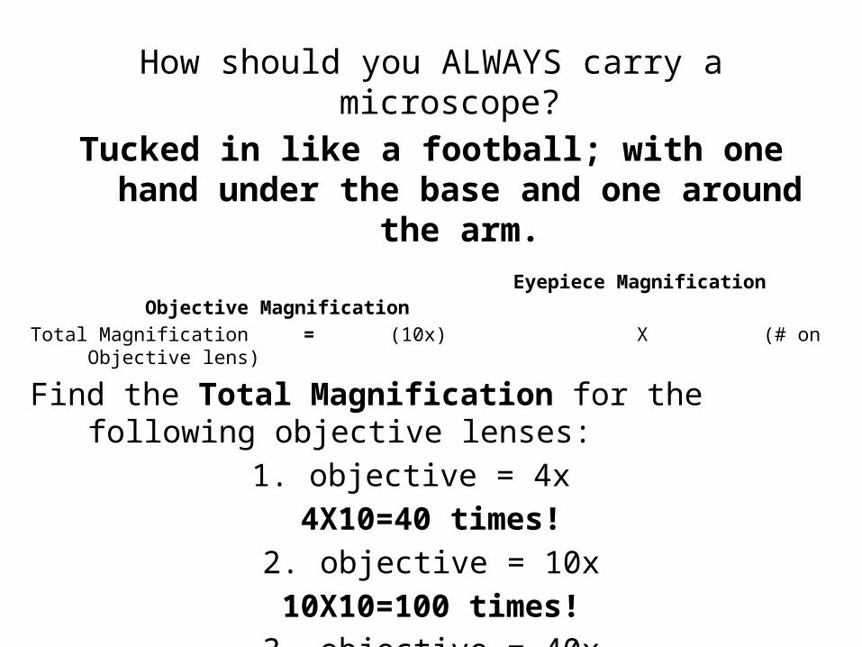
How should you ALWAYS carry a microscope? Tucked in like a football; with one hand under the
base and one around the arm. Eyepiece Magnification Objective MagnificationTotal Magnification = (10x) X (# on Objective lens)
Find the Total Magnification for the following objective lenses:
1. objective = 4x4X10=40 times!
2. objective = 10x10X10=100 times!3. objective = 40x40X10=400 times!
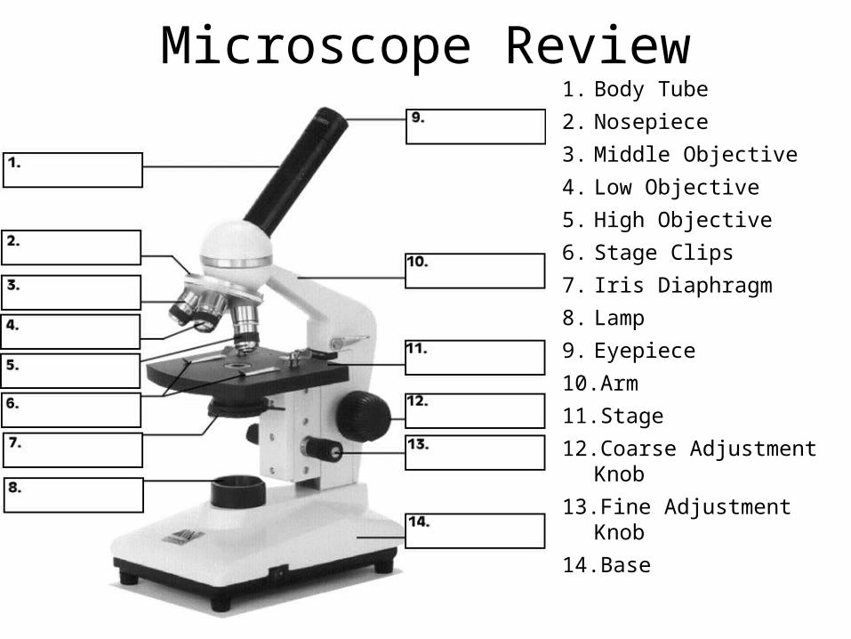
Microscope Review1. Body Tube2. Nosepiece3. Middle Objective4. Low Objective5. High Objective6. Stage Clips7. Iris Diaphragm8. Lamp9. Eyepiece10.Arm11. Stage12.Coarse Adjustment Knob13.Fine Adjustment Knob14.Base
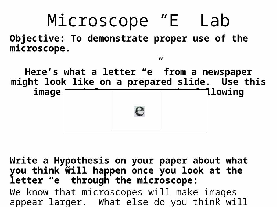
Objective: To demonstrate proper use of the microscope.
Here’s what a letter “e” from a newspaper might look like on a prepared slide. Use this image to help you answer
the following questions.
Write a Hypothesis on your paper about what you think will happen once you look at the letter “e” through the microscope: We know that microscopes will make images appear larger. What else do you think will happen to the image of the letter “e” when looked at through the microscope?
Microscope “E” Lab
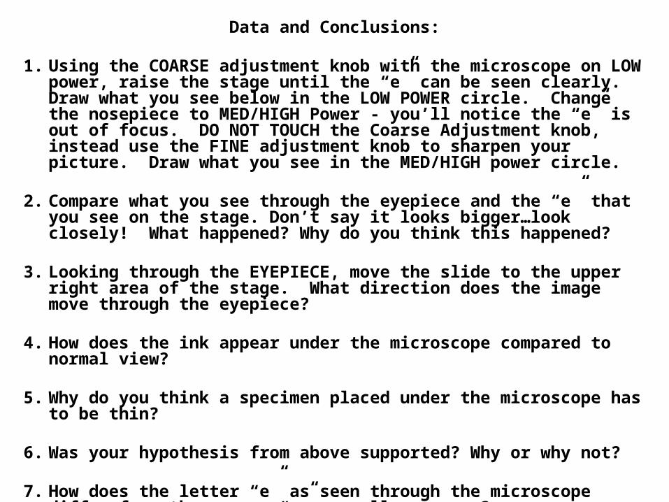
Data and Conclusions:
1. Using the COARSE adjustment knob with the microscope on LOW power, raise the stage until the “e” can be seen clearly. Draw what you see below in the LOW POWER circle. Change the nosepiece to MED/HIGH Power - you’ll notice the “e” is out of focus. DO NOT TOUCH the Coarse Adjustment knob, instead use the FINE adjustment knob to sharpen your picture. Draw what you see in the MED/HIGH power circle.
2. Compare what you see through the eyepiece and the “e” that you see on the stage. Don’t say it looks bigger…look closely! What happened? Why do you think this happened?
3. Looking through the EYEPIECE, move the slide to the upper right area of the stage. What direction does the image move through the eyepiece?
4. How does the ink appear under the microscope compared to normal view?
5. Why do you think a specimen placed under the microscope has to be thin?
6. Was your hypothesis from above supported? Why or why not?
7. How does the letter “e” as seen through the microscope differ from the way an “e” normally appears?
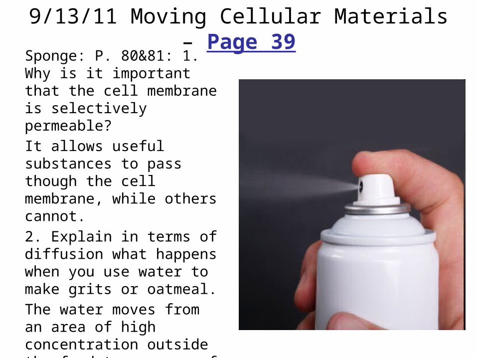
9/13/11 Moving Cellular Materials – Page 39Sponge: P. 80&81: 1. Why is it important that the cell membrane is selectively permeable?It allows useful substances to pass though the cell membrane, while others cannot.2. Explain in terms of diffusion what happens when you use water to make grits or oatmeal.The water moves from an area of high concentration outside the food to an area of low concentration inside the food.
Watch Tim and Moby from BrainPop talk about Moving Cellular Materials
Now answer the following questions on the left side:
1. What part of the cell helps the cell maintain homeostasis?
2. Can you apply your knowledge of the cell membrane to how a cell interacts with its environment?
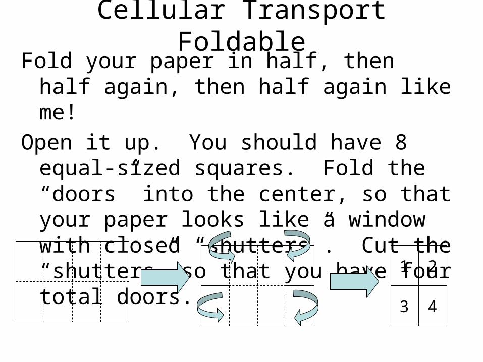
Cellular Transport FoldableFold your paper in half, then half again, then
half again like me!Open it up. You should have 8 equal-sized
squares. Fold the “doors” into the center, so that your paper looks like a window with closed “shutters”. Cut the “shutters” so that you have four total doors.
1 2
3 4
On the front of each door, explain the following processes:DOOR 1: OsmosisDOOR 2: DiffusionDOOR 3: Transport ProteinDOOR 4: Transport by EngulfingUnderneath each door, correctly illustrate the process. Use the textbook pages 80-85 to help you. Worth 100 points and to be TURNED IN tomorrow! After I grade them you will glue them to page 26.
Cellular Transport Foldable
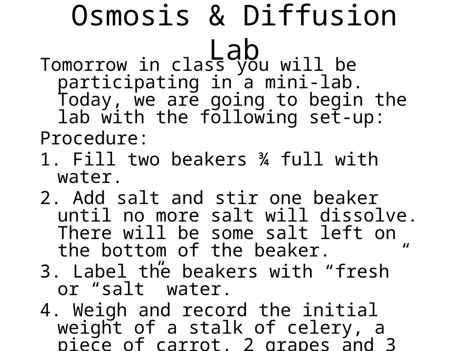
Osmosis & Diffusion LabTomorrow in class you will be participating in a
mini-lab. Today, we are going to begin the lab with the following set-up:
Procedure:1. Fill two beakers ¾ full with water.2. Add salt and stir one beaker until no more
salt will dissolve. There will be some salt left on the bottom of the beaker.
3. Label the beakers with “fresh” or “salt” water.4. Weigh and record the initial weight of a stalk
of celery, a piece of carrot, 2 grapes and 3 raisins and place in each beaker.
5. They will soak in the solutions overnight and we’ll measure them again tomorrow.
Exit Ticket: Lab – page 25What will happen to the carrots, celery, grapes & raisins in fresh water? What about in the salt
water? Do you expect all of the foods to act the same way?
Explain your answers.
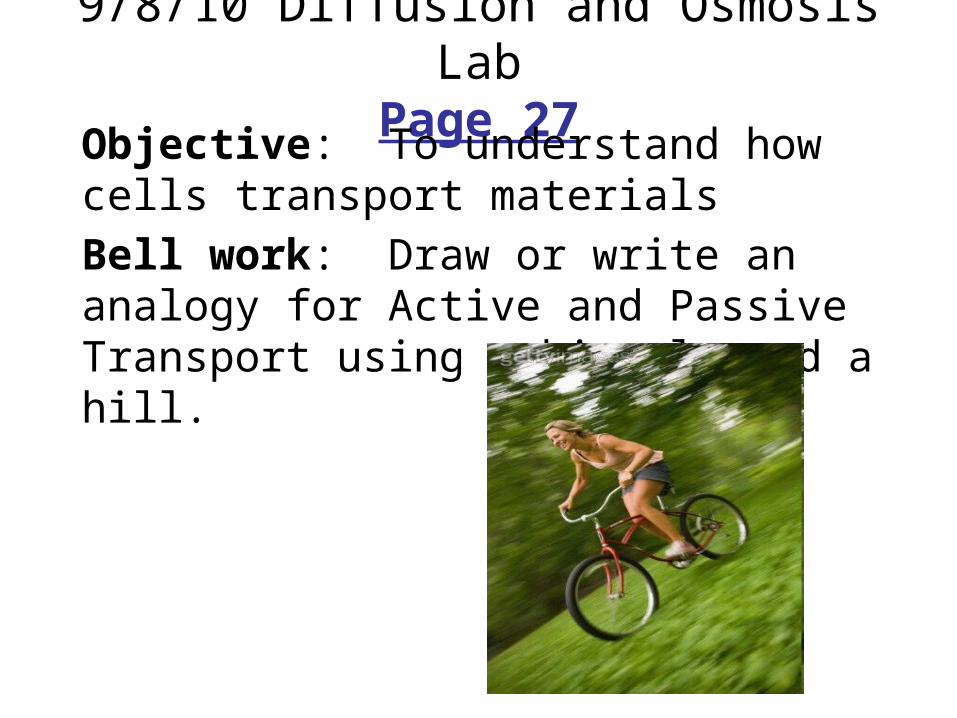
9/8/10 Diffusion and Osmosis LabPage 27
Objective: To understand how cells transport materialsBell work: Draw or write an analogy for Active and Passive Transport using a bicycle and a hill.
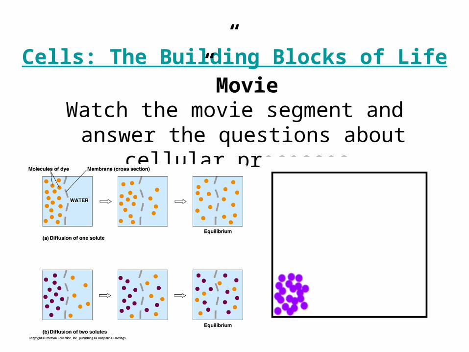
“Cells: The Building Blocks of Life” Movie
Watch the movie segment and answer the questions about cellular processes.
Diffusion and Osmosis LabPage 28
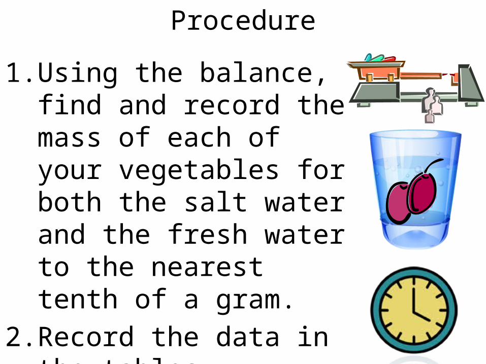
Procedure
1. Using the balance, find and record the mass of each of your vegetables for both the salt water and the fresh water to the nearest tenth of a gram.
2. Record the data in the tables provided.

Exit Ticket – page 27Using the graph paper provided, create two
BAR graphs: one for all salt water produce and one for all fresh water
produce. Each produce item should have two bars comparing initial
and final weight! Please color your bar
graph and also answer any questions on your handout from today’s
lab!
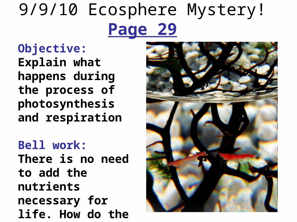
9/9/10 Ecosphere Mystery! Page 29Objective: Explain what happens during the process of photosynthesis and respiration
Bell work: There is no need to add the nutrients necessary for life. How do the shrimp and algae survive?
Graphic Organizer
Use the following words to fill in the organizer:
Glucose(2) carbon dioxide(2)oxygen(2)
Water(2) photosynthesis sugarSunlight mitochondria chloroplastEnergy ATP cellular respiration
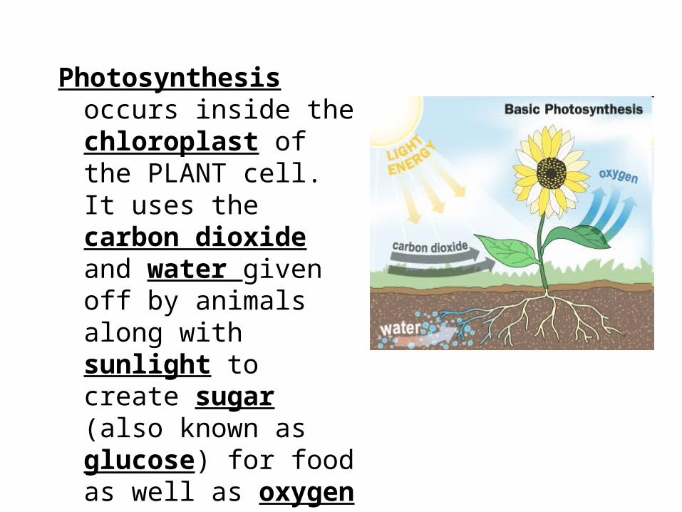
Photosynthesis occurs inside the chloroplast of the PLANT cell. It uses the carbon dioxide and water given off by animals along with sunlight to create sugar (also known as glucose) for food as well as oxygen as waste.
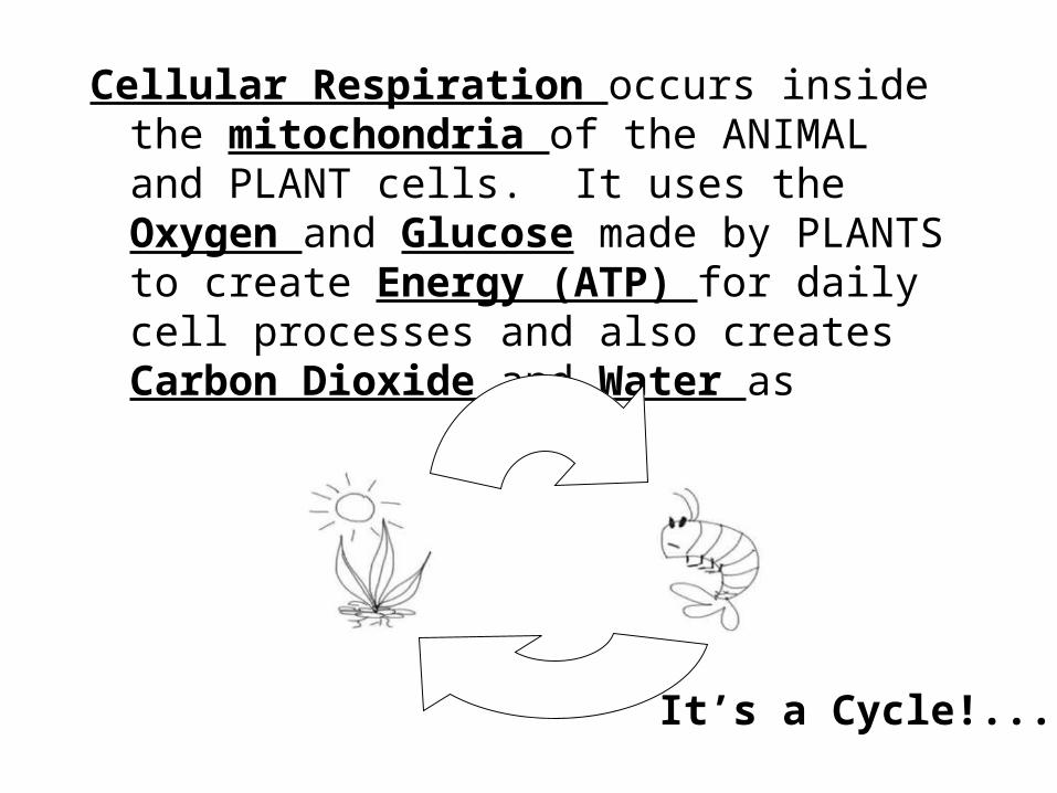
Cellular Respiration occurs inside the mitochondria of the ANIMAL and PLANT cells. It uses the Oxygen and Glucose made by PLANTS to create Energy (ATP) for daily cell processes and also creates Carbon Dioxide and Water as waste.
It’s a Cycle!...
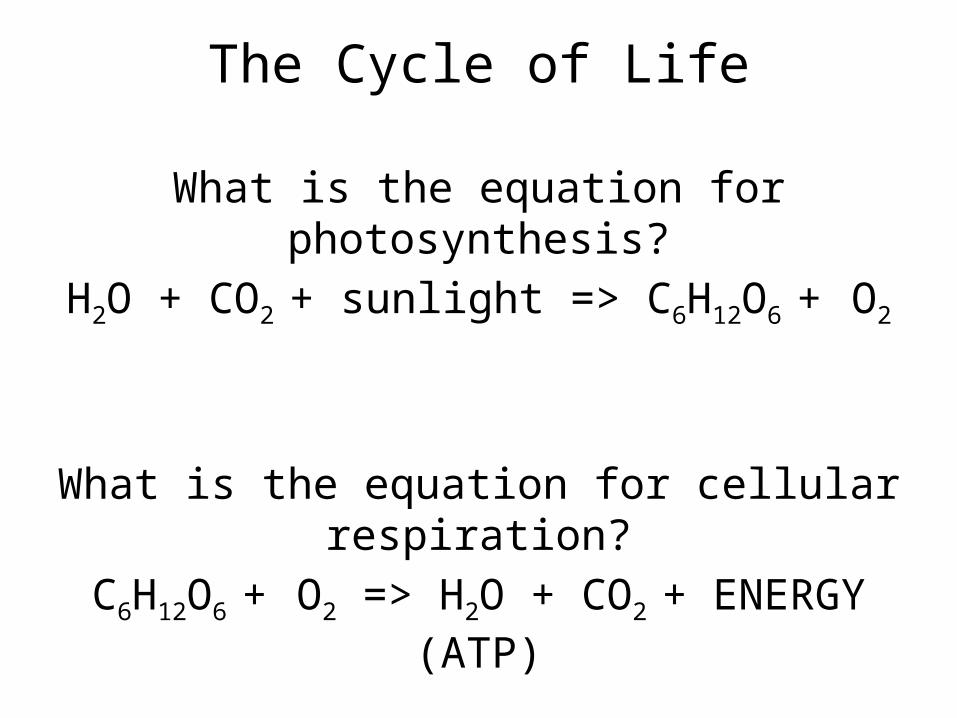
The Cycle of Life
What is the equation for photosynthesis?H2O + CO2 + sunlight => C6H12O6 + O2
What is the equation for cellular respiration?C6H12O6 + O2 => H2O + CO2 + ENERGY (ATP)
They’re backwards of one another!!!
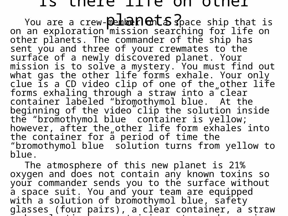
Is there life on other planets?You are a crew-member on a space ship that is on an
exploration mission searching for life on other planets. The commander of the ship has sent you and three of your crewmates to the surface of a newly discovered planet. Your mission is to solve a mystery. You must find out what gas the other life forms exhale. Your only clue is a CD video clip of one of the other life forms exhaling through a straw into a clear container labeled “bromothymol blue.” At the beginning of the video clip the solution inside the “bromothymol blue” container is yellow; however, after the other life form exhales into the container for a period of time the “bromothymol blue” solution turns from yellow to blue.
The atmosphere of this new planet is 21% oxygen and does not contain any known toxins so your commander sends you to the surface without a space suit. You and your team are equipped with a solution of bromothymol blue, safety glasses (four pairs), a clear container, a straw and a plastic bag containing water and a sprig of an aquatic plant. Each member of your team has been assigned one of the following roles: documentation officer, timekeeper, peacekeeper, or materials manager. You have 25 minutes from the time of your arrival on the new planet to solve the mystery and ready yourself to return to your ship.
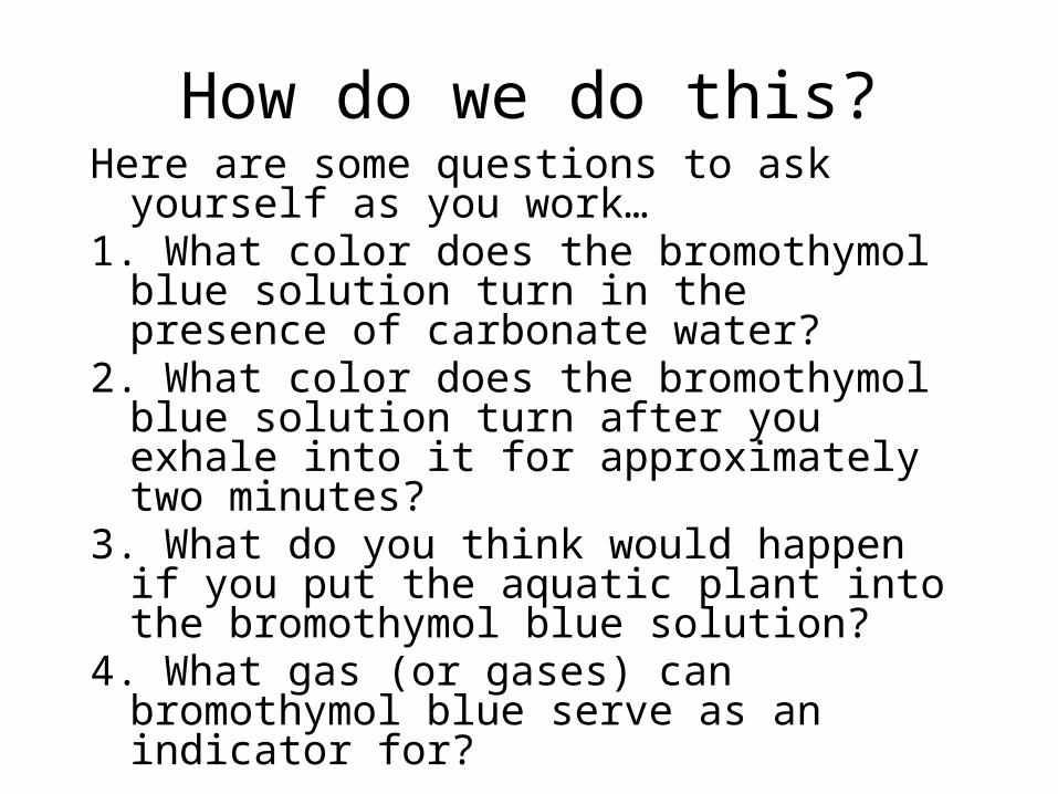
How do we do this?Here are some questions to ask yourself as you
work…1. What color does the bromothymol blue
solution turn in the presence of carbonate water?
2. What color does the bromothymol blue solution turn after you exhale into it for approximately two minutes?
3. What do you think would happen if you put the aquatic plant into the bromothymol blue solution?
4. What gas (or gases) can bromothymol blue serve as an indicator for?
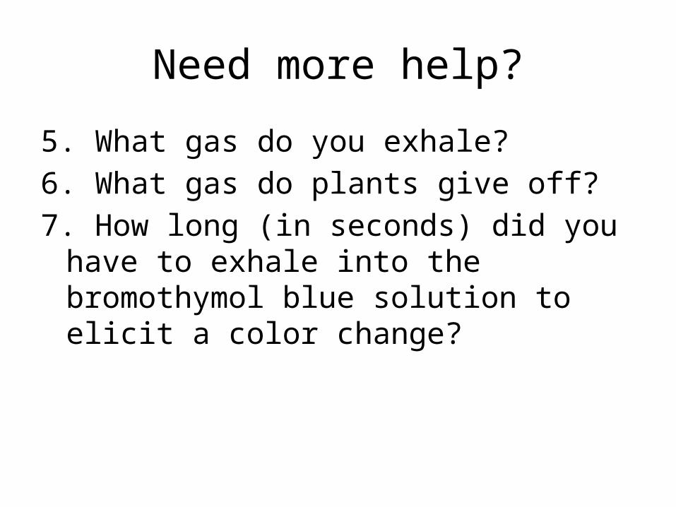
Need more help?
5. What gas do you exhale?6. What gas do plants give off?7. How long (in seconds) did you have to
exhale into the bromothymol blue solution to elicit a color change?
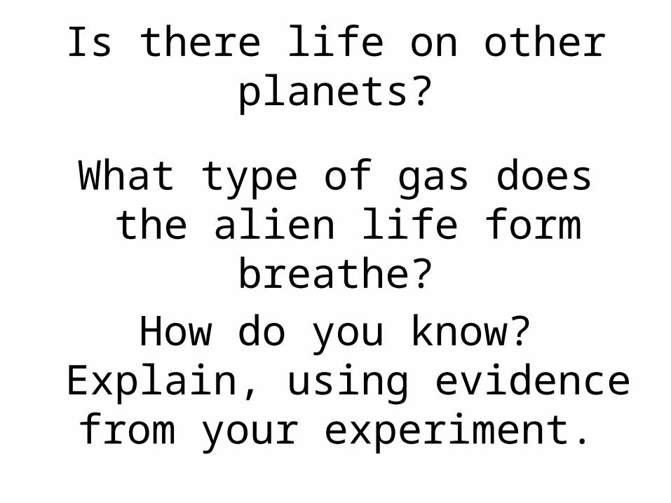
Is there life on other planets?
What type of gas does the alien life form breathe?
How do you know? Explain, using evidence from your
experiment.
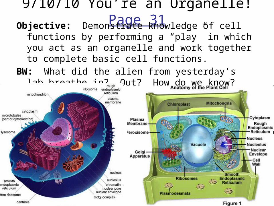
9/10/10 You’re an Organelle! Page 31Objective: Demonstrate knowledge of cell functions by
performing a “play” in which you act as an organelle and work together to complete basic cell functions.
BW: What did the alien from yesterday’s lab breathe in? Out? How do we know?
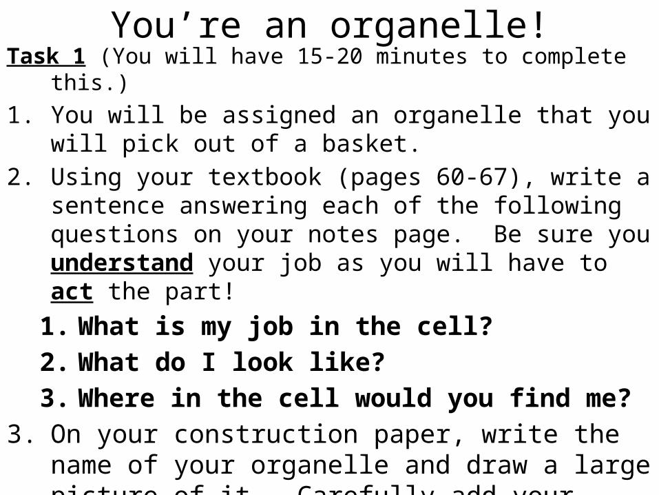
You’re an organelle!Task 1 (You will have 15-20 minutes to complete this.)1. You will be assigned an organelle that you will pick out of
a basket. 2. Using your textbook (pages 60-67), write a sentence
answering each of the following questions on your notes page. Be sure you understand your job as you will have to act the part!
1. What is my job in the cell?2. What do I look like?3. Where in the cell would you find me?
3. On your construction paper, write the name of your organelle and draw a large picture of it. Carefully add your string and wear it!
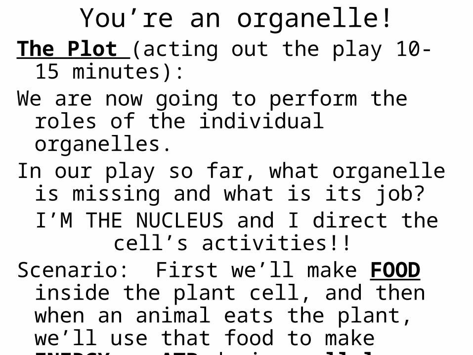
The Plot (acting out the play 10-15 minutes):We are now going to perform the roles of the
individual organelles. In our play so far, what organelle is missing
and what is its job?I’M THE NUCLEUS and I direct the cell’s
activities!! Scenario: First we’ll make FOOD inside the
plant cell, and then when an animal eats the plant, we’ll use that food to make ENERGY or ATP during cellular respiration! Then we’ll use that energy to make protein!
You’re an organelle!
You’re an organelle!
Mini-Lesson:Why is the location of the organelle within the cell
important?
Exit Tickets1) Compare the process of photosynthesis to
the process of baking a cake.
2) What organelle uses the food made during photosynthesis to produce energy?
3) Describe the relationship between the nucleus, endoplasmic reticulum and the ribosomes.

![Agenda - pastelglyph.krpastelglyph.kr/contents/commercial_pr.pdf · agenda 005 about pg overview organization history 016 pg portfolio #1 [핚국관광공사]2017홍보영상 #2 [jayjun]](https://img.pdfslide.us/doc/110x75/5cc8678f88c993a6188da488/agenda-agenda-005-about-pg-overview-organization-history-016-pg-portfolio.jpg)