Embed Size (px)
Citation preview
7thAUG 2015
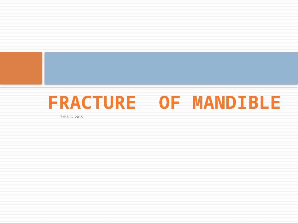
FRACTURE OF MANDIBLE
Importance of Facial injury
Soft tissue & bone- antr protection to cranium
Face appearance- Look Whole antr region with functions of daily
life - Sight ,smell,eating,breathing & Talking
Any significant impairment – Life style & Quality of Life

? Trauma care provider
Etiology Incidence Involv vital structures Life threating conditions
HEALING (TREATMENT)WITHOUT COMPLICATION Restore Pretrauma appearance and functions
Development
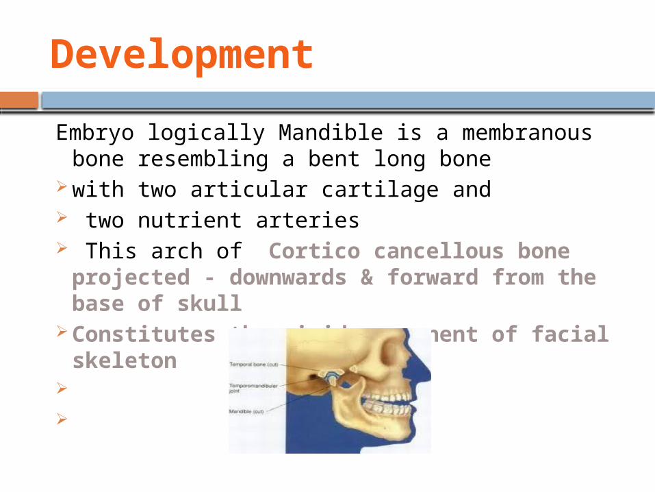
Embryo logically Mandible is a membranous bone resembling a bent long bone
with two articular cartilage and two nutrient arteries This arch of Cortico cancellous bone projected -
downwards & forward from the base of skull Constitutes the rigid component of facial skeleton
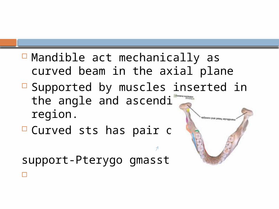
Mandible act mechanically as curved beam in the axial plane
Supported by muscles inserted in the angle and ascending ramus region.
Curved sts has pair of sling support-Pterygo gmasstric sling.
Force to Fracture (Nahum
1975) Curved beam like bone Condylar Neck #- 425 lb frontal impact
Symphysis-800-900 lb Front impact with both condylar #
Mandible more sensitive to lateral impact than frontal
impact( cushioned by opening and retrusion of jaw
Incidence & Etiology
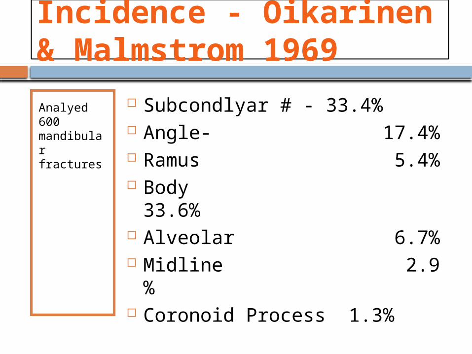
Incidence - Oikarinen & Malmstrom 1969
Analyed 600 mandibular fractures
Subcondlyar # - 33.4% Angle- 17.4% Ramus 5.4% Body 33.6% Alveolar 6.7% Midline 2.9 % Coronoid Process 1.3%
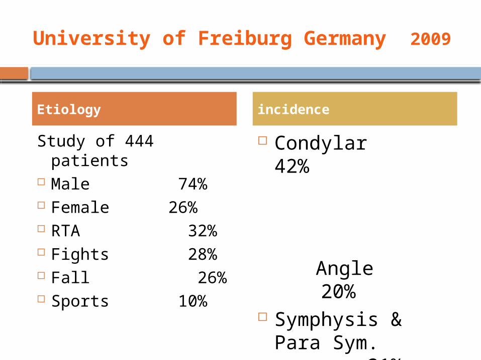
University of Freiburg Germany 2009
Study of 444 patients Male 74% Female 26% RTA 32% Fights 28% Fall 26% Sports 10%
Condylar 42%
Angle
20% Symphysis & Para
Sym. 21%
Etiology incidence
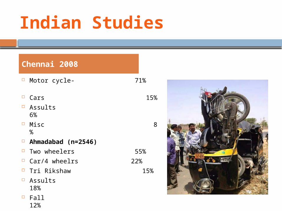
Indian Studies
Motor cycle- 71%
Cars 15% Assults 6% Misc 8 % Ahmadabad (n=2546) Two wheelers 55% Car/4 wheelrs 22% Tri Rikshaw 15% Assults 18% Fall 12%
Chennai 2008
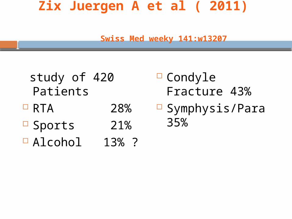
Zix Juergen A et al ( 2011) Swiss Med weeky 141:w13207
study of 420 Patients
RTA 28% Sports 21% Alcohol 13% ?
Condyle Fracture 43%
Symphysis/Para 35%

Level I trauma center AIIMS (2007-2010 ) CTR2014)
542 Patients RTA 54.6% Fall 22.3% Fight 18.5%
Location Body 29.6% Angle 24.6% Ramus 19.5% Dento Alv 14.6% Symphysis 11% Condyle 0.8%
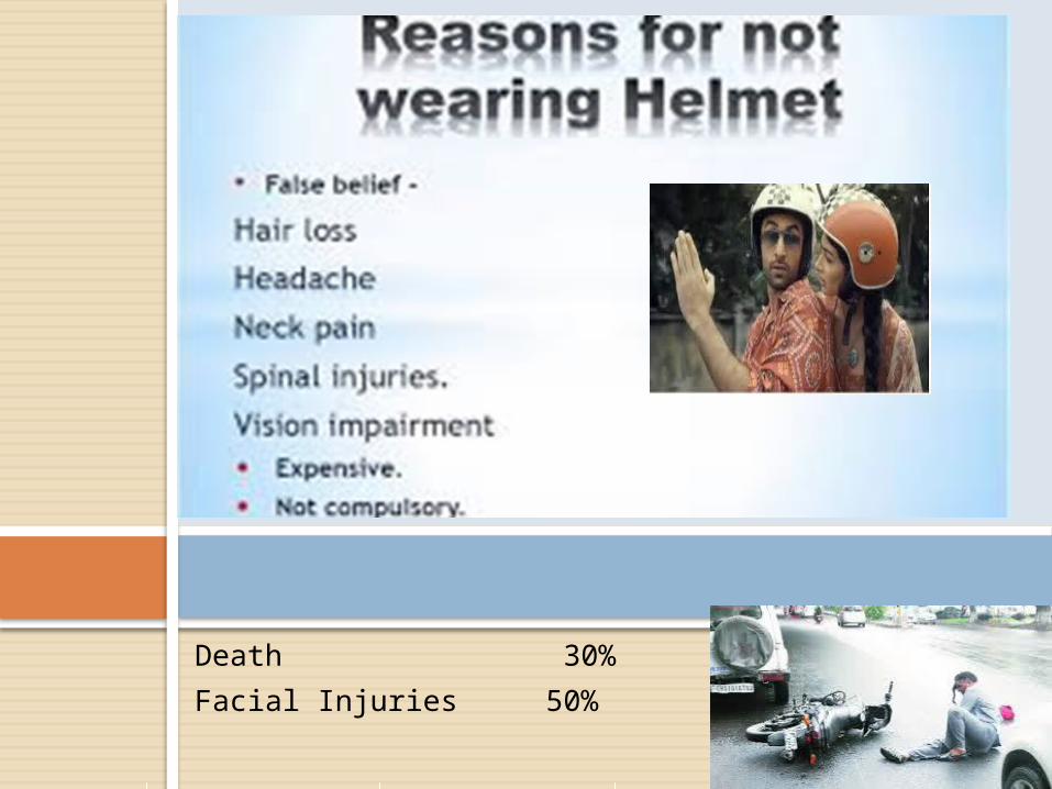
Death 30%
Facial Injuries 50%
Click icon to add picture

Helmet
More than 80 percent of all motorcycle crashes result in injury or death to the motorcyclist.
Per mile driven, a motorcyclist is 16 times more likely to die in a crash than an automobile driver. Wearing a motorcycle helmet reduces that risk by almost one-third (29 percent).
Head injury is a leading cause of death in motor cycle crashes. Riders who don�t wear helmets and who experience a crash are 40 percent more likely to sustain a fatal head injury.
A study of 900 motorcycle crashes (conducted by the University of Southern California) showed that wearing a helmet was the single most critical factor in preventing or reducing head and neck injuries among motorcycle drivers and passengers.
From 1984 through 1995, helmets saved the lives of more than 7,400 motorcyclists. But more than 6,300 additional deaths could have been prevented if all riders had been wearing helmets.
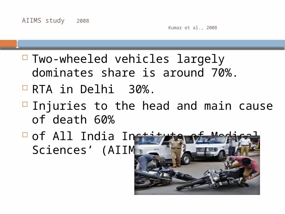
AIIMS study 2008 Kumar et al., 2008
Two-wheeled vehicles largely dominates share is around 70%.
RTA in Delhi 30%. Injuries to the head and main cause of
death 60% of All India Institute of Medical Sciences’
(AIIMS) admissions —
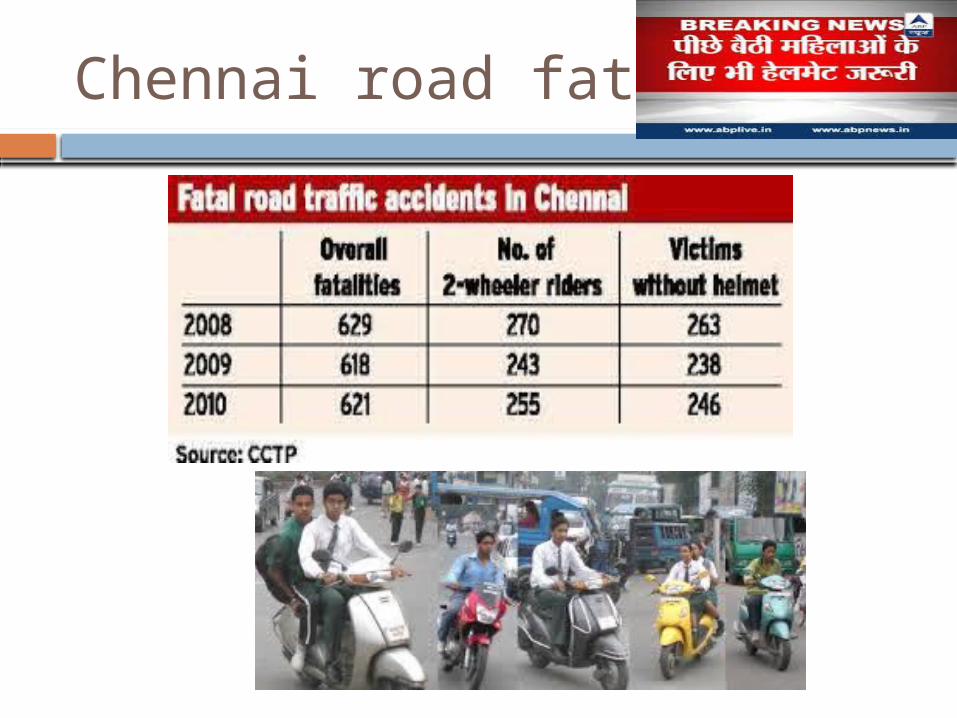
Chennai road fatalities
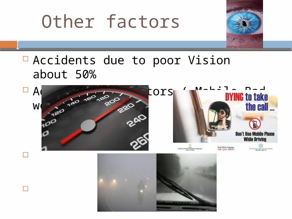
Other factors
Accidents due to poor Vision about 50% Adding other factors ( Mobile,Bad
weather )
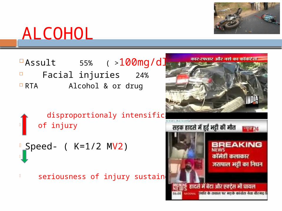
ALCOHOL
Assult 55% ( >100mg/dl Facial injuries 24% RTA Alcohol & or drug
disproportionaly intensification - of injury
- Speed- ( K=1/2 MV2)-
- seriousness of injury sustained
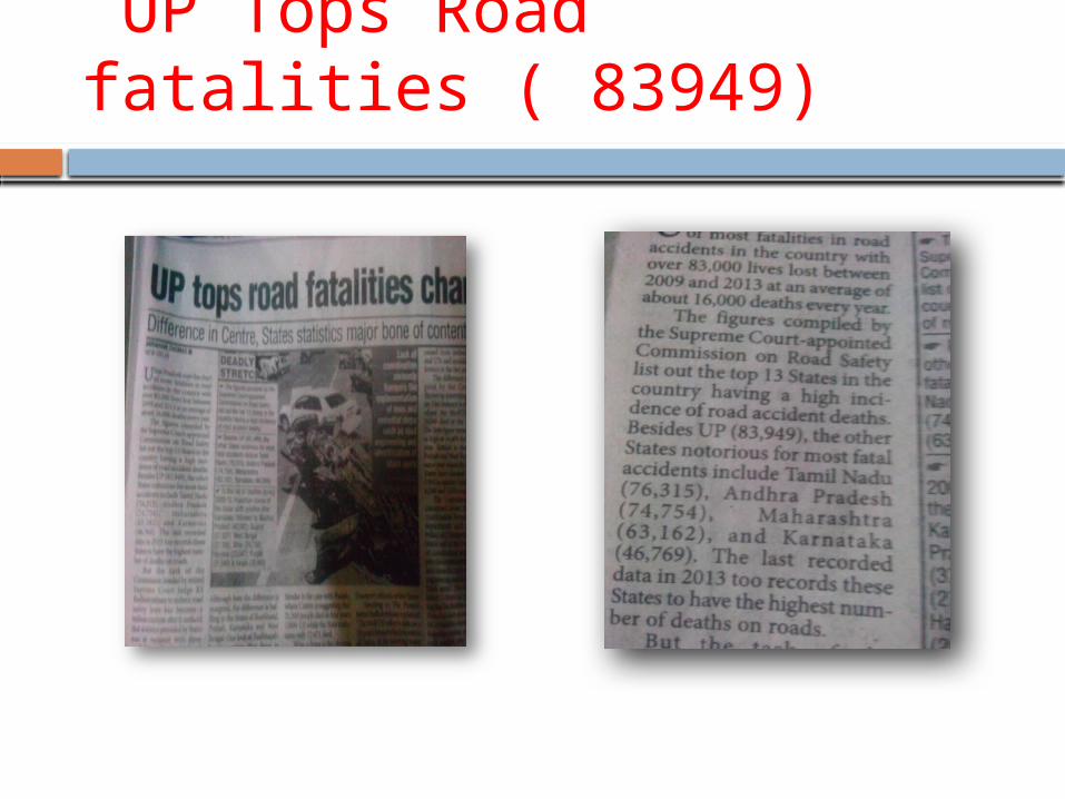
UP Tops Road fatalities ( 83949)
Urban Interpersonal violence
Reduced incidence of condylar fracture Increased Body Fracture
Ellis, Moos and El Attar 1985 Busuito, Smith & Robson 1986
Predictability
Not consistent within all groups and hospitals
Location and community demographics Trauma reported from industrial urban-
Altercations Rural setting- RTA
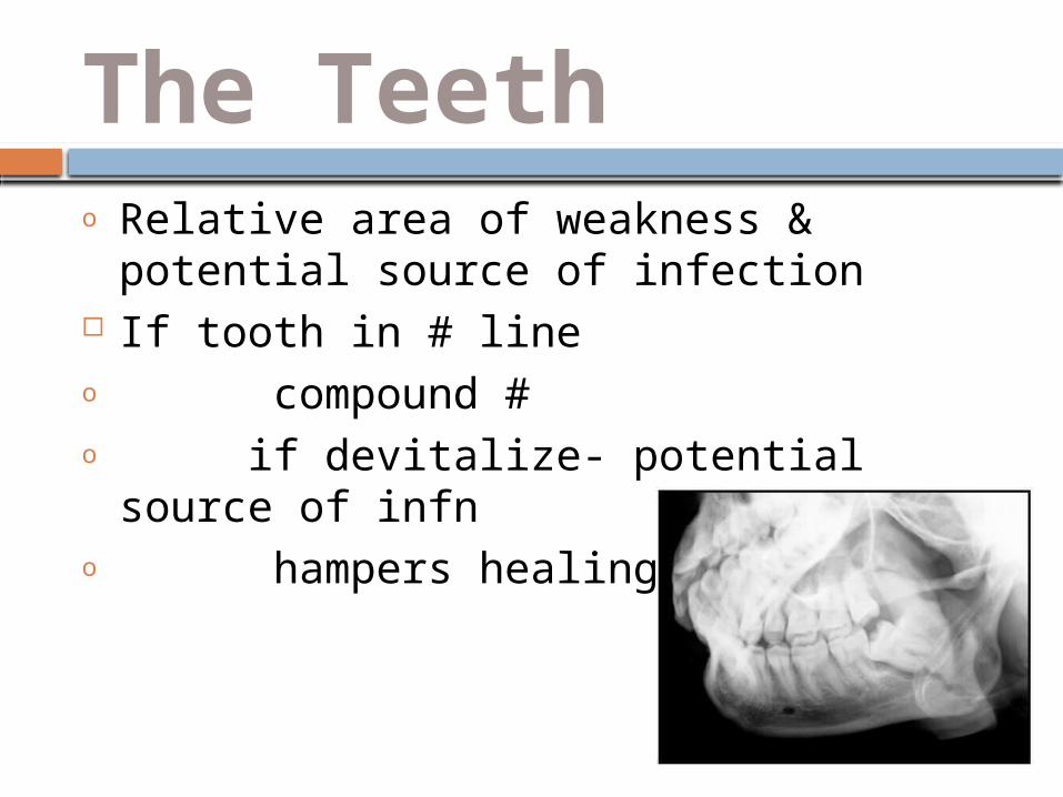
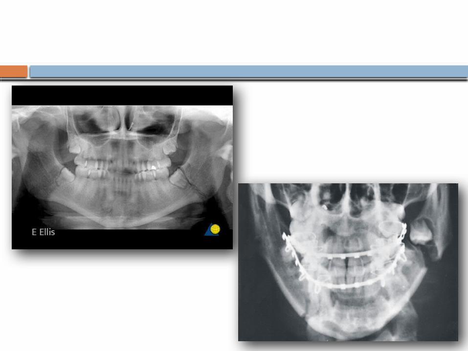
The Teetho Relative area of weakness & potential
source of infection If tooth in # lineo compound #o if devitalize- potential source of infno hampers healing process
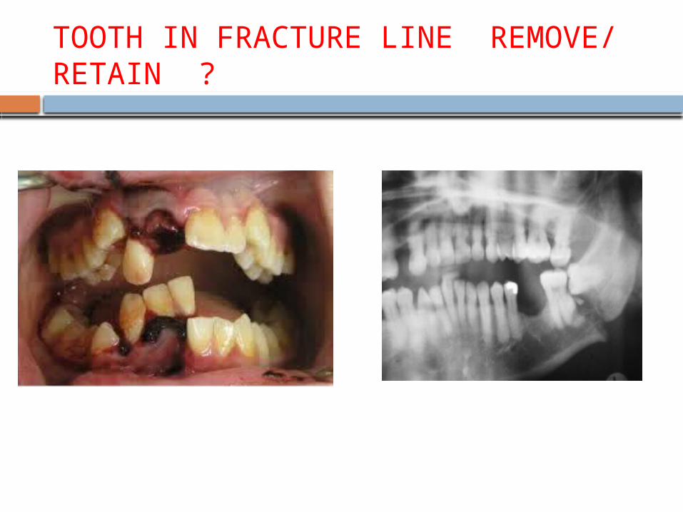
TOOTH IN FRACTURE LINE REMOVE/ RETAIN ?
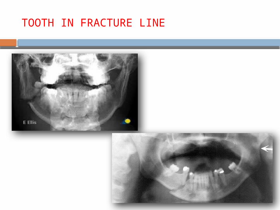
TOOTH IN FRACTURE LINE
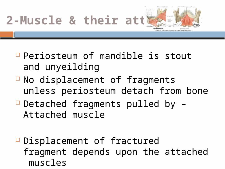
2-Muscle & their attachments
Periosteum of mandible is stout and unyeilding
No displacement of fragments unless periosteum detach from bone
Detached fragments pulled by – Attached muscle
Displacement of fractured fragment
depends upon the attached muscles

Muscle attachments
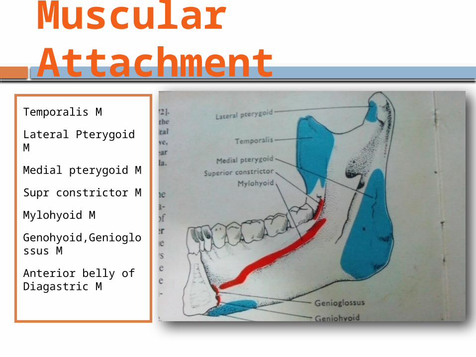
Temporalis M
Lateral Pterygoid M
Medial pterygoid M
Supr constrictor M
Mylohyoid M
Genohyoid,Genioglossus M
Anterior belly of Diagastric M
Muscular Attachment
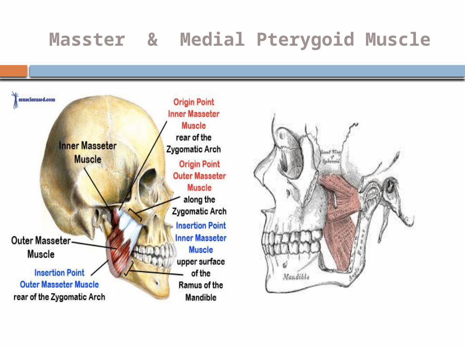
Masster & Medial Pterygoid Muscle
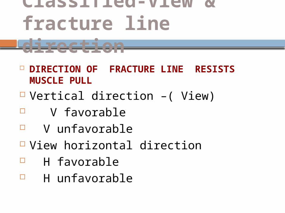
Classified-View & fracture line direction
DIRECTION OF FRACTURE LINE RESISTS MUSCLE PULL
Vertical direction –( View) V favorable V unfavorable View horizontal direction H favorable H unfavorable
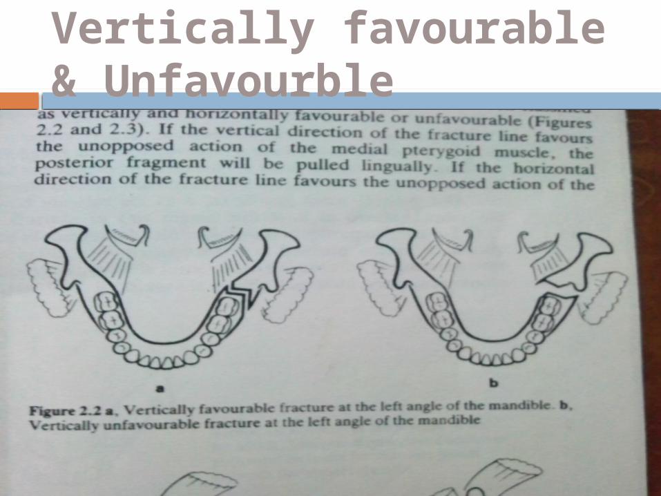
Vertically favourable & Unfavourble
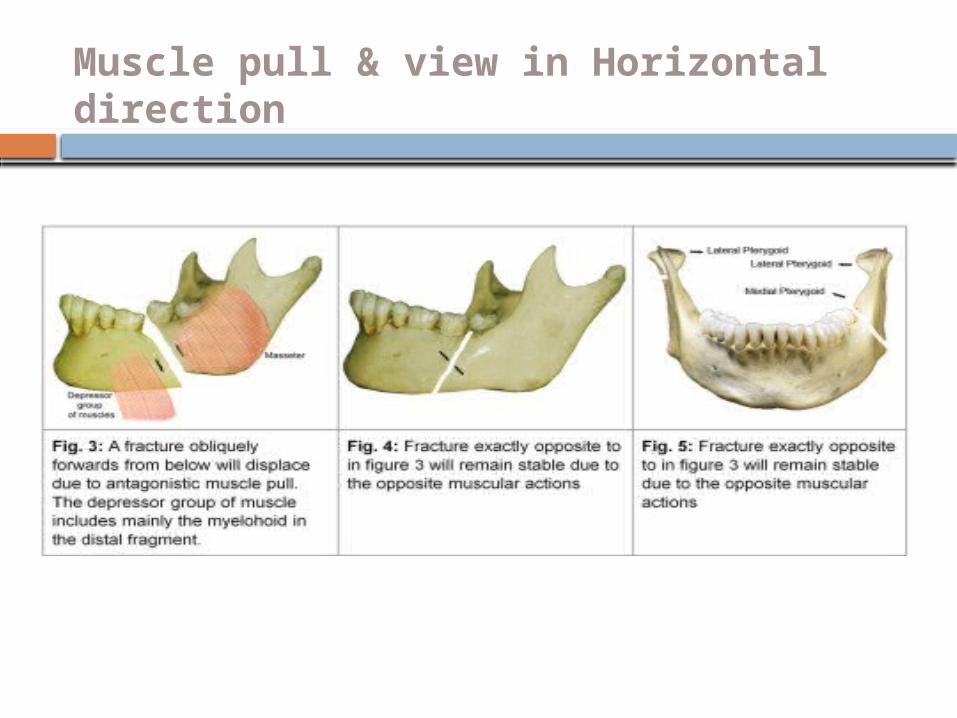
Muscle pull & view in Horizontal direction
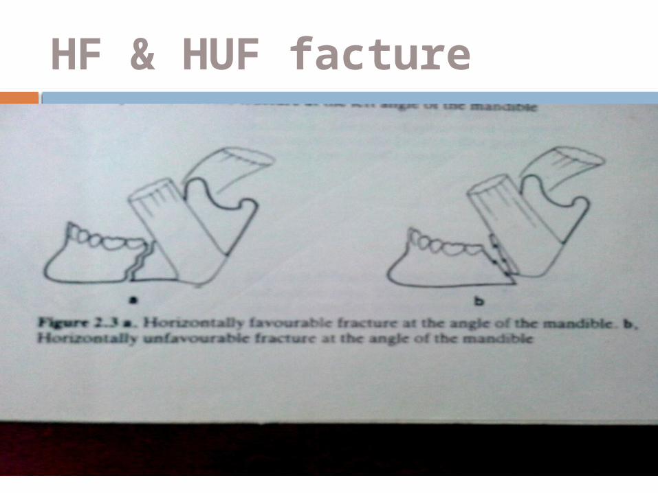
HF & HUF facture
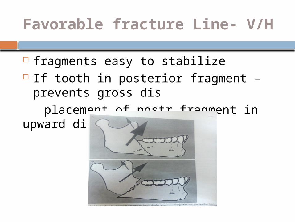
Favorable fracture Line- V/H
fragments easy to stabilize If tooth in posterior fragment – prevents
gross dis placement of postr fragment in upward direction
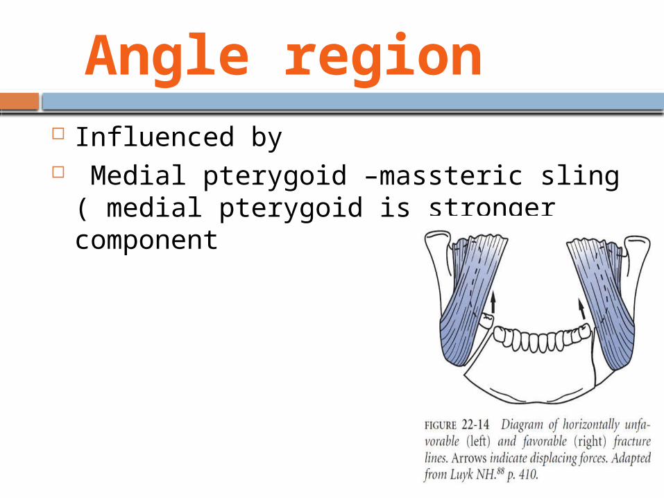
Angle region Influenced by Medial pterygoid –massteric sling
( medial pterygoid is stronger component
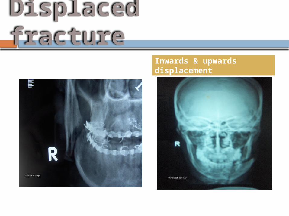
Displaced fracture
Inwards & upwards displacement
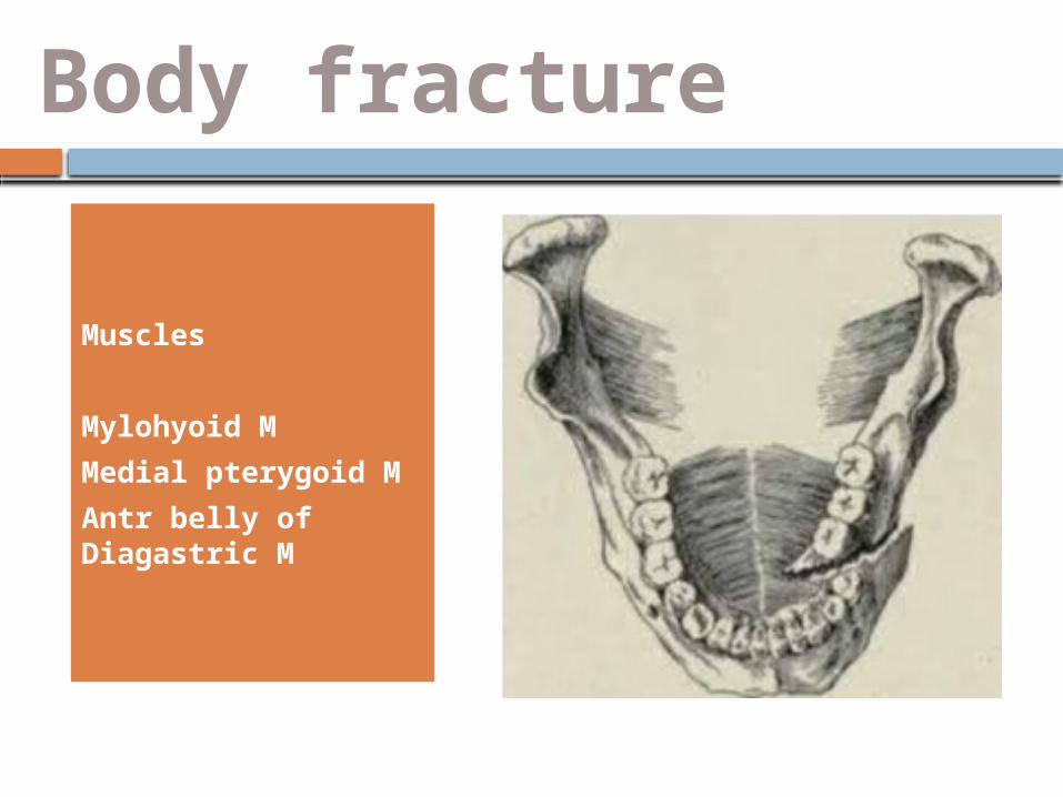
Body fracture
Muscles
Mylohyoid M
Medial pterygoid M
Antr belly of Diagastric M
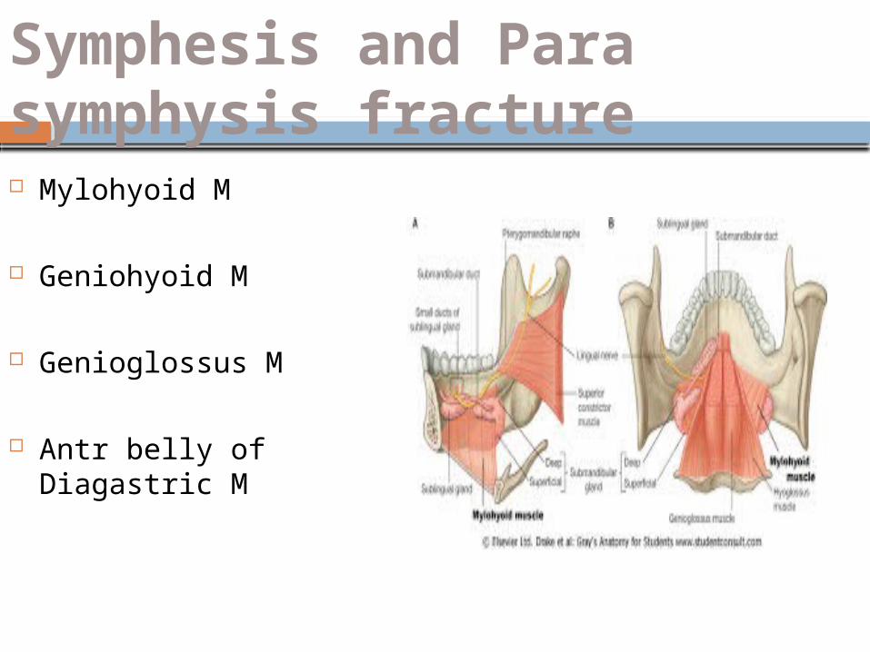
Symphesis and Para symphysis fracture Mylohyoid M
Geniohyoid M
Genioglossus M
Antr belly of Diagastric M
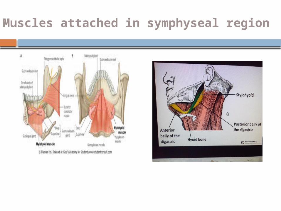
Muscles attached in symphyseal region
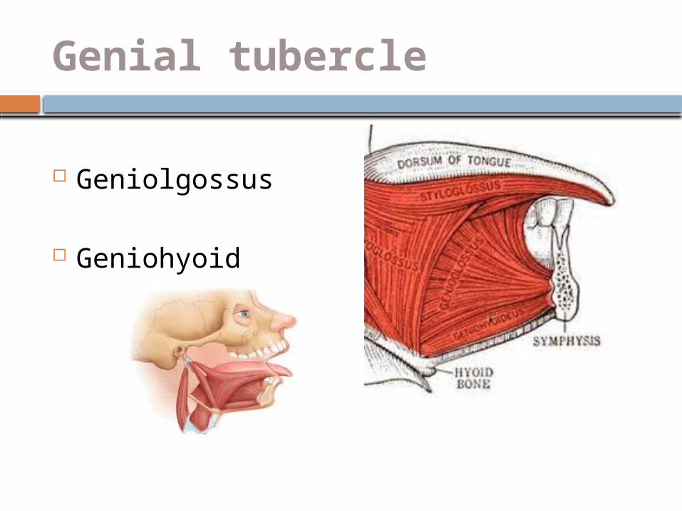
Genial tubercle
Geniolgossus
Geniohyoid
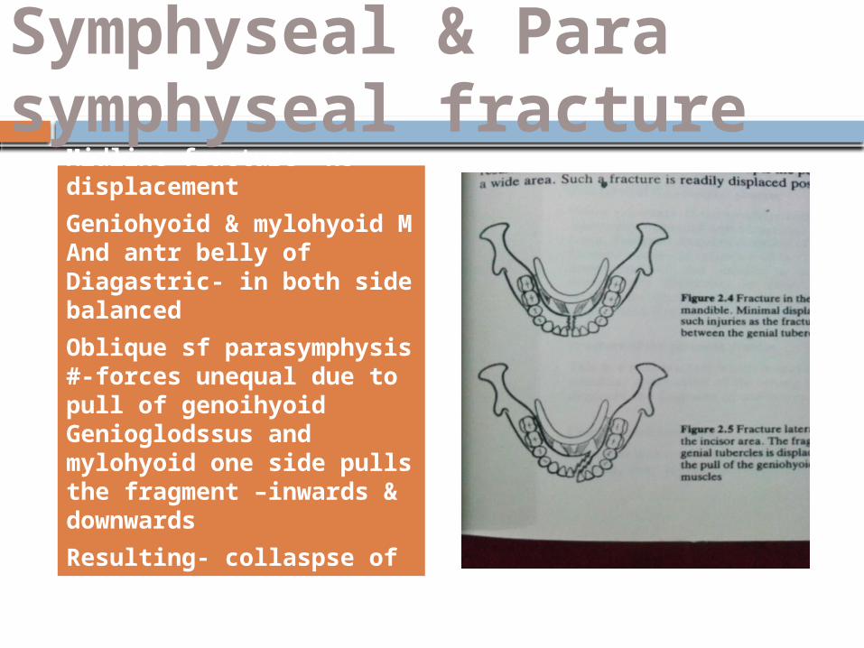
Symphyseal & Para symphyseal fracture
Midline fracture- no displacement
Geniohyoid & mylohyoid M And antr belly of Diagastric- in both side balanced
Oblique sf parasymphysis #-forces unequal due to pull of genoihyoid Genioglodssus and mylohyoid one side pulls the fragment –inwards & downwards
Resulting- collaspse of arch
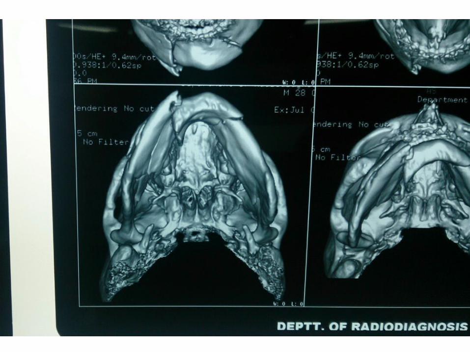
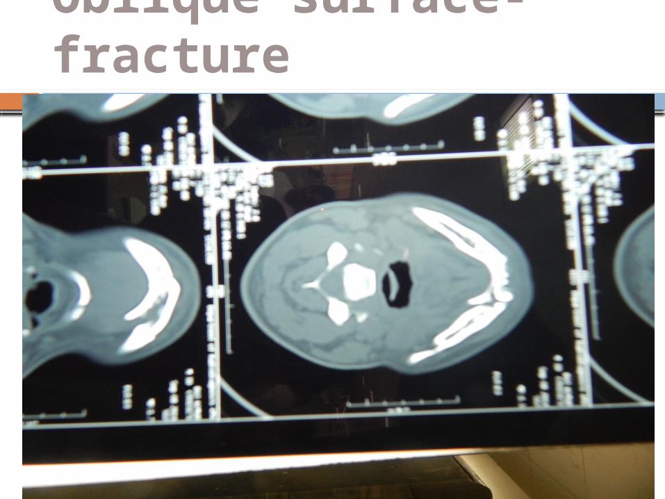
Oblique surface-fracture displacement
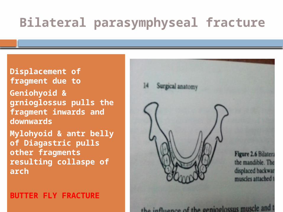
Bilateral parasymphyseal fracture
Displacement of fragment due to
Geniohyoid & grnioglossus pulls the fragment inwards and downwards
Mylohyoid & antr belly of Diagastric pulls other fragments resulting collaspe of arch
BUTTER FLY FRACTURE
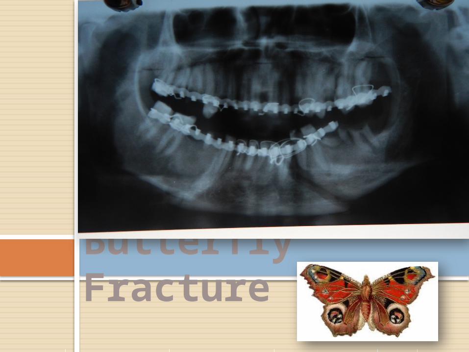
Butterfly Fracture
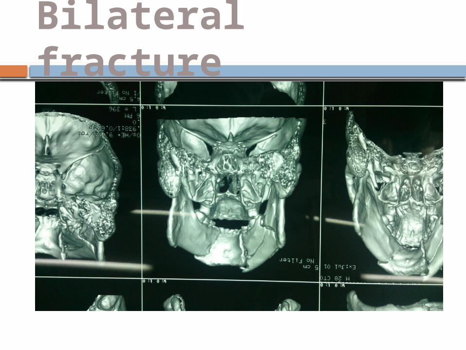
Bilateral fracture
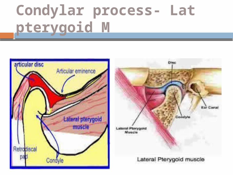
Condylar process- Lat pterygoid M
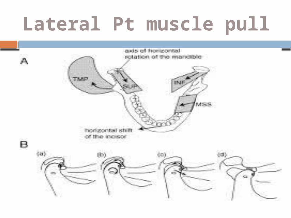
Lateral Pt muscle pull
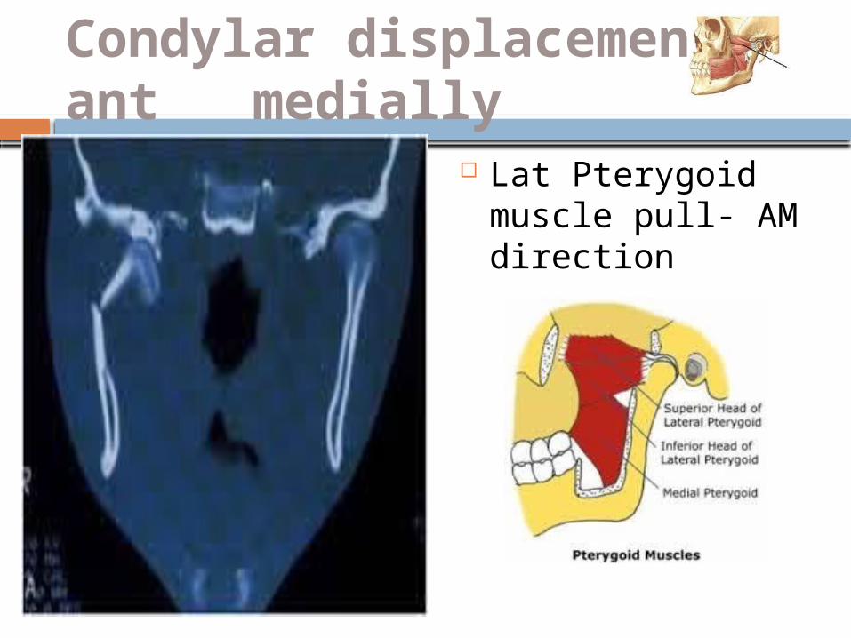
Condylar displacement-ant medially
Lat Pterygoid muscle pull- AM direction
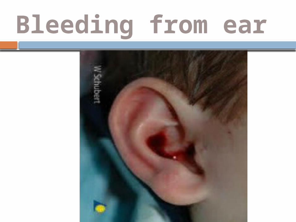
Bleeding from ear
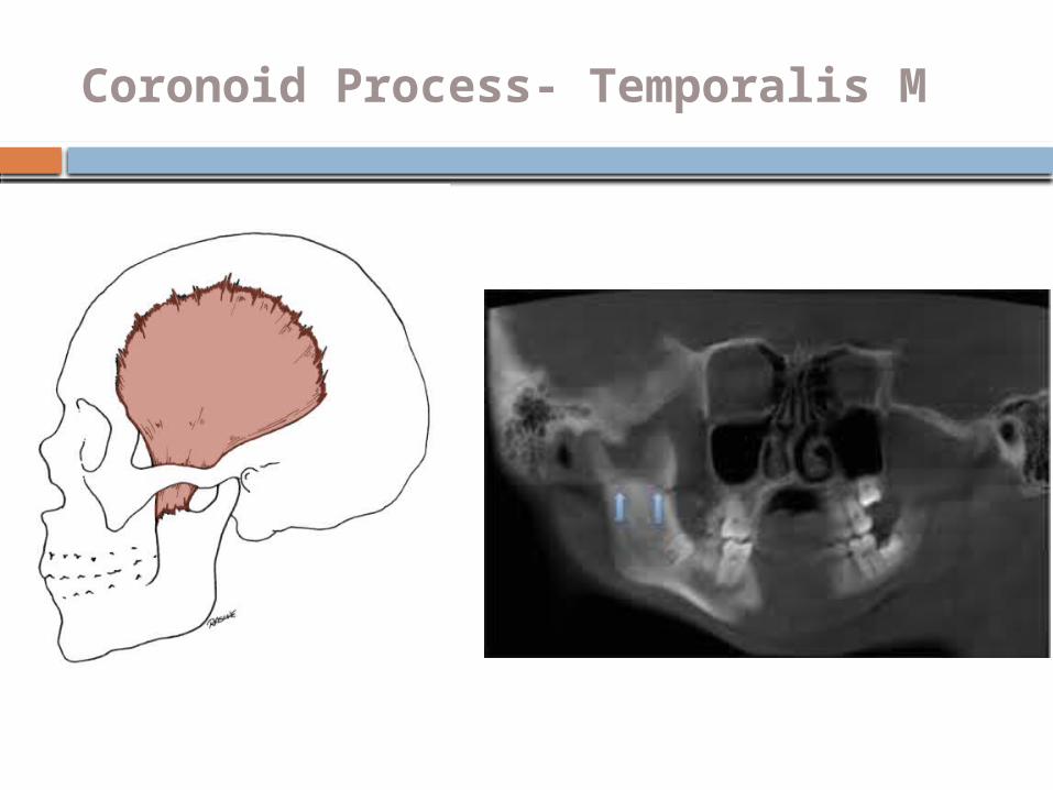
Coronoid Process- Temporalis M
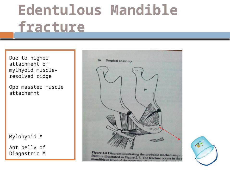
Edentulous Mandible fracture
Due to higher attachment of mylhyoid muscle-resolved ridge
Opp masster muscle attachemnt
Mylohyoid M
Ant belly of Diagastric M
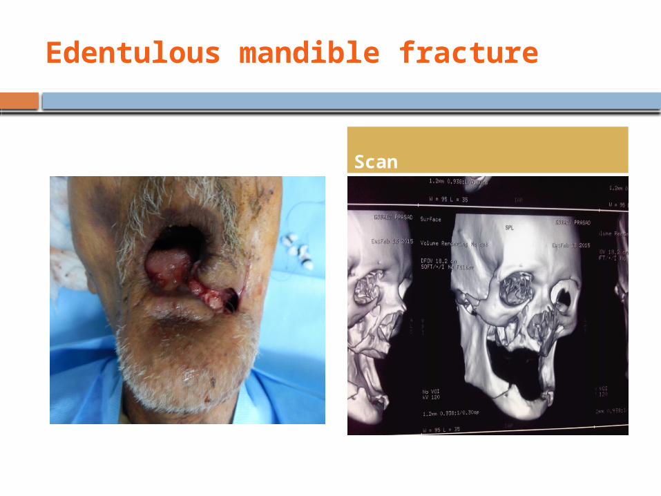
Edentulous mandible fracture
Scan
14th Aug
2015 In Continuation
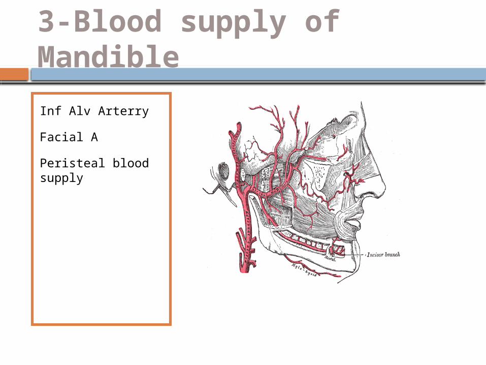
3-Blood supply of Mandible
Inf Alv Arterry
Facial A
Peristeal blood supply
Periosteal blood supply Lost with the tooth lost changes IAA gradually diminishes in size May eventually dis appear
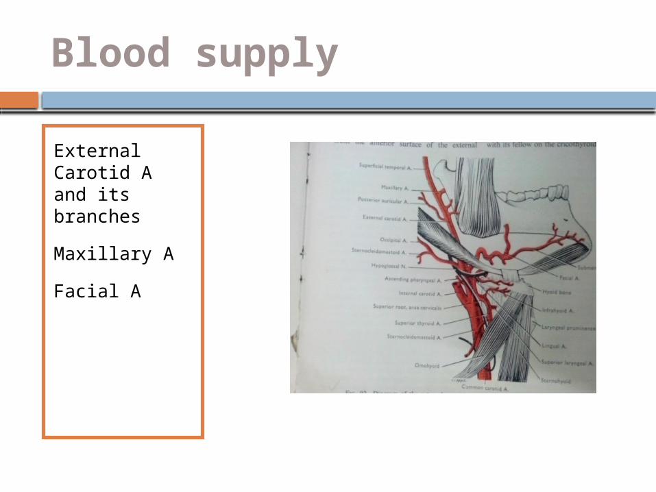
Blood supply
External Carotid A and its branches
Maxillary A
Facial A
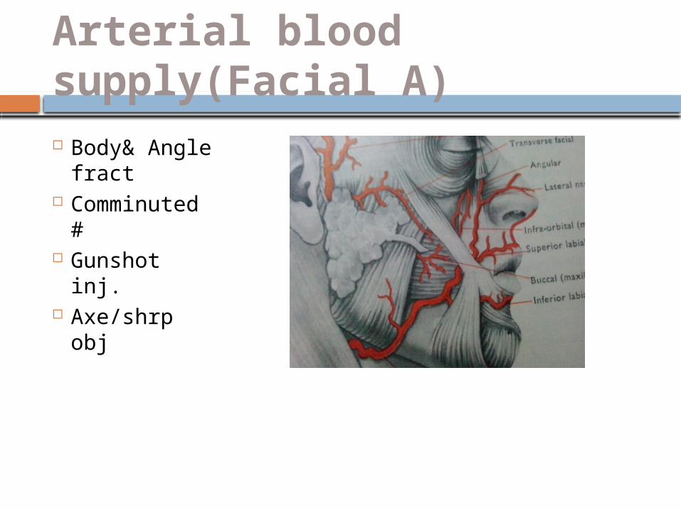
Arterial blood supply(Facial A) Body&
Angle fract Comminute
d # Gunshot
inj. Axe/shrp
obj
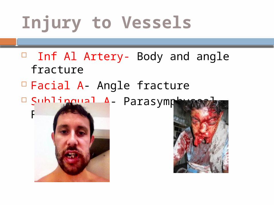
Injury to Vessels
Inf Al Artery- Body and angle fracture Facial A- Angle fracture Sublingual A- Parasymphyseal Fracture
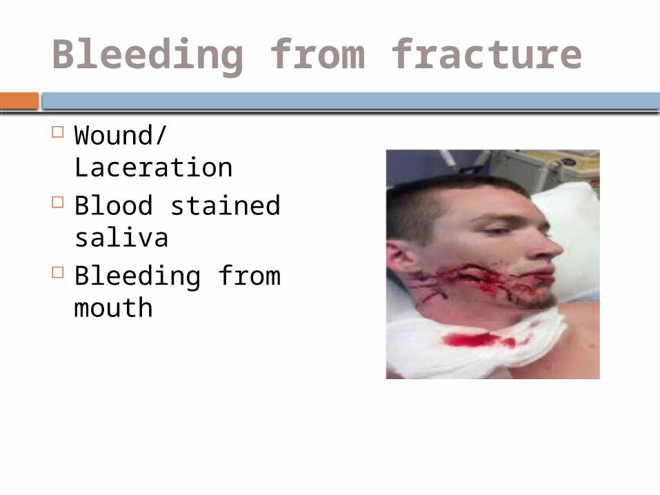
Bleeding from fracture
Wound/Laceration Blood stained
saliva Bleeding from
mouth
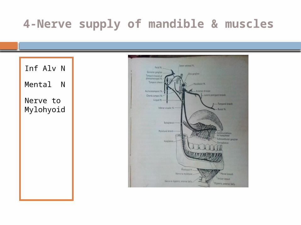
4-Nerve supply of mandible & muscles
Inf Alv N
Mental N
Nerve to Mylohyoid
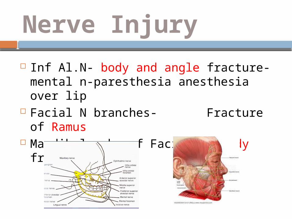
Nerve Injury
Inf Al.N- body and angle fracture-mental n-paresthesia anesthesia over lip
Facial N branches- Fracture of Ramus Mandibular br of Facial N- Body fracture
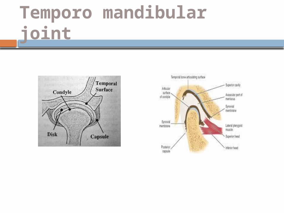
Temporo mandibular joint
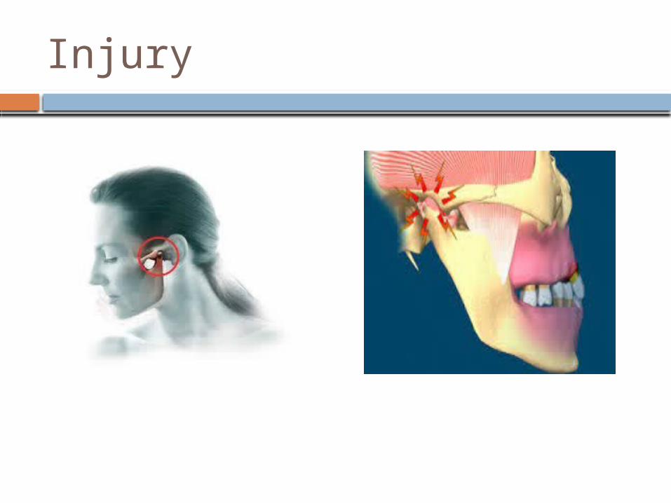
Injury
Effects of trauma-TMJ
TMJ effusion(-Fracture)
Heamorthrosis Meniscus
damage(IDTMJ) TMJ ankylosis
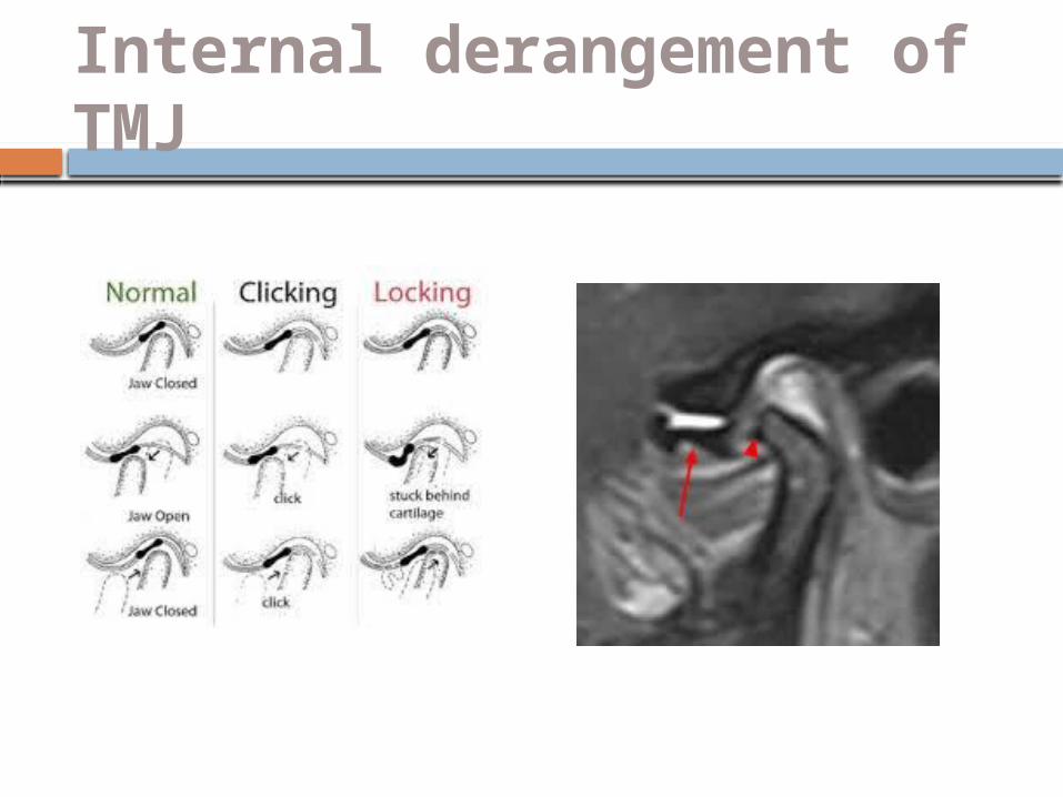
Internal derangement of TMJ
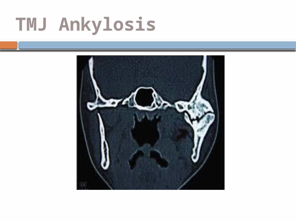
TMJ Ankylosis
Area of weakness-Vulnerable to Fracture
Junction of alv bone and basal mnd bone-so DA # are independent to mandibular fracture.
Symphyseal #-formed by the union of two half of mandible in centre at irst year of life. So area of weakness.
Parasymphyseal region- lateral to the mental prominence-presence of mental formen.
Body region-junction of thicker body with Ramus-angle regionand due to curvature of trajectories in this region.
Anatomical variations
Strength of lower jaw varies with presence r absence of tooth.
Presence of impacted tooth r long root of cannine make the area vulnerable for fracture.
Condylar region-slender neck of condyle render to fracture as a result of direct violence to chin.acts as safety mechanism to prevent injury to middle cranial fossa.
Curve of mandible is more distorted from trauma-so buccal and lingual plate fractures at different level-this may give appearance of double fracture.
Referrence Books-
Text book of Oral& MaxFac Surgery- N.Malik Kelly’s fractures of the mandible-Peter
Banks, Oral & Maxillofacial Surgery- R.J.Fonseca Principles of Internal fixation of the
Craniomaxillofacial Skeleton-Ehrenfeld,Manson& Prein.
Maxillofacial Surgery Reconstruction- Peter ward Booth
END