Embed Size (px)
Citation preview
7 RNA Synthesis and Processing
Chapt. 7 RNA synthesis and processing brief
Student learning outcomes:
1. Explain transcription in Prokaryotes
2*. Explain how Eukaryotic transcription is complex: RNA Polymerases, General Transcription Factors
3. Briefly explain complex regulation of transcription in Eukaryotes: many proteins
4*. Describe RNA processing: Cap, polyA, splicing
5. Describe RNA turnover, Nonsense-mediated decay
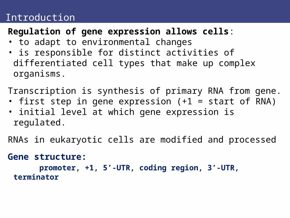
Introduction
Regulation of gene expression allows cells:• to adapt to environmental changes• is responsible for distinct activities of differentiated
cell types that make up complex organisms.
Transcription is synthesis of primary RNA from gene.• first step in gene expression (+1 = start of RNA) • initial level at which gene expression is regulated.
RNAs in eukaryotic cells are modified and processed
Gene structure: promoter, +1, 5’-UTR, coding region, 3’-UTR, terminator
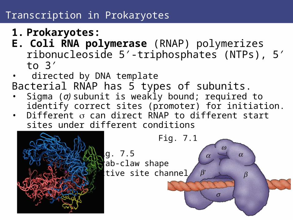
Transcription in Prokaryotes
1. Prokaryotes:E. Coli RNA polymerase (RNAP) polymerizes
ribonucleoside 5′-triphosphates (NTPs), 5′ to 3′• directed by DNA template Bacterial RNAP has 5 types of subunits.• Sigma (σ) subunit is weakly bound; required to identify
correct sites (promoter) for initiation. • Different can direct RNAP to different start sites under
different conditions
Fig. 7.1
Fig. 7.5Crab-claw shapeActive site channel
Transcription in Prokaryotes
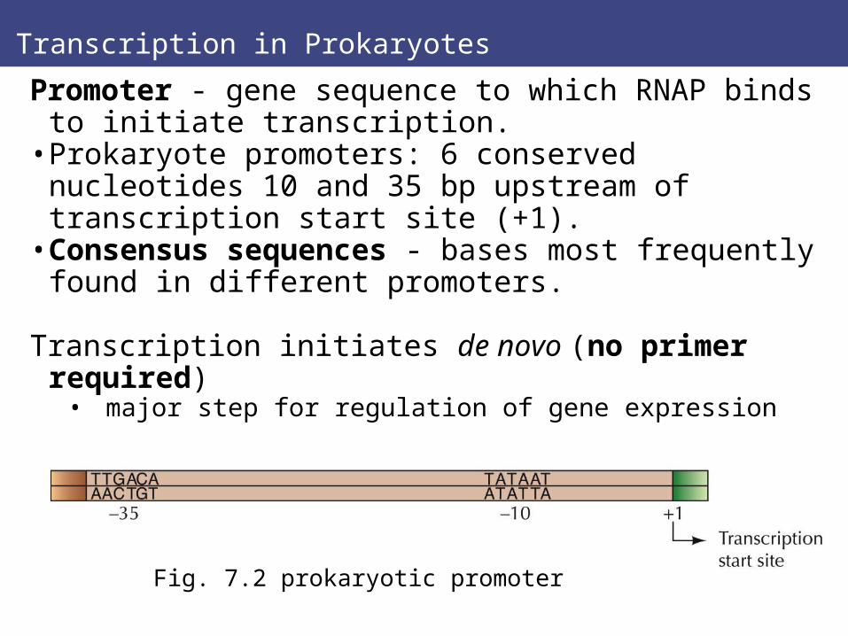
Promoter - gene sequence to which RNAP binds to initiate transcription.
• Prokaryote promoters: 6 conserved nucleotides 10 and 35 bp upstream of transcription start site (+1).
• Consensus sequences - bases most frequently found in different promoters.
Transcription initiates de novo (no primer required) • major step for regulation of gene expression
Fig. 7.2 prokaryotic promoter
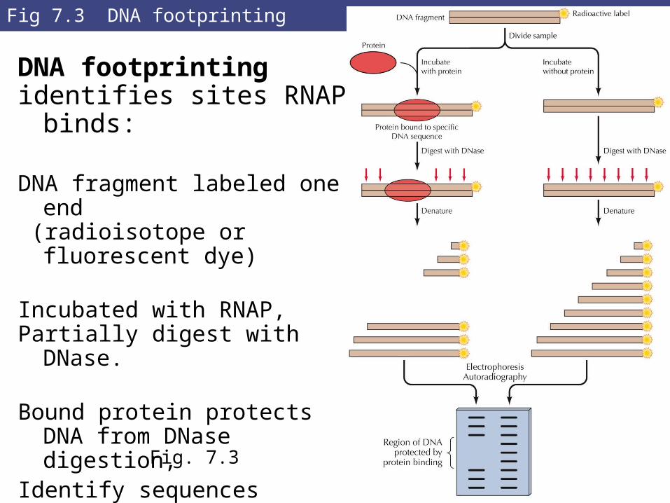
Fig 7.3 DNA footprinting
DNA footprinting identifies sites RNAP binds:
DNA fragment labeled one end (radioisotope or fluorescent dye)
Incubated with RNAP,Partially digest with DNase.
Bound protein protects DNA from DNase digestion,
Identify sequences compared to DNA with no protein.
Fig. 7.3
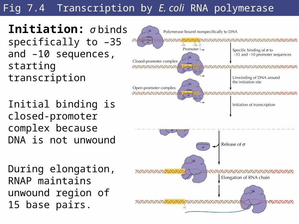
Fig 7.4 Transcription by E. coli RNA polymerase
Initiation: σ binds specifically to –35 and –10 sequences, starting transcription
Initial binding is closed-promoter complex because DNA is not unwound
During elongation,RNAP maintainsunwound region of 15 base pairs.
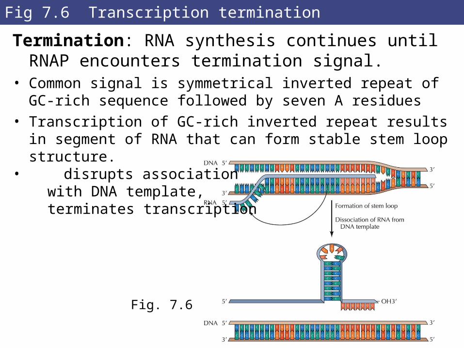
Fig 7.6 Transcription termination
Termination: RNA synthesis continues until RNAP encounters termination signal.
• Common signal is symmetrical inverted repeat of GC-rich sequence followed by seven A residues
• Transcription of GC-rich inverted repeat results in segment of RNA that can form stable stem loop structure.
• disrupts association with DNA template, terminates transcription
Fig. 7.6
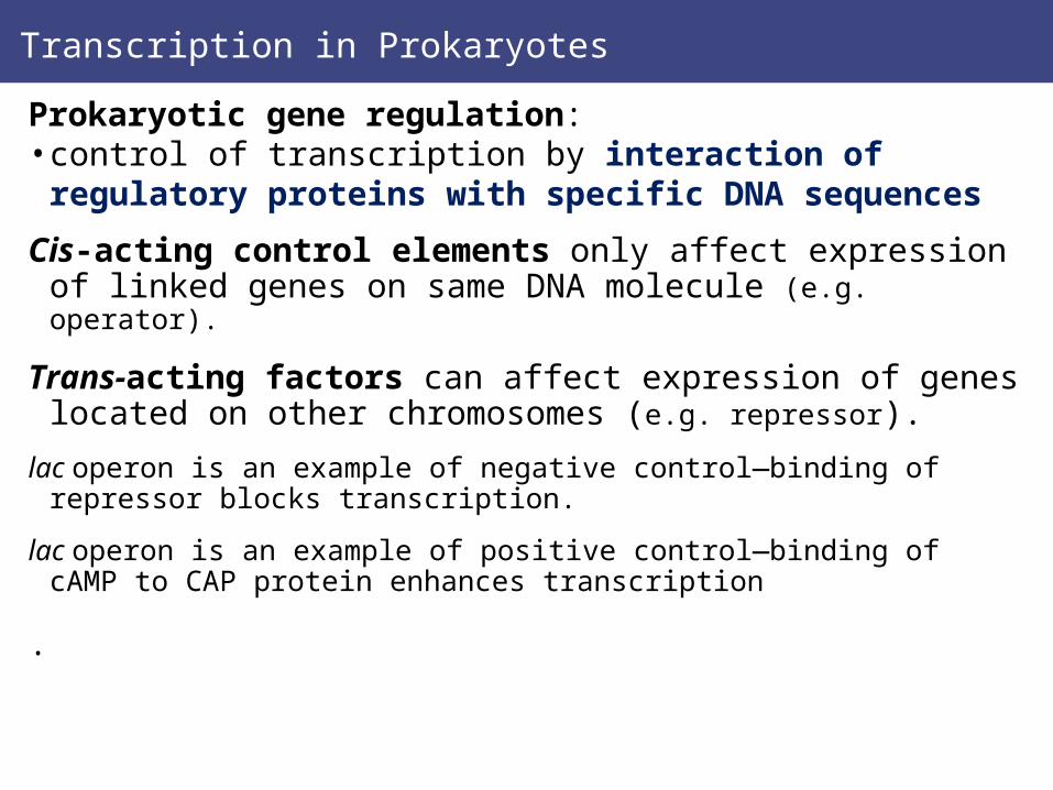
Transcription in Prokaryotes
Prokaryotic gene regulation: • control of transcription by interaction of regulatory
proteins with specific DNA sequences
Cis-acting control elements only affect expression of linked genes on same DNA molecule (e.g. operator).
Trans-acting factors can affect expression of genes located on other chromosomes (e.g. repressor).
lac operon is an example of negative control—binding of repressor blocks transcription.
lac operon is an example of positive control—binding of cAMP to CAP protein enhances transcription
.
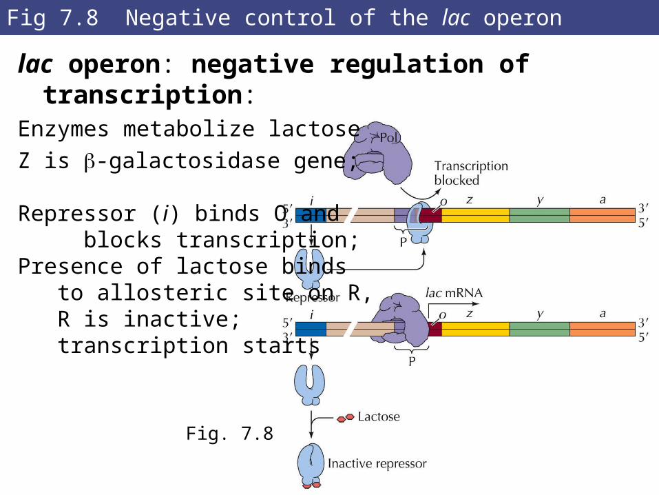
Fig 7.8 Negative control of the lac operon
lac operon: negative regulation of transcription:Enzymes metabolize lactose
Z is -galactosidase gene;
Repressor (i) binds O and blocks transcription;Presence of lactose binds to allosteric site on R, R is inactive; transcription starts
Fig. 7.8
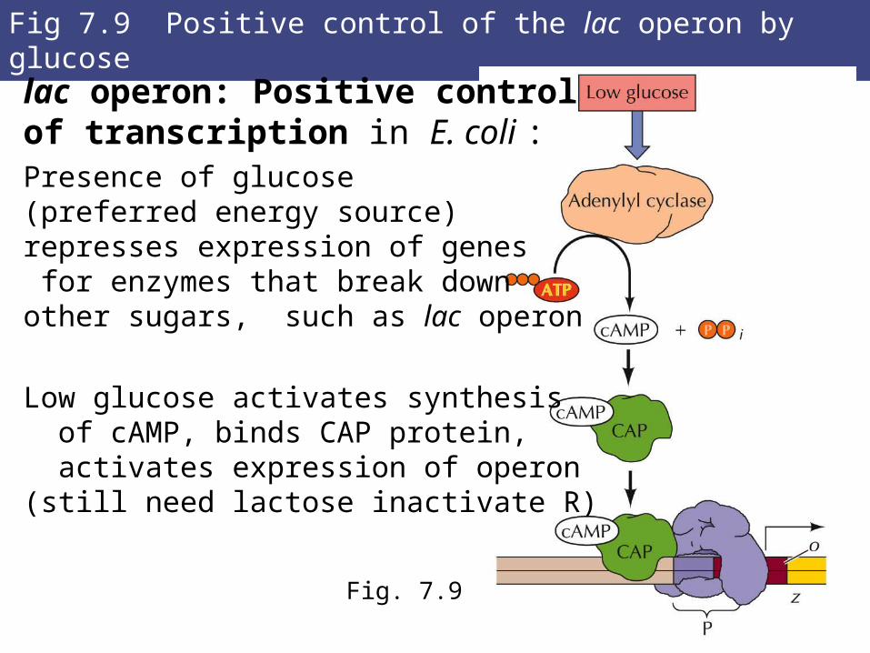
Fig 7.9 Positive control of the lac operon by glucose
lac operon: Positive controlof transcription in E. coli :Presence of glucose (preferred energy source) represses expression of genes for enzymes that break downother sugars, such as lac operon
Low glucose activates synthesis of cAMP, binds CAP protein, activates expression of operon(still need lactose inactivate R)
Fig. 7.9
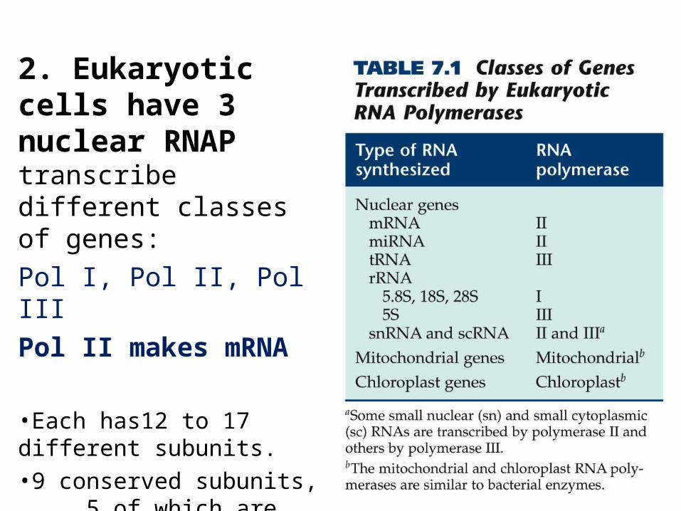
2. Eukaryotic cells have 3 nuclear RNAP transcribe different classes of genes:
Pol I, Pol II, Pol III
Pol II makes mRNA
•Each has12 to 17 different subunits.•9 conserved subunits, 5 of which are related to subunits of bacterial RNAP
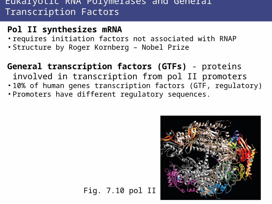
Eukaryotic RNA Polymerases and General Transcription Factors
Pol II synthesizes mRNA• requires initiation factors not associated with RNAP• Structure by Roger Kornberg – Nobel Prize
General transcription factors (GTFs) - proteins involved in transcription from pol II promoters
• 10% of human genes transcription factors (GTF, regulatory)• Promoters have different regulatory sequences.
Fig. 7.10 pol II
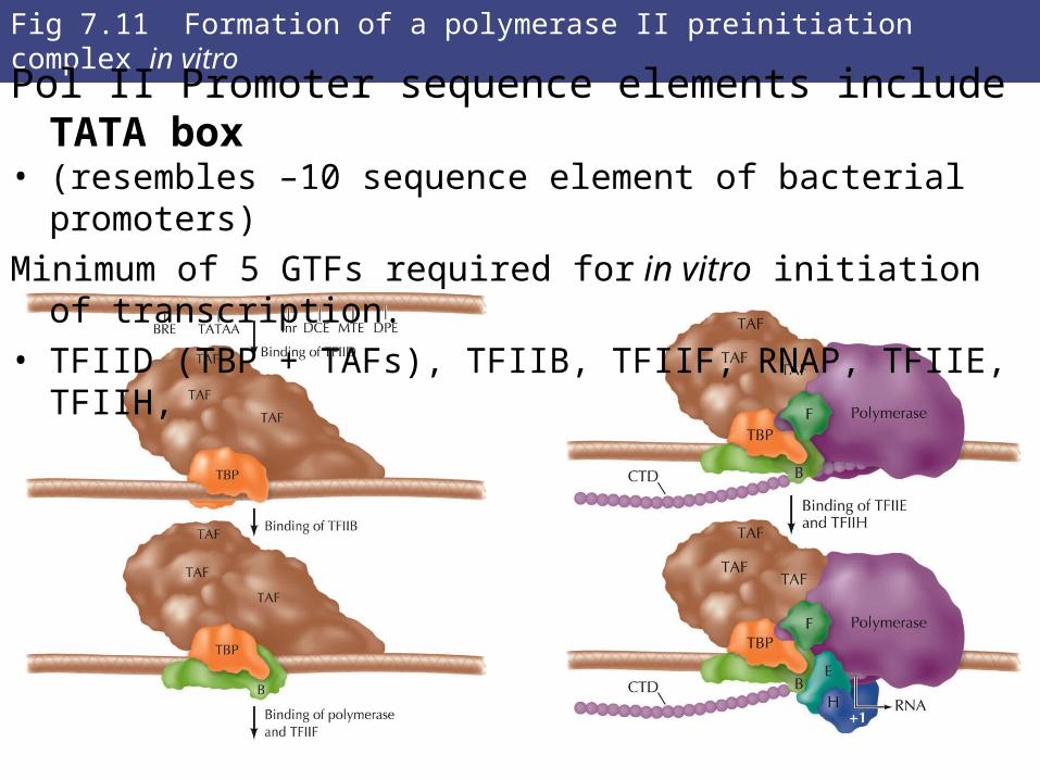
Fig 7.11 Formation of a polymerase II preinitiation complex in vitro
Pol II Promoter sequence elements include TATA box • (resembles –10 sequence element of bacterial promoters)
Minimum of 5 GTFs required for in vitro initiation of transcription.• TFIID (TBP + TAFs), TFIIB, TFIIF, RNAP, TFIIE, TFIIH,
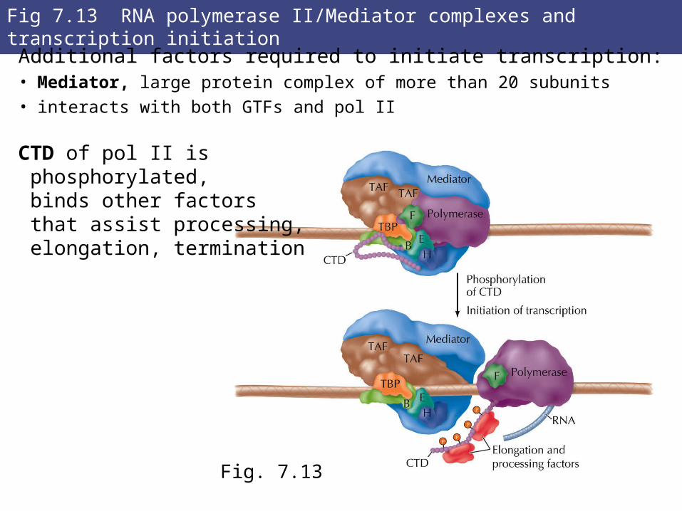
Fig 7.13 RNA polymerase II/Mediator complexes and transcription initiation
Additional factors required to initiate transcription:• Mediator, large protein complex of more than 20 subunits • interacts with both GTFs and pol II
CTD of pol II is phosphorylated, binds other factors that assist processing, elongation, termination
Fig. 7.13
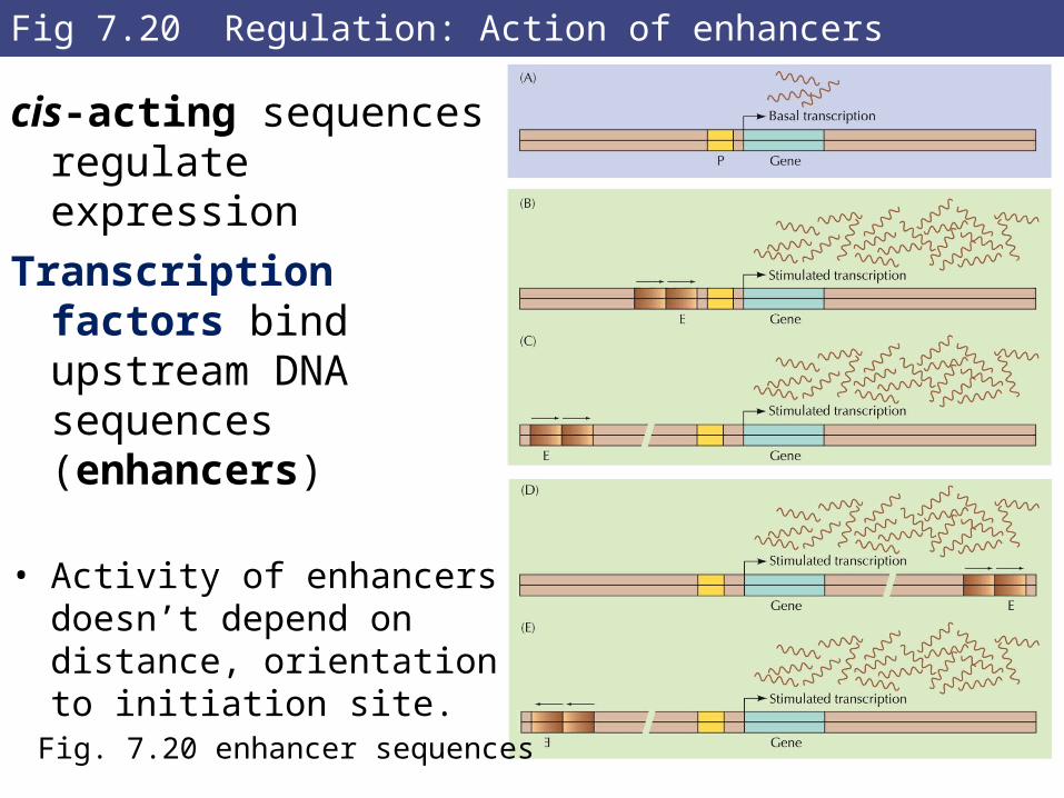
Fig 7.20 Regulation: Action of enhancers
cis-acting sequences regulate expression
Transcription factors bind upstream DNA sequences (enhancers)
• Activity of enhancers doesn’t
depend on distance, orientation to initiation site.
Fig. 7.20 enhancer sequences
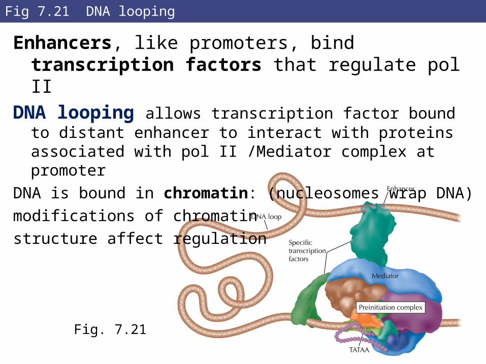
Fig 7.21 DNA looping
Enhancers, like promoters, bind transcription factors that regulate pol II
DNA looping allows transcription factor bound to distant enhancer to interact with proteins associated with pol II /Mediator complex at promoter
DNA is bound in chromatin: (nucleosomes wrap DNA)
modifications of chromatin
structure affect regulation
Fig. 7.21
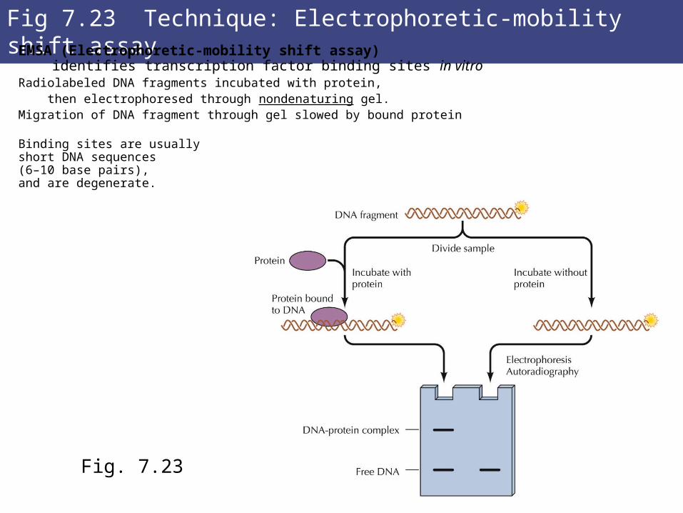
Fig 7.23 Technique: Electrophoretic-mobility shift assayEMSA (Electrophoretic-mobility shift assay) identifies transcription factor binding sites in vitroRadiolabeled DNA fragments incubated with protein, then electrophoresed through nondenaturing gel.Migration of DNA fragment through gel slowed by bound protein
Binding sites are usuallyshort DNA sequences (6–10 base pairs),and are degenerate.
Fig. 7.23
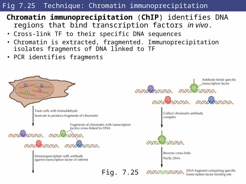
Fig 7.25 Technique: Chromatin immunoprecipitation
Chromatin immunoprecipitation (ChIP) identifies DNA regions that bind transcription factors in vivo.
• Cross-link TF to their specific DNA sequences • Chromatin is extracted, fragmented. Immunoprecipitation
isolates fragments of DNA linked to TF• PCR identifies fragments
Fig. 7.25
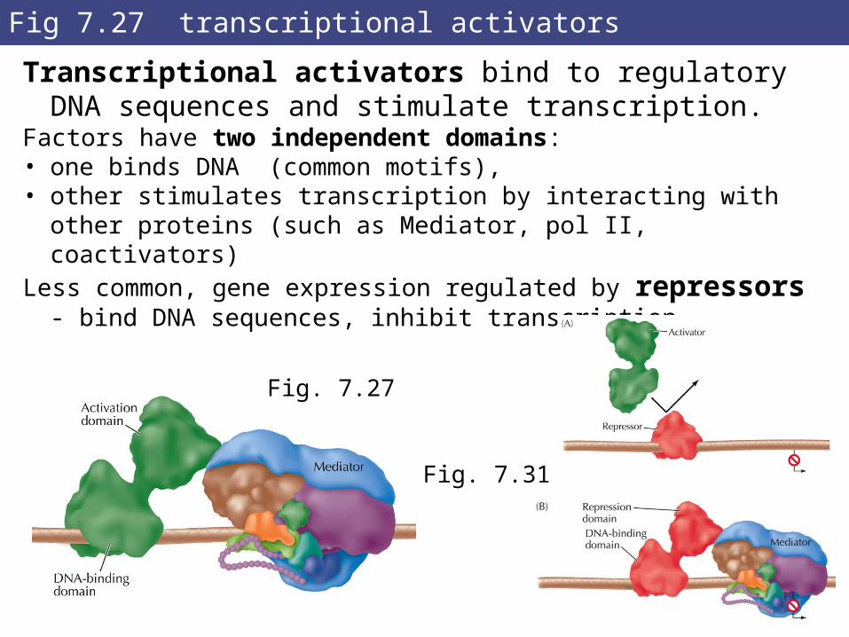
Fig 7.27 transcriptional activators
Transcriptional activators bind to regulatory DNA sequences and stimulate transcription.
Factors have two independent domains: • one binds DNA (common motifs), • other stimulates transcription by interacting with other
proteins (such as Mediator, pol II, coactivators)Less common, gene expression regulated by repressors -
bind DNA sequences, inhibit transcription
Fig. 7.27
Fig. 7.31
RNA Processing and Turnover
4. RNA processing and turnover:
Primary transcript of gene is only the first step:
Most newly-synthesized RNAs must be modified, except bacterial mRNAs which are used immediately for
protein synthesis while still being transcribed.
rRNAs and tRNAs must be processed in both prokaryotic and eukaryotic cells.
Regulation of processing provides another level of control of gene expression.
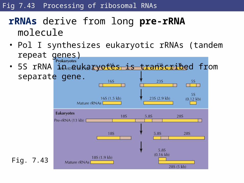
Fig 7.43 Processing of ribosomal RNAs
rRNAs derive from long pre-rRNA molecule• Pol I synthesizes eukaryotic rRNAs (tandem repeat genes)• 5S rRNA in eukaryotes is transcribed from separate gene.
Fig. 7.43
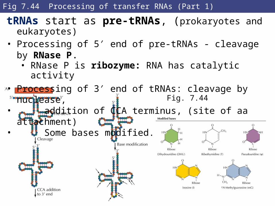
Fig 7.44 Processing of transfer RNAs (Part 1)
tRNAs start as pre-tRNAs, (prokaryotes and eukaryotes)
• Processing of 5′ end of pre-tRNAs - cleavage by RNase P.• RNase P is ribozyme: RNA has catalytic activity
• Processing of 3′ end of tRNAs: cleavage by nuclease, • addition of CCA terminus, (site of aa attachment)• Some bases modified.
Fig. 7.44
RNA Processing and Turnover
**Eukaryote pre-mRNAs extensively modified before export from nucleus:
• 5’-CAP, splicing, 3’-cleavage/polyA addition
Transcription and processing are coupled.
C-terminal domain (CTD) of RNA pol II plays key role in coordinating processes:
• After phosphorylation, binds other protein complexes:• Capping enzymes bind phosphorylated CTD after initiation• Cap added after transcription of first 20 to 30 nucleotides
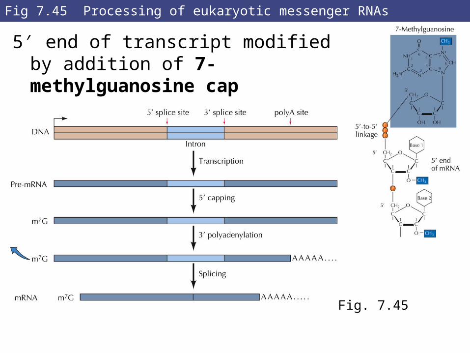
Fig 7.45 Processing of eukaryotic messenger RNAs
5′ end of transcript modified by addition of 7-methylguanosine cap
Fig. 7.45
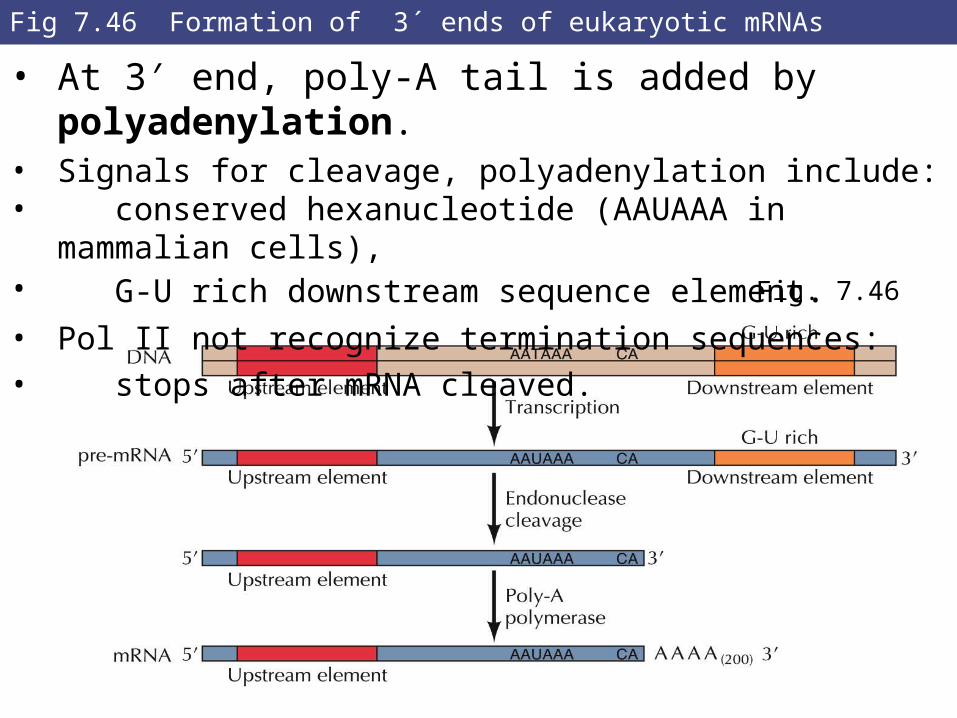
Fig 7.46 Formation of 3´ ends of eukaryotic mRNAs
• At 3′ end, poly-A tail is added by polyadenylation.• Signals for cleavage, polyadenylation include:• conserved hexanucleotide (AAUAAA in mammalian cells),• G-U rich downstream sequence element.• Pol II not recognize termination sequences: • stops after mRNA cleaved. Fig. 7.46
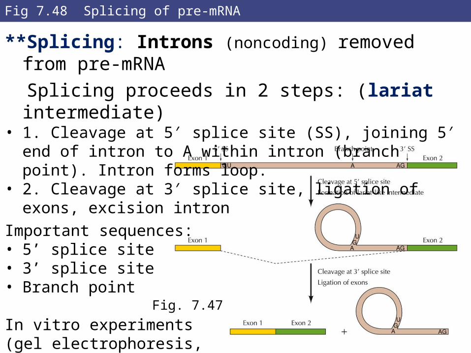
Fig 7.48 Splicing of pre-mRNA
**Splicing: Introns (noncoding) removed from pre-mRNA
Splicing proceeds in 2 steps: (lariat intermediate)• 1. Cleavage at 5′ splice site (SS), joining 5′ end of intron to A
within intron (branch point). Intron forms loop.• 2. Cleavage at 3′ splice site, ligation of exons, excision intron
Important sequences:• 5’ splice site• 3’ splice site• Branch point
In vitro experiments(gel electrophoresis,mutant sequences)
Fig. 7.47
RNA Processing and Turnover
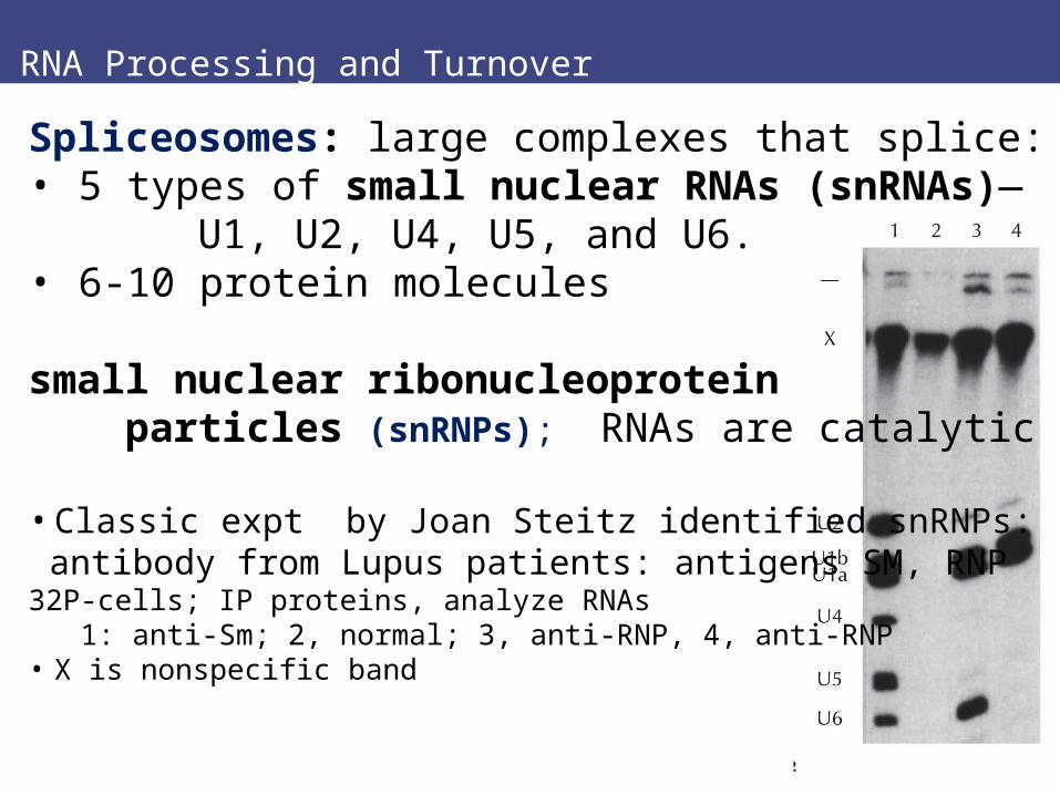
Spliceosomes: large complexes that splice: • 5 types of small nuclear RNAs (snRNAs)— U1, U2, U4, U5, and U6.• 6-10 protein molecules
small nuclear ribonucleoprotein particles (snRNPs); RNAs are catalytic
• Classic expt by Joan Steitz identified snRNPs: antibody from Lupus patients: antigens SM, RNP32P-cells; IP proteins, analyze RNAs 1: anti-Sm; 2, normal; 3, anti-RNP, 4, anti-RNP• X is nonspecific band
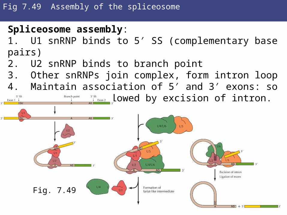
Fig 7.49 Assembly of the spliceosome
Spliceosome assembly:1. U1 snRNP binds to 5′ SS (complementary base pairs)2. U2 snRNP binds to branch point3. Other snRNPs join complex, form intron loop4. Maintain association of 5′ and 3′ exons: so can be ligated followed by excision of intron.
Fig. 7.49
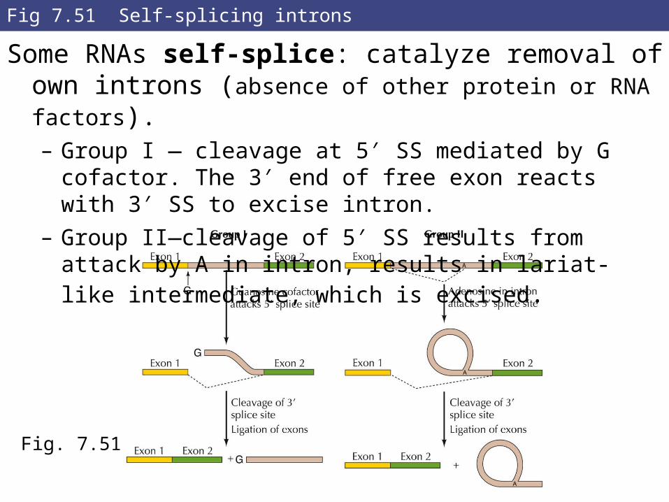
Fig 7.51 Self-splicing introns
Some RNAs self-splice: catalyze removal of own introns (absence of other protein or RNA factors).– Group I — cleavage at 5′ SS mediated by G cofactor. The 3′
end of free exon reacts with 3′ SS to excise intron.
– Group II—cleavage of 5′ SS results from attack by A in intron, results in lariat-like intermediate, which is excised.
Fig. 7.51
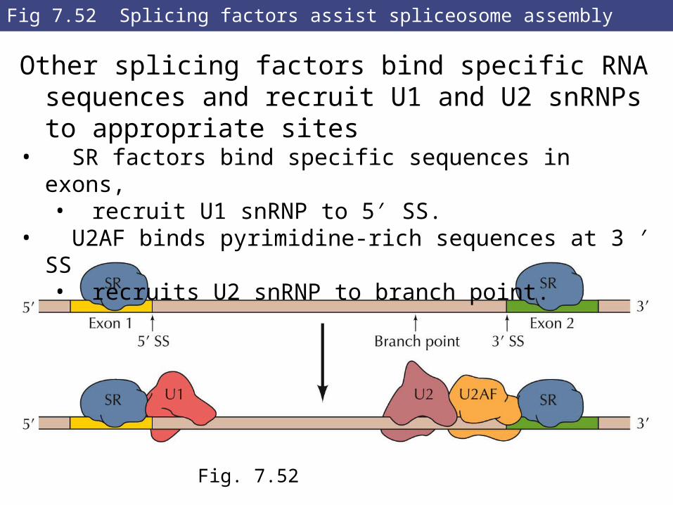
Fig 7.52 Splicing factors assist spliceosome assembly
Other splicing factors bind specific RNA sequences and recruit U1 and U2 snRNPs to appropriate sites
• SR factors bind specific sequences in exons,• recruit U1 snRNP to 5′ SS.
• U2AF binds pyrimidine-rich sequences at 3 ′ SS• recruits U2 snRNP to branch point.
Fig. 7.52
RNA Processing and Turnover
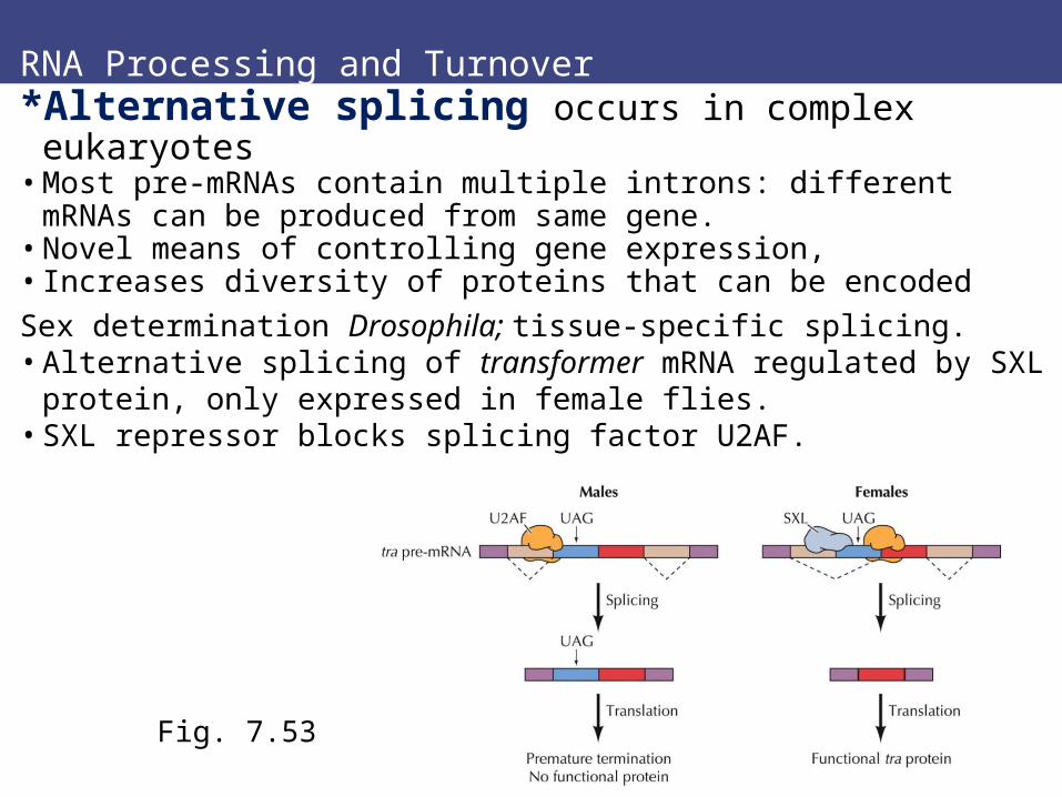
*Alternative splicing occurs in complex eukaryotes• Most pre-mRNAs contain multiple introns: different mRNAs can
be produced from same gene.• Novel means of controlling gene expression, • Increases diversity of proteins that can be encoded
Sex determination Drosophila; tissue-specific splicing.• Alternative splicing of transformer mRNA regulated by SXL
protein, only expressed in female flies.• SXL repressor blocks splicing factor U2AF.
Fig. 7.53
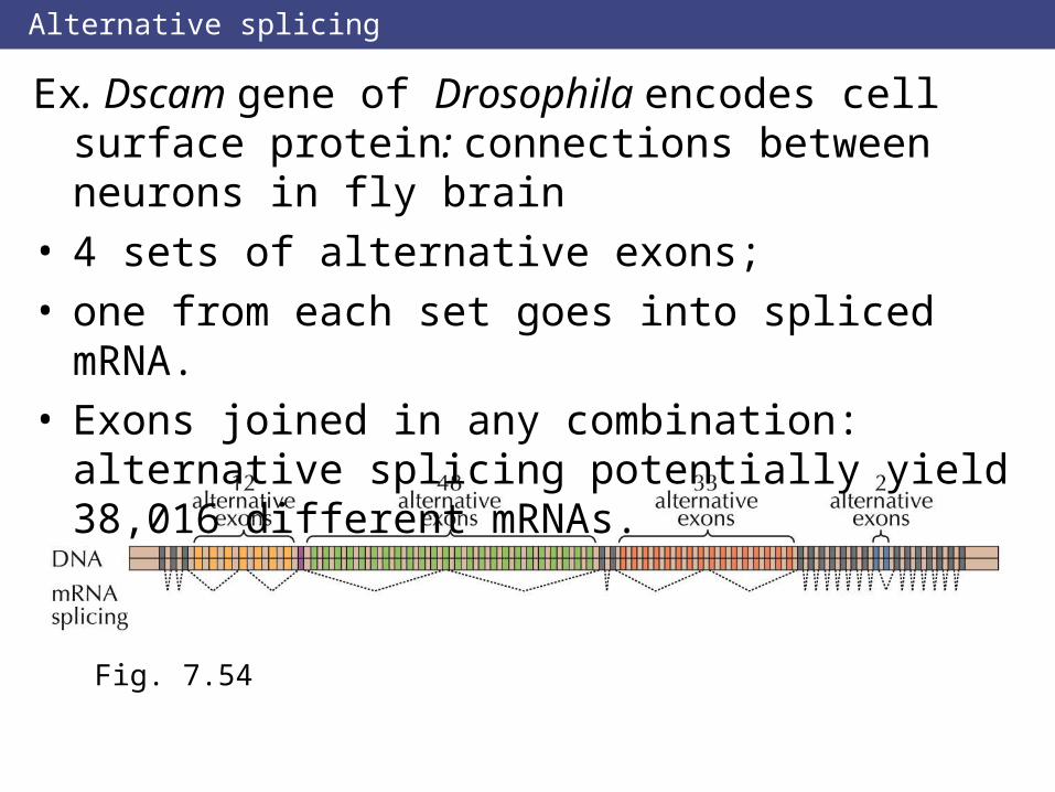
Alternative splicing
Ex. Dscam gene of Drosophila encodes cell surface protein: connections between neurons in fly brain
• 4 sets of alternative exons;• one from each set goes into spliced mRNA.• Exons joined in any combination: alternative splicing
potentially yield 38,016 different mRNAs.
Fig. 7.54
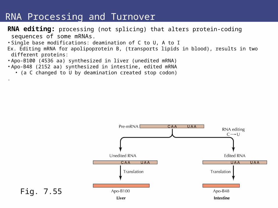
RNA Processing and TurnoverRNA editing: processing (not splicing) that alters protein-coding sequences of some
mRNAs.• Single base modifications: deamination of C to U, A to IEx. Editing mRNA for apolipoprotein B, (transports lipids in blood), results in two different proteins:• Apo-B100 (4536 aa) synthesized in liver (unedited mRNA)• Apo-B48 (2152 aa) synthesized in intestine, edited mRNA
• (a C changed to U by deamination created stop codon).
Fig. 7.55
RNA Processing and Turnover
Aberrant mRNAs can be degraded.
Nonsense-mediated mRNA decay eliminates mRNAs that lack complete open-reading frames.
• Ribosomes encounter premature termination codons, translation stops, defective mRNA degraded
• Ultimately, RNAs degraded in cytoplasm. • Rate of degradation controls gene expression
rRNAs and tRNAs very stable, (both prokaryotes, eukaryotes)• high levels of these RNAs (greater than 90% of all RNA)
Bacterial mRNAs rapidly degraded: t1/2 2 to 3 minutes. • Rapid turnover lets cell respond quickly to changes in environment, such as
nutrient availability.
.
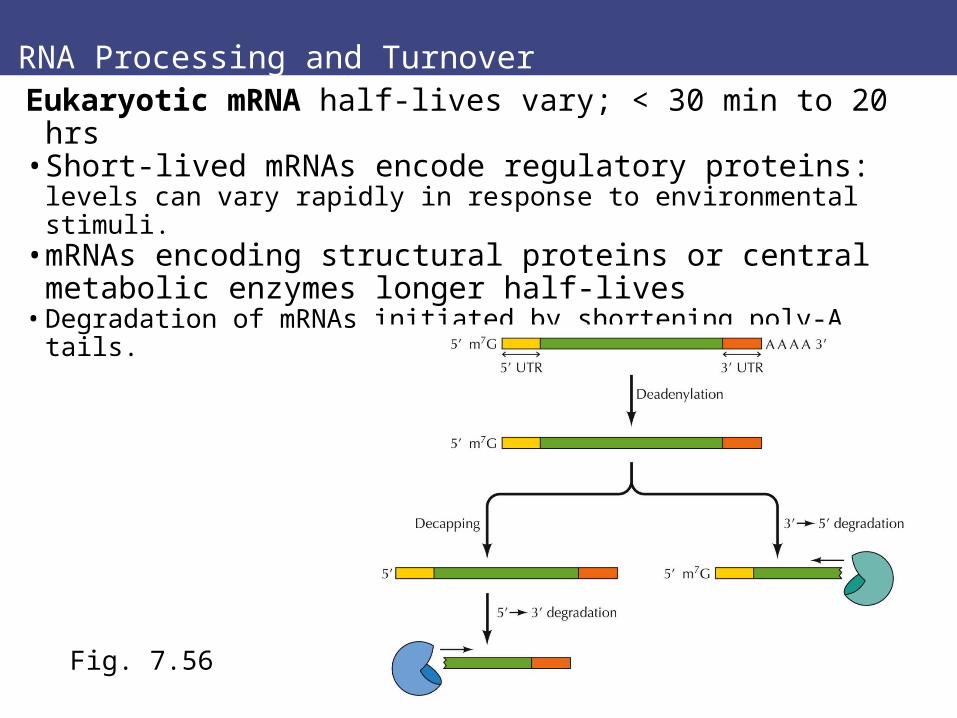
RNA Processing and Turnover
Eukaryotic mRNA half-lives vary; < 30 min to 20 hrs• Short-lived mRNAs encode regulatory proteins: levels
can vary rapidly in response to environmental stimuli.• mRNAs encoding structural proteins or central
metabolic enzymes longer half-lives• Degradation of mRNAs initiated by shortening poly-A tails.
Fig. 7.56
Review questions
1.How does footprinting identify protein binding sites?
6.How do enhancers differ from promoters as cis-acting regulatory sites
14. How are 2 structurally and functionally different forms of apolipoprotein B synthesized in liver and intestine?
15. What is nonsense-mediated mRNA decay and its significance?