Embed Size (px)
Citation preview
684
ASTROBIOLOGYVolume 7, Number 4, 2007© Mary Ann Liebert, Inc.DOI: 10.1089/ast.2006.0098
Special Paper
Ultrastructural and Geochemical Characterization ofArchean–Paleoproterozoic Graphite Particles:
Implications for Recognizing Traces of Life in HighlyMetamorphosed Rocks
JAMES D. SCHIFFBAUER,1 LEIMING YIN,2 ROBERT J. BODNAR,1 ALAN J. KAUFMAN,3FANWEI MENG,2 JIE HU,2 BING SHEN,1 XUNLAI YUAN,4 HUIMING BAO,5
and SHUHAI XIAO1
ABSTRACT
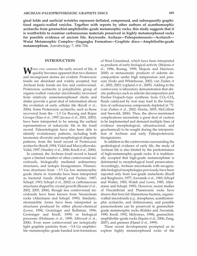
Abundant graphite particles occur in amphibolite-grade quartzite of the Archean–Paleopro-terozoic Wutai Metamorphic Complex in the Wutaishan area of North China. Petrographicthin section observations suggest that the graphite particles occur within and betweenquartzite clasts and are heterogeneous in origin. Using HF maceration techniques, the Wutaigraphite particles were extracted for further investigation. Laser Raman spectroscopic analy-sis of a population of extracted graphite discs indicated that they experienced a maximummetamorphic temperature of 513 � 50°C, which is consistent with the metamorphic grade ofthe host rock and supports their indigenicity. Scanning and transmission electron microscopyrevealed that the particles bear morphological features (such as hexagonal sheets of graphitecrystals) related to metamorphism and crystal growth, but a small fraction of them (graphitediscs) are characterized by a circular morphology, distinct marginal concentric folds, surficialwrinkles, and complex nanostructures. Ion microprobe analysis of individual graphite discsshowed that their carbon isotope compositions range from �7.4‰ to �35.9‰ V-PDB (ViennaPee Dee Belemnite), with an average of �20.3‰, which is comparable to bulk analysis of ex-tracted carbonaceous material.
The range of their size, ultrastructures, and isotopic signatures suggests that the morphol-ogy and geochemistry of the Wutai graphite discs were overprinted by metamorphism andtheir ultimate carbon source probably had diverse origins that included abiotic processes. Weconsidered both biotic and abiotic origins of the carbon source and graphite disc morpholo-gies and cannot falsify the possibility that some circular graphite discs characterized by mar-
1Department of Geosciences, Virginia Polytechnic Institute and State University, Blacksburg, Virginia.2Nanjing Institute of Geology and Paleontology, Nanjing, China.3Department of Geology, University of Maryland, College Park, Maryland.4State Key Laboratory of Palaeontology and Stratigraphy, Nanjing Institute of Geology and Palaeontology, Chi-
nese Academy of Sciences, Nanjing, China.5Department of Geology and Geophysics, Louisiana State University, Baton Rouge, Louisiana.
ARCHEAN–PALEOPROTEROZOIC GRAPHITE DISCS 685
INTRODUCTION
WHEN ONE ASSESSES the early record of life, itquickly becomes apparent that two distinct
and incongruent stories are evident: Proterozoicfossils are abundant and widely accepted, butArchean body fossils are few and controversial.Proterozoic acritarchs (a polyphyletic group oforganic-walled vesicular microfossils) recoveredfrom relatively unmetamorphosed cherts andshales provide a great deal of information aboutthe evolution of early cellular life (Knoll et al.,2006). Some Proterozoic acritarchs, such as thoserecovered from the �1.5 Ga Ruyang and RoperGroups (Xiao et al., 1997; Javaux et al., 2001, 2003),have been interpreted to be among the earliestrepresentatives of eukaryotic life in the fossilrecord. Paleontologists have also been able toidentify evolutionary patterns, including bothtaxonomic diversity and morphological disparitypatterns, from the fossil record of Proterozoicacritarchs (Knoll, 1994; Vidal and Moczydlowska-Vidal, 1997; Huntley et al., 2006; Knoll et al., 2006).
In contrast, the Archean fossil record is basedupon a limited number of often controversial mi-crofossils, biologically mediated sedimentarystructures, and isotopic biosignatures. Filamen-tous structures from �3.5 Ga low metamorphicgrade cherts in Australia have been interpretedas bacterial fossils (Schopf and Packer, 1987;Schopf, 1993; Schopf et al., 2002) or carbonaceousstructures shaped by crystal growth (Brasier et al.,2002, 2005, 2006), though less controversial mi-crofossils have been known from Neoarcheanrocks (Altermann and Schopf, 1995). Similarly,stromatolitic forms have been interpreted asstructures produced by either physio-chemical(Lowe, 1994; Grotzinger and Rothman, 1996;Grotzinger and Knoll, 1999) or biologicalprocesses (Hofmann et al., 1999; Allwood et al.,2006). Even more controversial are isotopicallylight graphite particles from �3.8 Ga amphibo-lite–metamorphic–grade banded iron-formations
of West Greenland, which have been interpretedas products of early biological activity (Mojzsis etal., 1996; Rosing, 1999; Mojzsis and Harrison,2000) or metasomatic products of siderite de-composition under high temperature and pres-sure (Fedo and Whitehouse, 2002; van Zuilen etal., 2002, 2003; Lepland et al., 2005). Adding to thecontroversy is laboratory demonstration that abi-otic pathways such as siderite decomposition andFischer-Tropsch–type synthesis from CO2–CH4fluids catalyzed by iron may lead to the forma-tion of carbonaceous compounds depleted in 13C(van Zuilen et al., 2002; Horita, 2005; McCollomand Seewald, 2006). Thus, the above-mentionedcomplications necessitate a great deal of cautionto be implemented and demand multiple lines ofevidence (morphological, ultrastructural, andgeochemical) to be sought during the interpreta-tion of Archean and early Paleoproterozoicbiosignatures.
In addition to the controversies surrounding thegeobiological evidence of early life, the study ofArchean life is also limited by the predominanceof high-metamorphic–grade rocks. It is tradition-ally accepted that high-grade metamorphism isdetrimental to morphological fossil preservation.Accordingly, Archean microfossils with recogniz-able biological morphologies previously have beenreported only from low-grade metacherts (Knolland Barghoorn, 1977; Awramik et al., 1983; Schopfand Walter, 1983; Walsh and Lowe, 1985; Alter-mann and Schopf, 1995). However, recent studiesof Precambrian and Phanerozoic rocks haveshown that bona fide filamentous bacteria, organic-walled microfossils (e.g., leiospheres, acanthomor-phic acritarchs, and chitinozoans), and possibleparaconodonts can be preserved in greenschist-grade metamorphic rocks (Kidder and Awramik,1990; Knoll, 1992; Molyneux, 1998), greenschist-amphibolite–grade rocks (Squire et al., 2006; Zang,2007), and gneisses (Hanel et al., 1999).
These recent developments prompted us toexplore highly metamorphosed rocks of the
ginal folds and surficial wrinkles represent deflated, compressed, and subsequently graphi-tized organic-walled vesicles. Together with reports by other authors of acanthomorphicacritarchs from greenschist-amphibolite–grade metamorphic rocks, this study suggests that itis worthwhile to examine carbonaceous materials preserved in highly metamorphosed rocksfor possible evidence of ancient life. Keywords: Archean—Paleoproterozoic—Acritarch—Wutai Metamorphic Complex—Jingangku Formation—Graphite discs—Amphibolite-grademetamorphism. Astrobiology 7, 684–704.
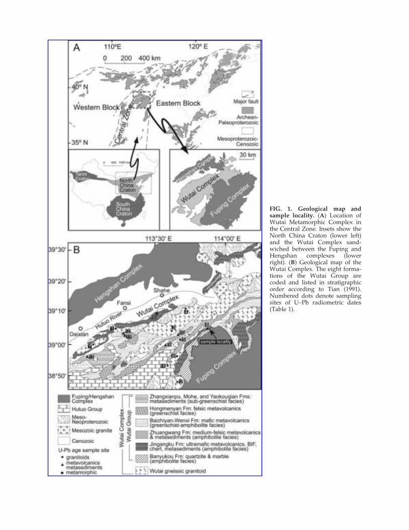
FIG. 1. Geological map andsample locality. (A) Location ofWutai Metamorphic Complex inthe Central Zone. Insets show theNorth China Craton (lower left)and the Wutai Complex sand-wiched between the Fuping andHengshan complexes (lowerright). (B) Geological map of theWutai Complex. The eight forma-tions of the Wutai Group arecoded and listed in stratigraphicorder according to Tian (1991).Numbered dots denote samplingsites of U–Pb radiometric dates(Table 1).
Archean–early Paleoproterozoic Wutai Metamor-phic Complex in the Wutaishan area of NorthChina, using a combination of light microscopy,electron microscopy, Raman spectroscopy, andion microprobe techniques. The immediate goalof this study was to characterize the morpholog-ical, ultrastructural, and geochemical features ofthe carbonaceous material recovered from theWutai Metamorphic Complex. Our study re-sulted in the recovery of abundant graphite par-ticles from amphibolite-grade quartzites of theWutai Metamorphic Complex in the Wutaishanarea of North China. Raman spectroscopy indi-cated that these graphite particles are indigenousto the host rock. Most graphite particles are ir-regular in shape and show hexagonal brokenedges, but a distinct population of graphite par-ticles can be characterized as circular discs withmarginal folds and surficial wrinkles. Althoughthe morphology of these graphite discs must have been overprinted by fragmentation, crystalgrowth, and other abiotic processes, the presenceof marginal folds and surficial wrinkles on circu-lar discs seems to suggest that they may repre-sent deflated, compressed, and subsequentlygraphitized organic-walled vesicles. Transmis-sion electron microscopy shows that they appearto consist of two graphitized layers separated byan electron-dense material trapped in between.Isotopic analysis of bulk carbonaceous materialgave a carbon isotope value of �21.3‰ V-PDB,but ion probe analysis of individual graphiteparticles gave a range of carbon isotope valuesbetween �7.4‰ and �35.9‰ V-PDB, with an av-erage of �20.3‰. At the present, the morpho-logical, ultrastructural, and geochemical evidencefor a biological origin is still equivocal, but thisstudy represents a first attempt to characterizecarbonaceous material from highly metamor-phosed Archean–early Paleoproterozoic rocks us-ing a combination of analytical tools.
GEOLOGICAL SETTING
The Wutai Metamorphic Complex is locatedbetween the Fuping and Hengshan metamorphiccomplexes, which together compose the middlesegment of the Trans-North China Orogen andconstitute the Hengshan-Wutai-Fuping moun-tain belt. The Trans-North China Orogen is a nar-row northeast-southwest zone, also known as theCentral Zone, which separates the eastern and
western blocks of the North China Craton (Fig.1A). Three different tectonic models have beenproposed for the development and evolution ofthe Hengshan-Wutai-Fuping mountain belt. Thefirst suggests that the Fuping and Hengshan com-plexes were part of a single continental block, andthe Wutai complex was formed in a late Archeanrift basin between the Fuping and Hengshancomplexes (Tian, 1991; Yuan and Zhang, 1993).The second model suggests that the Hengshan-Wutai-Fuping mountain belt represents a lateArchean continent-arc-continent collision system,with the Hengshan and Fuping complexes corre-sponding to two Archean continental blocks, andthe Wutai complex representing the trapped in-termediate island arc (Bai, 1986; Li et al., 1990; Baiet al., 1992; Wang et al., 1996). As a third model,recent data suggest that these complexes repre-sent a single late Archean to early Paleoprotero-zoic magmatic arc that underwent deformation,metamorphism, and exhumation, and was sub-sequently incorporated into the Trans-NorthChina Orogen in the development of the NorthChina Craton (Zhao et al., 2004).
The Wutai Metamorphic Complex is a green-stone terrane that consists of tonalitic-trond-hjemitic-granodioritic gneisses, granitoids, maficto felsic volcanic rocks, and metamorphosed vol-canic-sedimentary rocks (Polat et al., 2005). On thebasis of metamorphic facies and geological map-ping, the volcanic-sedimentary package of theWutai Metamorphic Complex has been classifiedas the Wutai Group (Fig. 1B), which is subdividedinto eight formations (Tian, 1991). The lower Wu-tai Group consists mainly of amphibolite, or-thogneiss, and metasedimentary rocks, includingbanded iron-formation and quartzite metamor-phosed to lower amphibolite facies. Petrographicstudy indicates that the lower Wutai Group washeated to a maximum temperature of 600–650°Cand buried to a maximum pressure of 10–12 kbar(Zhao et al., 1999). The middle Wutai Group iscomposed of greenschist facies tholeiites and fel-sic volcanics. The upper Wutai Group consists ofgreenschist to subgreenschist facies metasedi-ments and metavolcanics. The Wutai Metamor-phic Complex is unconformably overlain by theHutuo Group, which is comprised of subgreen-schist facies metasediments and minor maficvolcanics, including quartzites, slates, and meta-basalts.
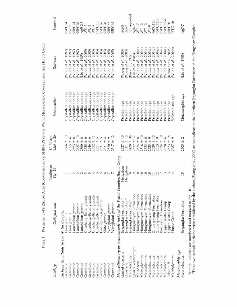
Recent geochronological data from SHRIMP(Sensitive High Resolution Ion Microprobe) U–Pb
ARCHEAN–PALEOPROTEROZOIC GRAPHITE DISCS 687
zircon analyses of samples from the lower, mid-dle, and upper subgroups of the Wutai Group(Wilde et al., 2004a), however, suggest that theseunits are roughly of similar age. For example,Wilde et al. (2004a) reported eight SHRIMP zir-con U–Pb ages from intermediate to felsic vol-canic rocks of the lower, middle, and upper sub-groups. These eight ages range from 2533 � 8 to2513 � 8 Ma, and together they give a weightedmean of 2523 � 3 Ma. These ages do not supportcorrelation between age and metamorphic gradeof the three subgroups as had been previouslysuggested (Bai, 1986; Tian, 1991). Therefore, meta-morphic grade cannot be used as a stratigraphicindicator within the Wutai Group (Wilde et al.,2004a), and the geochronological data cast doubton the superposition relationship of the unitswithin the Wutai Group (Bai, 1986; Tian, 1991).
Regardless of the controversy that surroundsthe stratigraphic relationship of the Wutai Group,there is ample geochronological data to suggestthat the Wutai Complex and Hutuo Group areArchean–early Paleoproterozoic in age (Table 1).The Wutai Group is penetrated by gneissic gran-itoids and granitoids that were emplaced pre-,syn-, and post-greenstone metamorphism (Tian,1991; Wilde et al., 2005). Extensive radiometricdating shows three episodes of granite intrusion,during 2560–2540 Ma, 2540–2515 Ma, and 2170–2120 Ma; the 2540–2515 Ma granitoids were in-terpreted as coeval with felsic volcanism in theWutai Complex (Wilde et al., 2005). Metavol-canics in the Wutai Group range from 2533 � 8Ma to 2513 � 8 Ma (Wilde et al., 2004a), whichfurther confirms an Archean age for the WutaiGroup. A volcanic ash from the overlying HutuoGroup gave a SHRIMP U–Pb zircon age of 2087 �9 Ma (Wilde et al., 2004b). Therefore, the age ofthe Wutai Metamorphic Complex is conserva-tively constrained from late Archean to early Pa-leoproterozoic.
Our samples were collected from a 10–30 cmthick bed of carbonaceous quartzite (Fig. 2C) in ageological unit mapped as the Jingangku For-mation of the Wutai Group, near the village ofShentangpu (39°07.386�N, 113°55.223�E; Fig. 2A).The Jingangku Formation consists of ultramaficvolcanics, amphibolite, iron-formation (Fig. 2B),volcanogenic massive sulfide deposit, and meta-sedimentary rocks, including micaschist, calc-sil-icate, and quartzite (Tian, 1991; Polat et al., 2005).The metavolcanics are interpreted as “remnantsof oceanic crust,” whereas the metasedimentary
rocks as “stable continental margin sediments”(Polat et al., 2005).
Some geologists (Wang et al., 2000) argue that thegeological unit mapped as the Jingangku Forma-tion may entirely or partly belong to the HutuoGroup. Because the Wutai Complex can interfingerwith the younger, less severely metamorphosedHutuo Group in the Wutaishan area (Wilde et al.,2004b), extreme care has been taken to ensure thatour collected samples belong to the Jingangku For-mation of the Wutai Complex. Our sampling lo-cality is ca. 25 km northeast of the Wutai-Hutuo in-terfingering zone (Wilde et al., 2004b). In addition,our sample locale was in close association with am-phibolite and banded iron-formation (Fig. 2B),which are characteristic lithologies of the Jin-gangku Formation of the Wutai Complex but notcharacteristic of the Hutuo Group. Furthermore,Raman geothermometery of isolated graphite andpetrographic analysis of our samples show thatthey are more akin to the amphibolite-grade meta-morphism of the Wutai Complex, but inconsistentwith the greenschist-grade metamorphism of theHutuo Group in eastern Wutaishan area (Tian,1991), where our samples were collected. More im-portantly, samples from the Jingangku Formationnear our sampling locality yielded a 2508 � 2 MaU–Pb age that is interpreted to date the peak meta-morphism of the Jingangku Formation (Table 1)(Liu et al., 1985). Pyrite from the Jingangku For-mation also yields 33S anomalies (Ding et al., 2004),which are consistent with its Archean age because33S anomalies are not known in geological samplesyounger than �2.4 Ga (Bekker et al., 2004). There-fore, our samples likely belong to the JingangkuFormation of the Wutai Complex, rather than toany stratigraphic units of the overlying HutuoGroup. Regardless, the conservative age estimate(Archean–Paleoproterozoic) of our samples standseven if they belong to the Hutuo Group (2087 � 9Ma; Wilde et al., 2004b).
METHODS
Standard petrographic thin sections were madefrom the Jingangku carbonaceous quartzite sam-ples and examined under a microscope usingboth plain and polarized light. In thin sections,quartzite clasts are set in a fine-grained carbona-ceous (primarily graphitic) matrix. Graphite par-ticles occur in both the matrix and clasts (Fig.3A–H), which confirms their indigenicity.
SCHIFFBAUER ET AL.688
TA
BL
E1.
PUB
LIS
HE
DU
–PB
ZIR
CO
NA
GE
S(C
ON
VE
NT
ION
AL
OR
SHR
IMP)
OF
TH
EW
UT
AI
ME
TA
MO
RP
HIC
CO
MP
LE
XA
ND
TH
EH
UT
UO
GR
OU
P
Loca
lity
inU
–Pb
age
Lith
olog
yG
eolo
gica
l un
itFi
g. 1
B(M
a) �
2�In
terp
reta
tion
Ref
eren
ceSa
mpl
e #
Arc
hean
Gra
nito
ids
in t
he W
utai
Com
plex
Gra
nito
idE
kou
gran
ite
125
66�
13
Cry
stal
lizat
ion
age
(Wild
e et
al.,
199
7)95
PC34
Gra
nito
idE
kou
gran
ite
125
55�
6C
ryst
alliz
atio
n ag
e(W
ilde
et a
l., 1
997)
95-1
9G
rani
toid
Lan
zhis
han
gran
ite
225
53�
8C
ryst
alliz
atio
n ag
e(W
ilde
et a
l., 1
997)
95PC
94G
rani
toid
Lan
zhis
han
gran
ite
225
37�
10
Cry
stal
lizat
ion
age
(Wild
e et
al.,
199
7)95
PC96
Gra
nito
idL
anzh
isha
n gr
anit
e2
2560
� 6
Cry
stal
lizat
ion
age
(Liu
et
al.,
1985
)A
g5-2
,5G
rani
toid
Che
chan
g-B
eita
i gr
anit
e3
2538
� 6
Cry
stal
lizat
ion
age
(Wild
e et
al.,
200
5)W
C-5
Gra
nito
idC
hech
ang-
Bei
tai
gran
ite
325
46�
6C
ryst
alliz
atio
n ag
e(W
ilde
et a
l., 2
005)
WC
-6G
rani
toid
Che
chan
g-B
eita
i gr
anit
e4
2552
� 1
1C
ryst
alliz
atio
n ag
e(W
ilde
et a
l., 2
005)
WC
-7G
rani
toid
Che
chan
g-B
eita
i gr
anit
e3
2551
� 5
Cry
stal
lizat
ion
age
(Wild
e et
al.,
200
5)95
PC6B
Gra
nito
idG
uang
min
gsi
gran
ite
525
31�
5C
ryst
alliz
atio
n ag
e(W
ilde
et a
l., 2
005)
95PC
76G
rani
toid
Shif
o gr
anit
e6
2531
� 4
Cry
stal
lizat
ion
age
(Wild
e et
al.,
200
5)95
PC98
Gra
nito
idW
angj
iahu
i gr
anit
e7
2520
� 9
Cry
stal
lizat
ion
age
(Wild
e et
al.,
200
5)95
PC62
Gra
nito
idW
angj
iahu
i gr
anit
e7
2517
� 1
2C
ryst
alliz
atio
n ag
e(W
ilde
et a
l., 2
005)
95PC
63
Met
ased
imen
tary
or
met
avol
cani
c ro
cks
of t
he W
utai
Com
plex
/Hut
uo G
roup
Gar
net
quar
tzit
eJi
ngan
gku
Form
atio
n*H
engs
han
2527
� 1
0Pr
otol
ith
age
(Wan
g et
al.,
200
0)H
G-5
Qua
rtzi
teJi
ngan
gku
Form
atio
n*H
engs
han
2501
� 1
5Pr
otol
ith
age
(Wan
g et
al.,
200
0)H
G-7
Met
avol
cani
csJi
ngan
gku
Form
atio
n8
2438
� 3
6Pr
otol
ith
age
(Bai
et
al.,
1992
)no
t re
port
edQ
uart
z ke
rato
phyr
eH
ongm
enya
n Fo
rmat
ion
925
22�
17
Prot
olit
h ag
e(L
iu e
t al
., 19
85)
Ag6
-2M
etad
acit
eH
ongm
enya
n Fo
rmat
ion
1025
24�
8Pr
otol
ith
age
(Wild
e et
al.,
200
4a)
WT
-17
Met
avol
cani
csH
ongm
enya
n Fo
rmat
ion
1025
16�
10
Prot
olit
h ag
e(W
ilde
et a
l., 2
004a
)W
T-1
2M
etav
olca
nics
Hon
gmen
yan
Form
atio
n10
2533
� 8
Prot
olit
h ag
e(W
ilde
et a
l., 2
004a
)W
T-1
3M
etav
olca
nics
Hon
gmen
yan
Form
atio
n10
2523
� 9
Prot
olit
h ag
e(W
ilde
et a
l., 2
004a
)W
T-9
Met
avol
cani
csZ
huan
gwan
g Fo
rmat
ion
1125
29�
10
Prot
olit
h ag
e(W
ilde
et a
l., 2
004a
)96
PC11
4M
etav
olca
nics
Zhu
angw
ang
Form
atio
n12
2513
� 8
Prot
olit
h ag
e(W
ilde
et a
l., 2
004a
)95
PC31
19M
etav
olca
nics
Bai
zhiy
an F
orm
atio
n12
2524
� 1
0Pr
otol
ith
age
(Wild
e et
al.,
200
4a)
95PC
3115
Met
avol
cani
csU
pper
Wut
ai G
roup
1325
28�
6Pr
otol
ith
age
(Wild
e et
al.,
200
4a)
95PC
355C
Fels
ic t
uff
Upp
er W
utai
Gro
up13
2523
� 1
8Pr
otol
ith
age
(Krö
ner
et a
l., 2
005)
95PC
30M
etab
olca
nics
Hut
uo G
roup
1420
87�
9V
olca
nic
ash
age
(Wild
e et
al.,
200
4b)
HT
G-1
0
Met
amor
phic
age
Met
avol
cani
csJi
ngan
gku
Form
atio
n15
2508
� 2
Met
amor
phic
age
(Liu
et
al.,
1985
)A
g7-7
Sam
ple
loca
litie
s ar
e nu
mbe
red
and
mar
ked
in F
ig. 1
B.
*The
se t
wo
sam
ple
hori
zons
wer
e co
nsid
ered
by
the
auth
ors
(Wan
g et
al.,
200
0) a
s eq
uiva
lent
s to
the
Nor
ther
n Ji
ngan
gku
Form
atio
n in
the
Hen
gsha
n C
ompl
ex.
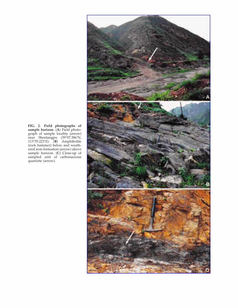
FIG. 2. Field photographs ofsample horizon. (A) Field photo-graph of sample locality (arrow)near Shentangpu (39°07.386’N,113°55.223’E). (B) Amphibolite(rock hammer) below and weath-ered iron-formation (arrow) abovesample horizon. (C) Close-up ofsampled unit of carbonaceousquartzite (arrow).
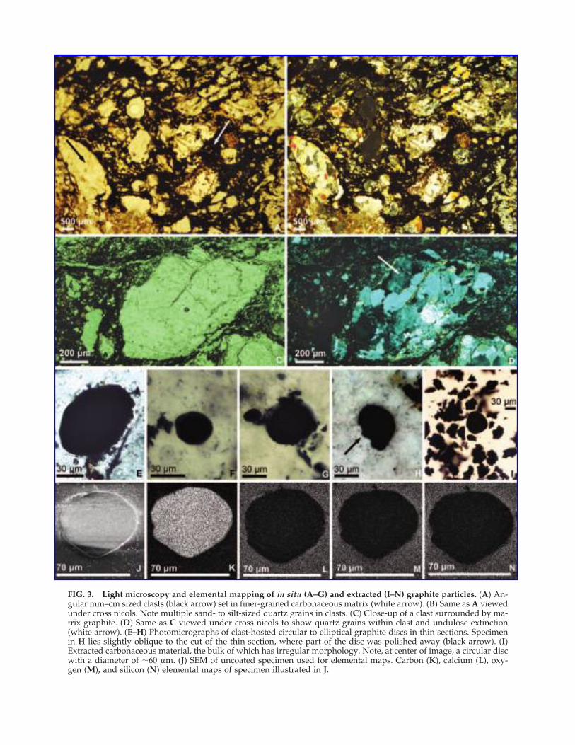
FIG. 3. Light microscopy and elemental mapping of in situ (A–G) and extracted (I–N) graphite particles. (A) An-gular mm–cm sized clasts (black arrow) set in finer-grained carbonaceous matrix (white arrow). (B) Same as A viewedunder cross nicols. Note multiple sand- to silt-sized quartz grains in clasts. (C) Close-up of a clast surrounded by ma-trix graphite. (D) Same as C viewed under cross nicols to show quartz grains within clast and undulose extinction(white arrow). (E–H) Photomicrographs of clast-hosted circular to elliptical graphite discs in thin sections. Specimenin H lies slightly oblique to the cut of the thin section, where part of the disc was polished away (black arrow). (I)Extracted carbonaceous material, the bulk of which has irregular morphology. Note, at center of image, a circular discwith a diameter of �60 �m. (J) SEM of uncoated specimen used for elemental maps. Carbon (K), calcium (L), oxy-gen (M), and silicon (N) elemental maps of specimen illustrated in J.
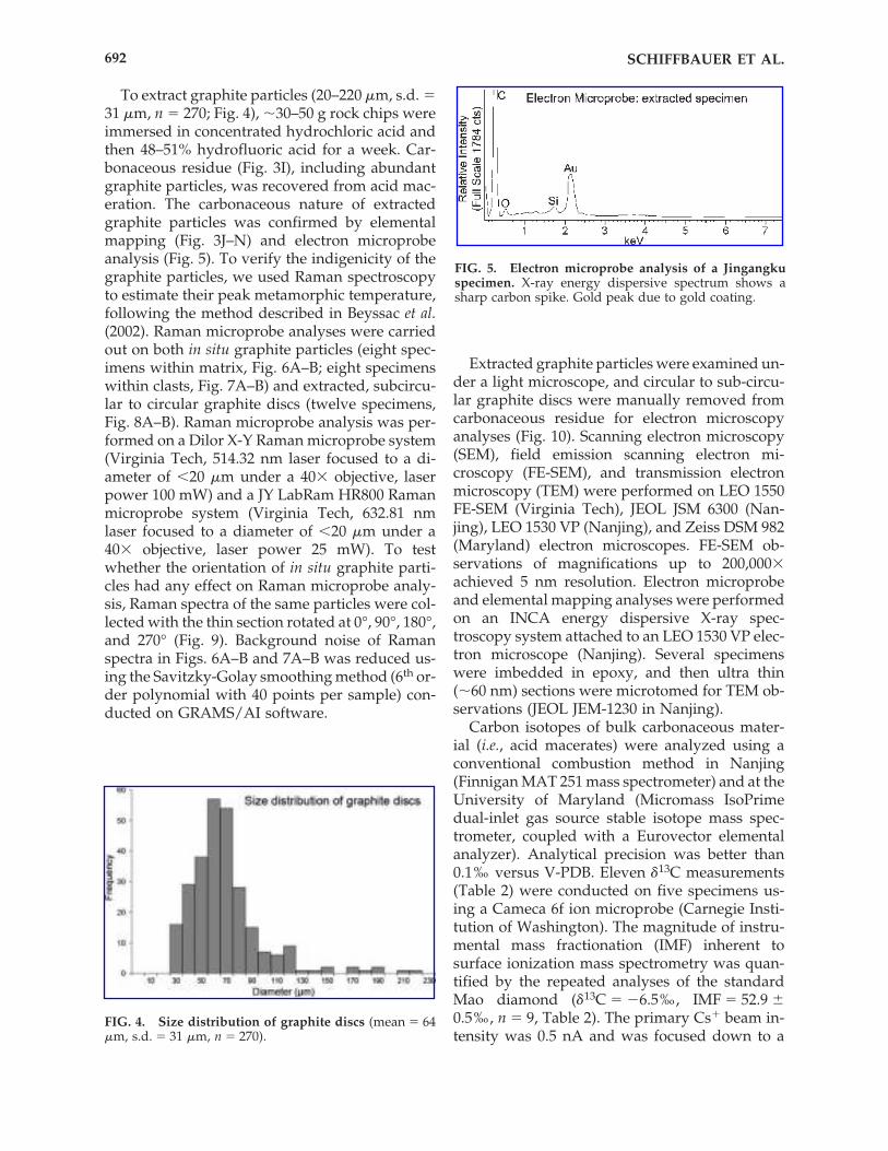
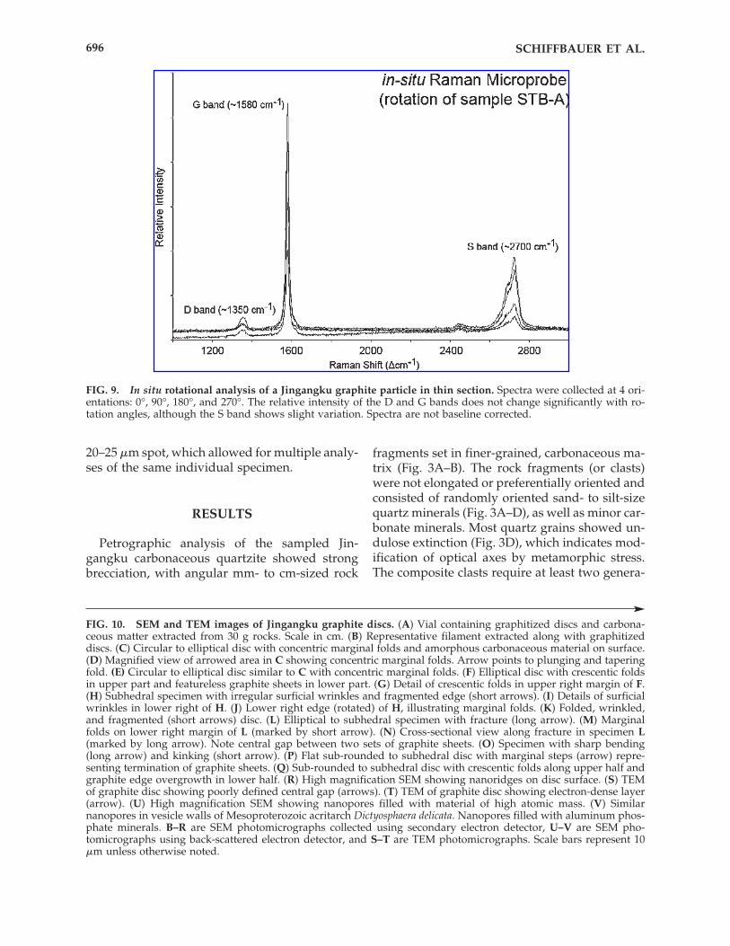
To extract graphite particles (20–220 �m, s.d. �31 �m, n � 270; Fig. 4), �30–50 g rock chips wereimmersed in concentrated hydrochloric acid andthen 48–51% hydrofluoric acid for a week. Car-bonaceous residue (Fig. 3I), including abundantgraphite particles, was recovered from acid mac-eration. The carbonaceous nature of extractedgraphite particles was confirmed by elementalmapping (Fig. 3J–N) and electron microprobeanalysis (Fig. 5). To verify the indigenicity of thegraphite particles, we used Raman spectroscopyto estimate their peak metamorphic temperature,following the method described in Beyssac et al.(2002). Raman microprobe analyses were carriedout on both in situ graphite particles (eight spec-imens within matrix, Fig. 6A–B; eight specimenswithin clasts, Fig. 7A–B) and extracted, subcircu-lar to circular graphite discs (twelve specimens,Fig. 8A–B). Raman microprobe analysis was per-formed on a Dilor X-Y Raman microprobe system(Virginia Tech, 514.32 nm laser focused to a di-ameter of �20 �m under a 40� objective, laserpower 100 mW) and a JY LabRam HR800 Ramanmicroprobe system (Virginia Tech, 632.81 nmlaser focused to a diameter of �20 �m under a40� objective, laser power 25 mW). To testwhether the orientation of in situ graphite parti-cles had any effect on Raman microprobe analy-sis, Raman spectra of the same particles were col-lected with the thin section rotated at 0°, 90°, 180°,and 270° (Fig. 9). Background noise of Ramanspectra in Figs. 6A–B and 7A–B was reduced us-ing the Savitzky-Golay smoothing method (6th or-der polynomial with 40 points per sample) con-ducted on GRAMS/AI software.
Extracted graphite particles were examined un-der a light microscope, and circular to sub-circu-lar graphite discs were manually removed fromcarbonaceous residue for electron microscopyanalyses (Fig. 10). Scanning electron microscopy(SEM), field emission scanning electron mi-croscopy (FE-SEM), and transmission electronmicroscopy (TEM) were performed on LEO 1550FE-SEM (Virginia Tech), JEOL JSM 6300 (Nan-jing), LEO 1530 VP (Nanjing), and Zeiss DSM 982(Maryland) electron microscopes. FE-SEM ob-servations of magnifications up to 200,000�achieved 5 nm resolution. Electron microprobeand elemental mapping analyses were performedon an INCA energy dispersive X-ray spec-troscopy system attached to an LEO 1530 VP elec-tron microscope (Nanjing). Several specimenswere imbedded in epoxy, and then ultra thin(�60 nm) sections were microtomed for TEM ob-servations (JEOL JEM-1230 in Nanjing).
Carbon isotopes of bulk carbonaceous mater-ial (i.e., acid macerates) were analyzed using aconventional combustion method in Nanjing(Finnigan MAT 251 mass spectrometer) and at theUniversity of Maryland (Micromass IsoPrimedual-inlet gas source stable isotope mass spec-trometer, coupled with a Eurovector elementalanalyzer). Analytical precision was better than0.1‰ versus V-PDB. Eleven �13C measurements(Table 2) were conducted on five specimens us-ing a Cameca 6f ion microprobe (Carnegie Insti-tution of Washington). The magnitude of instru-mental mass fractionation (IMF) inherent tosurface ionization mass spectrometry was quan-tified by the repeated analyses of the standardMao diamond (�13C � �6.5‰, IMF � 52.9 �0.5‰, n � 9, Table 2). The primary Cs� beam in-tensity was 0.5 nA and was focused down to a
FIG. 5. Electron microprobe analysis of a Jingangkuspecimen. X-ray energy dispersive spectrum shows asharp carbon spike. Gold peak due to gold coating.
FIG. 4. Size distribution of graphite discs (mean � 64�m, s.d. � 31 �m, n � 270).
SCHIFFBAUER ET AL.692
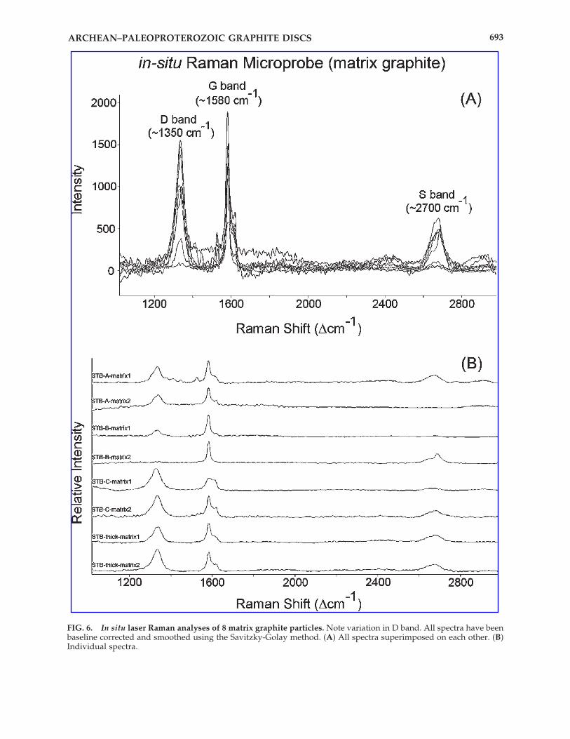
FIG. 6. In situ laser Raman analyses of 8 matrix graphite particles. Note variation in D band. All spectra have beenbaseline corrected and smoothed using the Savitzky-Golay method. (A) All spectra superimposed on each other. (B)Individual spectra.
ARCHEAN–PALEOPROTEROZOIC GRAPHITE DISCS 693
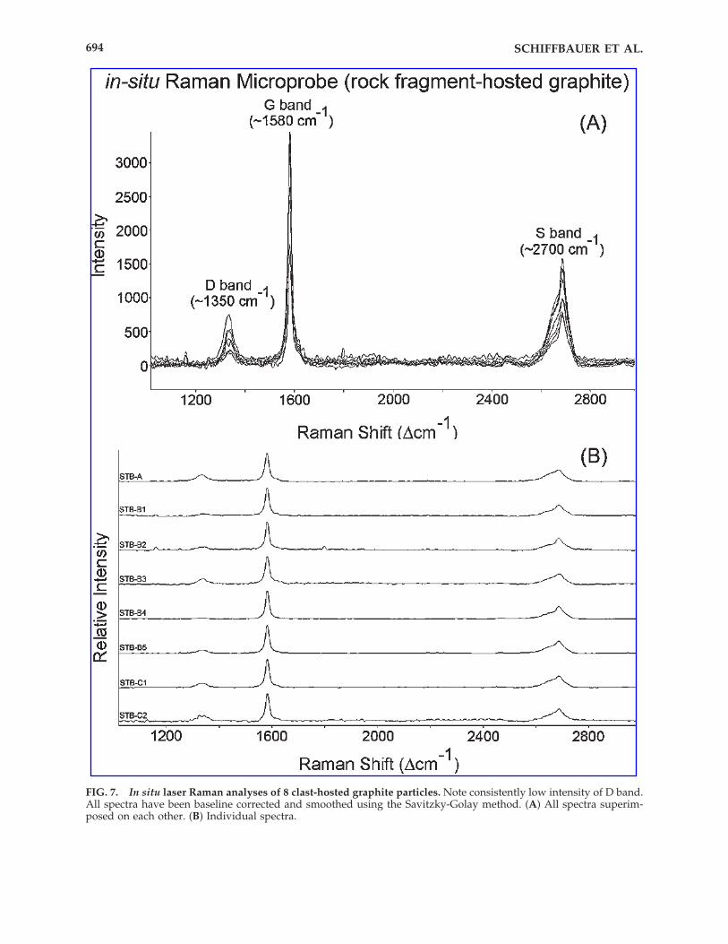
FIG. 7. In situ laser Raman analyses of 8 clast-hosted graphite particles. Note consistently low intensity of D band.All spectra have been baseline corrected and smoothed using the Savitzky-Golay method. (A) All spectra superim-posed on each other. (B) Individual spectra.
SCHIFFBAUER ET AL.694
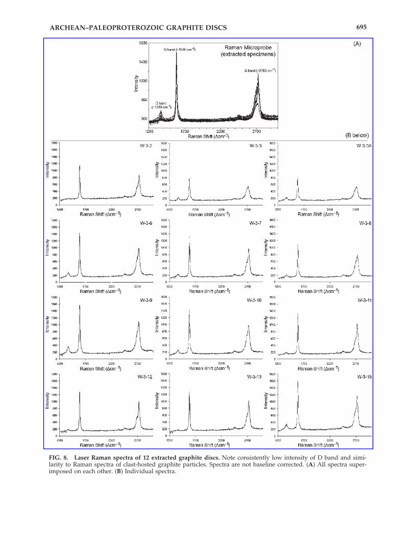
FIG. 8. Laser Raman spectra of 12 extracted graphite discs. Note consistently low intensity of D band and simi-larity to Raman spectra of clast-hosted graphite particles. Spectra are not baseline corrected. (A) All spectra super-imposed on each other. (B) Individual spectra.
ARCHEAN–PALEOPROTEROZOIC GRAPHITE DISCS 695
20–25 �m spot, which allowed for multiple analy-ses of the same individual specimen.
RESULTS
Petrographic analysis of the sampled Jin-gangku carbonaceous quartzite showed strongbrecciation, with angular mm- to cm-sized rock
fragments set in finer-grained, carbonaceous ma-trix (Fig. 3A–B). The rock fragments (or clasts)were not elongated or preferentially oriented andconsisted of randomly oriented sand- to silt-sizequartz minerals (Fig. 3A–D), as well as minor car-bonate minerals. Most quartz grains showed un-dulose extinction (Fig. 3D), which indicates mod-ification of optical axes by metamorphic stress.The composite clasts require at least two genera-
FIG. 9. In situ rotational analysis of a Jingangku graphite particle in thin section. Spectra were collected at 4 ori-entations: 0°, 90°, 180°, and 270°. The relative intensity of the D and G bands does not change significantly with ro-tation angles, although the S band shows slight variation. Spectra are not baseline corrected.
SCHIFFBAUER ET AL.696
FIG. 10. SEM and TEM images of Jingangku graphite discs. (A) Vial containing graphitized discs and carbona-ceous matter extracted from 30 g rocks. Scale in cm. (B) Representative filament extracted along with graphitizeddiscs. (C) Circular to elliptical disc with concentric marginal folds and amorphous carbonaceous material on surface.(D) Magnified view of arrowed area in C showing concentric marginal folds. Arrow points to plunging and taperingfold. (E) Circular to elliptical disc similar to C with concentric marginal folds. (F) Elliptical disc with crescentic foldsin upper part and featureless graphite sheets in lower part. (G) Detail of crescentic folds in upper right margin of F.(H) Subhedral specimen with irregular surficial wrinkles and fragmented edge (short arrows). (I) Details of surficialwrinkles in lower right of H. (J) Lower right edge (rotated) of H, illustrating marginal folds. (K) Folded, wrinkled,and fragmented (short arrows) disc. (L) Elliptical to subhedral specimen with fracture (long arrow). (M) Marginalfolds on lower right margin of L (marked by short arrow). (N) Cross-sectional view along fracture in specimen L(marked by long arrow). Note central gap between two sets of graphite sheets. (O) Specimen with sharp bending(long arrow) and kinking (short arrow). (P) Flat sub-rounded to subhedral disc with marginal steps (arrow) repre-senting termination of graphite sheets. (Q) Sub-rounded to subhedral disc with crescentic folds along upper half andgraphite edge overgrowth in lower half. (R) High magnification SEM showing nanoridges on disc surface. (S) TEMof graphite disc showing poorly defined central gap (arrows). (T) TEM of graphite disc showing electron-dense layer(arrow). (U) High magnification SEM showing nanopores filled with material of high atomic mass. (V) Similarnanopores in vesicle walls of Mesoproterozoic acritarch Dictyosphaera delicata. Nanopores filled with aluminum phos-phate minerals. B–R are SEM photomicrographs collected using secondary electron detector, U–V are SEM pho-tomicrographs using back-scattered electron detector, and S–T are TEM photomicrographs. Scale bars represent 10�m unless otherwise noted.
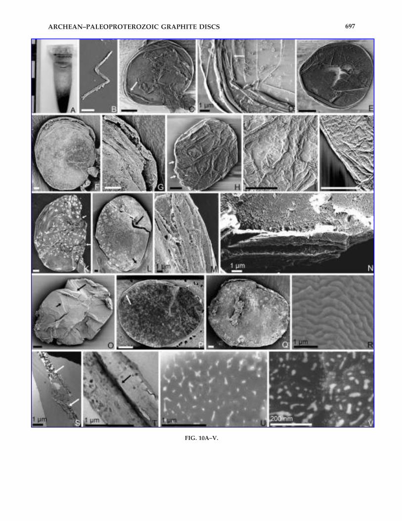
ARCHEAN–PALEOPROTEROZOIC GRAPHITE DISCS 697
FIG. 10A–V.
tions of fragmentation; the more recent fragmen-tation was probably tectonic brecciation becauseof the strong angularity, whereas the earlier frag-mentation appears to be sedimentary because ofthe moderate level of sorting. Heavy mineralanalysis is being conducted to test this interpre-tation.
Our samples are moderately carbonaceous (twomeasurements of 0.72% and 1.06% total organiccarbon (TOC), weight percentage determined bycombustion analyses of one randomly crushedsample, sample weights used for analysis �30 and�50 g). The carbonaceous material consistedmostly of graphite particles. Their indigenicityand graphite nature were verified by thin sectionpetrographic observations (Fig. 3A–H), elementalmapping (Fig. 3J–N), electron and Raman micro-probe analyses (Figs. 5–9), and scanning electronmicroscopy (Fig. 10C–R, 10U–V). Graphite parti-cles occur abundantly in the matrix between clasts(Fig. 3A–D), but less abundantly within clasts (Fig.3E–H). Some clast-hosted graphite particles arecircular to elliptical (Fig. 3E–H), but the morphol-ogy of matrix graphite is difficult to resolve un-der petrographic microscope because of the highconcentration and the opacity of carbonaceousmaterial in the matrix.
In situ Raman microprobe analyses of matrixgraphite showed highly variable spectra (eight
spectra collected, Fig. 6), often with a disorderedD band greater than or comparable to thegraphite G band. On the other hand, in situ Ra-man spectra of clast-hosted graphite (eight spec-tra collected, Fig. 7) were more consistent and il-lustrated a strong G band and a weak D band. Insitu samples were rotated 360°, with Raman spec-tra collected at each 90° interval; only minorchanges occured in the G- and D-band intensities,and slightly more so in S-band intensity (Fig. 9).Overall, all in situ Raman spectra had a relativelystrong G band, which suggests a high degree ofgraphite crystallinity (Wopenka and Pasteris,1993), though the differences between matrixgraphite and clast-hosted graphite may be in-dicative of their different origins.
Some graphite particles are circular (Fig.10C–U), and we term these particles graphite discs.Raman spectra of extracted graphite discs (Fig. 8)are highly consistent, comparable to those of clast-hosted graphite particles. Application of thegraphite Raman geothermometer (Wopenka andPasteris, 1993; Beyssac et al., 2002; Rahl et al., 2005)to these spectra suggests that these graphite discsexperienced peak metamorphic temperatures of513 � 50°C (n � 12), which is broadly consistentwith the amphibolite grade of the host rock, butslightly lower than the 600–650°C temperature es-timate based on mineral association (Zhao et al.,1999). Therefore, the carbonaceous precursors ofthese graphite discs were likely in place before orduring the amphibolite metamorphism.
When observed via scanning electron mi-croscopy, the extracted graphite particles aremostly irregularly shaped, with some discs (Fig.10C–Q) and rare filaments (Fig. 10B). The fila-ments, ca. 1.5 �m in width and tens of �m inlength, preserve no evidence for septation. Thediscs, which average about 60 �m in diameter(20–220 �m, s.d. � 31 �m, n � 270; Fig. 4) and 1–3�m in thickness (Fig. 10N, 10S–T), are circular,ovate, and slightly elliptical in morphology, andthey consist of graphite sheets (Fig. 10N–P). Thesediscs and filaments are broadly similar in mor-phology to the acritarchs and filaments from theoverlying Paleoproterozoic Hutuo Group in thesame geographic region (Sun and Zhu, 1998).However, the Hutuo discs and filaments arepoorly characterized; thus, at present, quantita-tive morphological comparisons cannot be ascer-tained.
Many specimens bear concentric (Fig. 10C–E)or crescentic (Fig. 10F–G, 10Q) marginal folds,
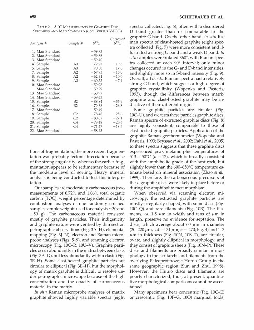
TABLE 2. �13C MEASUREMENTS OF GRAPHITE DISC
SPECIMENS AND MAO STANDARD (6.5% VERSUS V-PDB)
CorrectedAnalysis # Sample # �13C �13C
1. Mao Standard �59.832. Mao Standard �59.883. Mao Standard �59.404. Sample A3 �72.22 �19.35. Sample A3 �70.50 �17.67. Sample A2 �67.93 �15.08. Sample A2 �62.91 �10.09. Sample A2 �60.33 �7.4
10. Mao Standard �59.9811. Mao Standard �59.2913. Mao Standard �58.9714. Mao Standard �59.6315. Sample B2 �88.84 �35.916. Sample B2 �79.68 �26.817. Mao Standard �59.4918. Sample C2 �78.48 �25.619. Sample C2 �80.07 �27.120. Sample C4 �73.48 �20.621. Sample C4 �71.47 �18.522. Mao Standard �58.43
SCHIFFBAUER ET AL.698
ARCHEAN–PALEOPROTEROZOIC GRAPHITE DISCS 699
with isoclinal (Fig. 10F–G, 10L–M) or anticlinal(Fig. 10C–E) slopes. Some folds show plungingtermination into the surface of the disc (arrow inFig. 10D). Concentric and crescentic folds do notoccur in the center of these discs, which are ei-ther flat (Fig. 10C, 10E–F, 10L, 10P–Q) or coveredwith irregularly arranged, fine wrinkles (Fig.10H–J). The folds and wrinkles can be distin-guished, on the basis of electron shadows in SEMobservations using the secondary electron detec-tor, from steps and kinks resulting from termi-nation or dislocation of graphite sheets. At ex-tremely high magnification, the graphite discs arecharacterized by nanoscale (10–100 nm) ridgesand pores. The nanoridges bifurcate and anasto-mose (Fig. 10R), and their significance is obscure.The nanopores, when viewed with the backscat-ter detector, appear to be filled with an uniden-tified material that has an average atomic num-ber greater than graphite (Fig. 10U). Similarnanopores (Fig. 10V), though an order of magni-tude smaller in size, have been found in the vesi-cle walls of the Mesoproterozoic acritarch Dic-tyosphaera delicata, and are filled with aluminumphosphate minerals (Kaufman and Xiao, 2003).
Transmission electron microscopy showed thatsome discs appear to consist of two sets ofgraphite sheets, with a thin layer of electron-dense material between the sets (Fig. 10T). Thisis consistent with SEM observation of the natu-rally broken edge of a graphite disc (Fig. 10N),though in the latter specimen the two sets ofgraphite sheets are separated by a narrow gap.
Some morphological aspects of the graphitediscs—and most of the irregular graphite parti-cles—reflect graphite crystallization, overgrowth,inelastic deformation, and fragmentation. For ex-ample, the thickness of the discs may be variabledue to graphite overgrowth (Fig. 10Q). Thegraphite discs may become somewhat sub-rounded, subhedral, or angular (Fig. 10H, 10L,10P–Q), rather than curvilinear (Fig. 10C–G). Thetermination or dislocation of graphite sheets mayform steps (arrow in Fig. 10P). A few specimensshow very sharp bending (long arrow in Fig.10O), kinking (short arrow in Fig. 10O), fractur-ing (long arrow in Fig. 10L), and fragmentation(arrows in Fig. 10H, 10K). Some discs consist oftwo distinct sets of graphite sheets, separated bya gap of �1 �m (Fig. 10N); this gap may resultfrom physical separation along tabular cleavages.Features similar to these have been reported frommetamorphic graphites from Grenville marbles
and interpreted as overgrowths and deformationfeatures (Kretz, 1996).
Five extracted graphite discs were analyzed forcarbon isotopic signatures using a Cameca 6f ionmicroprobe. The results are intriguing but incon-clusive (Table 2). In most cases, multiple analyzedspots on the same individual disc showed lowvariability of �13C values (2–3‰, three speci-mens), but the other specimens demonstratedgreater intraspecimen variability (7–9‰, twospecimens). Additionally, the range of values be-tween different individuals spanned from �7.3 to�35.8‰ V-PDB. The mean �13C value of all ionprobe measurements (�20.3‰ V-PDB) is similarto the �21.3‰ V-PDB value determined by stan-dard techniques for bulk kerogen.
DISCUSSION
Graphite particles are common in Archean andyounger metamorphic rocks, such as marbles,schists, and gneisses (Rakovan and Jaszczak,2002; Ueno et al., 2002; van Zuilen et al., 2003;Satish-Kumar, 2005). Typically, metamorphicgraphite is found in a few varying forms: well-developed crystals (Palache, 1941), deformedcrystals (Kretz, 1996), crystals with microtopo-graphic growth spirals (Rakovan and Jaszczak,2002), crystals within graphite veins (along withmany other minerals including quartz, silliman-ite, ilmenite, and muscovite) (Rumble and Hoer-ing, 1986), and in spheroidal or spherule aggre-gates (Jaszczak, 1997).
Based on isotopic studies and Raman analysesin conjunction with high-resolution transmissionelectron microscopy, it has been suggested thatsyngenetic graphite can form within metasedi-mentary rocks during metamorphic heating fromthe progressive crystallization of organic carbonwithin precursor sediments, whereby the originalcarbonaceous material becomes ordered intocrystalline graphite (Weis et al., 1981; Wopenkaand Pasteris, 1993; Rantitsch et al., 2004). Alter-natively, some graphite forms are more likely tohave formed by carbon precipitation from car-bon-rich fluids (Rumble and Hoering, 1986;Satish-Kumar et al., 2001; Jaszczak and Rakovan,2002), by siderite decomposition (Fedo andWhitehouse, 2002; van Zuilen et al., 2002, 2003),or by Fischer-Tropsch precipitation from CO2–CH4 fluids (Horita, 2005; McCollom and Seewald,2006). Thus, the question arises whether the Jin-
gangku Formation graphite discs, characterizedby such morphological features as marginal con-centric folds, fine surficial wrinkles, and complexnanoridge and nanoporous structures, couldhave been morphologically shaped by metamor-phic processes. If the circular morphology andconcentric folds of these discs can be producedby metamorphism alone, then similar morpholo-gies should be observed in other metamorphicgraphite particles as well. However, to the best ofour knowledge, no other graphite crystals havebeen shown to have similar morphological fea-tures that so distinctively characterize the Jin-gangku graphite discs (Palache, 1941; Kretz, 1996;Jaszczak, 1997; Rakovan and Jaszczak, 2002; Uenoet al., 2002; van Zuilen et al., 2003; Satish-Kumar,2005). Instead, those graphite particles occur asinterstitial crystals or inclusions in the commonforms mentioned above, and their morphologiesare probably unrelated to biology, though theirultimate carbon source may or may not be bio-logical (Mojzsis et al., 1996; Fedo and Whitehouse,2002; van Zuilen et al., 2003). Thus, it seems asthough the morphology, micro-, and nano-scalestructures of the Jingangku graphite discs cannotbe accounted for by metamorphism alone.
The curvilinear margin of some Jingangkugraphite discs is also difficult to account for bymetamorphism alone. Abiotic precipitation ofgraphite is expected to produce hexagonal crys-tals. Such crystals may be deformed during meta-morphism, but circular discs with concentricfolds are not to be expected. If the Jingangkugraphite discs were derived from amorphouskerogen, graphite overgrowth during metamor-phism should reduce rather than enhance themarginal curvilinearity, and concentric folds arenot to be expected, either. One may argue thatgliding of graphite sheets in a directed stress en-vironment could seemingly form crescentic mar-ginal folds as illustrated in Fig. 10F–G, but thefolds should have much sharper crests (Kretz,1996; e.g., Fig. 10O) and the disc center shouldshow shearing in the same direction and magni-tude as the margin. Additionally, bending or glid-ing of graphite sheets is not expected to generateregular concentric marginal folds.
Our inability to account for all morphologiesof the Jingangku graphite discs by metamorphicprocesses alone compels us to consider other al-ternative interpretations. Is it possible that theJingangku discs and filaments are graphitized bi-ological structures? In this alternative interpreta-
tion, the filaments may represent filamentousbacteria, and the discs could represent originallyspheroidal vesicles with a recalcitrant organicwall, which were subsequently deflated, flat-tened, and graphitized during compaction, dia-genesis, and metamorphism. Their circular toovate shape, marginal concentric folds, and sur-ficial wrinkles are expected morphologies duringthe compression and elastic deformation of or-ganic-walled vesicles; these features are com-monly observed in compressed organic-walledmicrofossils in Proterozoic rocks (Schopf andKlein, 1992). Further, the nanoporous structuresare similar to those found in Mesoproterozoicacritarchs, i.e., Dictyosphaera delicata (Kaufmanand Xiao, 2003). In addition, the gap between twosets of graphite sheets in some specimens (Fig.10N), conservatively interpreted above as relatedto physical separation along tabular cleavages,may be alternatively interpreted as a gap betweentwo compressed vesicle walls. Moreover, TEMobservations show that some specimens have anelectron-dense central layer (Fig. 10T), rather thana gap; this layer cannot be interpreted as cleav-age separation but may represent materialtrapped within the compressed vesicle. Thus, wetentatively conclude that the graphite discs fromthe Jingangku Formation may represent deflated,flattened, and graphitized vesicles similar toacritarchs from younger rocks.
CONCLUSIONS
Key morphological features suggest biogenicity
Important features such as circular morphol-ogy, marginal folds, fine wrinkles, and trappedmaterials may have resulted from early diage-netic compression of spheroidal vesicles. Al-though these morphological features must havebeen altered during metamorphism and graphi-tization, hints of their presence may still bepreservable in a fraction of the Jingangkugraphite discs. Indeed, laboratory experimentsdemonstrate that Mesoproterozoic organic-walled microfossils (such as Dictyosphaera delicataand Shuiyousphaeridium macroreticulatum) heatedwithin the hosting rock to 500°C over periods ofup to 125 days show signs of graphitization,while retaining such features as discoidal mor-phology and concentric folds (Schiffbauer et al.,2006).
SCHIFFBAUER ET AL.700
ARCHEAN–PALEOPROTEROZOIC GRAPHITE DISCS 701
Geochemical evidence inconclusive but consistentwith biogenicity
A biological interpretation on the basis of mor-phological features described above implies thatthe ultimate carbon source of the Jingangkugraphite discs must be biological as well. Car-bonaceous material isolated from one of our sam-ples has a bulk �13C value of �21.3‰ V-PDB, asdetermined by conventional combustion meth-ods. Individual discs, measured using an ion mi-croprobe (Kaufman and Xiao, 2003), show a widerange of �13C values from �7.4‰ to �35.9‰ V-PDB (mean � �20.3‰; n � 11; Table 2). Three ofthe individually analyzed graphite discs showedlow variability (�3‰) of �13C values, but theother two specimens demonstrated much higherintraspecimen variability (7–9‰). These �13C val-ues are inconclusive, given what is known aboutcarbon isotope fractionations associated with abi-otic processes. From experimental analyses, mul-tiple abiotic pathways can lead to the synthesisof carbon compounds with �13C values as low as�60‰ due to kinetic isotope effects (Horita,2005); moreover, it has been suggested that car-bon compounds with �13C values as low as�30‰ (previously regarded as recycled sedi-mentary organic compounds) may be indigenousto the mantle (Horita, 2005). However, it is worthnoting that the Jingangku specimens have a muchwider �13C range and are more depleted in 13Cthan graphite particles found in granulite-grademetamorphic rocks (Farquhar et al., 1999; Santoshet al., 2003; Satish-Kumar, 2005), where isotopichomogenization is presumably stronger. Therange and heterogeneity of the Jingangku �13Cvalues may record partial isotopic exchange dur-ing metamorphism (Ueno et al., 2002), and thelowest measured values (e.g., �35.9‰) are betterapproximations of primary �13C values. If so,then the measured �13C values may be strongerevidence for biological source than they appear.
Jingangku graphite particles have complex origins
It needs to be stressed that, though some ir-regular graphite particles in the Jingangku sam-ples may be metamorphic or fragmentation prod-ucts from circular graphite discs, it is unlikely thatall of the graphite particles were derived fromcompressed vesicles. Indeed, carbonaceous ex-tracts from unmetamorphosed fossiliferous rocksof younger age mainly consist of amorphouskerogen, and only a small fraction is represented
by morphologically recognizable acritarchs. Noris the carbon source of all Jingangku graphite par-ticles biological. Petrographic evidence and Ra-man analyses discussed above suggest diverseorigins of Jingangku graphite particles. It appearsthat the extracted graphite discs may have comefrom the clasts because of (1) the in situ obser-vation that some graphite particles in clasts arecircular (Fig. 3E–H), (2) the similarity betweenRaman spectra of clast-hosted graphite and ex-tracted graphite discs (Figs. 7–8), and (3) the pos-sibility that quartzite clasts may provide a shieldagainst shearing deformation. However, this is-sue cannot be unambiguously resolved becauseof the limited resolution of in situ observationusing light microscopy and the high concentra-tion of matrix graphite. Thus, the Jingangkugraphite particles may have complex origins, andwe cannot conclusively disprove the possibilitythat both abiotic (e.g., decomposition of ferrouscarbonate minerals or Fischer–Tropsch precipita-tion from CO2–CH4 fluids catalyzed by Fe) andbiotic processes may have contributed to the car-bon source and morphology of the Jingangkugraphite particles.
Highly metamorphosed rocks could retain highlyaltered but morphologically and geochemicallyrecognizable signs of life
Despite the inconclusive nature of our inter-pretation, this study does suggest that, in oursearch for evidence of ancient life, more workshould be directed to carbonaceous material evenin highly metamorphosed rocks. Recent studiesof Precambrian and Phanerozoic metamorphicrocks have recovered bona fide filamentous bacte-ria, organic-walled microfossils (e.g., leiospheres,acanthomorphic acritarchs, and chitinozoans),and possible paraconodonts from greenschist-amphibolite-, and gneiss-grade metamorphicrocks (Kidder and Awramik, 1990; Knoll, 1992;Molyneux, 1998; Hanel et al., 1999; Squire et al.,2006; Zang, 2007). Thus, it is worthwhile to ex-plore whether the taphonomic window forArchean life is wider than previously thought.
ACKNOWLEDGMENTS
This research was supported by the NASAExobiology Program, National Science Founda-tion of China, Chinese Academy of Sciences, Chi-
nese Ministry of Science and Technology, StateKey Laboratory of Paleobiology and Stratigraphyof the Chinese Academy of Sciences, VirginiaTech ASPIRES and ICTAS programs, the VirginiaSpace Grant Consortium, the Paleontological So-ciety, and the Geological Society of America. Wethank Y. Tian for field assistance; W. Huang (mac-eration), C. Wang (TEM), C. Farley (laser Ramanmicroprobe), S. Mutchler (graphical assistance),E. Hauri and J. Wang (ion probe), and Y. Mao, J.Barry, and S.R.F. McCartney (SEM,Virginia TechInstitute for Critical Technologies and AppliedSciences) for technical help; and N. Butterfield, K.Grey, R. Law, R. Tracy, M. Kowalewski, and J.W.Huntley for discussion.
ABBREVIATIONS
FE-SEM, field emission scanning electron mi-croscopy; IMF, instrumental mass fractionation;SEM, scanning electron microscopy; SHRIMP,Sensitive High Resolution Ion Microprobe; TEM,transmission electron microscopy; TOC, total or-ganic carbon; V-PDB, Vienna Pee Dee Belemnite.
REFERENCES
Allwood, A.C., Walter, M.R., Kamber, B.S., Marshall, C.P.,and Burch, I.W. (2006) Stromatolite reef from the EarlyArchean era of Australia. Nature 441, 714–718.
Altermann, W. and Schopf, J.W. (1995) Microfossils fromthe Neoarchean Campbell Group, Griqualand West Se-quence of the Transvaal Supergroup, and their pale-oenvironmental and evolutionary implications. Pre-cambrian Res. 75, 65–90.
Awramik, S.M., Schopf, J.W., and Walter, M.R. (1983) Fil-amentous fossil bacteria from the Archean of WesternAustralia. Precambrian Res. 20, 357–374.
Bai, J. (1986) The Early Precambrian Geology of Wutaishan,Tianjin Science and Technology Press, Tianjin.
Bai, J., Wang, R.Z., and Guo, J.J. (1992) The Major GeologicEvents of Early Precambrian and Their Dating in Wutais-han Region, Geological Publishing House, Beijing.
Bekker, A., Holland, H.D., Wang, P.-L., Rumble III, D.,Stein, H.J., Hannah, J.L., Coetzee, L.L., and Beukes, N.J.(2004) Dating the rise of atmospheric oxygen. Nature427, 117–120.
Beyssac, O., Goffé, B., Chopin, C., and Rouzaud, J.N.(2002) Raman spectra of carbonaceous material inmetasediments: a new geothermometer. Journal of Meta-morphic Geology 20, 859–871.
Brasier, M., McLoughlin, N., Green, O., and Wacey, D.(2006) A fresh look at the fossil evidence for early Ar-
chaean cellular life. Philos. Trans. R. Soc. Lond., B, Biol.Sci. 361, 887–902.
Brasier, M.D., Green, O.R., Jephcoat, A.P., Kleppe, A.K.,Kranendonk, M.J.V., Lindsay, J.F., Steele, A., andGrassineau, N.V. (2002) Questioning the evidence forEarth’s oldest fossils. Nature 416, 76–81.
Brasier, M.D., Green, O.R., Lindsay, J.F., McLoughlin, N.,Steele, A., and Stoakes, C. (2005) Critical testing ofEarth’s oldest putative fossil assemblage from the 3.5Ga Apex chert, Chinaman Creek, Western Australia.Precambrian Res. 140, 55–102.
Ding, T., Wan, D., Zhang, Z., Wang, C., and Li, Y. (2004)Sulfur isotope anomaly discovered in sulfide bed oflater Archean Jingangku formation, Wutai group,Shanxi Province, China. Geochim. Cosmochim. Acta 68(Supplement 1), A789.
Farquhar, J., Hauri, E., and Wang, J. (1999) New insightsinto carbon fluid chemistry and graphite precipitation:SIMS analysis of granulite facies graphite from Pon-mudi, South India. Earth Planet. Sci. Lett. 171, 607–621.
Fedo, C.M. and Whitehouse, M.J. (2002) Metasomatic ori-gin of quartz-pyroxene rock, Akilia, Greenland, andimplications for Earth’s earliest life. Science 296, 1449–1452.
Grotzinger, J.P. and Knoll, A.H. (1999) Stromatolites inPrecambrian carbonates: evolutionary mileposts or en-vironmental dipsticks? Annu. Rev. Earth Planet. Sci. 27,313–358.
Grotzinger, J.P. and Rothman, D.H. (1996) An abioticmodel for stromatolite morphogenesis. Nature 383,423–425.
Hanel, M., Montenari, M., and Kalt, A. (1999) Determin-ing sedimentation ages of high-grade metamorphicgneisses by their palynological record: a case study inthe northern Schwarzwald (Variscan Belt, Germany).International Journal of Earth Sciences 88, 49–59.
Hofmann, H.J., Grey, K., Hickman, A.H., and Thorpe, R.I.(1999) Origin of 3.45 Ga coniform stromatolites in War-rawoona Group, Western Australia. Geol. Soc. Am. Bull.111, 1256–1262.
Horita, J. (2005) Some perspectives on isotope biosigna-tures for early life. Chem. Geol. 218, 171–186.
Huntley, J.W., Xiao, S., and Kowalewski, M. (2006) 1.3 bil-lion years of acritarch history: an empirical morpho-space approach. Precambrian Res. 144, 52–68.
Jaszczak, J.A. (1997) Unusual graphite crystals from theLime Crest quarry, Sparta, New Jersey. Rocks and Min-erals 72, 330–334.
Jaszczak, J.A. and Rakovan, J. (2002) Growth spirals ongraphite crystals from the Trotter Mine dump, Franklin,New Jersey. The Picking Table 43, 11–13.
Javaux, E.J., Knoll, A.H., and Walter, M.R. (2001) Mor-phological and ecological complexity in early eukary-otic ecosystems. Nature 412, 66–69.
Javaux, E.J., Knoll, A.H., and Walter, M.R. (2003) Recog-nizing and interpreting the fossils of early eukaryotes.Orig. Life Evol. Biosph. 33, 75–94.
Kaufman, A.J. and Xiao, S. (2003) High CO2 levels in theProterozoic atmosphere estimated from analyses of in-dividual microfossils. Nature 425, 279–282.
SCHIFFBAUER ET AL.702
ARCHEAN–PALEOPROTEROZOIC GRAPHITE DISCS 703
Kidder, D.L. and Awramik, S.M. (1990) Acritarchs inlower greenschist facies argillite of the middle Protero-zoic Libby Formation, Upper Belt Supergroup, Mon-tana. Palaios 5, 124–133.
Knoll, A.H. (1992) Microfossils in metasedimentary chertsof the Scotia Group, Prins Karls Forland, western Sval-bard. Palaeontology 35, 751–774.
Knoll, A.H. (1994) Proterozoic and Early Cambrian pro-tists: Evidence for accelerating evolutionary tempo.Proc. Natl. Acad. Sci. U.S.A. 91, 6743–6750.
Knoll, A.H. and Barghoorn, E.S. (1977) Archean micro-fossils showing cell-division from Swaziland System ofSouth Africa. Science 198, 396–398.
Knoll, A.H., Javaux, E.J., Hewitt, D., and Cohen, P. (2006)Eukaryotic organisms in Proterozoic oceans. Philos.Trans. R. Soc. Lond., B, Biol. Sci. 361, 1023–1038.
Kretz, R. (1996) Graphite deformation in marble and my-lonitic marble, Grenville Province, Canadian Shield.Journal of Metamorphic Geology 14, 399–412.
Kröner, A., Wilde, S.A., Li, J.H., and Wang, K.Y. (2005)Age and evolution of a late Archean to Paleoprotero-zoic upper to lower crustal section in the Wutais-han/Hengshan/Fuping terrain of northern China. Jour-nal of Asian Earth Sciences 24, 577–595.
Lepland, A., van Zuilen, M.A., Arrhenius, G., White-house, M.J., and Fedo, C.J. (2005) Questioning the evi-dence for Earth’s earliest life—Akilia revisited. Geology33(1), 77–79.
Li, J.L., Wang, K.Y., Wang, C.Q., Liu, X.H., and Zhao, Z.Y.(1990) Early Proterozoic collision orogenic belt in Wu-taishan area, China. Scientia Geologica Sinica 25, 1–11.
Liu, D.Y., Page, R.W., Compston, W., and Wu, J.S. (1985)U-Pb zircon geochronology of late Archean metamor-phic rocks in the Taihangshan-Wutaishan area, NorthChina. Precambrian Res. 27, 85–109.
Lowe, D.R. (1994) Abiotic origin of described stromato-lites older than 3.2 Ga. Geology 22, 387–390.
McCollom, T.M. and Seewald, J.S. (2006) Carbon isotopecomposition of organic compounds produced by abi-otic synthesis under hydrothermal conditions. EarthPlanet. Sci. Lett. 243, 74–84.
Mojzsis, S.J. and Harrison, T.M. (2000) Vestiges of a be-ginning: clues to the emergent biosphere recorded inthe oldest known sedimentary rocks. GSA Today 10, 1–6.
Mojzsis, S.J., Arrhenius, G., McKeegan, K.D., Harrison,T.M., Nutman, A.P., and Friend, C.R.L. (1996) Evidencefor life on Earth by 3800 million years ago. Nature 384,55–59.
Molyneux, S.G. (1998) An upper Dalradian microfossil re-assessed. J. Geol. Soc. London 155, 741–743.
Palache, C. (1941) Contributions to the mineralogy ofSterling Hill, New Jersey: Morphology of graphite,arsenopyrite, pyrite, and arsenic. Am. Mineral. 26, 709–717.
Polat, A., Kusky, T., Li, J., Fryer, B., Kerrich, R., andPatrick, K. (2005) Geochemistry of Neoarchean (ca.2.55–2.50 Ga) volcanic and ophiolitic rocks in the Wu-taishan greenstone belt, central orogenic belt, NorthChina craton: Implications for geodynamic setting andcontinental growth. GSA Bulletin 117, 1387–1399.
Rahl, J.M., Anderson, K.M., Brandon, M.T., and Fassoulas,C. (2005) Raman spectroscopic carbonaceous materialthermometry of low-grade metamorphic rocks: cali-bration and application to tectonic exhumation in Crete,Greece. Earth Planet. Sci. Lett. 240, 339–354.
Rakovan, J. and Jaszczak, J.A. (2002) Multiple length scalegrowth spirals on metamorphic graphite {001} surfacesstudied by atomic force microscopy. Am. Mineral. 87,17–24.
Rantitsch, G., Grogger, W., Teichert, C., Ebner, F., Hofer,C., Maurer, E., Schaffer, B., and Toth, M. (2004) Con-version of carbonaceous material to graphite within theGreywacke Zone of the Eastern Alps. International Jour-nal of Earth Sciences 93, 959–973.
Rosing, M.T. (1999) 13C-depleted carbon microparticles in3700-Ma sea-floor sedimentary rocks from WestGreenland. Science 283, 674–676.
Rumble, D. and Hoering, T.C. (1986) Carbon isotope geo-chemistry of graphite vein deposits from New Hamp-shire, U.S.A. Geochim. Cosmochim. Acta 50, 1239–1247.
Santosh, M., Wada, H., Satish-Kumar, M., and Binu-Lal,S.S. (2003) Carbon isotope “stratigraphy” in a singlegraphite crystal: implications for the crystal growthmechanism of fluid-deposited graphite. Am. Mineral.88, 1689–1696.
Satish-Kumar, M. (2005) Graphite-bearing CO2-fluid in-clusions in granulites: insights on graphite precipita-tion and carbon isotope evolution. Geochim. Cosmochim.Acta 69, 3841–3856.
Satish-Kumar, M., Wada, H., Santosh, M., and Yoshida,M. (2001) Fluid-rock history of granulite facies humite-marbles from Ambasamudram, southern India. Journalof Metamorphic Geology 19, 395–410.
Schiffbauer, J.D., Xiao, S., Bodnar, R.J., Yin, L., Yuan, X.,Meng, F., Hu, J., and Kaufman, A.J. (2006) Can organic-walled microfossils survive high metamorphic heating?Characterization of experimentally heated acritarchsusing Raman spectroscopy. [abstract]. GSA Abstractsand Programs 38(7), 305.
Schopf, J.W. (1993) Microfossils of the Early ArcheanApex chert: new evidence of the antiquity of life. Sci-ence 260, 640–646.
Schopf, J.W. and Klein, C. (1992) The Proterozoic Biosphere:A Multidisciplinary Study, Cambridge University Press,Cambridge.
Schopf, J.W., Kudryavtsev, A.B., Agresti, D.G., Wdowiak,T.J., and Czaja, A.D. (2002) Laser-Raman imagery ofEarth’s earliest fossils. Nature 416, 73–76.
Schopf, J.W. and Packer, B.M. (1987) Ealry Archean (3.3-billion- to 3.5-billion-year-old) microfossils from War-rawoona group, Australia. Science 237, 70–73.
Schopf, J.W. and Walter, M.R. (1983) Archean microfos-sils: new evidence of ancient microbes. In Earth’s Earli-est Biosphere: Its Origin and Evolution, edited by J.W.Schopf, Princeton University Press, Princeton, NJ, pp.214–239.
Squire, R.J., Stewart, I.R., and Zang, W.L. (2006)Acritarchs in polydeformed and highly altered Cam-brian rocks in western Victoria. Australian Journal ofEarth Sciences 53, 697–705.

Sun, S. and Zhu, S. (1998) The discovery of micropaleo-phytes from the Doucun Subgroup, Hutuo Group inWutai Mountain, Shanxi, China. Acta Micropalaeonto-logica Sinica 15, 286–293.
Tian, Y.Q. (1991) Geology and Gold Mineralization of Wu-tai–Hengshan Greenstone Belt. Shanxi Science and Tech-nology Press, Taiyuan.
Ueno, Y., Yurimoto, H., Yoshioka, H., Komiya, T., andMaruyama, S. (2002) Ion microprobe analysis ofgraphite from ca. 3.8 Ga metasediments, Isuasupracrustal belt, West Greenland: Relationship be-tween metamorphism and carbon isotopic composi-tion. Geochim. Cosmochim. Acta 66, 1257–1268.
van Zuilen, M.A., Lepland, A., and Arrhenius, G. (2002)Reassessing the evidence for the earliest traces of life.Nature 418, 627–630.
van Zuilen, M.A., Lepland, A., Teranes, J., Finarelli, J.,Wahlen, M., and Arrhenius, G. (2003) Graphite and car-bonates in the 3.8-Ga-old Isua Supracrustal Belt, south-ern West Greenland. Precambrian Res. 126, 331–348.
Vidal, G. and Moczydlowska-Vidal, M. (1997) Biodiver-sity, speciation, and extinction trends of Proterozoicand Cambrian phytoplankton. Paleobiology 23, 230–246.
Walsh, M.M. and Lowe, D.R. (1985) Filamentous micro-fossils from the 3,500-myr-old Onverwacht Group, Bar-berton Mountain Land, South Africa. Nature 314,530–532.
Wang, K.Y., Li, J.L., Hao, J., Li, J.H., and Zhou, S.P. (1996)The Wutaishan orogenic belt within the ShanxiProvince, northern China: a record of late Archaean col-lision tectonics. Precambrian Res. 78, 95–103.
Wang, K.Y., Hao, J., Wilde, S., and Cawood, P. (2000) Re-consideration of some key geological problems of lateArchean–early Proterozoic in the Wutaishan-Hengshanarea: constraints from SHRIMP U–Pb zircon data. Sci-entia Geologica Sinica 35, 175–184.
Weis, P.L., Friedman, I., and Gleason, J.P. (1981) The ori-gin of epigenetic graphite: evidence from isotopes.Geochim. Cosmochim. Acta 45, 2325–2332.
Wilde, S.A., Cawood, P., Wang, K.Y., and Nemchin, A.(1997) The relationship and timing of granitoid evolu-tion with respect to felsic volcanism in the Wutai Com-plex, North China Craton. Proceedings of the 30th Inter-national Geological Conference: Precambrian Geology andMetamorphic Petrology 17, 75–88.
Wilde, S.A., Cawood, P.A., Wang, K.Y., Nemchin, A., andZhao, G.C. (2004a) Determining Precambrian crustalevolution in China: a case-study from Wutaishan,Shanxi Province, demonstrating the application of pre-cise SHRIMP U–Pb geochronology. In Aspects of the Tec-
tonic Evolution of China, Special Publication 226, edited byJ. Malpas, C.J.N. Fletcher, J.R. Ali, and J.C. Aitchison,Geological Society of London, London, pp. 5–25.
Wilde, S.A., Zhao, G.C., Wang, K.Y., and Sun, M. (2004b)First SHRIMP zircon U–Pb ages for Hutuo Group inWutaishan: Further evidence for palaeoproterozoicamalgamation of North China Craton. Chin. Sci. Bull.49, 83–90.
Wilde, S.A., Cawood, P.A., Wang, K.Y., and Nemchin,A.A. (2005) Granitoid evolution in the Late ArchaeanWutai Complex, North China Craton. Journal of AsianEarth Sciences 24, 597–613.
Wopenka, B. and Pasteris, J.D. (1993) Structural charac-terization of kerogens to granulite-facies graphite: ap-plicability of Raman microprobe spectroscopy. Am.Mineral. 78, 533–557.
Xiao, S., Knoll, A.H., Kaufman, A.J., Yin, L., and Zhang,Y. (1997) Neoproterozoic fossils in Mesoproterozoicrocks? Chemostratigraphic resolution of a biostrati-graphic conundrum from the North China Platform.Precambrian Res. 84, 197–220.
Yuan, G.P. and Zhang, R.Y. (1993) The structural envi-ronment of the paleorift in Wutai greenstone belt.Shanxi Geology 8, 21–28.
Zang, W.-L. (2007) Deposition and deformation of late Ar-chaean sediments and preservation of microfossils inthe Harris Greenstone Domain, Gawler Craton, SouthAustralia. Precambrian Res. 156, 107–124.
Zhao, G., Cawood, P., and Lu, L. (1999) Petrology andP–T history of theWutai amphibolites: implications fortectonic evolution of the Wutai Complex, China. Pre-cambrian Res. 93, 181–199.
Zhao, G., Sun, M., Wilde, S.A., and Guo, J. (2004) LateArchean to Paleoproterozoic evolution of the Trans-North China Orogen: insights from synthesis of exist-ing data from the Hengshan-Wutai-Fuping belt. In As-pects of the Tectonic Evolution of China, Special Publication226. edited by J. Malpas, C.J.N. Fletcher, J.R. Ali, andJ.C. Aitchison, Geological Society of London, London,pp. 27–55.
Address reprint requests to:James D. Schiffbauer
4044 Derring HallDepartment of Geosciences
Virginia Polytechnic Institute and State UniversityBlacksburg VA 24061
E-mail: [email protected]
SCHIFFBAUER ET AL.704