Embed Size (px)
Citation preview
EL
SE
VIE
RJA
JS V
olu
me 3
NU
MB
ER 1
Janu
ary–A
pril 2016
PAG
ES 1–42
ISSN: 2214-9635
Journal of Arthroscopy AND Joint Surgery
Offi cial Journal of the International Society for Knowledge for Surgeons on Arthroscopy and Arthroplasty (ISKSAA)
Available online at www.sciencedirect.com
ScienceDirect
Volume 3 Number 1 January–April 2016
Indexed in Scopus
ISKSAA (International Society for Knowledge for Surgeons on Arthroscopy and Arthroplasty) is a society of orthopaedic surgeons from around the world to share and disseminate knowledge, support research and improve patient care in Arthroscopy and Arthroplasty. We are proud to announce that ISKSAA membership has crossed the 1200 mark (India & Overseas) making it the fastest growing Orthopaedic Association in the country in just over 3 years of its inception. With over 190000 hits from over 139 countries on the website www.isksaa.com & more and more interested people joining as members of ISKSAA, we do hope that ISKSAA will stand out as a major body to provide opportunities to our younger colleagues in training, education and fellowships.
Our Goals………
To provide health care education opportunities for increasing cognitive and psycho-motor skills in Arthroscopy and Arthroplasty
To provide CME programs for the ISKSAA members as well as other qualified professionals. To provide Clinical Fellowships in Arthroscopy and Arthroplasty To provide opportunities to organise and collaborate research projects To provide a versatile website for dissemination of knowledge
ISKSAA Life Membership
The membership is open to Orthopaedic Surgeons, Postgraduate Orthopaedic students and Allied medical personal interested in Arthroscopy & Arthroplasty.
Benefits of ISKSAA Life membership include…. Eligibility to apply for ISKSAA’s Prestigious Fellowship Programme. We are finalising affiliations with
ESSKA , ISAKOS , BOA , BASK , Wrightington and FLINDERS MEDICAL CENTRE , IMRI AUSTRALIA to provide more ISKSAA Fellowships in India , UK , USA , Australia and Europe . We awarded 14 ISKSAA Fellowships in Feb 2013, 6 ISKSAA IMRI fellowships in Feb 2014, 54 ISKSAA fellowships in September 2014, 22 ISKSAA Wrightington MCh. fellowships in December 2014 and 40 ISKSAA Fellowships in October 2015 and are awaiting the results of ISKSAA Wrightington MCh. fellowships in December 2015.
Free Subscription of ISKSAA’s official, SCOPUS INDEXED, peer reviewed, online scientific journal Journal of Arthroscopy and Joint Surgery (JAJS).
Only as a life member, you can enjoy the benefit of reduced Congress charges in ISKSAA Global Summit 2016 and participate in the Cadaveric workshops.
Member’s only section on the website which has access to the conference proceedings and live surgeries of ISKSAA 2012 , 2013 & 2014 along with a host of other educational material .
Important opportunity for interaction with world leaders in Arthroscopy & Arthroplasty. Opportunity to participate in ISKSAA courses and workshops
To enjoy all the benefits & privileges of an ISKSAA member, you are invited to apply for the Life membership of ISKSAA by going to the membership registration section of the website and entering all your details electronically. All details regarding membership application and payment options are available (www.isksaa.com)
ISKSAA GLOBAL SUMMIT 2016 FELLOWSHIPS
We are happy to announce over 50 Clinical Fellowships for ISKSAA 2016 Congress ranging from 2 weeks to 1 month in India and Abroad (UK, USA, Australia & Europe) only for ISKSAA Life members. Applications for Fellowships will open at www.isksaa.com from 1st July 2016 and will close on 31st August 2016. These fellowships will be focussed on Arthroscopy & Arthroplasty and Sports Medicine. Members with Submission or publication of an article in JAJS, completed an ISKSAA Indian Fellowship & earlier membership of ISKSAA will be preferred for the ISKSAA International Fellowships
Aims and ScopeJournal of Arthroscopy and Joint Surgery (JAJS) is committed to bring forth scientific manuscripts in the form of original research articles, current concept reviews, meta-analyses, case reports and letters to the editor. The focus of the Journal is to present wide-ranging, multi-disciplinary perspectives on the problems of the joints that are amenable with Arthroscopy and Arthroplasty. Though Arthroscopy and Arthroplasty entail surgical procedures, the Journal shall not restrict itself to these purely surgical procedures and will also encompass pharmacological, rehabilitative and physical measures that can prevent or postpone the execution of a surgical procedure. The Journal will also publish scientific research related to tissues other than joints that would ultimately have an effect on the joint function.
Author inquiriesYou can track your submitted article at http://www.elsevier.com/track-submission. You can track your accepted article at http://www.elsevier.com/trackarticle. You are also welcome to contact Customer Support via http://support.elsevier.com
Copyright© 2016, International Society for Knowledge for Surgeons on Arthroscopy and Arthroplasty. Published by Reed Elsevier India Pvt. Ltd. All rights reserved. Papers accepted for publication become the copyright of International Society for Knowledge for Surgeons on Arthroscopy and Arthroplasty, and authors will be asked to sign a transfer of copyright form, on receipt of the accepted manuscript by Elsevier. This enables the Publisher to administer copyright on behalf of the Authors, whilst allowing the continued use of the material by the Author for scholarly communication.This journal and the individual contributions contained in it are protected under copyright by Elsevier Ltd., and the following terms and conditions apply to their use:
PhotocopyingSingle photocopies of single articles may be made for personal use as allowed by national copyright laws. Permission of the Publisher and payment of a fee is required for all other photocopying, including multiple or systematic copying, copying for advertising or promotional purposes, resale, and all forms of document delivery. Special rates are available for educational institutions that wish to make photocopies for non-profit educational classroom use.For information on how to seek permission visit http://www.elsevier.com/permissions or call: (+44) 1865 843830 (UK) / (+1) 215 239 3804 (USA).
Derivative WorksSubscribers may reproduce table of contents or prepare lists of articles including abstracts for internal circulation within their institutions. Permission of the Publisher is required for resale or distribution outside the institution. Permission of the Publisher is required for all other derivative works, including compilations and translations (please consult www.elsevier.com/permissions).
Electronic Storage or UsagePermission of the Publisher is required to store or use electronically any material contained in this journal, including any article or part of an article (please consult www.elsevier.com/permissions).Except as outlined above, no part of this publication may be reproduced, stored in a retrieval system or transmitted in any form or by any means, electronic, mechanical, photocopying, recording or otherwise, without prior written permission of the Publisher.
NoticeNo responsibility is assumed by the Publisher for any injury and/or damage to persons or property as a matter of products liability, negligence or otherwise, or from any use or operation of any methods, products, instructions or ideas contained in the material herein. Because of rapid advances in the medical sciences, in particular, independent verification of diagnoses and drug dosages should be made.Although all advertising material is expected to conform to ethical (medical) standards, inclusion in this publication does not constitute a guarantee or endorsement of the quality or value of such product or of the claims made of it by its manufacturer.
Subscription informationThe Journal of Arthroscopy and Joint Surgery (ISSN: 2214-9635) is published thrice a year. The annual price for individual subscription based in India is INR 3600; and for international subscribers, the annual price is USD 60. For institutional subscription within and outside India, please contact the Publishers office at [email protected]. Further information is available on this journal and other Elsevier products through Elsevier’s website (http://www.elsevier.com). Subscriptions are accepted on a prepaid basis only and are entered on a calendar year basis. Issues are sent by standard mail. Priority rates are available upon request. Claims for missing issues should be made within six months of the date of dispatch.
Orders, claims, advertisement and journal enquiries: please contact
Editorial Office: Dr Pushpinder Singh Bajaj, Bajaj Specialist Clinics, B-7/5 Safdarjung Enclave, New Delhi – 110029. Tel: 41057555 / 41057556 / 41057557. Email: [email protected].
Publishing Office: Elsevier, A division of Reed Elsevier India Pvt. Ltd., 14th Floor, Building No.10B, DLF Cyber City, Phase-II, Gurgaon-122002, Haryana, India. Email: [email protected]
Journal of Arthroscopy and Joint SurgeryAn official publication of International Society for Knowledge for Surgeons on Arthroscopy and Arthroplasty
(ISSN: 2214–9635)
Volume 3, Number 1, January–April 2016

Editor-in-ChiefPROF RAVI GUPTA Chandigarh MR SANJEEV ANAND UK
Executive EditorPROF LALIT MAINI Delhi
Managing EditorDR PUSHPINDER BAJAJ Delhi
Deputy EditorDR AMITE PANKAJ Delhi
Journal of Arthroscopy and Joint SurgeryAn official publication of International Society for Knowledge for Surgeons on Arthroscopy and Arthroplasty
(ISSN: 2214–9635)
Volume 3, Number 1, January–April 2016
Section EditorsTrauma & Rehabilitation
DR ALEXANDER WOOD UK
HipDR AJAY AGGARWAL USA
Foot & AnkleDR MUNEESH BHATIA UK
Training & EducationDR JANAK MEHTA Australia
ArthroplastyDR MANOJ SOOD UK
Pediatric OrthopaedicsDR PARMANAND GUPTA Chandigarh
Orthopaedic OncologyDR MANISH PARUTHI Mumbai
Elbow, Wrist & HandDR RAJ MURALI UK
ShoulderDR AMOL TAMBE UK
Associate Editors
Editorial Board
Advisory Board
DR DINESH PATEL USADR PONKY FIRER South Africa
PROF JEGAN KRISHNAN AustraliaDR GURINDER BEDI Delhi
DR RAJESH SETHI UKDR DINSHAW PARDIWALA Mumbai
PROF GIANNOUDIS UKPROF AMAR RANGAN UK
DR KHALID MOHAMMAD New ZealandMR KAPIL KUMAR UK
DR MAKARAM SRINIVASAN UK
DR V BHALAIK UKDR PUNEET MONGA UK
DR TAOFEEK ADEYEMI NigeriaDR MS DHILLON Chandigarh
DR VIVEK PANDEY Karnataka
DR SUNDARARAJAN CoimbatoreDR ASHISH DEVGAN RohtakDR RAJU EASWARAN Delhi
DR RAHUL KHARE Delhi
DR ANDREAS SETTJE GermanyDR ANANT JOSHI Mumbai
DR ASHOK RAJGOPAL GurgaonDR ASHISH BABULKAR Pune
DR ASIT SHAH USADR ANIL BHAT Karnataka
MR BINOD SINGH UKDR BINU THOMAS Tamil NaduDR DAVID MARTIN AustraliaDR DAVID RAJAN Coimbatore
DR DENNY LIE SingaporeDR EDWARD T MAH Australia
DR GRAHAM MERCER South AustraliaDR H K WONG Hong Kong
DR HIROYUKI SUGAYA JapanDR HITESH GOPALAN Cochin
PROF J E MENDES PortugalDR JAAP WILLEMS Holland
DR JOHN EBNEZAR BangaloreDR JVS VIDYASAGAR Hyderabad
PROF LENNARD FUNK UKDR MARIO PENTA South Australia
DR NICK WALLWORK South AustraliaDR NIRBHAY SHAH RajkotDR PAOLO PALADINI Italy
DR PARAG SANCHETI PuneDR PETER CAMPBELL Australia
PROF PP KOTWAL Delhi
PROF RAJASEKARAN CoimbatoreMR RAM VENKATESH UK
MR R PANDEY UKPROF RAJ BAHADUR Chandigarh
MR ROBERT J GREGORY UKDR ROHIT ARORA AustriaDR SACHIN TAPASVI PuneDR SANJAY DESAI Mumbai
DR SANJAY GARUDE MumbaiDR SANJAY TRIVEDI Ahmedabad
DR SRIPATHI RAO KarnatakaPROF SUDHIR KAPOOR Delhi
MR VED GOSWAMI UKDR YOUNG LAE MOON Korea
Disclaimer: Although all advertising material is expected to conform to ethical (medical) standards, inclusion in the publication does not constitute a guarantee or endorsement of the quality or value of such product or of the claims made of it by its manufacturer. Please consult full prescribing information before issuing prescrip-tions for any products mentioned in this publication.
Printed at EIH Limited-Unit Printing Press, IMT Manesar, Gurgaon
Copyright (C) 2016, International Society for Knowledge for Surgeons on Arthroscopy and Arthroplasty. All rights reserved.
Published by Reed Elsevier India Pvt. Ltd.
No part of the publication may be transmitted in any form or by any means, electronic or mechanical, without written permission from the Editor-in-Chief.

Editorial
Editorial 1Ravi Gupta
Original Articles
Humeral avulsion of glenohumeral ligaments – Detection on magnetic resonance arthrography 3Rukhtam Saqib, Lennard Funk , Jonathan Harris
Arthroscopic fi xation using TightRope device for acute acromioclavicular joint disruptions 7Paras Gupta, Gagan Kansal, Shekhar Srivastav , Shekhar Agarwal
Arthroscopic stabilization of acute acromioclavicular joint dislocation with tightrope AC system: A tale of failures 13Shreesh Kumar Gangary , Sanjay Meena
Comparison of CT-based patient-specifi c templating and digital radiography templating in total knee arthroplasty 17Rutul R. Gandhi, Alfonso Manzotti, Norberto Confalonieri , Peitro Cerveri
A radiological comparative study between transtibial & anteromedial portal drilling of femoral tunnel in single bundle anterior cruciate ligament reconstruction: A comparison of four angles 22Siddharth M. Shetty, Vikram Shetty, Arjun Ballal, Jeetu Mohanchandran , Anoop Hegde
Mucoid degeneration of the anterior cruciate ligament: Partial arthroscopic debridement and outcomes 28Gagan Khanna, Rajan Sharma, Aditya Bhardwaj, Harjot S. Gurdutta, Deepak K. Agrawal , Abhishek S. Rathore
Case Reports
Intra-articular lipoma arborescence of the knee – A rare clinical entity 34Vivek Pandey, Arun Gupta, Smita Kulshrestha , Lipisha Agarwal
Vertical intraarticular dislocation of patella 39Amit Chauhan, Shanmuga Maheswaran , Sanjeev Anand
Table of Contents
Journal of Arthroscopy and Joint SurgeryAn official publication of International Society for Knowledge for Surgeons on Arthroscopy and Arthroplasty
(ISSN: 2214–9635)
Volume 3, Number 1, January–April 2016

j o u rn a l o f a r t h r o s c o p y an d j o i n t s u r g e r y 3 ( 2 0 1 6 ) 1 – 2
Available online at www.sciencedirect.com
ScienceDirect
journal homepage: www.elsevier.com/locate/jajs
Editorial
The current issue of JAJS contains two original papersreporting the outcome of surgical management of acromio-clavicular (A.C) joint injuries by arthroscopic tight ropestabilization. Both the studies are prospective studies andhave 10 and 11 patients, respectively. Both the papers haveincluded Rockwood grade III to grade V A.C joint injuries. Thesimilar surgical technique used by both the authors, however,has not produced similar outcomes. The first series of Guptaet al. has reported seven excellent, two good and one fairresult. While Gangary et al. in their series have reported onegood, seven satisfactory and two poor outcomes. The secondstudy has reported six failures of a total number of 11shoulders. Two shoulders required revision surgery. Of the sixfailed cases, four belonged to grade III injury.
Arthroscopic assisted reconstruction with non-rigid cor-aco-clavicular (CC) lacing is a relatively newmethodof surgicalstabilization of the A.C joint. The main advantages of thismethod include better cosmetic result, shorter time of surgery,no intra-operative fluoroscopy and reduced post-surgicalmorbidity due to minimal invasive nature of the surgery.1,2
However, themethod involves a higher cost of the implant andrequires a surgeon well versed with the procedure ofarthroscopy.3
Some of the recent studies have shown successful outcomewith coraco-clavicular lacing procedures including tightrope.1,2,4–6 On the contrary, there are studies which havereported unfavorable results with these surgical procedures.Clavert et al. in a prospective multi-centric study of 116patients have reported 50% significant persistent dislocationsafter arthroscopic endobutton coraco-clavicular procedureswith a complication rate of 22.4%.7 Similarly, Barth et al. in amulticenteric study have concluded that coracoclavicularstabilization alone is not sufficient irrespective of the implantused.8
Loriaut et al. have also reported 7% patients requiringrevision surgery because of persistence of dislocation afterarthroscopic assisted reconstruction of A.C. joint.9
Thus the results of arthroscopic non-rigid CC fixation, inthe current literature are mixed. Whether the diagonallyopposite outcomes are related to variations in patientselection or execution of the surgical technique is stillunknown.
But a universal similarity in all the studies emanating fromsingle centers is that the series are small with less number ofpatients thus reducing the power of study. Reporting of largercase series from individual centers will certainly take out thefactor of the learning curve of the surgeon, in addition toenhancing the power of the study thus eliminating the chancefactor.
r e f e r e n c e s
1. Darabos N, Vlahovic I, Gusic N, Darabos A, Bakota B, Miklic D.Is AC TightRope fixation better than Bosworth screw fixationfor minimally invasive operative treatment of Rockwood IIIAC joint injury? Injury. 2015;46(suppl 6):S113–S118.
2. Vrgoč G, Japjec M, Jurina P, et al. Operative treatment of acuteacromioclavicular dislocations Rockwood III andV-Comparativestudy between K-wires combined with FiberTape(®) vs.TightRope System(®). Injury. 2015;46(suppl. 6):S107–S112.
3. Horst K, Dienstknecht T, Pishnamaz M, Sellei RM, Kobbe P,Pape HC. Operative treatment of acute acromioclavicular jointinjuries graded Rockwood III and IV: risks and benefits in tightrope technique vs. k-wire fixation. Patient Saf Surg. 2013;7:18.
4. Andreani L, Bonicoli E, Parchi P, Piolanti N, Michele L.Acromio-clavicular repair using two different techniques. EurJ Orthop Surg Traumatol. 2014;24(2):237–242.
5. Jensen G, Ellwein A, Voigt C, Katthagen JC, Lill H. Injuries ofthe acromioclavicular joint: hook plate versus arthroscopy.Unfallchirurg. 2015;118(12):1041–1055.
6. Natera-Cisneros L, Sarasquete-Reiriz J, Escolà-Benet A,Rodriguez-Miralles J. Acute high-grade acromioclavicular jointinjuries treatment: arthroscopic non-rigid coracoclavicularfixation provides better quality of life outcomes than hookplate ORIF. Orthop Traumatol Surg Res. 2016;102(1):31–39.
7. Clavert P, Meyer A, Boyer P, Gastaud O, Barth J, Duparc F.Complication rates and types of failure after arthroscopicacute acromioclavicular dislocation fixation. Prospectivemulticenter study of 116 cases. Orthop Traumatol Surg Res.2015;101(8 suppl.):S313–S316.
8. Barth J, Duparc F, Andrieu K, et al. Is coracoclavicularstabilisation alone sufficient for the endoscopic treatment ofsevere acromioclavicular joint dislocation (Rockwood typesIII, IV, and V)? Orthop Traumatol Surg Res. 2015;101(8 suppl.):S297–S303.
9. Loriaut P, Casabianca L, Alkhaili J, et al. Arthroscopictreatment of acute acromioclavicular dislocations using a

j o u r n a l o f a r t h r o s c o p y and j o i n t s u r g e r y 3 ( 2 0 1 6 ) 1 – 22
double button device: clinical and mri results. OrthopTraumatol Surg Res. 2015;101(8):895–901.
Ravi Gupta, MS, DNB, FAMS, FIMSAProfessor, Department of Orthopaedics,Government Medical College Hospital,
Chandigarh 160031, IndiaE-mail address: [email protected]
Received 7 March 2016Received in revised form 9 March 2016
Accepted 15 March 2016
http://dx.doi.org/10.1016/S2214-9635(16)30003-72214-9635/
j o u rn a l o f a r t h r o s c o p y an d j o i n t s u r g e r y 3 ( 2 0 1 6 ) 3 – 6
Available online at www.sciencedirect.com
ScienceDirect
journal homepage: www.elsevier.com/locate/jajs
Original Article
Humeral avulsion of glenohumeral
ligaments – Detection on magneticresonance arthrographyRukhtam Saqib a,*, Lennard Funk b, Jonathan Harris c
a Foundation Year 2, Stepping Hill Hospital, Stockport, UKbConsultant Orthopaedic Surgeon, Wrightington Hospital, UKcConsultant Musculoskeletal Radiologist, Salford Royal Hospital, Manchester, UK
a r t i c l e i n f o
Article history:
Received 6 December 2015
Accepted 22 February 2016
Available online 22 March 2016
Keywords:
Arthroscopy
Magnetic resonance arthrography
HAGL
Sensitivity
Prevalence
a b s t r a c t
Background: Humeral avulsion of the glenohumeral ligaments (HAGL) is an important cause
of shoulder instability, with magnetic resonance arthrography (MRA) routinely being used
for diagnosis. Our aim was to compare the diagnostic value of MRA to shoulder arthroscopy
for the detection of HAGL lesions and to calculate its prevalence.
Methods: Patients who underwent a shoulder arthroscopy with a single surgeon and pre-
operative MRA between February 2011 and March 2012 for instability were identified. MRAs
were reported by experienced musculoskeletal radiologists and compared to arthroscopy
findings for the presence of HAGL lesions. Sensitivity, specificity, positive and negative
predictive values, prevalence and positive and negative likelihood ratios were calculated.
Results: A total of 194 patients were identified with a HAGL lesion prevalence of 4.64% on
arthroscopy. The sensitivity of MRA in detecting HAGL was 0.44 (CI: 0.14–0.79) and the
specificity was 0.97 (CI: 0.94–0.99). The positive predictive value was 0.44 (CI: 0.14–0.79) and
negative predictive value was 0.97 (CI: 0.94–0.99). The positive likelihood ratio was 16.44 (CI:
5.30–51.00) and negative likelihood ratio was 0.57 (CI: 0.32–1.02).
Conclusions: MRA appears to be specific and accurate in excluding HAGL lesions, but not
sensitive. HAGLs were associated with numerous other injuries such as bankart, SLAP and
Hill–Sach lesions. The prevalence of 4.64% is comparable to previous studies.
# 2016 International Society for Knowledge for Surgeons on Arthroscopy and Arthro-
lsevier, a division of Reed Elsevier India, Pvt. Ltd. All rights reserved.
plasty. Published by E2
1. Introduction
Humeral avulsion of glenohumeral ligaments (HAGL) is animportant cause of shoulder instability.1 Instability usuallyarises as a result of acute trauma from glenohumeral subluxa-tion or dislocation, with a combination of hyperabduction
* Corresponding author. Tel.: +44 7719491775; fax: +44 7719491775.E-mail address: [email protected] (R. Saqib).
http://dx.doi.org/10.1016/j.jajs.2016.02.0012214-9635/# 2016 International Society for Knowledge for Surgeons onReed Elsevier India, Pvt. Ltd. All rights reserved.
and external rotation. MRA is well established in assessingglenohumeral pathology but its role in identifyingHAGL lesionsisunder-reported in literature.3 Janaetal. andCarlsondescribedthe J-sign referring to the conversion of the U-shaped axillarypouch to a J-shape as the inferior glenohumeral ligament (IGHL)complex drops inferiorly.3,4 Other characteristics includeincreased intensity, thickening of the inferior capsule, a
Arthroscopy and Arthroplasty. Published by Elsevier, a division of
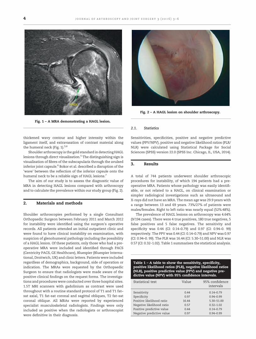
[(Fig._1)TD$FIG]
Fig. 1 – A MRA demonstrating a HAGL lesion.
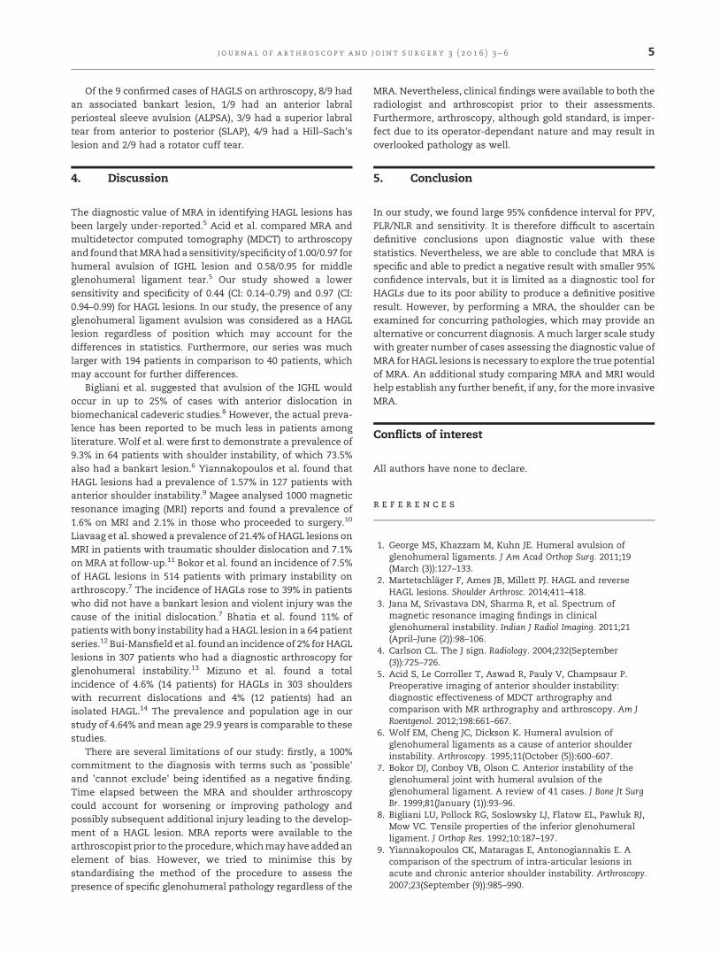
[(Fig._2)TD$FIG]
Fig. 2 – A HAGL lesion on shoulder arthroscopy.
j o u r n a l o f a r t h r o s c o p y and j o i n t s u r g e r y 3 ( 2 0 1 6 ) 3 – 64
thickened wavy contour and higher intensity within theligament itself, and extravasation of contrast material alongthe humeral neck (Fig. 1).3,4
Shoulder arthroscopy is the gold standard indetectingHAGLlesions through direct visualisation.5 The distinguishing sign isvisualisation of fibres of the subscapularis through the avulsedinferior joint capsule.6 Bokor et al. described a disruption of the'wave' between the reflection of the inferior capsule onto thehumeral neck to be a reliable sign of HAGL lesions.7
The aim of our study is to assess the diagnostic value ofMRA in detecting HAGL lesions compared with arthroscopyand to calculate the prevalence within our study group (Fig. 2).
Table 1 – A table to show the sensitivity, specificity,positive likelihood ratios (PLR), negative likelihood ratios(NLR), positive predictive value (PPV) and negative pre-dictive value (NPV) with 95% confidence intervals.
Statistical test Value 95% confidenceintervals
Sensitivity 0.44 0.14–0.79Specificity 0.97 0.94–0.99Positive likelihood ratio 16.44 5.30–51.00Negative likelihood ratio 0.57 0.32–1.02Positive predictive value 0.44 0.14–0.79Negative predictive value 0.97 0.94–0.99
2. Materials and methods
Shoulder arthroscopies performed by a single ConsultantOrthopaedic Surgeon between February 2011 and March 2012for instability were identified using the surgeon's operativerecords. All patients attended an initial outpatient clinic andwere found to have clinical instability on examination, withsuspicion of glenohumeral pathology including the possibilityof a HAGL lesion. Of these patients, only those who had a pre-operative MRA were included and identified through PACS(Centricity PACS, GE Healthcare), Bluespier (Bluespier Interna-tional, Droitwich, UK) and clinic letters. Patients were includedregardless of demographics, background, side of operation orindication. The MRAs were requested by the OrthopaedicSurgeon to ensure that radiologists were made aware of thepositive clinical findings on the request forms. The investiga-tions and procedureswere conducted over three hospital sites.1.5T MRI scanners with gadolinium as contrast were usedthroughout with a routine standard protocol of T1 and T1 fat–sat axial, T1 fat–sat coronal and sagittal obliques, T2 fat–satcoronal oblique. All MRAs were reported by experiencedspecialist musculoskeletal radiologists. Findings were onlyincluded as positive when the radiologists or arthroscopistwere definitive in their diagnosis.
2.1. Statistics
Sensitivities, specificities, positive and negative predictivevalues (PPV/NPV), positive and negative likelihood ratios (PLR/NLR) were calculated using Statistical Package for SocialSciences (SPSS) version 22.0 (SPSS Inc. Chicago, IL, USA, 2014).
3. Results
A total of 744 patients underwent shoulder arthroscopicprocedures for instability, of which 194 patients had a pre-operative MRA. Patients whose pathology was easily identifi-able, or not related to a HAGL, on clinical examination orsimpler radiological investigations such as ultrasound andX-rays did not have anMRA. Themean agewas 29.9 years witha range between 13 and 69 years. 73%/27% of patients weremales/females. Right to left ratio was nearly equal (52%:48%).
The prevalence of HAGL lesions on arthroscopy was 4.64%(9/194 cases). There were 4 true positives, 180 true negatives, 5false positives and 5 false negatives. The sensitivity andspecificity was 0.44 (CI: 0.14–0.79) and 0.97 (CI: 0.94–0. 99)respectively. The PPVwas 0.44 (CI: 0.14–0.79) and NPVwas 0.97(CI: 0.94–0. 99). The PLR was 16.44 (CI: 5.30–51.00) and NLR was0.57 (CI: 0.32–1.02). Table 1 summarises the statistical analysis.
j o u r n a l o f a r t h r o s c o p y and j o i n t s u r g e r y 3 ( 2 0 1 6 ) 3 – 6 5
Of the 9 confirmed cases of HAGLS on arthroscopy, 8/9 hadan associated bankart lesion, 1/9 had an anterior labralperiosteal sleeve avulsion (ALPSA), 3/9 had a superior labraltear from anterior to posterior (SLAP), 4/9 had a Hill–Sach'slesion and 2/9 had a rotator cuff tear.
4. Discussion
The diagnostic value of MRA in identifying HAGL lesions hasbeen largely under-reported.5 Acid et al. compared MRA andmultidetector computed tomography (MDCT) to arthroscopyand found thatMRAhada sensitivity/specificity of 1.00/0.97 forhumeral avulsion of IGHL lesion and 0.58/0.95 for middleglenohumeral ligament tear.5 Our study showed a lowersensitivity and specificity of 0.44 (CI: 0.14–0.79) and 0.97 (CI:0.94–0.99) for HAGL lesions. In our study, the presence of anyglenohumeral ligament avulsion was considered as a HAGLlesion regardless of position which may account for thedifferences in statistics. Furthermore, our series was muchlarger with 194 patients in comparison to 40 patients, whichmay account for further differences.
Bigliani et al. suggested that avulsion of the IGHL wouldoccur in up to 25% of cases with anterior dislocation inbiomechanical cadeveric studies.8 However, the actual preva-lence has been reported to be much less in patients amongliterature. Wolf et al. were first to demonstrate a prevalence of9.3% in 64 patients with shoulder instability, of which 73.5%also had a bankart lesion.6 Yiannakopoulos et al. found thatHAGL lesions had a prevalence of 1.57% in 127 patients withanterior shoulder instability.9 Magee analysed 1000 magneticresonance imaging (MRI) reports and found a prevalence of1.6% on MRI and 2.1% in those who proceeded to surgery.10
Liavaag et al. showed a prevalence of 21.4% of HAGL lesions onMRI in patients with traumatic shoulder dislocation and 7.1%on MRA at follow-up.11 Bokor et al. found an incidence of 7.5%of HAGL lesions in 514 patients with primary instability onarthroscopy.7 The incidence of HAGLs rose to 39% in patientswho did not have a bankart lesion and violent injury was thecause of the initial dislocation.7 Bhatia et al. found 11% ofpatientswith bony instability had aHAGL lesion in a 64 patientseries.12 Bui-Mansfield et al. found an incidence of 2% for HAGLlesions in 307 patients who had a diagnostic arthroscopy forglenohumeral instability.13 Mizuno et al. found a totalincidence of 4.6% (14 patients) for HAGLs in 303 shoulderswith recurrent dislocations and 4% (12 patients) had anisolated HAGL.14 The prevalence and population age in ourstudy of 4.64% andmean age 29.9 years is comparable to thesestudies.
There are several limitations of our study: firstly, a 100%commitment to the diagnosis with terms such as 'possible'and 'cannot exclude' being identified as a negative finding.Time elapsed between the MRA and shoulder arthroscopycould account for worsening or improving pathology andpossibly subsequent additional injury leading to the develop-ment of a HAGL lesion. MRA reports were available to thearthroscopist prior to the procedure,whichmayhave added anelement of bias. However, we tried to minimise this bystandardising the method of the procedure to assess thepresence of specific glenohumeral pathology regardless of the
MRA. Nevertheless, clinical findings were available to both theradiologist and arthroscopist prior to their assessments.Furthermore, arthroscopy, although gold standard, is imper-fect due to its operator-dependant nature and may result inoverlooked pathology as well.
5. Conclusion
In our study, we found large 95% confidence interval for PPV,PLR/NLR and sensitivity. It is therefore difficult to ascertaindefinitive conclusions upon diagnostic value with thesestatistics. Nevertheless, we are able to conclude that MRA isspecific and able to predict a negative result with smaller 95%confidence intervals, but it is limited as a diagnostic tool forHAGLs due to its poor ability to produce a definitive positiveresult. However, by performing a MRA, the shoulder can beexamined for concurring pathologies, which may provide analternative or concurrent diagnosis. A much larger scale studywith greater number of cases assessing the diagnostic value ofMRA for HAGL lesions is necessary to explore the true potentialof MRA. An additional study comparing MRA and MRI wouldhelp establish any further benefit, if any, for themore invasiveMRA.
Conflicts of interest
All authors have none to declare.
r e f e r e n c e s
1. George MS, Khazzam M, Kuhn JE. Humeral avulsion ofglenohumeral ligaments. J Am Acad Orthop Surg. 2011;19(March (3)):127–133.
2. Martetschläger F, Ames JB, Millett PJ. HAGL and reverseHAGL lesions. Shoulder Arthrosc. 2014;411–418.
3. Jana M, Srivastava DN, Sharma R, et al. Spectrum ofmagnetic resonance imaging findings in clinicalglenohumeral instability. Indian J Radiol Imaging. 2011;21(April–June (2)):98–106.
4. Carlson CL. The J sign. Radiology. 2004;232(September(3)):725–726.
5. Acid S, Le Corroller T, Aswad R, Pauly V, Champsaur P.Preoperative imaging of anterior shoulder instability:diagnostic effectiveness of MDCT arthrography andcomparison with MR arthrography and arthroscopy. Am JRoentgenol. 2012;198:661–667.
6. Wolf EM, Cheng JC, Dickson K. Humeral avulsion ofglenohumeral ligaments as a cause of anterior shoulderinstability. Arthroscopy. 1995;11(October (5)):600–607.
7. Bokor DJ, Conboy VB, Olson C. Anterior instability of theglenohumeral joint with humeral avulsion of theglenohumeral ligament. A review of 41 cases. J Bone Jt SurgBr. 1999;81(January (1)):93–96.
8. Bigliani LU, Pollock RG, Soslowsky LJ, Flatow EL, Pawluk RJ,Mow VC. Tensile properties of the inferior glenohumeralligament. J Orthop Res. 1992;10:187–197.
9. Yiannakopoulos CK, Mataragas E, Antonogiannakis E. Acomparison of the spectrum of intra-articular lesions inacute and chronic anterior shoulder instability. Arthroscopy.2007;23(September (9)):985–990.

j o u r n a l o f a r t h r o s c o p y and j o i n t s u r g e r y 3 ( 2 0 1 6 ) 3 – 66
10. Magee T. Prevalence of HAGL lesions and associatedabnormalities on shoulder MR examination. Skelet Radiol.2014;43(March (3)):307–313.
11. Liavaag S, Stiris MG, Svenningsen S, Enger M, Pripp AH, BroxJI. Capsular lesions with glenohumeral ligament injuries inpatients with primary shoulder dislocation: magneticresonance imaging and magnetic resonance arthrographyevaluation. Scand J Med Sci Sports. 2011;21(December (6)):e291–e297.
12. Bhatia DN, DasGupta B. Surgical treatment of significantglenoid bone defects and associated humeral avulsions of
glenohumeral ligament (HAGL) lesions in anterior shoulderinstability. Knee Surg Sports Traumatol Arthrosc. 2013;21(July(7)):1603–1609.
13. Bui-Mansfield LT, Taylor DC, Uhorchak JM, Tenuta JJ.Humeral avulsions of the glenohumeral ligament: imagingfeatures and a review of the literature. Am J Roentgenol.2002;179:649–655.
14. Mizuno N, Yoneda M, Hayashida K, Nakagawa S, Mae T,Izawa K. Recurrent anterior shoulder dislocation caused by amidsubstance complete capsular tear. J Bone Jt Surg Am.2005;87(December (12)):2717–2723.
j o u r n a l o f a r t h r o s c o p y and j o i n t s u r g e r y 3 ( 2 0 1 6 ) 7 – 1 2
Available online at www.sciencedirect.com
ScienceDirect
journal homepage: www.elsevier.com/locate/jajs
Original Article
Arthroscopic fixation using TightRope device for
acute acromioclavicular joint disruptionsParas Gupta a,*, Gagan Kansal a, Shekhar Srivastav b, Shekhar Agarwal c
aClinical Fellow, Delhi Institute of Trauma & Orthopedics, Sant Parmanand Hospital, New Delhi, IndiabConsultant, Delhi Institute of Trauma & Orthopedics, Sant Parmanand Hospital, New Delhi, IndiacDirector & HOD, Delhi Institute of Trauma & Orthopedics, Sant Parmanand Hospital, New Delhi, India
a r t i c l e i n f o
Article history:
Received 11 January 2016
Accepted 13 January 2016
Available online 15 February 2016
Keywords:
AC joint dislocation
Arthroscopic surgery
Shoulder arthroscopy
TightRope device
Outcome analysis
a b s t r a c t
Background: Acromioclavicular (AC) joint dislocation is a common shoulder injury that
affects young adults. The treatment of these injuries depends on dislocation grade, the
patient's complaints and time since injury.
Materials: Patients with acute AC joint injuries (Rockwood grades III–V) of less than 4 weeks
duration were included in the study. We had 10 cases of AC joint injuries, which fulfilled the
inclusion criteria. All patients were diagnosed based on history, examination and plain
radiographs. Grading was done based on plain radiographs. Patients with grades III–V were
selected for TightRope fixation. All 10 patients with AC joint injury underwent arthroscopic
AC joint fixation using TightRope. Functional outcome of patients was done using constant
shoulder score preoperatively and postoperatively. All patients were followed up for at least
one year. We were able to achieve satisfactory reduction in all the patients.
Results: Meanconstant scoreat thefinal follow-upwas92.2 (range76–97). Themeandifference
in constant score between operated and normal shoulderwas 9.8. On thebasis of difference of
constant score, 7 patients had excellent results, 2 had good results and 1 had fair result.
Conclusion: Arthroscopic fixation of acute AC joint dislocation using the TightRope device is a
simple, reproducible, minimal invasive technique that enables a rapid return to activity for
the acute injury.
# 2016 International Society for Knowledge for Surgeons on Arthroscopy and Arthro-
plasty. Published by Elsevier, a division of Reed Elsevier India, Pvt. Ltd. All rights reserved.
1. Introduction
Acromioclavicular (AC) joint dislocation is a common shoulderinjury that typically affects young adults.1,2 The treatment ofthese injuries depends on dislocation grade, the patient'scomplaints and time since injury. For Rockwood I and II
* Corresponding author. Tel.: +91 9717260996.E-mail address: [email protected] (P. Gupta).
http://dx.doi.org/10.1016/j.jajs.2016.01.0022214-9635/# 2016 International Society for Knowledge for Surgeons onReed Elsevier India, Pvt. Ltd. All rights reserved.
dislocations, non-operative management shows very satisfy-ing results. Acute Rockwood IV and V dislocations are a clearindication for surgery.3 The treatment guidelines for Rock-wood III dislocations are not uniform.4 While some publica-tions postulate conservative treatment to be superior, othersreport better outcome with operative treatment of Rockwoodtype III dislocations.5,6
Arthroscopy and Arthroplasty. Published by Elsevier, a division of
[(Fig._1)TD$FIG]
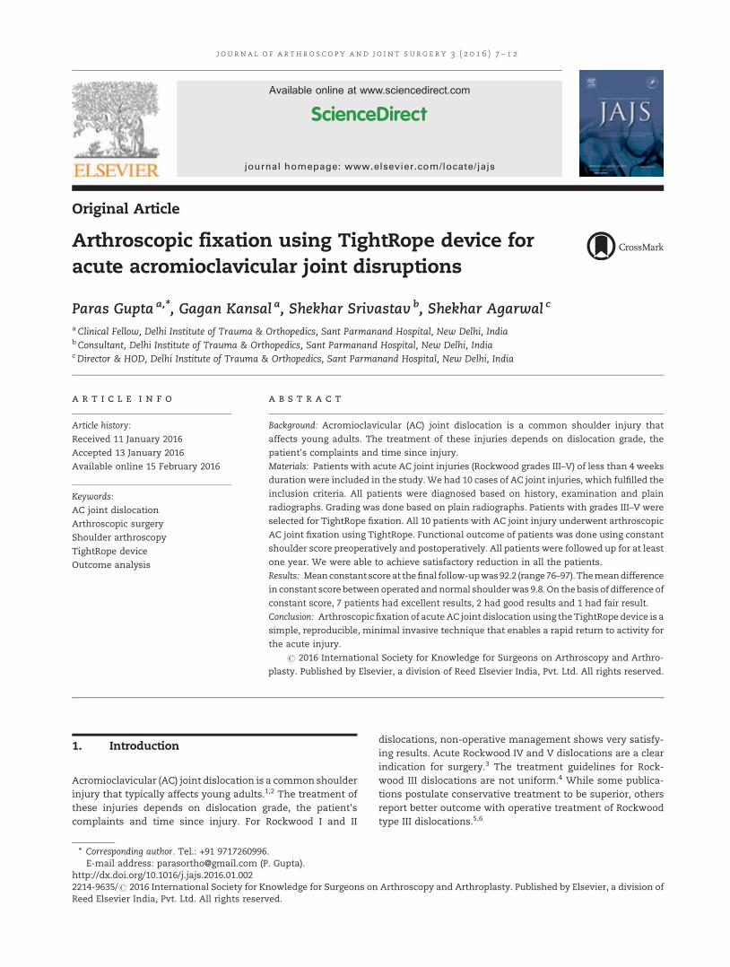
Fig. 1 – Arthroscopic view of base of coracoid after clearanceof rotator interval.
j o u rn a l o f a r t h r o s c o p y and j o i n t s u r g e r y 3 ( 2 0 1 6 ) 7 – 1 28
Conventionally open procedures have been considered aspreferred modality of treatment in AC dislocations. However,recent years have seen an upsurge in the use of arthroscopictechniques for management of acute AC joint injuries.7 A goldstandard for the reconstruction of coracoclavicular (CC)complex is yet to emerge. Most techniques not only fail torecreate the original anatomy, but often involve the use ofmaterials that are not strong enough tomaintain the reductionduring healing process.7 A new option is offered by theTightRopeTM system (Arthrex, Naples, USA), which consists of2metal buttons (1 circular and 1 oblong), joined by a continuousloop of Fibrewire suture.8 The device can be used to fix the ACjoint using an arthroscopic technique. This technique has beendescribed as a simple, reproducible, minimal invasive proce-dure for acute AC joint fixation that enables a rapid return toactivity for the acute injury. It also leavesminimal scarring anddoes not require any metalwork removal. We describe ourexperience of treating 10 patients with acute AC joint disloca-tion with arthroscopic TightRopeTM fixation.
[(Fig._2)TD$FIG]
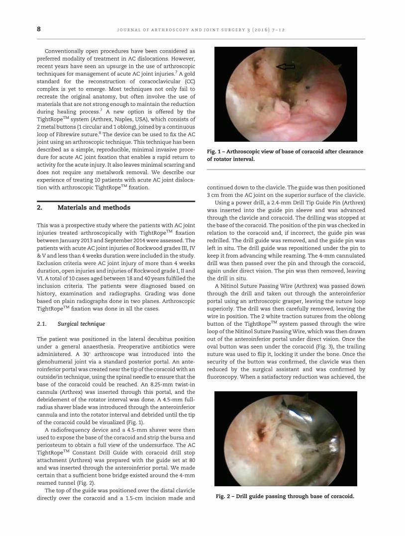
Fig. 2 – Drill guide passing through base of coracoid.
2. Materials and methods
This was a prospective study where the patients with AC jointinjuries treated arthroscopically with TightRopeTM fixationbetween January 2013 and September 2014 were assessed. Thepatients with acute AC joint injuries of Rockwood grades III, IV&V and less than 4weeks durationwere included in the study.Exclusion criteria were AC joint injury of more than 4 weeksduration, open injuries and injuries of Rockwood grade I, II andVI. A total of 10 cases aged between 18 and 40 years fulfilled theinclusion criteria. The patients were diagnosed based onhistory, examination and radiographs. Grading was donebased on plain radiographs done in two planes. ArthroscopicTightRopeTM fixation was done in all the cases.
2.1. Surgical technique
The patient was positioned in the lateral decubitus positionunder a general anaesthesia. Preoperative antibiotics wereadministered. A 308 arthroscope was introduced into theglenohumeral joint via a standard posterior portal. An ante-roinferior portalwas created near the tip of the coracoidwith anoutside/in technique, using the spinal needle to ensure that thebase of the coracoid could be reached. An 8.25-mm twist-incannula (Arthrex) was inserted through this portal, and thedebridement of the rotator interval was done. A 4.5-mm full-radius shaver blade was introduced through the anteroinferiorcannula and into the rotator interval and debrided until the tipof the coracoid could be visualized (Fig. 1).
A radiofrequency device and a 4.5-mm shaver were thenused to expose the base of the coracoid and strip the bursa andperiosteum to obtain a full view of the undersurface. The ACTightRopeTM Constant Drill Guide with coracoid drill stopattachment (Arthrex) was prepared with the guide set at 80and was inserted through the anteroinferior portal. We madecertain that a sufficient bone bridge existed around the 4-mmreamed tunnel (Fig. 2).
The top of the guide was positioned over the distal clavicledirectly over the coracoid and a 1.5-cm incision made and
continued down to the clavicle. The guidewas then positioned3 cm from the AC joint on the superior surface of the clavicle.
Using a power drill, a 2.4-mm Drill Tip Guide Pin (Arthrex)was inserted into the guide pin sleeve and was advancedthrough the clavicle and coracoid. The drilling was stopped atthe base of the coracoid. The position of the pinwas checked inrelation to the coracoid and, if incorrect, the guide pin wasredrilled. The drill guide was removed, and the guide pin wasleft in situ. The drill guide was repositioned under the pin tokeep it from advancing while reaming. The 4-mm cannulateddrill was then passed over the pin and through the coracoid,again under direct vision. The pin was then removed, leavingthe drill in situ.
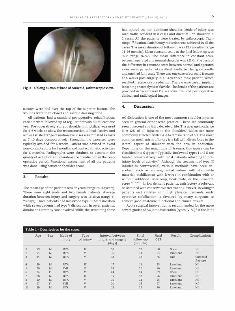
A Nitinol Suture Passing Wire (Arthrex) was passed downthrough the drill and taken out through the anteroinferiorportal using an arthroscopic grasper, leaving the suture loopsuperiorly. The drill was then carefully removed, leaving thewire in position. The 2 white traction sutures from the oblongbutton of the TightRopeTM system passed through the wireloop of the Nitinol Suture PassingWire, whichwas then drawnout of the anteroinferior portal under direct vision. Once theoval button was seen under the coracoid (Fig. 3), the trailingsuture was used to flip it, locking it under the bone. Once thesecurity of the button was confirmed, the clavicle was thenreduced by the surgical assistant and was confirmed byfluoroscopy. When a satisfactory reduction was achieved, the
[(Fig._3)TD$FIG]
Fig. 3 – Oblong button at base of coracoid, arthroscopic view.
j o u r n a l o f a r t h r o s c o p y and j o i n t s u r g e r y 3 ( 2 0 1 6 ) 7 – 1 2 9
sutures were tied over the top of the superior button. Thewounds were then closed and aseptic dressing done.
All patients had a standard postoperative rehabilitation.Patients were followed up at regular intervals till at least oneyear. Post-operatively, sling or shoulder immobilizer was usedfor 4–6 weeks to allow the reconstruction to heal. Passive andactive assisted range ofmotion exercises was initiated as earlyas 7–10 days postoperatively. Strengthening exercises weretypically avoided for 6 weeks. Patient was advised to avoidnon-contact sports for 3months and contact athletic activitiesfor 6 months. Radiographs were obtained to ascertain thequality of reduction andmaintenance of reduction in the post-operative period. Functional assessment of all the patientswas done using constant shoulder score.
3. Results
The mean age of the patients was 32 years (range 24–40 years).There were eight male and two female patients. Averageduration between injury and surgery was 16 days (range 6–28 days). Three patients had Rockwood type III AC dislocationwhile seven patients had type V dislocation. In seven patients,dominant extremity was involved while the remaining three
Table 1 – Descriptives for the cases.
Age Sex Mode ofinjury
Typeof injury
Interval betweinjury and surg
(days)
1 29 M RTA III 102 24 M RTA V 63 34 M RTA V 18
4 29 M RTA III 175 36 M Fall V 286 34 F RTA V 247 28 M RTA III 158 40 M RTA V 109 27 F Fall V 2010 39 M RTA V 12
had injured the non-dominant shoulder. Mode of injury wasroad traffic accident in 8 cases and direct fall on shoulder in2 cases. All the patients were treated by arthroscopic Tigh-tRopeTM fixation. Satisfactory reduction was achieved in all thecases. The mean duration of follow-up was 12.7 months (range11–16 months). Mean constant score at the final follow-up was92.2 (range 76–97). The mean difference in constant scorebetween operated and normal shoulder was 9.8. On the basis ofthe difference in constant score between normal and operatedsides, sevenpatientshadexcellent results, twohadgood resultsand one had fair result. There was one case of coracoid fractureat 4 weeks post-surgery in a 34-year-old male patient, whichresulted in some lossof reduction. Therewasnocaseof implantlooseningor osteolysis of clavicle. Thedetails of thepatients areprovided in Table 1 and Fig. 4 shows pre- and post-operativeclinical and radiological images.
4. Discussion
AC dislocation is one of the most common shoulder injuriesseen in general orthopaedic practice. These are commonlyseen in second and third decade of life. The average incidenceis 9–12% of all injuries to the shoulder.9 Males are morecommonly affected, with male to female ratio of 5:1. The mostcommon mechanism of injury is a fall with direct force to thelateral aspect of shoulder with the arm in adduction.Depending on the magnitude of trauma, this injury can beclassified into 6 types.10 Typically, Rockwood types I and II aretreated conservatively, with most patients returning to pre-injury levels of activity.11 Although the treatment of type IIIinjuries is controversial, various methods have been de-scribed, such as an augmented suture with absorbablematerial, stabilization with k-wires in combination with orwithout additional wire loop, hook plate, or the Bosworthscrew.4,12–15,17 In low demand patients, satisfactory results canbe obtainedwith conservative treatment. However, in youngerpatients and athletes with high physical demands, earlyoperative stabilization is favoured by many surgeons toachieve good anatomic, functional and clinical results.
Acute surgical intervention is recommended for the moresevere grades of AC joint dislocation (types IV–VI).3 If the joint
enery
Finalfollow-up(months)
FinalCSS
Result Complications
12 88 Good Nil14 94 Excellent Nil12 76 Fair Coracoid
fracture12 95 Excellent Nil11 96 Excellent Nil14 89 Good Nil12 96 Excellent Nil12 95 Excellent Nil16 97 Excellent Nil12 96 Excellent Nil
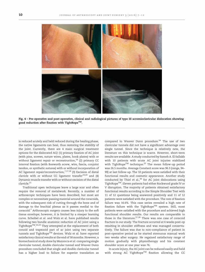
[(Fig._4)TD$FIG]
Fig. 4 – Pre-operative and post-operative, clinical and radiological pictures of type III acromioclavicular dislocation showinggood reduction after fixation with TightRopeTM.
j o u rn a l o f a r t h r o s c o p y and j o i n t s u r g e r y 3 ( 2 0 1 6 ) 7 – 1 210
is reduced acutely and held reduced during the healing phase,the native ligaments can heal, thus restoring the stability ofthe joint. Currently, there are 4 main surgical treatmentoptions for the dislocated ACJ: (1) primary fixation of AC joint(with pins, screws, suture wires, plates, hook plates) with orwithout ligament repair or reconstruction,16 (2) primary CCinterval fixation (with Bosworth screw, wire, fascia, conjointtendon, or synthetic sutures) with or without incorporation ofAC ligament repair/reconstruction,7,17,18 (3) Excision of distalclavicle with or without CC ligament transfer19,20 and (4)Dynamicmuscle transfer with or without excision of the distalclavicle.21
Traditional open techniques leave a large scar and oftenrequire the removal of metalwork. Recently, a number ofarthroscopic techniques have been described, but most arecomplex or necessitate passingmaterial around the coracoids,with the subsequent risk of cutting through the bone and ofdamage to the brachial plexus as it passes medial to thecoracoid.7 Arthroscopic surgery causes less injury to the softtissue envelope; however, it is limited by a steeper learningcurve. Scheibel et al. and Walz et al. have published resultsfollowing two bundle anatomic reduction of AC joint using aTightRopeTM.22,23 They attempted at the replacement of bothconoid and trapezoid part of ac joint using two separatetunnels and TightRopeTM devices. Walz et al. have reportedsatisfactory clinical results at the endof 58months. However, abiomechanical study done byMazzocca et al. comparing singleclavicular tunnel, double clavicular tunnel and Weaver Dunnprocedure concluded that single and double clavicular tunnelhas a higher load to failure for superior translation as
compared to Weaver Dunn procedure.24 The use of twoclavicular tunnels did not have a significant advantage oversingle tunnel. Since the technique is relatively new, theliterature on this technique is scarce. However, short-termresults are available. A study conducted by SamehA. El Sallakhwith 10 patients with acute AC joint injuries stabilizedwith TightRopeTM technique.25 The mean follow-up periodwas 26.5 months. Average Constant score was 96.3 (range, 94–99) at last follow-up. The 10 patients were satisfied with theirfunctional results and cosmetic appearance. Another studyconducted by Thiel et al.,26 for AC joint dislocations usingTightRopeTM. Eleven patients had either Rockwood grade IV orV disruption. The majority of patients obtained satisfactoryfunctional results according to the Simple Shoulder Test with11 of 12 questions being answered positively and 11 of 12patients were satisfied with the procedure. The rate of fixationfailure was 16.6%. This case series revealed a high rate offixation failure with the TightRopeTM system. Still, mostpatients were satisfied with the procedure and achieved highfunctional shoulder results. Our results are comparable tothose in the literature.27,28 There was one case of coracoidfracture in our study. The fracture occurred at 4 weeks post-opresulting in shoulder stiffness and was managed conserva-tively. The failure was due to non-compliance of patient inpost-operative period as he started strenuous manual worktwo weeks after surgery. He regained satisfactory range ofmotion gradually with physiotherapy and his constantshoulder score at one year was 76.
In acute injury, the AC joint can be reduced easily and heldwith strong AC TightRopeTM fixation allowing the CC
j o u r n a l o f a r t h r o s c o p y and j o i n t s u r g e r y 3 ( 2 0 1 6 ) 7 – 1 2 11
ligaments to heal. As it is an elastic fixation, its removal is notrequired. Cautionshouldbeobservedwhile drilling the tunnelin coracoids. It should be centred at the base or elsemay causecoracoid fracture. As it is a minimal invasive technique,recovery is relatively fast and the scars are cosmeticallyacceptable. Most patients scored high on functional scoringand were happy with both the functional and cosmeticresults. The main drawbacks of our study were smallersample size and relatively shorter follow-up. However, theearly results with this small group were quite encouraging.Studies with larger number of cases, longer follow-up andcontrol groups are needed to assess the long-term outcomeand efficacy of this procedure.
5. Conclusion
Arthroscopic fixation of acute AC joint dislocation using theTightRopeTM Device is a simple, reproducible, minimalinvasive technique for acute AC joint fixation that enable arapid return to activity for the acute injury.
Disclaimer
None of the authors received payments or services, eitherdirectly or indirectly (i.e., via his or her institution), froma thirdparty in support of an aspect of this work. In addition, none ofthe authors, or his or her institution, has had a financialrelationship, in the thirty-six months prior to submission ofthis work, with an entity in the biomedical arena that could beperceived to influence or have the potential to influence whatis written in this work. Also, none of the authors has a patentor patents, planned, pending, or issued, that is broadlyrelevant to the work. No author has had any other relation-ships, or has engaged in any other activities, that could beperceived to influence or have the potential to influence whatis written in this work.
Conflict of interest
All authors have none to declare.
r e f e r e n c e s
1. Clayton RA, Court-Brown CM. The epidemiology ofmusculoskeletal tendinous and ligamentous injuries. Injury.2008;39(12):1338–1344.
2. Rockwood CA, Williams G, Young D, Disorders of theacromioclavicular joint. 2nd ed. Rockwood CA, Matsen FA,eds. The Shoulder. vol. 1. Philadelphia: WB Saunders; 1998:483–553.
3. Mazzocca AD, Santangelo SA, Johnson ST, Rios CG,Dumonski ML. A biomechanical evaluation of an anatomicalcoracoclavicular ligament reconstruction. Am J Sports Med.2006;34(2):236–246;ĆBradley JP, Elkousy H. Decision making:operative versus non-operative treatment ofacromioclavicular joint injuries. Clin Sports Med. 2003;22(2):277–290.
4. Galpin RD, Hawkins RJ, Grainger RW. A comparative analysisof operative versus non-operative treatment of grade IIIacromioclavicular separations. Clin Orthop Relat Res.1985;193:150–155.
5. Hootman JM. Acromioclavicular dislocation: conservative orsurgical therapy. J Athl Train. 2004;39(1):10–11.
6. Ceccarelli E, Bondi R, Alviti F, et al. Treatment of acute gradeIII acromioclavicular dislocation: a lack of evidence. J OrthopTraumatol. 2008;9(2):105–108.
7. Wolf EM, Pennington WT. Arthroscopic reconstruction foracromioclavicular joint dislocation. Arthroscopy. 2001;17(5):558–563.
8. Salzmann GM, Walz L, Schoettle PB, et al. Arthroscopicanatomical reconstruction of the acromioclavicular joint.Acta Orthop Belg. 2008;74(3):397–400.
9. Lemos MJ. The evaluation and treatment of the injuredacromioclavicular joint in athletes. Am J Sports Med. 1998;26(1):137–144.
10. Rockwood Jr CA, Williams GR, Young DC, Disorders of theacromioclavicular joint. 2nd ed. Rockwood CA, Matsen FA,eds. The Shoulder. vol. 1. Philadelphia, PA: WB Saunders;1998: 483–553.
11. Dias JJ, Steingold RF, Richardson RA, Tesfayohannes B, GreggPJ. The conservative treatment of acromioclaviculardislocation: review after five years. J Bone Joint Surg Br.1987;69(5):719–722.
12. Leidel BA, Braunstein V, Kirchhoff C, et al. Consistency oflong-term outcome of acute Rockwood grade IIIacromioclavicular joint separations after K-wiretransfixation. J Trauma. 2009;66(6):1666–1671.
13. Ejam S, Lind T, Falkenberg B. Surgical treatment of acuteand chronic acromioclavicular dislocation Tossy type IIIand V using the Hook plate. Acta Orthop Belg. 2008;74(4):441–445.
14. Eberle C, Fodor P, Metzger U. Hook plate (so-called Balserplate) or tension banding with the Bosworth screw incomplete acromioclavicular dislocation and clavicularfracture. Z Unfallchir Versicherungsmed. 1992;85(3):134–139.
15. Weitzmann G. Treatment of acute acromioclavicular jointdislocation by a modified Bosworth method report ontwenty-four cases. J Bone Joint Surg Am. 1967;49:1167–1178.
16. Phillips AM, Smart C, Groom AFG. Acromioclaviculardislocation: conservative or surgical therapy. Clin OrthopRelat Res. 1998;353:10–17.
17. Bosworth BM. Acromioclavicular separations: new methodof repair. Surg Gynecol Obstet. 1941;73:866–871.
18. Larsen E, Peterson V. Operative treatment of chronicacromioclavicular dislocations. Injury. 1987;18(1):55–56.
19. Weaver JK, Dunn HK. Treatment of acromioclavicularinjuries, specially complete acromioclavicular separation. JBone Joint Surg Am. 1972;54(6):1187–1194.
20. Mumford EB. Acromioclavicular dislocation. J Bone Joint Surg.1941;23:799–802.
21. Berson BL, Gilbert MS, Green S. Acromioclaviculardislocations: treatment by transfer of the conjoint tendonand distal end of the coracoids process to the clavicle. ClinOrthop. 1978;135:157–164.
22. Scheibel M, Dröschel S, Gerhardt C, Kraus N.Arthroscopically assisted stabilization of acute high-gradeacromioclavicular joint separations. Am J Sports Med. 2011;39(July (7)):1507–1516.
23. Walz LSG, Eichhorn S, Imhoff AB, et al. The anatomicreconstruction of AC joint dislocation using tightropedevices – a biomechanical study. Am J Sports Med. 2008;36(12):2398–2406.
24. Mazzocca AD, Arciero RA, Bicos J. Evaluation and treatmentof acromioclavicular joint injuries. Am J Sports Med. 2007;35(2):316–329.

j o u rn a l o f a r t h r o s c o p y and j o i n t s u r g e r y 3 ( 2 0 1 6 ) 7 – 1 212
25. El Sallakh SA. Evaluation of arthroscopic stabilization ofacute acromioclavicular joint dislocation using theTightRopeTM system. Orthopedics. 2012;35(January (1)):e18–e22.
26. Thiel E, Mutnal A, Gilot GJ. Surgical outcome followingarthroscopic fixation of acromioclavicular joint disruptionwith the tightrope device. Orthopedics. 2011;34(July (7)):e267–e274.
27. Richards A, Tennent TD. Arthroscopic stabilization of acuteacromioclavicular joint dislocation using the TightRopesystem. Tech Shoulder Elbow Surg. 2008;9(2):51–54.
28. Vieira LAG, Visco A, Fernandes LFD, Cordero NGG.Arthroscopic treatment of acromioclavicular jointdislocation by TightRope technique (Arthrex). Rev BrasOrtoped. 2009;44(1):52–56.
j o u rn a l o f a r t h r o s c o p y an d j o i n t s u r g e r y 3 ( 2 0 1 6 ) 1 3 – 1 6
Available online at www.sciencedirect.com
ScienceDirect
journal homepage: www.elsevier.com/locate/jajs
Original Article
Arthroscopic stabilization of acute
acromioclavicular joint dislocation with tightropeAC system: A tale of failuresShreesh Kumar Gangary b, Sanjay Meena a,*aDepartment of Orthopaedics, SGT Medical College, SGT University, Gurgaon, IndiabDepartment of Orthopaedics, ESI Hospital, Okhla, 110020 New Delhi, India
a r t i c l e i n f o
Article history:
Received 13 August 2015
Accepted 4 November 2015
Available online 15 December 2015
Keywords:
Acromioclavicular joint dislocation
Tightrope
Arthroscopy
Shoulder
a b s t r a c t
Background: Stabilization of acromioclavicular joint is a challenging technique with several
methods described in literature from non-biological methods to biological fixation of AC
joint. Arthroscopic fixation of AC joint is a newer technique with limited literature available.
The aim of our study is to evaluate the results of arthroscopic stabilization of acute
acromioclavicular joint with tightrope.
Methods: From February 2013 till August 2013, arthroscopic stabilization of acute ACJ
dislocation was performed in 11 patients. The group consisted of eight men and three
women with an average age of 34.2 years. The Rockwood type III to type V ACJ dislocations
(III, 6; IV, 2; V, 3) were indicated for surgery. The average interval between injury and surgery
was 5.4 days. In all cases, a second-generation tightrope implant was inserted by the
Endobutton technique joining the distal end of the clavicle and the coracoid process. The
results were evaluated using the UCLA Shoulder Scale at 10 months after surgery.
Results: All 11 patients returned to their preoperative activities without any restriction
of shoulder motion within 5 months of surgery. The average postoperative UCLA score
was 30.3 points (range 27–34). Radiographic evidence of the loss of partial reduction, with no
effect on the clinical outcome, was recorded in 5 patients (45%) and loss of full reduction
noted in5 (45%)patientsduringpostoperative rehabilitation.Onepatientwas lost in follow-up.
Conclusion: Arthroscopic stabilization of acute ACJ dislocations using a single tightrope
implant is a minimally invasive surgical technique, but less satisfactory result may be
because of non-biological nature of fixation. Non-biological AC joint fixation is not a good
method of fixation of a biological AC joint.
# 2015 International Society for Knowledge for Surgeons on Arthroscopy and Arthro-
plasty. Published by Elsevier, a division of Reed Elsevier India, Pvt. Ltd. All rights reserved.
* Corresponding author. Tel.: +91 9968444612.E-mail address: [email protected] (S. Meena).
http://dx.doi.org/10.1016/j.jajs.2015.11.0032214-9635/# 2015 International Society for Knowledge for Surgeons on Arthroscopy and Arthroplasty. Published by Elsevier, a division ofReed Elsevier India, Pvt. Ltd. All rights reserved.
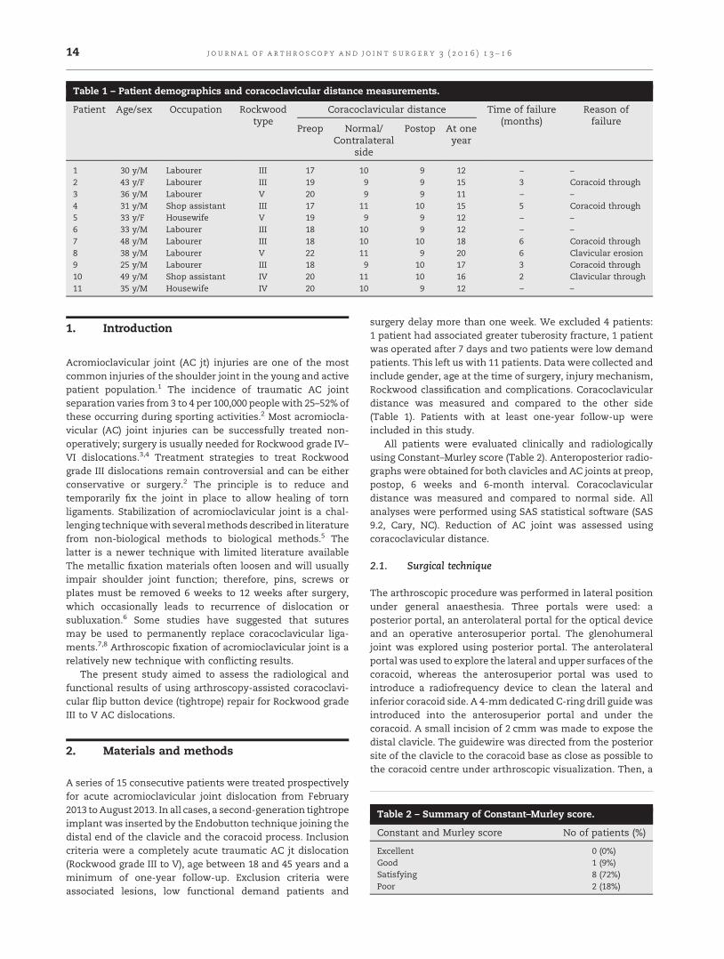
Table 1 – Patient demographics and coracoclavicular distance measurements.
Patient Age/sex Occupation Rockwoodtype
Coracoclavicular distance Time of failure(months)
Reason offailure
Preop Normal/Contralateral
side
Postop At oneyear
1 30 y/M Labourer III 17 10 9 12 – –
2 43 y/F Labourer III 19 9 9 15 3 Coracoid through3 36 y/M Labourer V 20 9 9 11 – –
4 31 y/M Shop assistant III 17 11 10 15 5 Coracoid through5 33 y/F Housewife V 19 9 9 12 – –
6 33 y/M Labourer III 18 10 9 12 – –
7 48 y/M Labourer III 18 10 10 18 6 Coracoid through8 38 y/M Labourer V 22 11 9 20 6 Clavicular erosion9 25 y/M Labourer III 18 9 10 17 3 Coracoid through10 49 y/M Shop assistant IV 20 11 10 16 2 Clavicular through11 35 y/M Housewife IV 20 10 9 12 – –
j o u r n a l o f a r t h r o s c o p y and j o i n t s u r g e r y 3 ( 2 0 1 6 ) 1 3 – 1 614
1. Introduction
Acromioclavicular joint (AC jt) injuries are one of the mostcommon injuries of the shoulder joint in the young and activepatient population.1 The incidence of traumatic AC jointseparation varies from 3 to 4 per 100,000 peoplewith 25–52% ofthese occurring during sporting activities.2 Most acromiocla-vicular (AC) joint injuries can be successfully treated non-operatively; surgery is usually needed for Rockwood grade IV–VI dislocations.3,4 Treatment strategies to treat Rockwoodgrade III dislocations remain controversial and can be eitherconservative or surgery.2 The principle is to reduce andtemporarily fix the joint in place to allow healing of tornligaments. Stabilization of acromioclavicular joint is a chal-lenging techniquewith severalmethodsdescribed in literaturefrom non-biological methods to biological methods.5 Thelatter is a newer technique with limited literature availableThe metallic fixation materials often loosen and will usuallyimpair shoulder joint function; therefore, pins, screws orplates must be removed 6 weeks to 12 weeks after surgery,which occasionally leads to recurrence of dislocation orsubluxation.6 Some studies have suggested that suturesmay be used to permanently replace coracoclavicular liga-ments.7,8 Arthroscopic fixation of acromioclavicular joint is arelatively new technique with conflicting results.
The present study aimed to assess the radiological andfunctional results of using arthroscopy-assisted coracoclavi-cular flip button device (tightrope) repair for Rockwood gradeIII to V AC dislocations.
Table 2 – Summary of Constant–Murley score.
Constant and Murley score No of patients (%)
Excellent 0 (0%)Good 1 (9%)Satisfying 8 (72%)Poor 2 (18%)
2. Materials and methods
A series of 15 consecutive patients were treated prospectivelyfor acute acromioclavicular joint dislocation from February2013 toAugust 2013. In all cases, a second-generation tightropeimplant was inserted by the Endobutton technique joining thedistal end of the clavicle and the coracoid process. Inclusioncriteria were a completely acute traumatic AC jt dislocation(Rockwood grade III to V), age between 18 and 45 years and aminimum of one-year follow-up. Exclusion criteria wereassociated lesions, low functional demand patients and
surgery delay more than one week. We excluded 4 patients:1 patient had associated greater tuberosity fracture, 1 patientwas operated after 7 days and two patients were low demandpatients. This left us with 11 patients. Data were collected andinclude gender, age at the time of surgery, injury mechanism,Rockwood classification and complications. Coracoclaviculardistance was measured and compared to the other side(Table 1). Patients with at least one-year follow-up wereincluded in this study.
All patients were evaluated clinically and radiologicallyusing Constant–Murley score (Table 2). Anteroposterior radio-graphs were obtained for both clavicles and AC joints at preop,postop, 6 weeks and 6-month interval. Coracoclaviculardistance was measured and compared to normal side. Allanalyses were performed using SAS statistical software (SAS9.2, Cary, NC). Reduction of AC joint was assessed usingcoracoclavicular distance.
2.1. Surgical technique
The arthroscopic procedure was performed in lateral positionunder general anaesthesia. Three portals were used: aposterior portal, an anterolateral portal for the optical deviceand an operative anterosuperior portal. The glenohumeraljoint was explored using posterior portal. The anterolateralportal was used to explore the lateral and upper surfaces of thecoracoid, whereas the anterosuperior portal was used tointroduce a radiofrequency device to clean the lateral andinferior coracoid side. A 4-mmdedicated C-ring drill guidewasintroduced into the anterosuperior portal and under thecoracoid. A small incision of 2 cmm was made to expose thedistal clavicle. The guidewire was directed from the posteriorsite of the clavicle to the coracoid base as close as possible tothe coracoid centre under arthroscopic visualization. Then, a
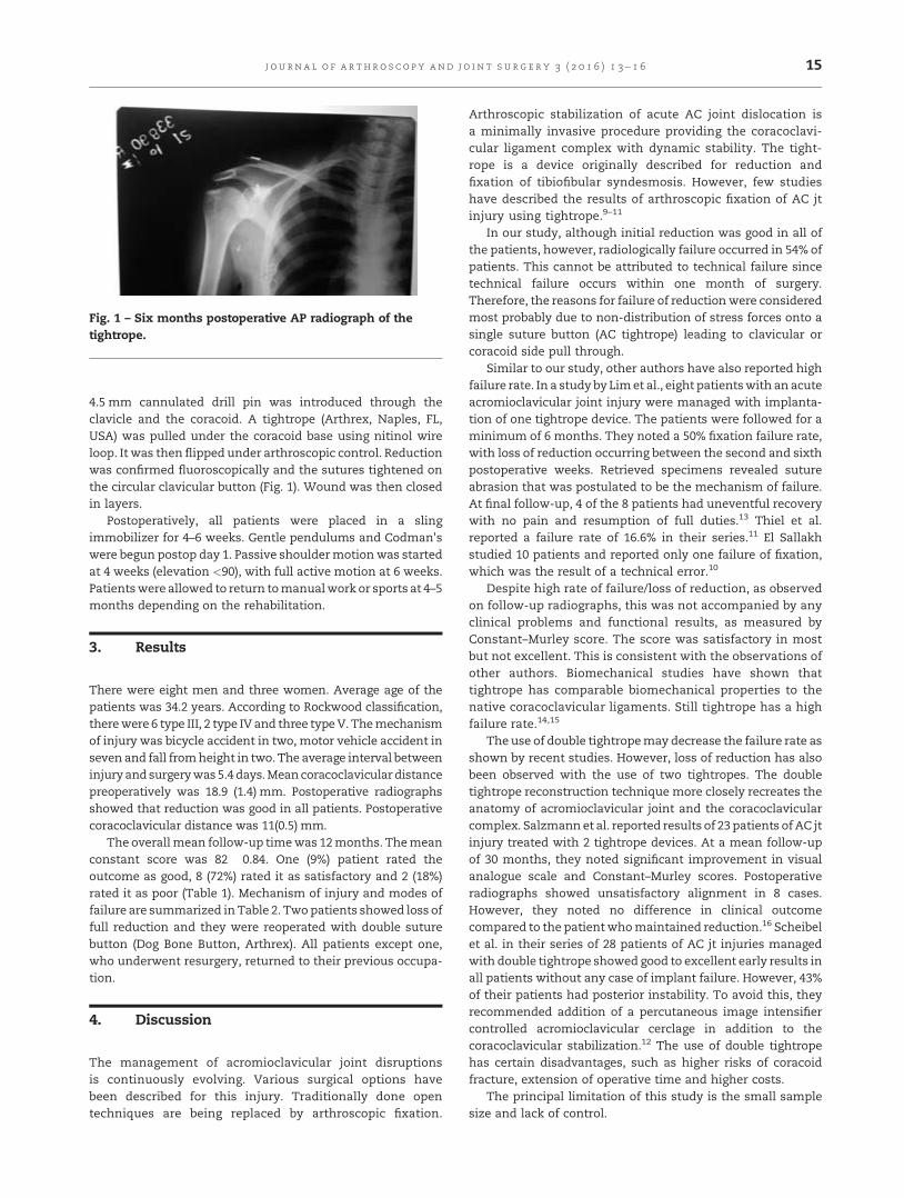
[(Fig._1)TD$FIG]
Fig. 1 – Six months postoperative AP radiograph of thetightrope.
j o u rn a l o f a r th r o s c o p y and j o i n t s u r g e r y 3 ( 2 0 1 6 ) 1 3 – 1 6 15
4.5 mm cannulated drill pin was introduced through theclavicle and the coracoid. A tightrope (Arthrex, Naples, FL,USA) was pulled under the coracoid base using nitinol wireloop. It was then flipped under arthroscopic control. Reductionwas confirmed fluoroscopically and the sutures tightened onthe circular clavicular button (Fig. 1). Wound was then closedin layers.
Postoperatively, all patients were placed in a slingimmobilizer for 4–6 weeks. Gentle pendulums and Codman'swere begun postop day 1. Passive shouldermotionwas startedat 4 weeks (elevation <90), with full active motion at 6 weeks.Patientswere allowed to return tomanualwork or sports at 4–5months depending on the rehabilitation.
3. Results
There were eight men and three women. Average age of thepatients was 34.2 years. According to Rockwood classification,therewere 6 type III, 2 type IV and three typeV. Themechanismof injury was bicycle accident in two, motor vehicle accident inseven and fall fromheight in two. The average interval betweeninjuryandsurgerywas 5.4days.Meancoracoclavicular distancepreoperatively was 18.9 (1.4) mm. Postoperative radiographsshowed that reduction was good in all patients. Postoperativecoracoclavicular distance was 11(0.5) mm.
The overallmean follow-up timewas 12months. Themeanconstant score was 82 � 0.84. One (9%) patient rated theoutcome as good, 8 (72%) rated it as satisfactory and 2 (18%)rated it as poor (Table 1). Mechanism of injury and modes offailure are summarized in Table 2. Twopatients showed loss offull reduction and they were reoperated with double suturebutton (Dog Bone Button, Arthrex). All patients except one,who underwent resurgery, returned to their previous occupa-tion.
4. Discussion
The management of acromioclavicular joint disruptionsis continuously evolving. Various surgical options havebeen described for this injury. Traditionally done opentechniques are being replaced by arthroscopic fixation.
Arthroscopic stabilization of acute AC joint dislocation isa minimally invasive procedure providing the coracoclavi-cular ligament complex with dynamic stability. The tight-rope is a device originally described for reduction andfixation of tibiofibular syndesmosis. However, few studieshave described the results of arthroscopic fixation of AC jtinjury using tightrope.9–11
In our study, although initial reduction was good in all ofthe patients, however, radiologically failure occurred in 54% ofpatients. This cannot be attributed to technical failure sincetechnical failure occurs within one month of surgery.Therefore, the reasons for failure of reductionwere consideredmost probably due to non-distribution of stress forces onto asingle suture button (AC tightrope) leading to clavicular orcoracoid side pull through.
Similar to our study, other authors have also reported highfailure rate. In a study by Limet al., eight patientswith an acuteacromioclavicular joint injury were managed with implanta-tion of one tightrope device. The patients were followed for aminimum of 6 months. They noted a 50% fixation failure rate,with loss of reduction occurring between the second and sixthpostoperative weeks. Retrieved specimens revealed sutureabrasion that was postulated to be the mechanism of failure.At final follow-up, 4 of the 8 patients had uneventful recoverywith no pain and resumption of full duties.13 Thiel et al.reported a failure rate of 16.6% in their series.11 El Sallakhstudied 10 patients and reported only one failure of fixation,which was the result of a technical error.10
Despite high rate of failure/loss of reduction, as observedon follow-up radiographs, this was not accompanied by anyclinical problems and functional results, as measured byConstant–Murley score. The score was satisfactory in mostbut not excellent. This is consistent with the observations ofother authors. Biomechanical studies have shown thattightrope has comparable biomechanical properties to thenative coracoclavicular ligaments. Still tightrope has a highfailure rate.14,15
The use of double tightropemay decrease the failure rate asshown by recent studies. However, loss of reduction has alsobeen observed with the use of two tightropes. The doubletightrope reconstruction technique more closely recreates theanatomy of acromioclavicular joint and the coracoclavicularcomplex. Salzmann et al. reported results of 23 patients of AC jtinjury treated with 2 tightrope devices. At a mean follow-upof 30 months, they noted significant improvement in visualanalogue scale and Constant–Murley scores. Postoperativeradiographs showed unsatisfactory alignment in 8 cases.However, they noted no difference in clinical outcomecompared to the patient whomaintained reduction.16 Scheibelet al. in their series of 28 patients of AC jt injuries managedwith double tightrope showed good to excellent early results inall patients without any case of implant failure. However, 43%of their patients had posterior instability. To avoid this, theyrecommended addition of a percutaneous image intensifiercontrolled acromioclavicular cerclage in addition to thecoracoclavicular stabilization.12 The use of double tightropehas certain disadvantages, such as higher risks of coracoidfracture, extension of operative time and higher costs.
The principal limitation of this study is the small samplesize and lack of control.
j o u r n a l o f a r t h r o s c o p y and j o i n t s u r g e r y 3 ( 2 0 1 6 ) 1 3 – 1 616
5. Conclusion
Arthroscopic stabilization of acute ACJ dislocations using asingle tightrope implant is a minimally invasive surgicaltechnique but with less satisfactory result, and may bebecause of non-biological nature, technical failure or implantfailure.
Conflicts of interest
All authors have none to declare.
r e f e r e n c e s
1. Clayton RA, Court-Brown CM. The epidemiology ofmusculoskeletal tendinous and ligamentous injuries. Injury.2008;39(12):1338–1344.
2. Rockwood CA, Williams GR, Young DC. Fractures in Adults:Acromioclavicular Injuries. Philadelphia, PA: Lippincott-Raven;1996:1341–1413.
3. Horn JS. The traumatic anatomy and treatment of acuteacromioclavicular dislocation. J Bone Joint Surg Br.1954;36:194–1201.
4. Lemos MJ. The evaluation and treatment of the injuredacromioclavicular joint in athletes. Am J Sports Med.1998;26:137–144.
5. Fauci F, Merolla G, Paladini P, Campi F, Porcellini G. Surgicaltreatment of chronic acromioclavicular dislocation withbiologic graft vs synthetic ligament: a prospectiverandomized comparative study. J Orthop Trauma. 2013;14(December (4)):283–290.
6. Jafary D, Keihan Shokouh H, Najd Mazhar F, Shariat ZadehH, Mochtary T. Clinical and radiological results of fixation ofacromioclavicular joint dislocation by hook plates retained
for more than five months. Trauma Mon. 2014;19(April (2)):e13728.
7. Mardani-Kivi M, Mirbolook A, Salariyeh M, Hashemi-Motlagh K, Saheb-Ekhtiari K. The comparison of ethibondsutures and semitendinosus autograft in the surgicaltreatment of acromioclavicular dislocation. Acta OrthopTraumatol Turc. 2013;47(5):307–310.
8. El Shewy MT, El Azizi H. Suture repair using loop techniquein cases of acute complete acromioclavicular jointdislocation. J Orthop Trauma. 2011;12(March (1)):29–35.
9. Hou Z, Graham J, Zhang Y, et al. Comparison of single andtwo tunnels techniques during open treatment ofacromioclavicular joint disruption. BMC Surg. 2014;14:53.
10. El Sallakh SA. Evaluation of arthroscopic stabilization ofacute acromioclavicular joint dislocation using the tightropesystem. Orthopaedics. 2012;35(1):e18–e22.
11. Thiel E, Mutnal A, Gilot GJ. Surgical outcome followingarthroscopic fixation of acromioclavicular joint disruptionwith the tightrope device. Orthopedics. 2011;34(7):e267–e274.
12. Scheibel M, Dröschel S, Gerhardt C, Kraus N.Arthroscopically assisted stabilization of acute high gradeacromioclavicular joint separations. Am J Sports Med. 2011;39(July (7)):1507–1516.
13. Lim YW, Sood A, Van Riet RP. Acromioclavicular jointreduction, repair and reconstruction using metallicbuttons—early results and complications. Tech ShoulderElbow Surg. 2007;8(4):213–221.
14. Nüchtern JV, Sellenschloh K, Bishop N, et al. Biomechanicalevaluation of 3 stabilization methods on acromioclavicularjoint dislocations. Am J Sports Med. 2013;41(June (6)):1387–1394.
15. Lädermann A, Gueorguiev B, Stimec B, Fasel J, Rothstock S,Hoffmeyer P. Acromioclavicular joint reconstruction: acomparative biomechanical study of three techniques. JShoulder Elbow Surg. 2013;22(February (2)):171–178.
16. Salzmann GM, Walz L, Buchmann S, Glabgly P, Venjakob A,Imhoff AB. Arthroscopically assisted 2 bundle anatomicalreduction of acute acromioclavicular joint separations. Am JSports Med. 2010;38(June (6)):1179–1187.
j o u rn a l o f a r t h r o s c o p y an d j o i n t s u r g e r y 3 ( 2 0 1 6 ) 1 7 – 2 1
Available online at www.sciencedirect.com
ScienceDirect
journal homepage: www.elsevier.com/locate/jajs
Original Article
Comparison of CT-based patient-specific templating
and digital radiography templating in total kneearthroplastyRutul R. Gandhi a,*, Alfonso Manzotti b, Norberto Confalonieri b,c,Peitro Cerveri d
a Fellow in Arthroplasty and Computer Assisted Surgery, Orthopedics and Traumatology, CTO Hospital, via Bignami,Milan, Italyb Professor, 1st Department, Orthopedics and Traumatology, CTO Hospital, via Bignami, Milan, ItalycHead, 1st Department, Orthopedics and Traumatology, CTO Hospital, via Bignami, Milan, ItalydDepartment of Electronics, Information and Bioengineering Politecnico di Milano, Milan, Italy
a r t i c l e i n f o
Article history:
Received 22 November 2015
Accepted 13 January 2016
Available online 5 March 2016
Keywords:
Knee arthroplasty
Digital templating
Patient-specific templating
CT templating
Templating accuracy
a b s t r a c t
Aim: The aim of the prospective study is to evaluate and compare the accuracy of digital
templating andCT-based templating in preoperative planning, in determining the size of the
femoral and tibial component in total knee arthroplasty.
Materials and methods: A prospective study was conducted to compare the accuracy in
predicting the size of the prosthetic components in total knee replacement in 81 patients.
Preoperatively, all the patients underwent the same standard protocol including digital
radiographs with calibration and a CT scan. A dedicated IMPAX digital software was used to
template the radiographs. TheCT-based planningwas performed on 3D reconstruction of CT
scans as per standardized protocol for patient-specific instrumentation.
Result: The planning of digital radiography indicates the correct size in 71% of the cases for
the femoral component and 47% for the tibial component. CT-based planning reached an
accuracy of 93% for the femur and 54% for the tibia in predicting the exact size. The accuracy
reaches 100% for both components if considered themaximum error of one size in CT-based
planning. There were no surgical complications in any of the cases.
Discussion: The improvement in the ability to predict the size of the prosthetic components
obtained by the CT images is statistically significant compared to that obtained by the
radiographic study to predict the size of the tibial and femoral component. CT can indicate
the number of size within the maximum error of measurement of one size in 100% of cases
and this can be helpful to the surgeon and the organization in terms of trays to be sterilized,
OR turnover, and cost savings. It appears that CT-based patient-specific templating is
relatively easy to use, less invasive, saves time, and improves the accuracy in the positioning
ponents.
nal Society for Knowledge for Surgeons on Arthroscopy and Arthro-
# 2016 Internatioplasty. Published by E
of the prosthetic com
lsevier, a division of Reed Elsevier India, Pvt. Ltd. All rights reserved.
* Corresponding author at: Parekh's Hospital, Ahmedabad, India. Tel.: +91 9925037911.E-mail address: [email protected] (R.R. Gandhi).
http://dx.doi.org/10.1016/j.jajs.2016.01.0012214-9635/# 2016 International Society for Knowledge for Surgeons on Arthroscopy and Arthroplasty. Published by Elsevier, a division ofReed Elsevier India, Pvt. Ltd. All rights reserved.
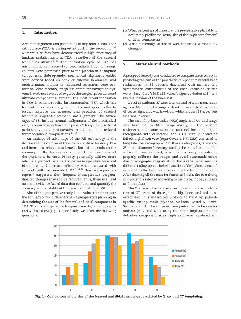
j o u r n a l o f a r t h r o s c o p y and j o i n t s u r g e r y 3 ( 2 0 1 6 ) 1 7 – 2 118
1. Introduction
Accurate alignment and positioning of implants in total kneearthroplasty (TKA) is an important goal of the procedure.1–7
Numerous studies have demonstrated a high frequency ofimplant malalignment in TKA, regardless of the surgicaltechniques utilized.8–10 The innovation cycle of TKA hasmirrored this fundamental concept. Initially, free-hand surgi-cal cuts were performed prior to the placement of implantcomponents. Subsequently, mechanical alignment guideswere devised based on bony or external landmarks, andpredetermined angular or measured resections were per-formed. More recently, imageless computer navigation sys-tems have been developed to guide the surgical procedure andultimate component alignment. The most recent innovationin TKA is patient-specific instrumentation (PSI), which hasbeen introduced as a next generation technology in an effort tofurther improve the accuracy and precision of surgicaltechnique, implant placement, and alignment. The advan-tages of PSI include normal realignment of the mechanicalaxis,minimized resection of the patient's bony tissue, reducedperioperative and postoperative blood loss, and reducedthromboembolic complications.11–13
An anticipated advantage of the PSI technology is thedecrease in the number of trays to be sterilized for every TKAand hence the related cost benefit. But this depends on theaccuracy of the technology to predict the exact size ofthe implant to be used. PSI may potentially achieve morereliable alignment parameters, decrease operative time andblood loss, and increase efficiency when compared withconventionally instrumented TKA.1,14–18 However, a previousreport18 suggested that frequent intraoperative surgeon-directed changes may still be required. Thus, there is a needfor more evidence-based data that evaluate and quantify theaccuracy and reliability of CT-based templating in PSI.
Aim of this prospective study is to evaluate and comparethe accuracy of two different types of preoperative planning, indetermining the size of the femoral and tibial component inTKA. The two compared techniques were digital radiographyand CT-based PSI (Fig. 1). Specifically, we asked the followingquestions:
[(Fig._1)TD$FIG]Fig. 1 – Comparison of the size of the femoral and tibial
(1) W
co
hat percentage of timeswas the preoperative plan able toaccurately predict the actual size of the implanted femoralor tibial components?
(2) W
hat percentage of knees was implanted without anychanges?2. Materials and methods
Aprospective studywas conducted to compare the accuracy inpredicting the size of the prosthetic components in total kneereplacement in 81 patients diagnosed with primary andsymptomatic osteoarthritis of the knee. Inclusion criteriawere ‘‘Easy Knee’’: BMI ≤35, varus/valgus deviation ≤158, andresidual flexion of the knee ≥908.
Out of 81 patients, 37 were women and 44 were men; meanage was 68.5 years, the range extended from 53 to 79 years. In48 cases, right side was involved, while in other 33 cases, leftside was involved.
The mean hip–knee–ankle (HKA) angle is 177.48 and rangewas from 172 to 1848. Preoperatively, all the patientsunderwent the same standard protocol including digitalradiographs with calibration and a CT scan. A dedicatedIMPAX digital software (Agfa-Gevaert, NV, USA) was used totemplate the radiographs. On these radiographs, a sphere,25 mm in diameter (size suggested by the manufacturer of thesoftware), was included, which is necessary in order toproperly calibrate the images and avoid systematic errorsdue to radiographicmagnification, that is variable between thedifferent radiographs. The best position of the sphere ismedialor lateral to the knee, as close as possible to the bone level.After drawing all the axes for femur and tibia, the best fittingcomponent is selected according to the make, model, and sizeof the implant.
The CT-based planning was performed on 3D reconstruc-tion of CT scans of three joints: hip, knee, and ankle, asestablished in standardized protocol to build up patient-specific cutting mask (MyKnee, Medacta, Castel S. Pietro,Switzerland). All the surgeries were performed by two seniorauthors (M.A. and N.C.) using the same implant, and thedefinitive component sizes implanted were registered and
mponent predicted by X-ray and CT templating.
j o u rn a l o f a r th r o s c o p y and j o i n t s u r g e r y 3 ( 2 0 1 6 ) 1 7 – 2 1 19
compared with the sizes suggested by both planning techni-ques considering also the range of error. Results' analysis wascarried out using nonparametric tests.
3. Result
The planning of digital radiography indicates the correct sizein 71% of the cases for the femoral component and 47% for thetibial component. The accuracy reaches 92–95% for bothcomponents if we consider the maximum error of one size. Byevaluating the correlation of the two components together, itappears that in 65% of cases the planning digital X-ray showsexactly the prosthetic components to be implanted; thispercentage becomes 93% when considering a margin of errorof up to one size (Table 1).
CT-based planning reached an accuracy of 93% for thefemur and 67% for the tibia considering the exact size. Theaccuracy reaches 100% for both components if considered themaximum error of one size. By evaluating the correlation ofthe two components together, it appears that in 80% of cases,the CT-based planning predicts exactly the prosthetic compo-nents to be implanted; this percentage becomes 100% as amargin of error of up to one size (Table 1).
The mean postoperative HKA angle was 177.538 (range:173–183).
There is a statistically significant difference in X-ray-basedplanning and CT-based planning for predicting femur size(p < 0.01), while there was no statistical significance as far astibia was concerned. When comparing both femur and tibia,CT-based planning showed statistically significant accurateresults compared to X-ray planning (p < 0.01).
There were no surgical complications, including bleeding,wound complications, arterial or venous thromboembolicdisease, vascular injury, neural deficit, ligament injury,instability, stiffness, fracture, infection, osteolysis, or implantloosening during any of the cases.
4. Discussion
The digital templating is a technique of preoperative studyused to try to predict in advance the size of the prosthesis to beimplanted. This method is spreading in orthopedic surgery
Table 1 – Correlation between the X-ray and CT-based templat
Planning error(no. planned sizevs installed size)
Femur and tibia
Rx CT-based
�2 0 0 0�1 18 (11.2%) 26 (16.0%) 10 105 (64.8%) 130 (80.2%) 58+1 28 (17.2%) 6 (3.8%) 15+2 11 (6.8%) 0 7Overall 162 162 81Within �1 151 (93%) 162 (100%) 73Over �1 11 (6.7%) 0 7
andhas largely replaced the study of analogX-rays and acetatetemplating.
Several studies have shownagoodability topredict, throughplanningofdigital radiography, the sizeof the tibial and femoralcomponents of the prosthesis to be implanted.19–21 In addition,the planning can provide an estimate of the angle of the bonecuts to be performed in the operating room, which can help inmaking a decision on how to proceed during surgery. It alsoallows for prevention of possible errors or technical difficultiesat the time of surgery.20
However, previous studies have suggested that templatingwith regard to implant size selection may provide inaccurateresults and the findings are not reproducible (Table 2).22–26
Trickett et al.22 showed accuracy to the exact size by 50%,rising to 98% if we considered a maximum error of one size.The femoral component was generally scheduled with higheraccuracy compared to that of the tibial component.
Heal and Blewitt23 found preoperative radiological templat-ing to be accurate in just 57% of cases and thus questioned itsusefulness in preoperative management.
Hsu et al.,26 studying 48 patients undergoing total kneereplacement, as well as confirming that 54% of the size of theprosthesiswas correctlypredictedby thedigital templating, alsostated that there were no statistically significant differenceswhen digital planning was done by an experienced surgeon inprosthetic surgery of the knee than one with less experience.
Kobayashi et al.27 have finally compared the accuracy oftraditional two-dimensional X-ray and three-dimensional CTscan, while getting superior results with CT, and showed thatthe improvement was not statistically significant and thiswould seem to indicate the non-necessity of preoperative CTscan in TKA.
It is therefore unclearwhether these emerging technologiesoffer a real cost benefit or result in an improved outcome.Hence, the present study tried to evaluate the usefulness andreliability of the preoperative CT-based templating to predictthe component size compared to the digital templating.
In our study, the improvement in the ability to predict thesize of the prosthetic components obtained by theCT images isstatistically significant compared to that obtained by theradiographic study to predict the size of the tibial and femoralcomponent.
In fact, it was possible to predict the size of the prostheticimplant with digital radiograph in 72% of cases for the femoral
e size and final implant size used.
Femur Tibia
Rx CT-based Rx CT-based
0 0 0(1.2%) 2 (2.5%) 17 (21.0%) 24 (29.6%)(71.7%) 76 (93.8%) 47 (58.0%) 54 (66.7%)(18.5%) 3 (3.7%) 13 (16.0%) 3 (3.7%)(8.6%) 0 4 (5.0%) 0
81 81 81(90%) 81 (100%) 77 (95%) 81 (100%)(8.6%) 0 4 (4.9%) 0
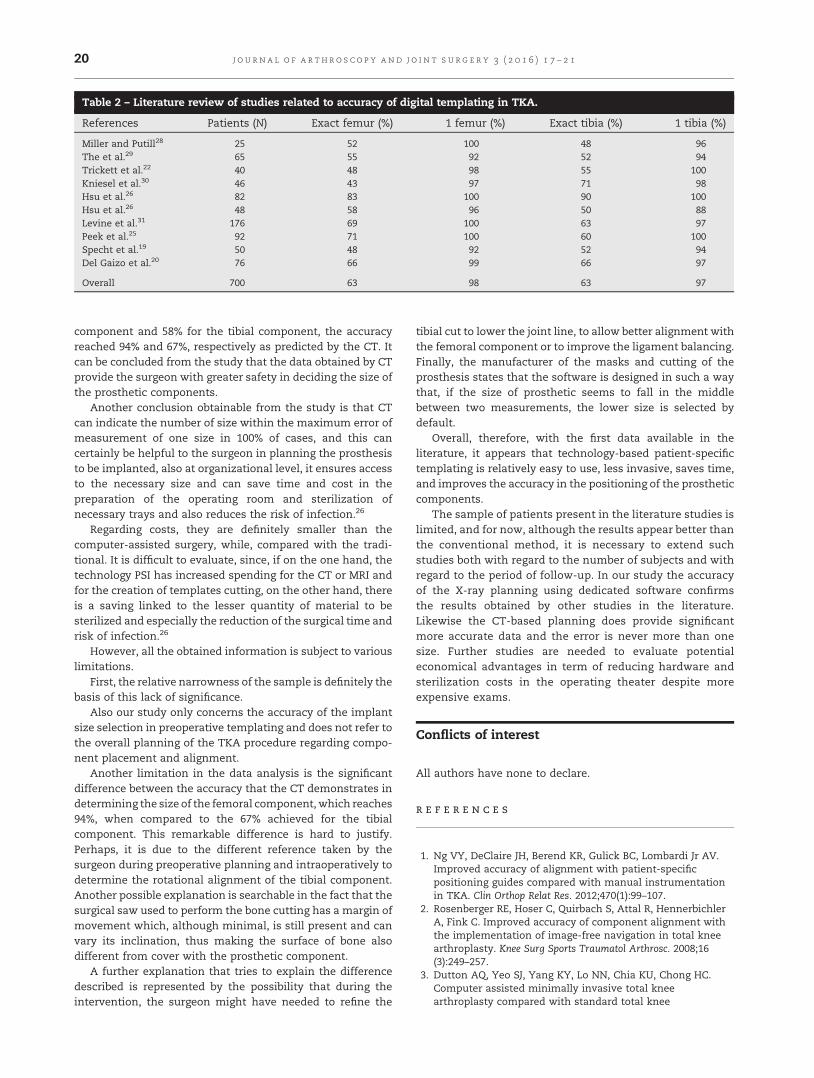
Table 2 – Literature review of studies related to accuracy of digital templating in TKA.
References Patients (N) Exact femur (%) �1 femur (%) Exact tibia (%) �1 tibia (%)
Miller and Putill28 25 52 100 48 96The et al.29 65 55 92 52 94Trickett et al.22 40 48 98 55 100Kniesel et al.30 46 43 97 71 98Hsu et al.26 82 83 100 90 100Hsu et al.26 48 58 96 50 88Levine et al.31 176 69 100 63 97Peek et al.25 92 71 100 60 100Specht et al.19 50 48 92 52 94Del Gaizo et al.20 76 66 99 66 97
Overall 700 63 98 63 97
j o u r n a l o f a r t h r o s c o p y and j o i n t s u r g e r y 3 ( 2 0 1 6 ) 1 7 – 2 120
component and 58% for the tibial component, the accuracyreached 94% and 67%, respectively as predicted by the CT. Itcan be concluded from the study that the data obtained by CTprovide the surgeon with greater safety in deciding the size ofthe prosthetic components.
Another conclusion obtainable from the study is that CTcan indicate the number of size within the maximum error ofmeasurement of one size in 100% of cases, and this cancertainly be helpful to the surgeon in planning the prosthesisto be implanted, also at organizational level, it ensures accessto the necessary size and can save time and cost in thepreparation of the operating room and sterilization ofnecessary trays and also reduces the risk of infection.26
Regarding costs, they are definitely smaller than thecomputer-assisted surgery, while, compared with the tradi-tional. It is difficult to evaluate, since, if on the one hand, thetechnology PSI has increased spending for the CT or MRI andfor the creation of templates cutting, on the other hand, thereis a saving linked to the lesser quantity of material to besterilized and especially the reduction of the surgical time andrisk of infection.26
However, all the obtained information is subject to variouslimitations.
First, the relative narrowness of the sample is definitely thebasis of this lack of significance.
Also our study only concerns the accuracy of the implantsize selection in preoperative templating and does not refer tothe overall planning of the TKA procedure regarding compo-nent placement and alignment.
Another limitation in the data analysis is the significantdifference between the accuracy that the CT demonstrates indetermining the size of the femoral component, which reaches94%, when compared to the 67% achieved for the tibialcomponent. This remarkable difference is hard to justify.Perhaps, it is due to the different reference taken by thesurgeon during preoperative planning and intraoperatively todetermine the rotational alignment of the tibial component.Another possible explanation is searchable in the fact that thesurgical saw used to perform the bone cutting has a margin ofmovement which, although minimal, is still present and canvary its inclination, thus making the surface of bone alsodifferent from cover with the prosthetic component.
A further explanation that tries to explain the differencedescribed is represented by the possibility that during theintervention, the surgeon might have needed to refine the
tibial cut to lower the joint line, to allow better alignment withthe femoral component or to improve the ligament balancing.Finally, the manufacturer of the masks and cutting of theprosthesis states that the software is designed in such a waythat, if the size of prosthetic seems to fall in the middlebetween two measurements, the lower size is selected bydefault.
Overall, therefore, with the first data available in theliterature, it appears that technology-based patient-specifictemplating is relatively easy to use, less invasive, saves time,and improves the accuracy in the positioning of the prostheticcomponents.
The sample of patients present in the literature studies islimited, and for now, although the results appear better thanthe conventional method, it is necessary to extend suchstudies both with regard to the number of subjects and withregard to the period of follow-up. In our study the accuracyof the X-ray planning using dedicated software confirmsthe results obtained by other studies in the literature.Likewise the CT-based planning does provide significantmore accurate data and the error is never more than onesize. Further studies are needed to evaluate potentialeconomical advantages in term of reducing hardware andsterilization costs in the operating theater despite moreexpensive exams.
Conflicts of interest
All authors have none to declare.
r e f e r e n c e s
1. Ng VY, DeClaire JH, Berend KR, Gulick BC, Lombardi Jr AV.Improved accuracy of alignment with patient-specificpositioning guides compared with manual instrumentationin TKA. Clin Orthop Relat Res. 2012;470(1):99–107.
2. Rosenberger RE, Hoser C, Quirbach S, Attal R, HennerbichlerA, Fink C. Improved accuracy of component alignment withthe implementation of image-free navigation in total kneearthroplasty. Knee Surg Sports Traumatol Arthrosc. 2008;16(3):249–257.
3. Dutton AQ, Yeo SJ, Yang KY, Lo NN, Chia KU, Chong HC.Computer assisted minimally invasive total kneearthroplasty compared with standard total knee
j o u rn a l o f a r th r o s c o p y and j o i n t s u r g e r y 3 ( 2 0 1 6 ) 1 7 – 2 1 21
arthroplasty. A prospective, randomized study. J Bone JointSurg Am. 2008;90(1):2–9.
4. Chin PL, Yang KY, Yeo SJ, Lo NN. Randomized control trialcomparing radiographic total knee arthroplasty implantplacement using computer navigation versus conventionaltechnique. J Arthroplasty. 2005;20(5):618–626.
5. Hernández-Vaquero D, Suarez-Vazquez A, Sandoval-GarciaMA, Noriega-Fernandez A. Computer assistance increasesprecision of component placement in total kneearthroplasty with articular deformity. Clin Orthop Relat Res.2010;468(5):1237–1241.
6. Kim YH, Kim JS, Yoon SH. Alignment and orientation of thecomponents in total knee replacement with and withoutnavigation support: a prospective, randomised study. J BoneJoint Surg Br. 2007;89(4):471–476.
7. Park SE, Lee CT. Comparison of robotic-assisted andconventional manual implantation of a primary total kneearthroplasty. J Arthroplasty. 2007;22(7):1054–1059.
8. Patil S, D'Lima DD, Fait JM, Colwell Jr CW. Improving tibialcomponent coronal alignment during total kneearthroplasty with use of a tibial planning device. J Bone JointSurg. 2007;89(2):381–387.
9. Mizu-uchi H, Matsuda S, Miura H, Higaki H, Okazaki K,Iwamoto Y. The effect of ankle rotation on cutting of thetibia in total knee arthroplasty. J Bone Joint Surg. 2006;88(12):2632–2636.
10. Matziolis G, Krocker D, Weiss U, Tohtz S, Perka C. Aprospective, randomized study of computer-assisted andconventional total knee arthroplasty: three-dimensionalevaluation of implant alignment and rotation. J Bone JointSurg. 2007;89(2):236–243.
11. Lombardi Jr AV, Berend KR, Adams JB. Patient specificapproach in total knee arthroplasty. Orthopedics. 2008;31(9):927–930.
12. Daniilidis K, Tibesku CO. A comparison of conventional andpatient specific instruments in total knee arthroplasty. IntOrthop. 2014;38(3):503–508.
13. Conteduca F, Iorio R, Mazza D, Ferretti A. Patient specificinstruments in arthroplasty. Int Orthop. 2014;38(2):259–265.
14. Howell SM, Kuznik K, Hull ML, Siston RA. Results of an initialexperience with custom-fit positioning total kneearthroplasty in a series of 48 patients. Orthopedics. 2008;31(9):857–863.
15. Watters TS, Mather III RC, Browne JA, Berend KR, LombardiJr AV, Bolognesi MP. Analysis of procedure-related costs andproposed benefits of using patient-specific approach in totalknee arthroplasty. J Surg Orthop Adv. 2011;20(2):112–116.
16. Spencer BA, Mont MA, McGrath MS, Boyd B, Mitrick MF.Initial experience with custom-fit total knee replacement:
intra-operative events and long-leg coronal alignment. IntOrthop. 2009;33(6):1571–1575.
17. Heyse TJ, Tibesku CO. Improved femoral componentrotation in TKA using patient-specific instrumentation.Knee. 2014;21(1):268–271.
18. Stronach BM, Pelt CE, Erickson J, Peters CL. Patient-specifictotal knee arthroplasty requires frequent surgeon-directedchanges. Clin Orthop Relat Res. 2013;471(1):169–174.
19. Specht LM, Levitz S, Iorio R, Healy WL, Tilzey JF. Acomparison of acetate and digital templating for total kneearthroplasty. Clin Orthop Relat Res. 2007;464:179–183.
20. Del Gaizo D, Soileau ES, Lachiewicz PF. Value of preoperativetemplating for primary total knee arthroplasty. J Knee Surg.2009;22(4):284–293.
21. Parvizi J. Digital templating: here to stay. Am J Orthop (BelleMead NJ). 2011;40(8):394.
22. Trickett RW, Hodgson P, Forster MC, Robertson A. Thereliability and accuracy of digital templating in total kneereplacement. J Bone Joint Surg Br. 2009;91(7):903–906.
23. Heal J, Blewitt N. Kinemax total knee arthroplasty: trial bytemplate. J Arthroplasty. 2002;17:90–94.
24. Arora J, Sharma S, Blyth M. The role of preoperativetemplating in primary total knee replacement. Knee SurgSports Traumatol Arthrosc. 2005;13(3):187–189.
25. Peek AC, Bloch B, Auld J. How useful is templating fortotal knee replacement component sizing? Knee. 2012;19(4):266–269.
26. Hsu AR, Gross CE, Bhatia S, Levine BR. Template-directedinstrumentation in total knee arthroplasty: cost savingsanalysis. Orthopedics. 2012;35(11):e1596–e1600.
27. Kobayashi A, Ishii Y, Takeda M, Noguchi H, Higuchi H,Toyabe S. Comparison of analog 2D and digital 3Dpreoperative templating for predicting implant size intotal knee arthroplasty. Comput Aided Surg. 2012;17(2):96–101.
28. Miller AG, Purtill JJ. Accuracy of digital templating in totalknee arthroplasty. Am J Orthop (Belle Mead NJ). 2012;41(11):510–512.
29. The B, Diercks RL, van Ooijen PM, van Horn JR. Comparisonof analog and digital preoperative planning in total hip andknee arthroplasties. A prospective study of 173 hips and 65total knees. Acta Orthop. 2005;76(1):78–84.
30. Kniesel B, Konstantinidis L, Hirschmüller A, Südkamp N,Helwig P. Digital templating in total knee and hipreplacement: an analysis of planning accuracy. Int Orthop.2014;38(4):733–739.
31. Levine B, Fabi D, Deirmengian C. Digital templating inprimary total hip and knee arthroplasty. Orthopedics. 2010;33(11):797.
j o u r n a l o f a r t h r o s c o p y and j o i n t s u r g e r y 3 ( 2 0 1 6 ) 2 2 – 2 7
Available online at www.sciencedirect.com
ScienceDirect
journal homepage: www.elsevier.com/locate/jajs
Original Article
A radiological comparative study between
transtibial & anteromedial portal drilling of femoraltunnel in single bundle anterior cruciate ligamentreconstruction: A comparison of four anglesSiddharth M. Shetty a, Vikram Shetty a, Arjun Ballal b,*,Jeetu Mohanchandran d, Anoop Hegde c
aAssociate Professor, K.S Hegde Medical Academy, Mangalore 575018, IndiabRegistrar, ARS Hospital, Tirupur 641604, IndiacRegistrar, Orthopaedic Surgeon, ARS Hospital, Tirupur 641604, IndiadPostgraduate, Department Of Orthopaedic surgery, K.S Hegde Medical Academy, Mangalore 575018, India
a r t i c l e i n f o
Article history:
Received 15 August 2015
Accepted 12 November 2015
Available online 9 December 2015
Keywords:
Anterior cruciate ligament
Transtibial
Transportal
Condylar tunnel angle
Shaft tunnel angle
Tunnel screw angle
a b s t r a c t
Femoral tunnel drilling for graft insertion is performed by transtibial or transportal tech-
nique.
Aim: Main objective of this study was to compare the postoperative anteroposterior and
lateral radiographs after arthroscopic ACL reconstruction by transtibial and transportal
techniques by measuring four angles.
Materials and methods: A retrospective study of 60 patients was conducted, who were further
subdivided into two groups containing 30 each. The first group had patients who had
undergone arthroscopic ACL reconstruction by transtibial technique and the second group
had patients who had undergone ACL reconstruction by transportal technique. Post-opera-
tive anteroposterior radiographs (AP) and lateral radiographs of the operated knees were
compared. Condylar tunnel (CT) and tunnel screw (TS) angles were measured in AP radio-
graphs and compared. In lateral radiographs, shaft tunnel (ST) and TS angles weremeasured
and compared. 'Student t-test' was used in calculating the results.
Results: Mean of the angles measured were tabulated and compared. CT angle was noted to
be more acute in transportal group (38.4 � 8.7568) as compared to transtibial group (46.97
� 12.7548) with a significant 'p-value' (0.004). ST angle was noted to be more obtuse in
the transportal group (119.57 � 11.2128) as compared to transtibial group (113.17 � 12.7938)
with a significant 'p-value' (0.044).
This infers that transportal technique oriented the graft more horizontal as compared to
transtibial technique.
es when compared in AP radiographs showed that transportal tech-
ad more acute angulation as compared to transtibial group (12.57
nique (5.5 � 4.6448) h� 8.2878) with a signifi
Tunnel screw angl
cant 'p-value' (<0.001). Same results were obtained when the lateral
* Corresponding author. Tel.: +91 7708913999.E-mail address: ArjunBallalb*[email protected]">ArjunBallalb*[email protected] ().
http://dx.doi.org/10.1016/j.jajs.2015.11.0112214-9635/# 2015 International Society for Knowledge for Surgeons on Arthroscopy and Arthroplasty. Published by Elsevier, a division ofReed Elsevier India, Pvt. Ltd. All rights reserved.
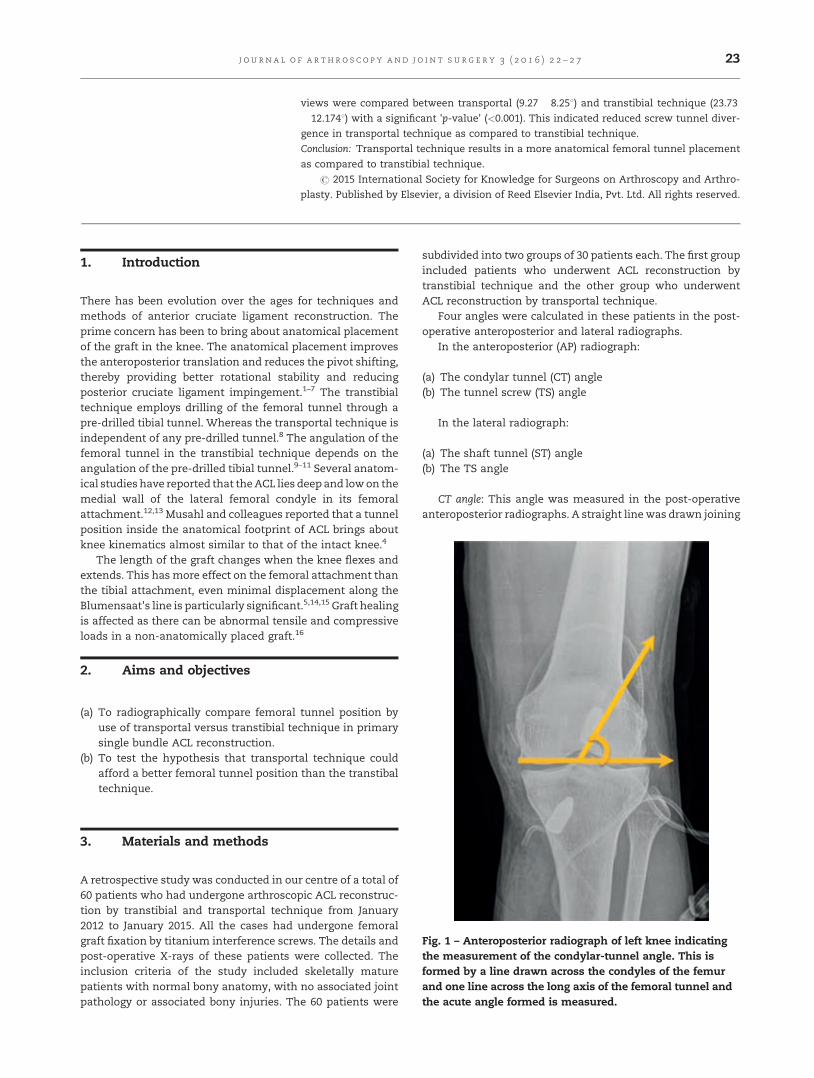
views were compared between transportal (9.27 � 8.258) and transtibial technique (23.73
� 12.1748) with a significant 'p-value' (<0.001). This indicated reduced screw tunnel diver-
gence in transportal technique as compared to transtibial technique.
Conclusion: Transportal technique results in a more anatomical femoral tunnel placement
as compared to transtibial technique.
# 2015 International Society for Knowledge for Surgeons on Arthroscopy and Arthro-
plasty. Published by Elsevier, a division of Reed Elsevier India, Pvt. Ltd. All rights reserved.
j o u rn a l o f a r th r o s c o p y and j o i n t s u r g e r y 3 ( 2 0 1 6 ) 2 2 – 2 7 23
1. Introduction
[(Fig._1)TD$FIG]
There has been evolution over the ages for techniques andmethods of anterior cruciate ligament reconstruction. Theprime concern has been to bring about anatomical placementof the graft in the knee. The anatomical placement improvesthe anteroposterior translation and reduces the pivot shifting,thereby providing better rotational stability and reducingposterior cruciate ligament impingement.1–7 The transtibialtechnique employs drilling of the femoral tunnel through apre-drilled tibial tunnel. Whereas the transportal technique isindependent of any pre-drilled tunnel.8 The angulation of thefemoral tunnel in the transtibial technique depends on theangulation of the pre-drilled tibial tunnel.9–11 Several anatom-ical studies have reported that theACL lies deep and lowon themedial wall of the lateral femoral condyle in its femoralattachment.12,13 Musahl and colleagues reported that a tunnelposition inside the anatomical footprint of ACL brings aboutknee kinematics almost similar to that of the intact knee.4
The length of the graft changes when the knee flexes andextends. This has more effect on the femoral attachment thanthe tibial attachment, even minimal displacement along theBlumensaat's line is particularly significant.5,14,15 Graft healingis affected as there can be abnormal tensile and compressiveloads in a non-anatomically placed graft.16
2. Aims and objectives
(a) T
o radiographically compare femoral tunnel position byuse of transportal versus transtibial technique in primarysingle bundle ACL reconstruction.(b) T
o test the hypothesis that transportal technique couldafford a better femoral tunnel position than the transtibaltechnique.3. Materials and methods
Fig. 1 – Anteroposterior radiograph of left knee indicatingthe measurement of the condylar-tunnel angle. This isformed by a line drawn across the condyles of the femurand one line across the long axis of the femoral tunnel andthe acute angle formed is measured.
A retrospective study was conducted in our centre of a total of60 patients who had undergone arthroscopic ACL reconstruc-tion by transtibial and transportal technique from January2012 to January 2015. All the cases had undergone femoralgraft fixation by titanium interference screws. The details andpost-operative X-rays of these patients were collected. Theinclusion criteria of the study included skeletally maturepatients with normal bony anatomy, with no associated jointpathology or associated bony injuries. The 60 patients were
subdivided into two groups of 30 patients each. The first groupincluded patients who underwent ACL reconstruction bytranstibial technique and the other group who underwentACL reconstruction by transportal technique.
Four angles were calculated in these patients in the post-operative anteroposterior and lateral radiographs.
In the anteroposterior (AP) radiograph:
(a) T
he condylar tunnel (CT) angle (b) T he tunnel screw (TS) angleIn the lateral radiograph:
(a) T
he shaft tunnel (ST) angle (b) T he TS angleCT angle: This angle was measured in the post-operativeanteroposterior radiographs. A straight line was drawn joining
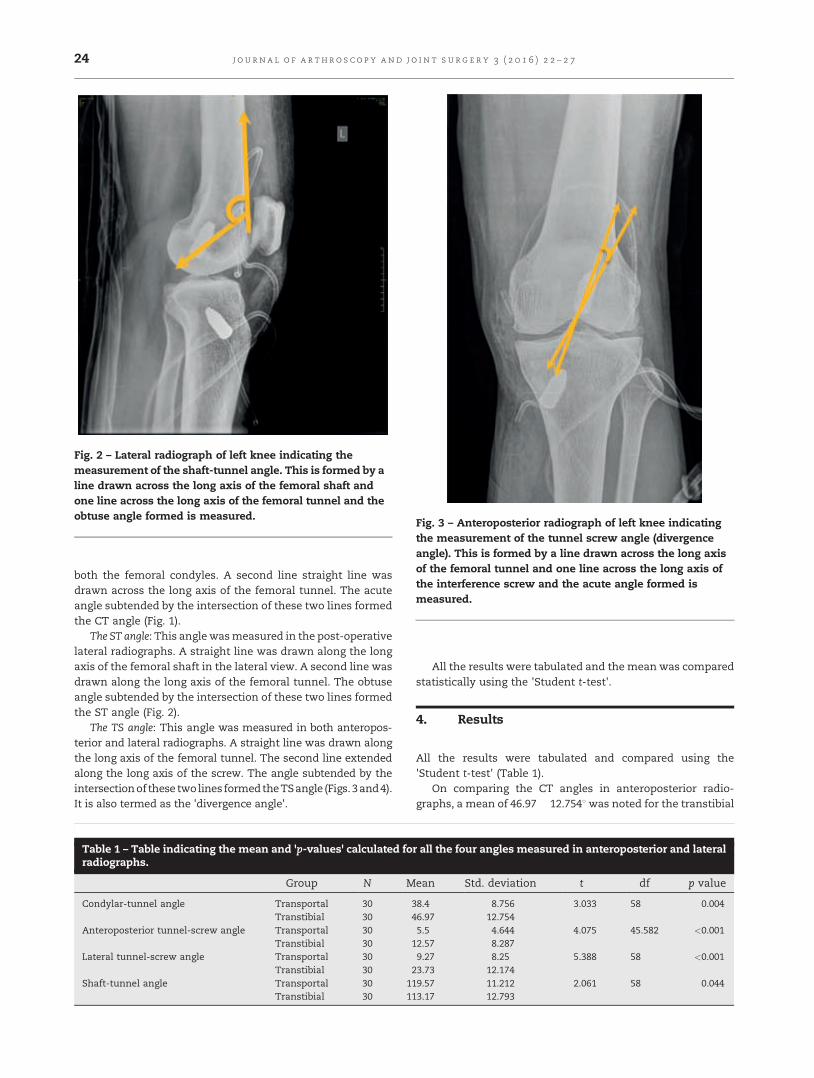
[(Fig._2)TD$FIG]
Fig. 2 – Lateral radiograph of left knee indicating themeasurement of the shaft-tunnel angle. This is formed by aline drawn across the long axis of the femoral shaft andone line across the long axis of the femoral tunnel and theobtuse angle formed is measured.
[(Fig._3)TD$FIG]
Fig. 3 – Anteroposterior radiograph of left knee indicatingthe measurement of the tunnel screw angle (divergenceangle). This is formed by a line drawn across the long axisof the femoral tunnel and one line across the long axis ofthe interference screw and the acute angle formed ismeasured.
j o u r n a l o f a r t h r o s c o p y and j o i n t s u r g e r y 3 ( 2 0 1 6 ) 2 2 – 2 724
both the femoral condyles. A second line straight line wasdrawn across the long axis of the femoral tunnel. The acuteangle subtended by the intersection of these two lines formedthe CT angle (Fig. 1).
The ST angle: This angle wasmeasured in the post-operativelateral radiographs. A straight line was drawn along the longaxis of the femoral shaft in the lateral view. A second line wasdrawn along the long axis of the femoral tunnel. The obtuseangle subtended by the intersection of these two lines formedthe ST angle (Fig. 2).
The TS angle: This angle was measured in both anteropos-terior and lateral radiographs. A straight line was drawn alongthe long axis of the femoral tunnel. The second line extendedalong the long axis of the screw. The angle subtended by theintersectionof these twolines formedtheTSangle (Figs. 3and4).It is also termed as the 'divergence angle'.
Table 1 – Table indicating the mean and 'p-values' calculated foradiographs.
Group N M
Condylar-tunnel angle Transportal 30 3Transtibial 30 4
Anteroposterior tunnel-screw angle Transportal 30Transtibial 30 1
Lateral tunnel-screw angle Transportal 30Transtibial 30 2
Shaft-tunnel angle Transportal 30 11Transtibial 30 11
All the results were tabulated and the mean was comparedstatistically using the 'Student t-test'.
4. Results
All the results were tabulated and compared using the'Student t-test' (Table 1).
On comparing the CT angles in anteroposterior radio-graphs, a mean of 46.97 � 12.7548was noted for the transtibial
r all the four angles measured in anteroposterior and lateral
ean Std. deviation t df p value
8.4 8.756 �3.033 58 0.0046.97 12.7545.5 4.644 �4.075 45.582 <0.0012.57 8.2879.27 8.25 �5.388 58 <0.0013.73 12.1749.57 11.212 2.061 58 0.0443.17 12.793
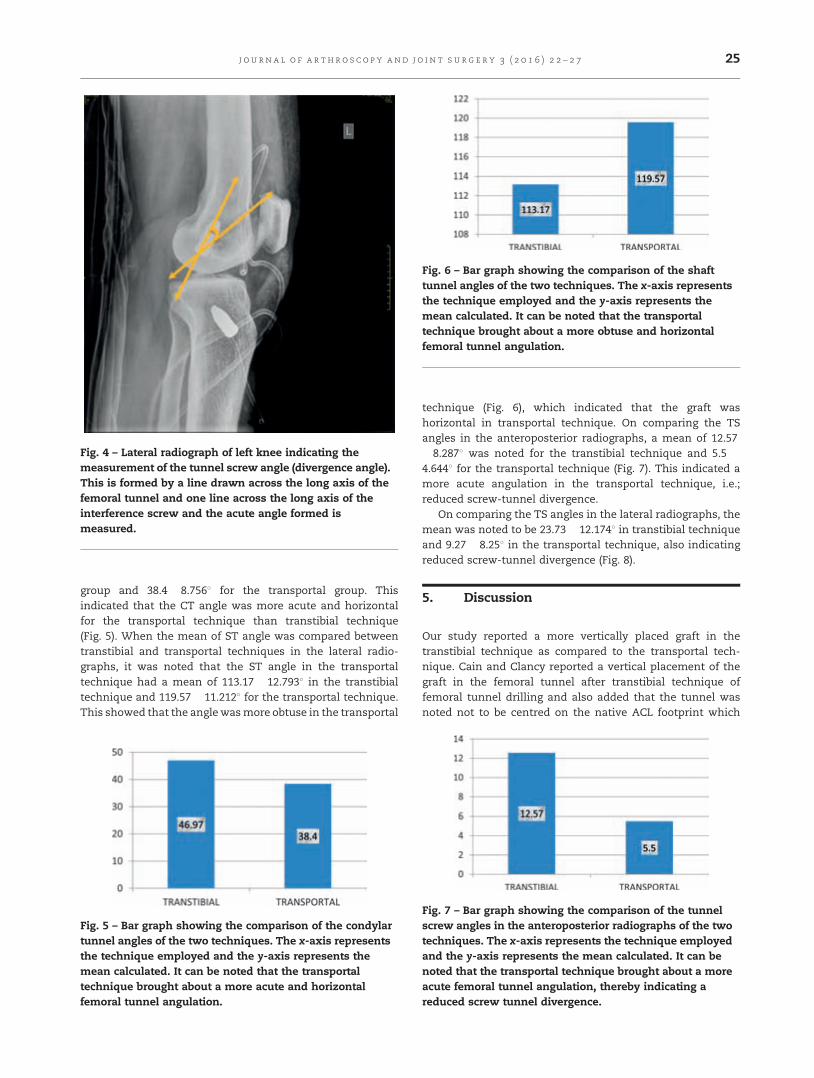
[(Fig._6)TD$FIG]
Fig. 6 – Bar graph showing the comparison of the shafttunnel angles of the two techniques. The x-axis representsthe technique employed and the y-axis represents themean calculated. It can be noted that the transportaltechnique brought about a more obtuse and horizontalfemoral tunnel angulation.
[(Fig._4)TD$FIG]
Fig. 4 – Lateral radiograph of left knee indicating themeasurement of the tunnel screw angle (divergence angle).This is formed by a line drawn across the long axis of thefemoral tunnel and one line across the long axis of theinterference screw and the acute angle formed ismeasured.
j o u rn a l o f a r th r o s c o p y and j o i n t s u r g e r y 3 ( 2 0 1 6 ) 2 2 – 2 7 25
group and 38.4 � 8.7568 for the transportal group. Thisindicated that the CT angle was more acute and horizontalfor the transportal technique than transtibial technique(Fig. 5). When the mean of ST angle was compared betweentranstibial and transportal techniques in the lateral radio-graphs, it was noted that the ST angle in the transportaltechnique had a mean of 113.17 � 12.7938 in the transtibialtechnique and 119.57 � 11.2128 for the transportal technique.This showed that the anglewasmore obtuse in the transportal
[(Fig._5)TD$FIG]
Fig. 5 – Bar graph showing the comparison of the condylartunnel angles of the two techniques. The x-axis representsthe technique employed and the y-axis represents themean calculated. It can be noted that the transportaltechnique brought about a more acute and horizontalfemoral tunnel angulation.
technique (Fig. 6), which indicated that the graft washorizontal in transportal technique. On comparing the TSangles in the anteroposterior radiographs, a mean of 12.57� 8.2878 was noted for the transtibial technique and 5.5 �4.6448 for the transportal technique (Fig. 7). This indicated amore acute angulation in the transportal technique, i.e.;reduced screw-tunnel divergence.
On comparing the TS angles in the lateral radiographs, themean was noted to be 23.73 � 12.1748 in transtibial techniqueand 9.27 � 8.258 in the transportal technique, also indicatingreduced screw-tunnel divergence (Fig. 8).
5. Discussion
Our study reported a more vertically placed graft in thetranstibial technique as compared to the transportal tech-nique. Cain and Clancy reported a vertical placement of thegraft in the femoral tunnel after transtibial technique offemoral tunnel drilling and also added that the tunnel wasnoted not to be centred on the native ACL footprint which[(Fig._7)TD$FIG]
Fig. 7 – Bar graph showing the comparison of the tunnelscrew angles in the anteroposterior radiographs of the twotechniques. The x-axis represents the technique employedand the y-axis represents the mean calculated. It can benoted that the transportal technique brought about a moreacute femoral tunnel angulation, thereby indicating areduced screw tunnel divergence.
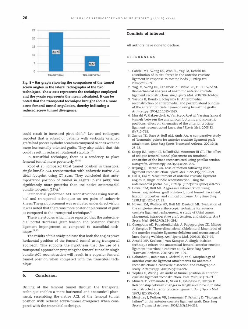
[(Fig._8)TD$FIG]
Fig. 8 – Bar graph showing the comparison of the tunnelscrew angles in the lateral radiographs of the twotechniques. The x-axis represents the technique employedand the y-axis represents the mean calculated. It can benoted that the transportal technique brought about a moreacute femoral tunnel angulation, thereby indicating areduced screw tunnel divergence.
j o u r n a l o f a r t h r o s c o p y and j o i n t s u r g e r y 3 ( 2 0 1 6 ) 2 2 – 2 726
could result in increased pivot shift.17 Lee and colleaguesreported that a subset of patients with vertically orientedgrafts had poorer Lysholmscores as compared to oneswith themore horizontally oriented grafts. They also added that thiscould result in reduced rotational stability.18
In transtibial technique, there is a tendency to placefemoral tunnel more posteriorly.19–22
Kopf et al. compared tibial tunnel position in transtibialsingle bundle ACL reconstruction with cadaveric native ACLtibial footprint using CT scan. They concluded that ante-roposterior position of tunnel in sagittal plane (48%) wassignificantly more posterior than the native anteromedialbundle footprint (25%).21
Steiner et al. performed ACL reconstructions using transti-bial and transportal techniques on ten pairs of cadavericknees. The graft placement was evaluated under direct vision.It was noted that there was more posterior placement of graftas compared to the transportal technique.23
There are studies which have reported that the anterome-dial portal decreases graft tension and posterior cruciateligament impingement as compared to transtibial tech-nique.24,25
The results of this study indicate that both the angles provehorizontal position of the femoral tunnel using transportalapproach. This supports the hypothesis that the use of atransportal approach in preparing the femoral tunnel in singlebundle ACL reconstruction will result in a superior femoraltunnel position when compared with the transtibial tech-nique.
6. Conclusion
Drilling of the femoral tunnel through the transportaltechnique enables a more horizontal and anatomical place-ment, resembling the native ACL of the femoral tunnelposition with reduced screw-tunnel divergence when com-pared with the transtibial technique.
Conflicts of interest
All authors have none to declare.
r e f e r e n c e s
1. Gabriel MT, Wong EK, Woo SL, Yagi M, Debski RE.Distribution of in situ forces in the anterior cruciateligament in response to rotator loads. J Orthop Res.2004;22:85–89.
2. Yagi M, Wong EK, Kanamori A, Debski RE, Fu FH, Woo SL.Biomechanical analysis of anatomic anterior cruciateligament reconstruction. Am J Sports Med. 2002;30:660–666.
3. Yasuda K, Kondo E, Ichiyama H. Anteromedialreconstruction of anteromedial and posterolateral bundlesof the anterior cruciate ligament using hamstring grafts.Arthroscopy. 2004;20:1015–1025.
4. Musahl V, Plakseychuk A, VanScyoc A, et al. Varying femoraltunnels between the anatomical footprint and isometricpositions: effect on kinematics of the anterior cruciateligament-reconstructed knee. Am J Sports Med. 2005;33(5):712–718.
5. Zavras TD, Race A, Bull AM, Amis AA. A comparative studyof 'isometric' points for anterior cruciate ligament graftattachment. Knee Surg Sports Traumatol Arthrosc. 2001;9(1):28–33.
6. Scopp JM, Jasper LE, Belkoff SM, Moorman III CT. The effectof oblique femoral tunnel placement on rotationalconstraint of the knee reconstructed using patellar tendonautografts. Arthroscopy. 2004;20(3):294–299.
7. Irrgang JJ, Harner CD. Loss of motion following kneeligament reconstruction. Sports Med. 1995;19(2):150–159.
8. Dai X, Cai Y. Measurement of anterior cruciate ligamentangles in single-bundle reconstruction using theanteromedial portal. Am J Orthop. (June):2012;(June):268–272.
9. Howell SM, Hull ML. Aggressive rehabilitation usinghamstring tendons: graft construct, tibial tunnel placement,fixation properties, and clinical outcome. Am J Knee Surg.1998;11(2):120–127. 23.
10. Howell SM, Wallace MP, Hull ML, Deutsch ML. Evaluation ofthe single-incision arthroscopic technique for anteriorcruciate ligament replacement. A study of tibial tunnelplacement, intraoperative graft tension, and stability. Am JSports Med. 1999;27(3):284–293.
11. Georgoulis AD, Papadonikolakis A, Papageorgiou CD, MitsouA, Stergiou N. Three-dimensional tibiofemoral kinematics ofthe anterior cruciate ligament-deficient and reconstructedknee during walking. Am J Sports Med. 2003;31(1):75–79.
12. Arnold MP, Kooloos J, van Kampen A. Single-incisiontechnique misses the anatomical femoral anterior cruciateligament insertion: a cadaver study. Knee Surg SportsTraumatol Arthrosc. 2001;9(4):194–199.
13. Colombet P, Robinson J, Christel P, et al. Morphology ofanterior cruciate ligament attachments for anatomicreconstruction: a cadaveric dissection and radiographicstudy. Arthroscopy. 2006;22(9):984–992.
14. Topliss C, Webb J. An audit of tunnel position in anteriorcruciate ligament reconstruction. Knee. 2001;8(1):59–63.
15. Muneta T, Yamamoto H, Sakai H, Ishibashi T, Furuya K.Relationship between changes in length and force in in vitroreconstructed anterior cruciate ligament. Am J Sports Med.1993;21(2):299–304.
16. Ménétrey J, Duthon VB, Laumonier T, Fritschy D. ‘‘Biologicalfailure’’ of the anterior cruciate ligament graft. Knee SurgSports Traumatol Arthrosc. 2008;16(3):224–231.
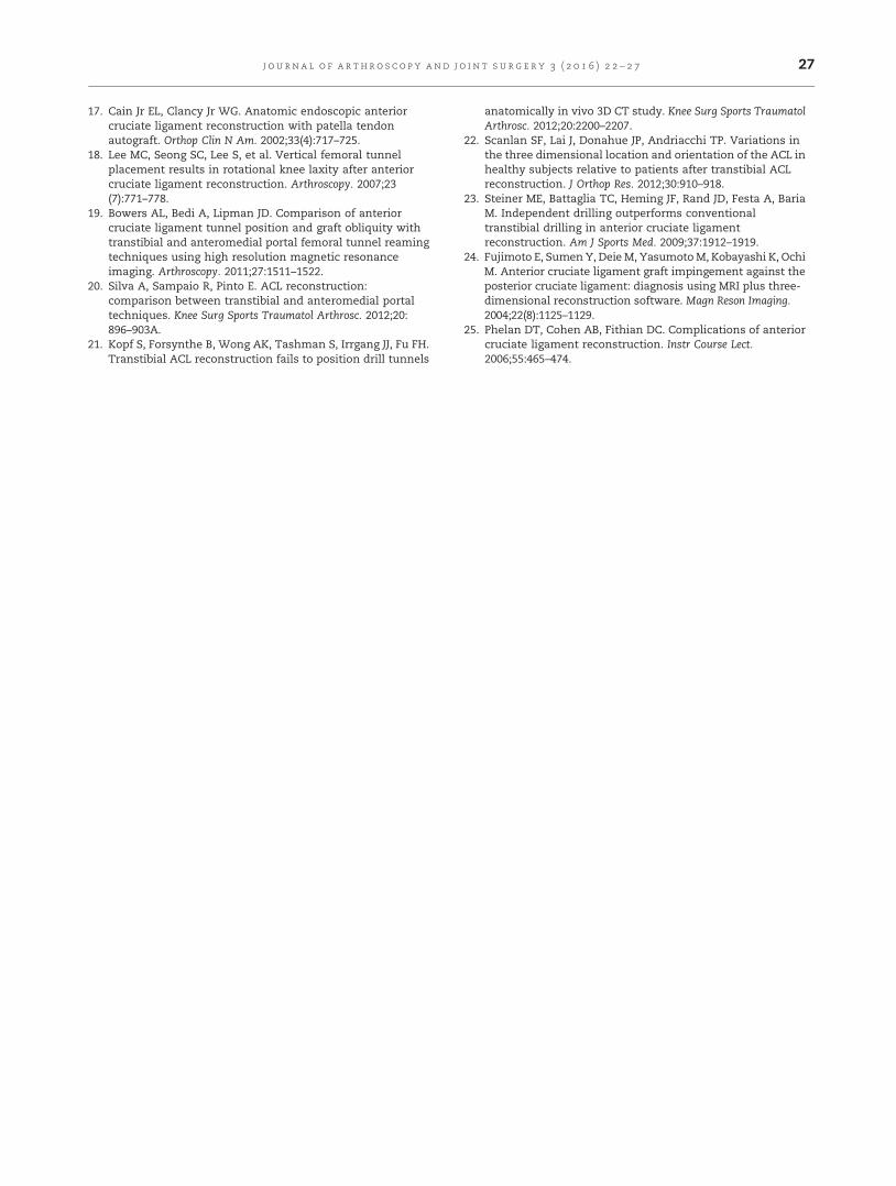
j o u rn a l o f a r th r o s c o p y and j o i n t s u r g e r y 3 ( 2 0 1 6 ) 2 2 – 2 7 27
17. Cain Jr EL, Clancy Jr WG. Anatomic endoscopic anteriorcruciate ligament reconstruction with patella tendonautograft. Orthop Clin N Am. 2002;33(4):717–725.
18. Lee MC, Seong SC, Lee S, et al. Vertical femoral tunnelplacement results in rotational knee laxity after anteriorcruciate ligament reconstruction. Arthroscopy. 2007;23(7):771–778.
19. Bowers AL, Bedi A, Lipman JD. Comparison of anteriorcruciate ligament tunnel position and graft obliquity withtranstibial and anteromedial portal femoral tunnel reamingtechniques using high resolution magnetic resonanceimaging. Arthroscopy. 2011;27:1511–1522.
20. Silva A, Sampaio R, Pinto E. ACL reconstruction:comparison between transtibial and anteromedial portaltechniques. Knee Surg Sports Traumatol Arthrosc. 2012;20:896–903A.
21. Kopf S, Forsynthe B, Wong AK, Tashman S, Irrgang JJ, Fu FH.Transtibial ACL reconstruction fails to position drill tunnels
anatomically in vivo 3D CT study. Knee Surg Sports TraumatolArthrosc. 2012;20:2200–2207.
22. Scanlan SF, Lai J, Donahue JP, Andriacchi TP. Variations inthe three dimensional location and orientation of the ACL inhealthy subjects relative to patients after transtibial ACLreconstruction. J Orthop Res. 2012;30:910–918.
23. Steiner ME, Battaglia TC, Heming JF, Rand JD, Festa A, BariaM. Independent drilling outperforms conventionaltranstibial drilling in anterior cruciate ligamentreconstruction. Am J Sports Med. 2009;37:1912–1919.
24. Fujimoto E, Sumen Y, Deie M, YasumotoM, Kobayashi K, OchiM. Anterior cruciate ligament graft impingement against theposterior cruciate ligament: diagnosis using MRI plus three-dimensional reconstruction software. Magn Reson Imaging.2004;22(8):1125–1129.
25. Phelan DT, Cohen AB, Fithian DC. Complications of anteriorcruciate ligament reconstruction. Instr Course Lect.2006;55:465–474.
j o u r n a l o f a r t h r o s c o p y and j o i n t s u r g e r y 3 ( 2 0 1 6 ) 2 8 – 3 3
Available online at www.sciencedirect.com
ScienceDirect
journal homepage: www.elsevier.com/locate/jajs
Original Article
Mucoid degeneration of the anterior cruciate
ligament: Partial arthroscopic debridement andoutcomesGagan Khanna, Rajan Sharma, Aditya Bhardwaj *, Harjot S. Gurdutta,Deepak K. Agrawal, Abhishek S. Rathore
Department of Orthopaedics, Sri Guru Ram Dass Institute of Medical Sciences and Research, Vallah, Amritsar, India
a r t i c l e i n f o
Article history:
Received 28 May 2015
Accepted 7 December 2015
Available online 12 January 2016
Keywords:
Anterior cruciate ligament
Mucoid degeneration
Arthroscopic debridement
Notchplasty
Celery stalk
a b s t r a c t
Background: Mucoid degeneration of the anterior cruciate ligament (ACL) is a common
pathology but is often unknown and underdiagnosed. Mucinous material within the sub-
stance of ACL produces pain and limited motion in the knee. The purpose of this study was
to diagnose mucoid degeneration of ACL and to assess the effectiveness of arthroscopic
treatment in these patients.
Materials and methods: Between 2011 and 2014, 13 patients were diagnosed to be suffering
from mucoid degeneration of ACL on the basis of magnetic resonance imaging (MRI),
histopathology, and arthroscopy findings. All the patients had clinical symptoms of central
knee pain behind patella and were unable to extend knees fully because of pain without
instability. The aim of surgery was to remove as much of the degenerative mass as possible
without having to sacrifice the entire ACL. Thus, the remaining ACL consisted of some intact
anteromedial or posterolateral portion of the ACL interspersed with degenerate ACL tissue.
Copious debridement of mucoid hypertrophied lesions of the ACL was performed.
Results: Mean follow-up was of 8.4 months (range 6–12 months) and all except one patient
had a full range of painless motion. All patients have resumed their normal daily activities.
None complained of any instability. Postoperatively, 12 knees showed complete pain relief
and 1 showed pain improvement by at least 4 Visual Analogue Scale (VAS) grades and
preoperative average International Knee Documentation Committee (IKDC) score 8 was
36.39 which improved postoperatively to the average 73.18.
Conclusions: Mucoid degeneration of the ACL should be suspected in patients presenting
pain on terminal extension or flexion without preceding trauma. Prior knowledge of
condition with high index of suspicion and careful interpretation of MRI can establish
the diagnosis preoperatively. Arthroscopic debridement with or without notchplasty gives
excellent functional results.
nal Society for Knowledge for Surgeons on Arthroscopy and Arthro-
lsevier, a division of Reed Elsevier India, Pvt. Ltd. All rights reserved.
plasty. Published by E# 2015 Internatio
* Corresponding author. Tel.: +91 9876118678.E-mail address: [email protected] (A. Bhardwaj).
http://dx.doi.org/10.1016/j.jajs.2015.12.0012214-9635/# 2015 International Society for Knowledge for Surgeons on Arthroscopy and Arthroplasty. Published by Elsevier, a division ofReed Elsevier India, Pvt. Ltd. All rights reserved.
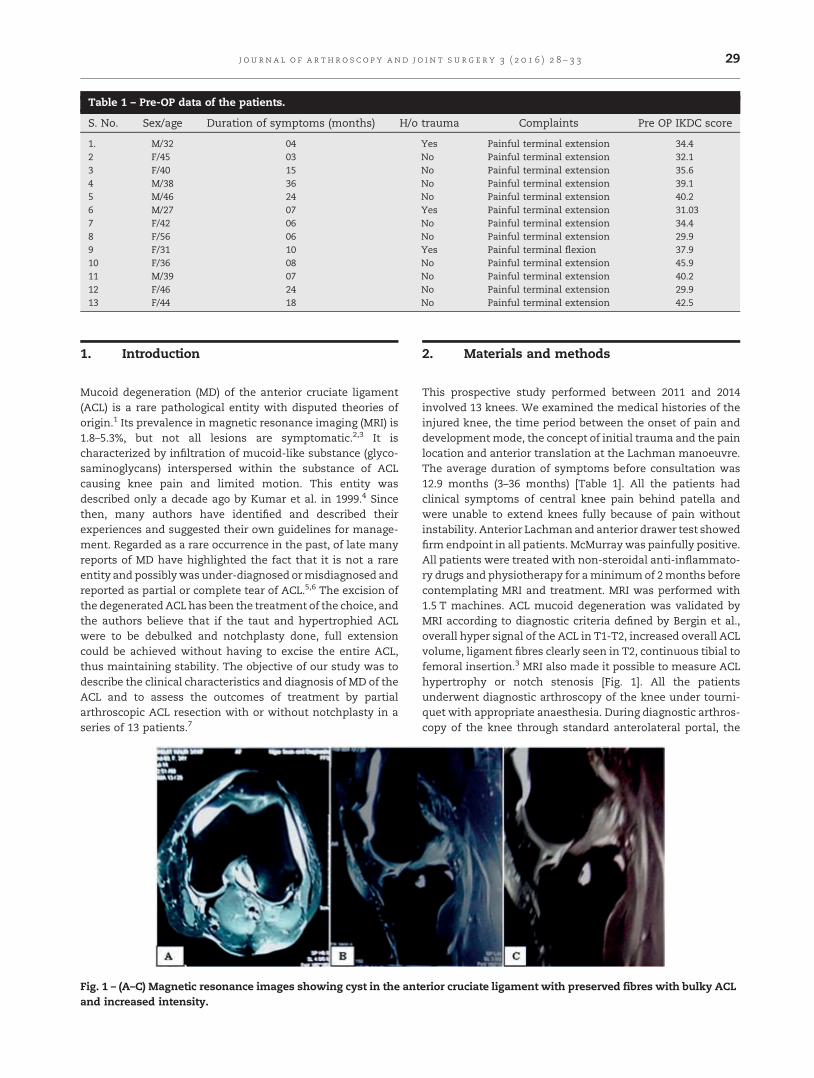
Table 1 – Pre-OP data of the patients.
S. No. Sex/age Duration of symptoms (months) H/o trauma Complaints Pre OP IKDC score
1. M/32 04 Yes Painful terminal extension 34.42 F/45 03 No Painful terminal extension 32.13 F/40 15 No Painful terminal extension 35.64 M/38 36 No Painful terminal extension 39.15 M/46 24 No Painful terminal extension 40.26 M/27 07 Yes Painful terminal extension 31.037 F/42 06 No Painful terminal extension 34.48 F/56 06 No Painful terminal extension 29.99 F/31 10 Yes Painful terminal flexion 37.910 F/36 08 No Painful terminal extension 45.911 M/39 07 No Painful terminal extension 40.212 F/46 24 No Painful terminal extension 29.913 F/44 18 No Painful terminal extension 42.5
j o u rn a l o f a r th r o s c o p y and j o i n t s u r g e r y 3 ( 2 0 1 6 ) 2 8 – 3 3 29
1. Introduction
Mucoid degeneration (MD) of the anterior cruciate ligament(ACL) is a rare pathological entity with disputed theories oforigin.1 Its prevalence in magnetic resonance imaging (MRI) is1.8–5.3%, but not all lesions are symptomatic.2,3 It ischaracterized by infiltration of mucoid-like substance (glyco-saminoglycans) interspersed within the substance of ACLcausing knee pain and limited motion. This entity wasdescribed only a decade ago by Kumar et al. in 1999.4 Sincethen, many authors have identified and described theirexperiences and suggested their own guidelines for manage-ment. Regarded as a rare occurrence in the past, of late manyreports of MD have highlighted the fact that it is not a rareentity and possiblywas under-diagnosed ormisdiagnosed andreported as partial or complete tear of ACL.5,6 The excision ofthe degeneratedACL has been the treatment of the choice, andthe authors believe that if the taut and hypertrophied ACLwere to be debulked and notchplasty done, full extensioncould be achieved without having to excise the entire ACL,thus maintaining stability. The objective of our study was todescribe the clinical characteristics and diagnosis of MD of theACL and to assess the outcomes of treatment by partialarthroscopic ACL resection with or without notchplasty in aseries of 13 patients.7[(Fig._1)TD$FIG]
Fig. 1 – (A–C) Magnetic resonance images showing cyst in the antand increased intensity.
2. Materials and methods
This prospective study performed between 2011 and 2014involved 13 knees. We examined the medical histories of theinjured knee, the time period between the onset of pain anddevelopment mode, the concept of initial trauma and the painlocation and anterior translation at the Lachman manoeuvre.The average duration of symptoms before consultation was12.9 months (3–36 months) [Table 1]. All the patients hadclinical symptoms of central knee pain behind patella andwere unable to extend knees fully because of pain withoutinstability. Anterior Lachman and anterior drawer test showedfirm endpoint in all patients. McMurraywas painfully positive.All patients were treated with non-steroidal anti-inflammato-ry drugs and physiotherapy for aminimumof 2months beforecontemplating MRI and treatment. MRI was performed with1.5 T machines. ACL mucoid degeneration was validated byMRI according to diagnostic criteria defined by Bergin et al.,overall hyper signal of the ACL in T1-T2, increased overall ACLvolume, ligament fibres clearly seen in T2, continuous tibial tofemoral insertion.3 MRI also made it possible to measure ACLhypertrophy or notch stenosis [Fig. 1]. All the patientsunderwent diagnostic arthroscopy of the knee under tourni-quet with appropriate anaesthesia. During diagnostic arthros-copy of the knee through standard anterolateral portal, the
erior cruciate ligament with preserved fibres with bulky ACL
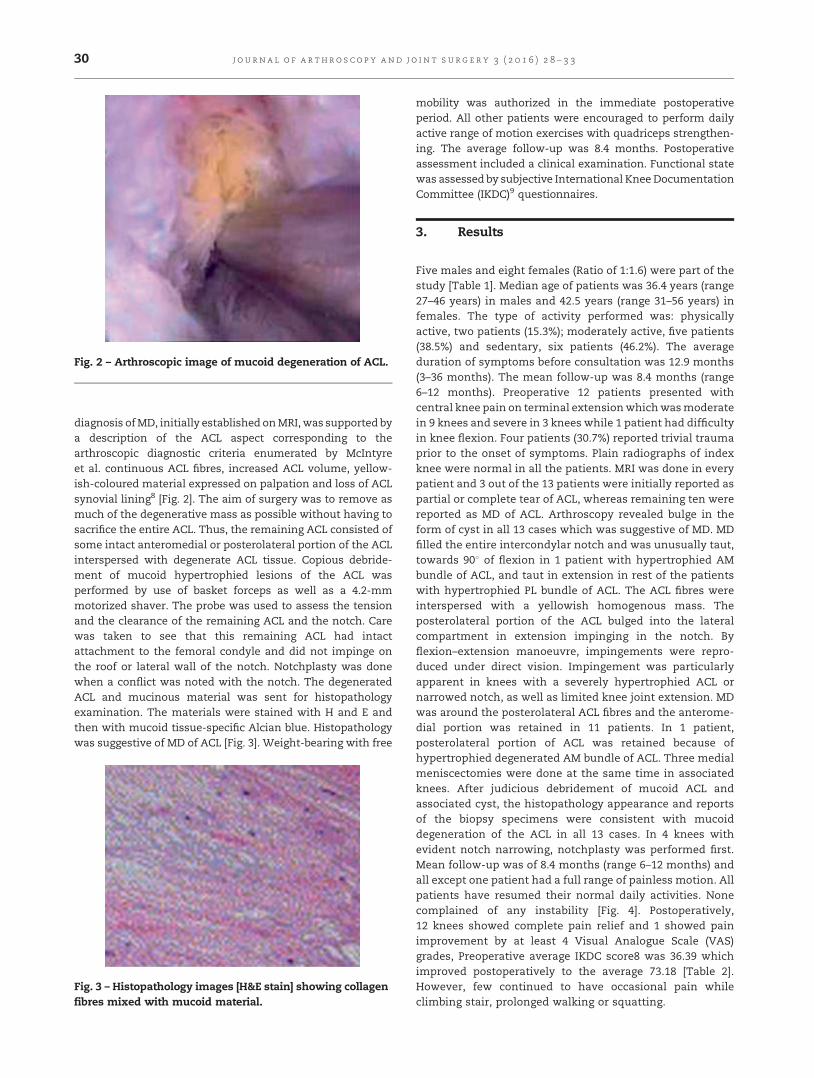
[(Fig._2)TD$FIG]
Fig. 2 – Arthroscopic image of mucoid degeneration of ACL.
j o u r n a l o f a r t h r o s c o p y and j o i n t s u r g e r y 3 ( 2 0 1 6 ) 2 8 – 3 330
diagnosis ofMD, initially established onMRI, was supported bya description of the ACL aspect corresponding to thearthroscopic diagnostic criteria enumerated by McIntyreet al. continuous ACL fibres, increased ACL volume, yellow-ish-coloured material expressed on palpation and loss of ACLsynovial lining8 [Fig. 2]. The aim of surgery was to remove asmuch of the degenerative mass as possible without having tosacrifice the entire ACL. Thus, the remaining ACL consisted ofsome intact anteromedial or posterolateral portion of the ACLinterspersed with degenerate ACL tissue. Copious debride-ment of mucoid hypertrophied lesions of the ACL wasperformed by use of basket forceps as well as a 4.2-mmmotorized shaver. The probe was used to assess the tensionand the clearance of the remaining ACL and the notch. Carewas taken to see that this remaining ACL had intactattachment to the femoral condyle and did not impinge onthe roof or lateral wall of the notch. Notchplasty was donewhen a conflict was noted with the notch. The degeneratedACL and mucinous material was sent for histopathologyexamination. The materials were stained with H and E andthen with mucoid tissue-specific Alcian blue. Histopathologywas suggestive of MD of ACL [Fig. 3]. Weight-bearing with free[(Fig._3)TD$FIG]
Fig. 3 – Histopathology images [H&E stain] showing collagenfibres mixed with mucoid material.
mobility was authorized in the immediate postoperativeperiod. All other patients were encouraged to perform dailyactive range of motion exercises with quadriceps strengthen-ing. The average follow-up was 8.4 months. Postoperativeassessment included a clinical examination. Functional statewas assessed by subjective International Knee DocumentationCommittee (IKDC)9 questionnaires.
3. Results
Five males and eight females (Ratio of 1:1.6) were part of thestudy [Table 1]. Median age of patients was 36.4 years (range27–46 years) in males and 42.5 years (range 31–56 years) infemales. The type of activity performed was: physicallyactive, two patients (15.3%); moderately active, five patients(38.5%) and sedentary, six patients (46.2%). The averageduration of symptoms before consultation was 12.9 months(3–36 months). The mean follow-up was 8.4 months (range6–12 months). Preoperative 12 patients presented withcentral knee pain on terminal extensionwhichwasmoderatein 9 knees and severe in 3 knees while 1 patient had difficultyin knee flexion. Four patients (30.7%) reported trivial traumaprior to the onset of symptoms. Plain radiographs of indexknee were normal in all the patients. MRI was done in everypatient and 3 out of the 13 patients were initially reported aspartial or complete tear of ACL, whereas remaining ten werereported as MD of ACL. Arthroscopy revealed bulge in theform of cyst in all 13 cases which was suggestive of MD. MDfilled the entire intercondylar notch and was unusually taut,towards 908 of flexion in 1 patient with hypertrophied AMbundle of ACL, and taut in extension in rest of the patientswith hypertrophied PL bundle of ACL. The ACL fibres wereinterspersed with a yellowish homogenous mass. Theposterolateral portion of the ACL bulged into the lateralcompartment in extension impinging in the notch. Byflexion–extension manoeuvre, impingements were repro-duced under direct vision. Impingement was particularlyapparent in knees with a severely hypertrophied ACL ornarrowed notch, as well as limited knee joint extension. MDwas around the posterolateral ACL fibres and the anterome-dial portion was retained in 11 patients. In 1 patient,posterolateral portion of ACL was retained because ofhypertrophied degenerated AM bundle of ACL. Three medialmeniscectomies were done at the same time in associatedknees. After judicious debridement of mucoid ACL andassociated cyst, the histopathology appearance and reportsof the biopsy specimens were consistent with mucoiddegeneration of the ACL in all 13 cases. In 4 knees withevident notch narrowing, notchplasty was performed first.Mean follow-up was of 8.4 months (range 6–12 months) andall except one patient had a full range of painless motion. Allpatients have resumed their normal daily activities. Nonecomplained of any instability [Fig. 4]. Postoperatively,12 knees showed complete pain relief and 1 showed painimprovement by at least 4 Visual Analogue Scale (VAS)grades, Preoperative average IKDC score8 was 36.39 whichimproved postoperatively to the average 73.18 [Table 2].However, few continued to have occasional pain whileclimbing stair, prolonged walking or squatting.
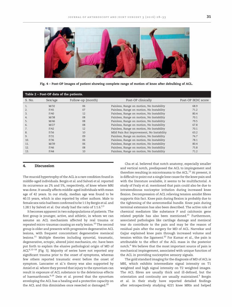
[(Fig._4)TD$FIG]
Fig. 4 – Post-OP images of patient showing complete range of motion of knee after debulking of ACL.
Table 2 – Post-OP data of the patients.
S. No. Sex/age Follow-up (month) Post-OP clinically Post-OP IKDC score
1. M/32 06 Painless, Range on motion, No Instability 68.92. F/45 07 Painless, Range on motion, No Instability 71.23. F/40 12 Painless, Range on motion, No Instability 80.44. M/38 08 Painless, Range on motion, No Instability 70.15. M/46 08 Painless, Range on motion, No Instability 79.56. M/27 08 Painless, Range on motion, No Instability 67.87. F/42 12 Painless, Range on motion, No Instability 70.18. F/56 10 Mild Pain But Improvement, No Instability 63.29. F/31 09 Painless, Range on motion, No Instability 74.710. F/36 06 Painless, Range on motion, No Instability 78.111. M/39 06 Painless, Range on motion, No Instability 80.412. F/46 08 Painless, Range on motion, No Instability 75.813. F/44 10 Painless, Range on motion, No Instability 71.2
j o u rn a l o f a r th r o s c o p y and j o i n t s u r g e r y 3 ( 2 0 1 6 ) 2 8 – 3 3 31
4. Discussion
Themucoid hypertrophy of the ACL is a rare condition found inmiddle-aged individuals. Bergin et al. and Salvati et al. reportedits occurrence as 2% and 5%, respectively, of knee where MRIwas done. It usually affectsmiddle-aged individualswithmeanage of 42 years. In our study, median age was found to be40.15 years, which is also reported by other authors. Male tofemale sex ratiohadbeenconfirmed tobe1:1 byBerginet al. and1.28:1 by Salvati et al. Our study had the ratio of 1:1.6.2,3
It becomes apparent in two subpopulations of patients. Thefirst group is younger, active, and athletic, in whom we canassume an ACL mechanism affected by real trauma orrepeatedmicro-traumas causing an early lesion.10 The secondgroup is older and presents with progressive degenerative ACLlesions, with frequent concomitant degenerative meniscallesions.11 Multiple theories including synovial, traumatic,degenerative, ectopic, altered joint mechanics, etc. have beenput forth to explain the elusive pathological origin of MD ofACL6,12–18 [Fig. 5]. Majority of series have not reported anysignificant trauma prior to the onset of symptoms, whereasfew others reported traumatic event before the onset ofsymptom. Lancaster et al.'s theory was also supported byAmiel et al. where they proved that injury to the synovium canresult in exposure of ACL substance to the deleterious effectsof haemarthrosis.6,19 Deie et al. proved that the synoviumenveloping the ACL has a healing and a protective capacity onthe ACL and this diminishes once resected or damaged.20
Cha et al. believed that notch anatomy, especially smallerand vertical notch, predisposed the ACL to impingement andtherefore resulting in microtrauma to the ACL.13 At present, itis difficult to point out a single lone cause for the knee pain andwith the literature available, it seems to be multifactorial. Astudy of Fealy et al. mentioned that pain could also be due tointratendinous nociceptor irritation during increased kneeflexion. Decompression of ACL relieving tension amidst fibressupports this fact. Knee pain during flexion is probably due tothe tightening of the anteromedial bundle. Knee pain duringterminal extension has also been described. The active role ofchemical mediators like substance P and calcitonin generelated peptide has also been mentioned.21 Furthermore,associated pathologies like cartilage damage and meniscaltear do contribute to the pain and may be the reason ofresidual pain after the surgery for MD of ACL. Narvekar andGajjar explained knee pain through increased volume andtension within the ligament.22 For Kumar et al., the pain isattributable to the effect of the ACL mass in the posteriornotch.4 We believe that the most important source of pain ismechanical impingement, associated with unique function ofthe ACL in providing nociceptive sensory signals.
The gold standard imaging for the diagnosis of MD of ACL isMRI, which exhibits intermediate signal intensity on T1weighted and high signal intensity on T2 weighted images.The ACL fibres are usually thick and ill-defined, but theorientation and continuity are usually maintained.5 Berginet al. in their study have reported detailed findingsafter retrospectively studying 4221 knee MRIs and helped
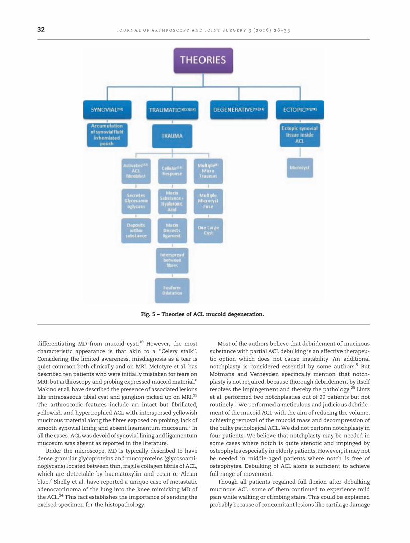
[(Fig._5)TD$FIG]
Fig. 5 – Theories of ACL mucoid degeneration.
j o u r n a l o f a r t h r o s c o p y and j o i n t s u r g e r y 3 ( 2 0 1 6 ) 2 8 – 3 332
differentiating MD from mucoid cyst.10 However, the mostcharacteristic appearance is that akin to a ‘‘Celery stalk’’.Considering the limited awareness, misdiagnosis as a tear isquiet common both clinically and on MRI. McIntyre et al. hasdescribed ten patients who were initially mistaken for tears onMRI, but arthroscopy and probing expressed mucoid material.8
Makino et al. have described the presence of associated lesionslike intraosseous tibial cyst and ganglion picked up on MRI.23
The arthroscopic features include an intact but fibrillated,yellowish and hypertrophied ACL with interspersed yellowishmucinous material along the fibres exposed on probing, lack ofsmooth synovial lining and absent ligamentum mucosum.5 Inall the cases, ACLwasdevoidof synovial lining and ligamentummucosum was absent as reported in the literature.
Under the microscope, MD is typically described to havedense granular glycoproteins and mucoproteins (glycosoami-noglycans) located between thin, fragile collagen fibrils of ACL,which are detectable by haematoxylin and eosin or Alcianblue.7 Shelly et al. have reported a unique case of metastaticadenocarcinoma of the lung into the knee mimicking MD ofthe ACL.24 This fact establishes the importance of sending theexcised specimen for the histopathology.
Most of the authors believe that debridement of mucinoussubstance with partial ACL debulking is an effective therapeu-tic option which does not cause instability. An additionalnotchplasty is considered essential by some authors.5 ButMotmans and Verheyden specifically mention that notch-plasty is not required, because thorough debridement by itselfresolves the impingement and thereby the pathology.25 Lintzet al. performed two notchplasties out of 29 patients but notroutinely.1 We performed a meticulous and judicious debride-ment of the mucoid ACL with the aim of reducing the volume,achieving removal of the mucoid mass and decompression ofthe bulky pathological ACL.We did not performnotchplasty infour patients. We believe that notchplasty may be needed insome cases where notch is quite stenotic and impinged byosteophytes especially in elderly patients. However, itmay notbe needed in middle-aged patients where notch is free ofosteophytes. Debulking of ACL alone is sufficient to achievefull range of movement.
Though all patients regained full flexion after debulkingmucinous ACL, some of them continued to experience mildpain while walking or climbing stairs. This could be explainedprobably because of concomitant lesions like cartilage damage
j o u rn a l o f a r th r o s c o p y and j o i n t s u r g e r y 3 ( 2 0 1 6 ) 2 8 – 3 3 33
in patellofemoral or tibiofemoral joint ormeniscal tear. Dejouret al. demonstrated positive anterior drawer in 36% andpositive pivot shift in 55% of his patients after debridement.26
None demonstrated a positive pivot shift. None developedinstability until final follow-up.
However, it is premature to say that these patients will notdevelop instability in future, as all of these were patients withsedentary activities except one who plays badminton. Wheth-er patients whose occupation requires heavy demand fromknee or athletes would not develop instability in future afterpartial debulking, is a matter of debate. Another notionaddressed by this work is that of secondary instability. It ismentioned elsewhere only byMcIntyre et al. who reported onecase of atraumatic ACL rupture at 1 postoperative year afterpartial resection.8 Our results indicate that postoperativelaxity, largely asymptomatic, can increase anterior laxity overtime and evoke instability.
5. Conclusion
Mucoid degeneration of the ACL should be suspected inpatients presenting pain on terminal extension or flexionwithout preceding trauma. Prior knowledge of condition withhigh index of suspicion and careful interpretation of MRI canestablish the diagnosis preoperatively. Arthroscopic debride-ment with or without notchplasty gives excellent functionalresults.
Conflicts of interest
All authors have none to declare.
r e f e r e n c e s
1. Lintz F, Pujol N, Dejour D, Boisrenoult P, Beaufils P. Anteriorcruciate ligament mucoid degeneration: selecting the besttreatment option. Orthop Traumatol Surg Res. 2010;96:400–406.
2. Salvati F, Rossi F, Limbucci N, Pistoia ML, Barile A,Masciocchi C. Mucoid metaplastic-degeneration of anteriorcruciate ligament. J Sports Med Phys Fitness. 2008;48:483–487.
3. Bergin D, Morrison WB, Carrino JA, Nallamshetty SN,Bartolozzi AR. Anterior cruciate ligament ganglia andmucoid degeneration: coexistence and clinical correlation.Am J Roentgenol. 2004;182:1283–1287.
4. Kumar A, Bickerstaff DR, Grimwood JS, Suvarna SK. Mucoidcystic degeneration of the cruciate ligament. J Bone Joint SurgBr. 1999;81:304–305.
5. Pandey V, Suman C, Sharma S, Rao SP, Kiran Acharya K,Sambaji C. Mucoid degeneration of the anterior cruciateligament: management and outcome. Indian J Orthop. 2014;48(2):197–202.
6. Lancaster TF, Kirby AB, Beall DP, Wolff JD, Wu DH. Mucoiddegeneration of the anterior cruciate ligament: a case report.J Okla State Med Assoc. 2004;97:326–328.
7. Chudasama CH, Chudasama VC, Prabhakar MM.Arthroscopic management of mucoid degeneration of
anterior cruciate ligament. Indian J Orthop. 2012;46(5):561–565.
8. McIntyre J, Moelleken S, Tirman P. Mucoid degeneration ofthe anterior cruciate ligament mistaken for ligamentoustears. Skeletal Radiol. 2001;30:312–315.
9. Rossi MJ, Lubowitz JH, Guttmann D. Development andvalidation of the International Knee DocumentationCommittee subjective knee form. Am J Sports Med.2002;30:152.
10. Scranton Jr PE, Farrar EL. Mucoid degeneration of thepatellar ligament in athletes. J Bone Joint Surg Am.1992;74:435–437.
11. Cha JH, Lee SH, Shin MJ, Choi BK, Bin SI. Relationshipbetween mucoid hypertrophy of the anterior cruciateligament (ACL) andmorphologic change of the intercondylarnotch: MRI and arthroscopy correlation. Skeletal Radiol.2008;37:821–826.
12. Diard F, Chateil JF, Hauger O, Moinard M. Para-articular andintraosseous synovial cysts and articular mucoid cysts.J Radiol. 1999;80:679–696.
13. Rolf C, Watson TP. Case report: Intratendinous ganglion ofthe anterior cruciate ligament in a young footballer. J OrthopSurg Res. 2006;1:11.
14. Bui-Mansfield LT, Youngberg RA. Intraarticular ganglia ofthe knee: prevalence, presentation, etiology, andmanagement. Am J Roentgenol. 1997;168:123–127.
15. Fernandes JL, Viana SL, Mendonça JL, et al. Mucoiddegeneration of the anterior cruciate ligament: magneticresonance imaging findings of an underdiagnosed entity.Acta Radiol. 2008;49:75–79.
16. Krudwig WK, Schulte KK, Heinemann C. Intraarticularganglion cysts of the knee joint: a report of 85 cases andreview of the literature. Knee Surg Sports Traumatol Arthrosc.2004;12:123–129.
17. Melloni P, Valls R, Yuguero M, Sáez A. Mucoid degenerationof the anterior cruciate ligament with erosion of the lateralfemoral condyle. Skeletal Radiol. 2004;33:359–362.
18. Hensen JJ, Coerkamp EG, Bloem JL, De Schepper AM. Mucoiddegeneration of the anterior cruciate ligament. JBR-BTR.2007;90:192–193.
19. Amiel D, Billings Jr E, Harwood FL. Collagenase activity inanterior cruciate ligament: protective role of the synovialsheath. J Appl Physiol (1985). 1990;69:902–906.
20. Deie M, Ochi M, Ikuta Y. High intrinsic healing potential ofhuman anterior cruciate ligament. Organ cultureexperiments. Acta Orthop Scand. 1995;66:28–32.
21. Fealy S, Kenter K, Dines JS, Warren RF. Mucoid degenerationof the anterior cruciate ligament. Arthroscopy. 2001;17:E37.
22. Narvekar A, Gajjar S. Mucoid degeneration of the anteriorcruciate ligament. Arthroscopy. 2004;20:141–146.
23. Makino A, Pascual-Garrido C, Rolón A, Isola M, Muscolo DL.Mucoid degeneration of the anterior cruciate ligament: MRI,clinical, intraoperative, and histological findings. Knee SurgSports Traumatol Arthrosc. 2011;19:408–411.
24. Shelly MJ, Dheer S, Kavanagh EC. Metastaticadenocarcinoma of the lung mimicking mucoiddegeneration of the anterior cruciate ligament. Ir J Med Sci.2010;179:309–311.
25. Motmans R, Verheyden F. Mucoid degeneration of theanterior cruciate ligament. Knee Surg Sports TraumatolArthrosc. 2009;17:737–740.
26. Dejour D, Cohn J, Tavernier T. La dégénérescence spontanéedu ligament croisé antérieur: étude radio clinique etanatomo-pathologique. Rev Chir Orthop Reparatrice ApparMot. 2005;91:67.
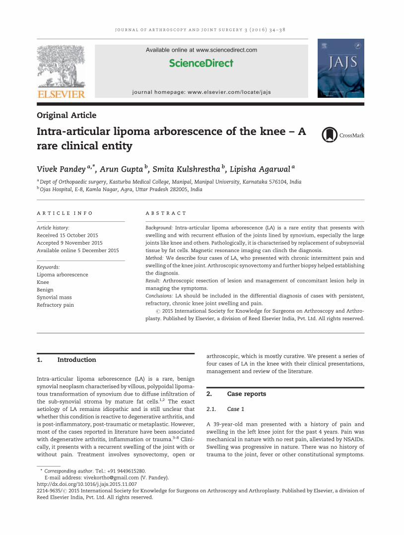
j o u r n a l o f a r t h r o s c o p y and j o i n t s u r g e r y 3 ( 2 0 1 6 ) 3 4 – 3 8
Available online at www.sciencedirect.com
ScienceDirect
journal homepage: www.elsevier.com/locate/jajs
Original Article
Intra-articular lipoma arborescence of the knee – A
rare clinical entityVivek Pandey a,*, Arun Gupta b, Smita Kulshrestha b, Lipisha Agarwal a
aDept of Orthopaedic surgery, Kasturba Medical College, Manipal, Manipal University, Karnataka 576104, IndiabOjas Hospital, E-8, Kamla Nagar, Agra, Uttar Pradesh 282005, India
a r t i c l e i n f o
Article history:
Received 15 October 2015
Accepted 9 November 2015
Available online 5 December 2015
Keywords:
Lipoma arborescence
Knee
Benign
Synovial mass
Refractory pain
a b s t r a c t
Background: Intra-articular lipoma arborescence (LA) is a rare entity that presents with
swelling and with recurrent effusion of the joints lined by synovium, especially the large
joints like knee and others. Pathologically, it is characterised by replacement of subsynovial
tissue by fat cells. Magnetic resonance imaging can clinch the diagnosis.
Method: We describe four cases of LA, who presented with chronic intermittent pain and
swelling of the knee joint. Arthroscopic synovectomy and further biopsy helped establishing
the diagnosis.
Result: Arthroscopic resection of lesion and management of concomitant lesion help in
managing the symptoms.
Conclusions: LA should be included in the differential diagnosis of cases with persistent,
refractory, chronic knee joint swelling and pain.
# 2015 International Society for Knowledge for Surgeons on Arthroscopy and Arthro-
plasty. Published by Elsevier, a division of Reed Elsevier India, Pvt. Ltd. All rights reserved.
1. Introduction
Intra-articular lipoma arborescence (LA) is a rare, benignsynovial neoplasm characterised by villous, polypoidal lipoma-tous transformation of synovium due to diffuse infiltration ofthe sub-synovial stroma by mature fat cells.1,2 The exactaetiology of LA remains idiopathic and is still unclear thatwhether this condition is reactive to degenerative arthritis, andis post-inflammatory, post-traumatic or metaplastic. However,most of the cases reported in literature have been associatedwith degenerative arthritis, inflammation or trauma.3–8 Clini-cally, it presents with a recurrent swelling of the joint with orwithout pain. Treatment involves synovectomy, open or
* Corresponding author. Tel.: +91 9449615280.E-mail address: [email protected] (V. Pandey).
http://dx.doi.org/10.1016/j.jajs.2015.11.0072214-9635/# 2015 International Society for Knowledge for Surgeons onReed Elsevier India, Pvt. Ltd. All rights reserved.
arthroscopic, which is mostly curative. We present a series offour cases of LA in the knee with their clinical presentations,management and review of the literature.
2. Case reports
2.1. Case 1
A 39-year-old man presented with a history of pain andswelling in the left knee joint for the past 4 years. Pain wasmechanical in nature with no rest pain, alleviated by NSAIDs.Swelling was progressive in nature. There was no history oftrauma to the joint, fever or other constitutional symptoms.
Arthroscopy and Arthroplasty. Published by Elsevier, a division of
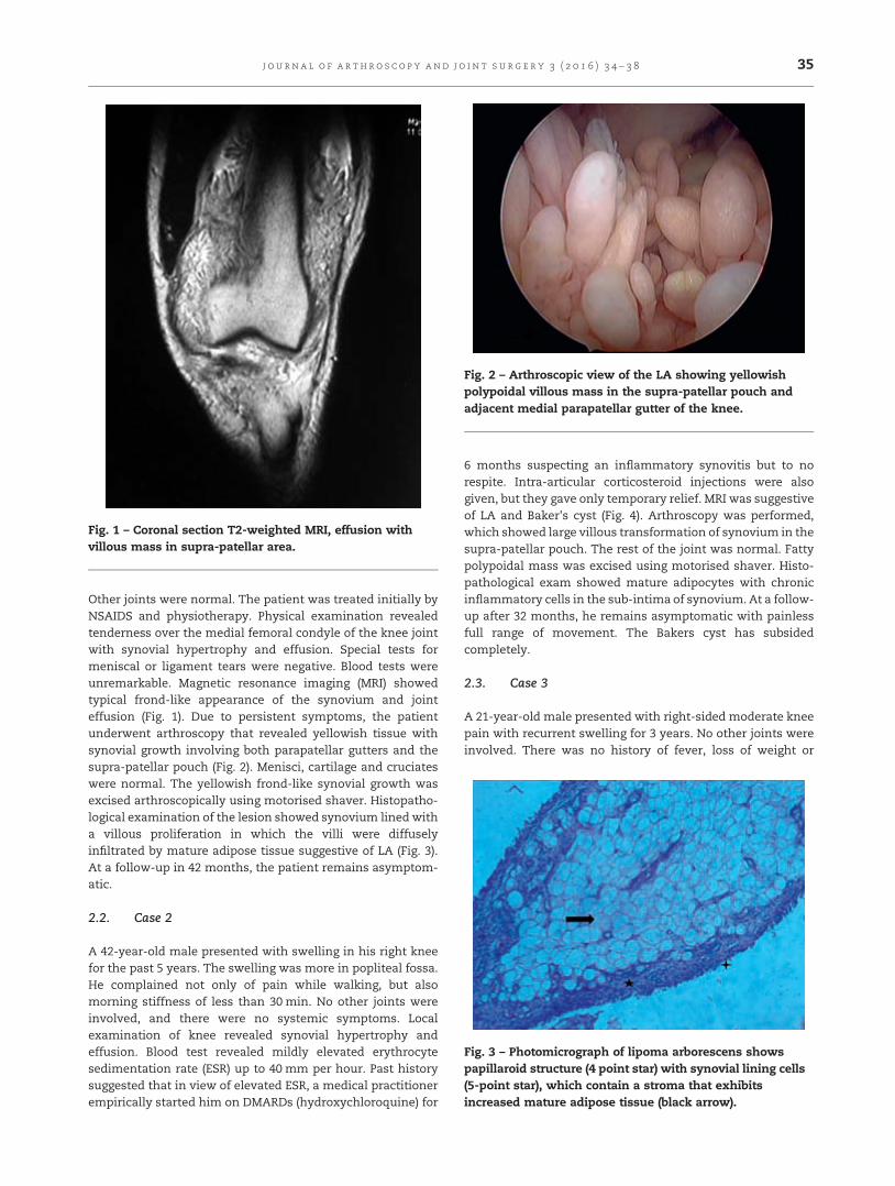
[(Fig._1)TD$FIG]
Fig. 1 – Coronal section T2-weighted MRI, effusion withvillous mass in supra-patellar area.
[(Fig._2)TD$FIG]
Fig. 2 – Arthroscopic view of the LA showing yellowishpolypoidal villous mass in the supra-patellar pouch andadjacent medial parapatellar gutter of the knee.
[(Fig._3)TD$FIG]
Fig. 3 – Photomicrograph of lipoma arborescens showspapillaroid structure (4 point star) with synovial lining cells(5-point star), which contain a stroma that exhibitsincreased mature adipose tissue (black arrow).
j o u rn a l o f a r th r o s c o p y and j o i n t s u r g e r y 3 ( 2 0 1 6 ) 3 4 – 3 8 35
Other joints were normal. The patient was treated initially byNSAIDS and physiotherapy. Physical examination revealedtenderness over the medial femoral condyle of the knee jointwith synovial hypertrophy and effusion. Special tests formeniscal or ligament tears were negative. Blood tests wereunremarkable. Magnetic resonance imaging (MRI) showedtypical frond-like appearance of the synovium and jointeffusion (Fig. 1). Due to persistent symptoms, the patientunderwent arthroscopy that revealed yellowish tissue withsynovial growth involving both parapatellar gutters and thesupra-patellar pouch (Fig. 2). Menisci, cartilage and cruciateswere normal. The yellowish frond-like synovial growth wasexcised arthroscopically using motorised shaver. Histopatho-logical examination of the lesion showed synovium lined witha villous proliferation in which the villi were diffuselyinfiltrated by mature adipose tissue suggestive of LA (Fig. 3).At a follow-up in 42 months, the patient remains asymptom-atic.
2.2. Case 2
A 42-year-old male presented with swelling in his right kneefor the past 5 years. The swelling was more in popliteal fossa.He complained not only of pain while walking, but alsomorning stiffness of less than 30 min. No other joints wereinvolved, and there were no systemic symptoms. Localexamination of knee revealed synovial hypertrophy andeffusion. Blood test revealed mildly elevated erythrocytesedimentation rate (ESR) up to 40 mm per hour. Past historysuggested that in view of elevated ESR, a medical practitionerempirically started him on DMARDs (hydroxychloroquine) for
6 months suspecting an inflammatory synovitis but to norespite. Intra-articular corticosteroid injections were alsogiven, but they gave only temporary relief. MRI was suggestiveof LA and Baker's cyst (Fig. 4). Arthroscopy was performed,which showed large villous transformation of synovium in thesupra-patellar pouch. The rest of the joint was normal. Fattypolypoidal mass was excised using motorised shaver. Histo-pathological exam showed mature adipocytes with chronicinflammatory cells in the sub-intima of synovium. At a follow-up after 32 months, he remains asymptomatic with painlessfull range of movement. The Bakers cyst has subsidedcompletely.
2.3. Case 3
A 21-year-old male presented with right-sided moderate kneepain with recurrent swelling for 3 years. No other joints wereinvolved. There was no history of fever, loss of weight or
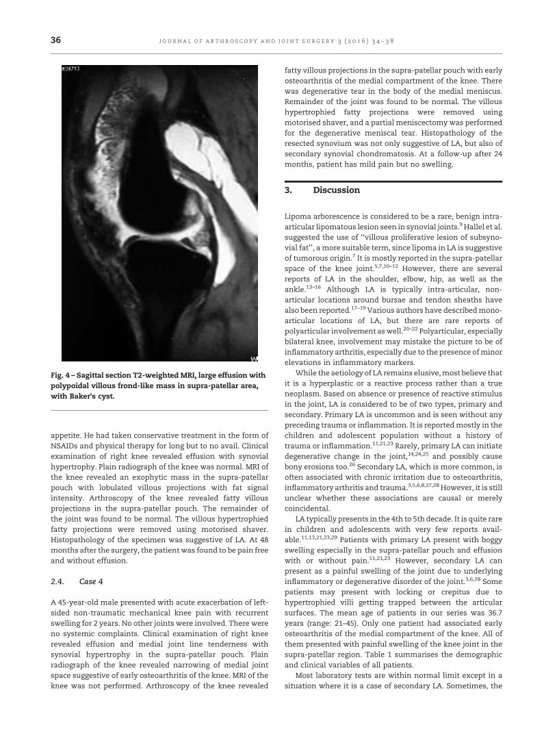
[(Fig._4)TD$FIG]
Fig. 4 – Sagittal section T2-weighted MRI, large effusion withpolypoidal villous frond-like mass in supra-patellar area,with Baker's cyst.
j o u r n a l o f a r t h r o s c o p y and j o i n t s u r g e r y 3 ( 2 0 1 6 ) 3 4 – 3 836
appetite. He had taken conservative treatment in the form ofNSAIDs and physical therapy for long but to no avail. Clinicalexamination of right knee revealed effusion with synovialhypertrophy. Plain radiograph of the knee was normal. MRI ofthe knee revealed an exophytic mass in the supra-patellarpouch with lobulated villous projections with fat signalintensity. Arthroscopy of the knee revealed fatty villousprojections in the supra-patellar pouch. The remainder ofthe joint was found to be normal. The villous hypertrophiedfatty projections were removed using motorised shaver.Histopathology of the specimen was suggestive of LA. At 48months after the surgery, the patient was found to be pain freeand without effusion.
2.4. Case 4
A 45-year-old male presented with acute exacerbation of left-sided non-traumatic mechanical knee pain with recurrentswelling for 2 years. No other joints were involved. There wereno systemic complaints. Clinical examination of right kneerevealed effusion and medial joint line tenderness withsynovial hypertrophy in the supra-patellar pouch. Plainradiograph of the knee revealed narrowing of medial jointspace suggestive of early osteoarthritis of the knee. MRI of theknee was not performed. Arthroscopy of the knee revealed
fatty villous projections in the supra-patellar pouch with earlyosteoarthritis of the medial compartment of the knee. Therewas degenerative tear in the body of the medial meniscus.Remainder of the joint was found to be normal. The villoushypertrophied fatty projections were removed usingmotorised shaver, and a partial meniscectomy was performedfor the degenerative meniscal tear. Histopathology of theresected synovium was not only suggestive of LA, but also ofsecondary synovial chondromatosis. At a follow-up after 24months, patient has mild pain but no swelling.
3. Discussion
Lipoma arborescence is considered to be a rare, benign intra-articular lipomatous lesion seen in synovial joints.9 Hallel et al.suggested the use of ‘‘villous proliferative lesion of subsyno-vial fat’’, a more suitable term, since lipoma in LA is suggestiveof tumorous origin.7 It is mostly reported in the supra-patellarspace of the knee joint.5,7,10–12 However, there are severalreports of LA in the shoulder, elbow, hip, as well as theankle.13–16 Although LA is typically intra-articular, non-articular locations around bursae and tendon sheaths havealso been reported.17–19 Various authors have describedmono-articular locations of LA, but there are rare reports ofpolyarticular involvement aswell.20–22 Polyarticular, especiallybilateral knee, involvement may mistake the picture to be ofinflammatory arthritis, especially due to the presence ofminorelevations in inflammatory markers.
While the aetiology of LA remains elusive,most believe thatit is a hyperplastic or a reactive process rather than a trueneoplasm. Based on absence or presence of reactive stimulusin the joint, LA is considered to be of two types, primary andsecondary. Primary LA is uncommon and is seen without anypreceding trauma or inflammation. It is reportedmostly in thechildren and adolescent population without a history oftrauma or inflammation.11,21,23 Rarely, primary LA can initiatedegenerative change in the joint,14,24,25 and possibly causebony erosions too.26 Secondary LA, which is more common, isoften associated with chronic irritation due to osteoarthritis,inflammatory arthritis and trauma.3,5,6,8,27,28 However, it is stillunclear whether these associations are causal or merelycoincidental.
LA typically presents in the 4th to 5th decade. It is quite rarein children and adolescents with very few reports avail-able.11,13,21,23,29 Patients with primary LA present with boggyswelling especially in the supra-patellar pouch and effusionwith or without pain.11,21,23 However, secondary LA canpresent as a painful swelling of the joint due to underlyinginflammatory or degenerative disorder of the joint.3,6,28 Somepatients may present with locking or crepitus due tohypertrophied villi getting trapped between the articularsurfaces. The mean age of patients in our series was 36.7years (range: 21–45). Only one patient had associated earlyosteoarthritis of the medial compartment of the knee. All ofthem presented with painful swelling of the knee joint in thesupra-patellar region. Table 1 summarises the demographicand clinical variables of all patients.
Most laboratory tests are within normal limit except in asituation where it is a case of secondary LA. Sometimes, the
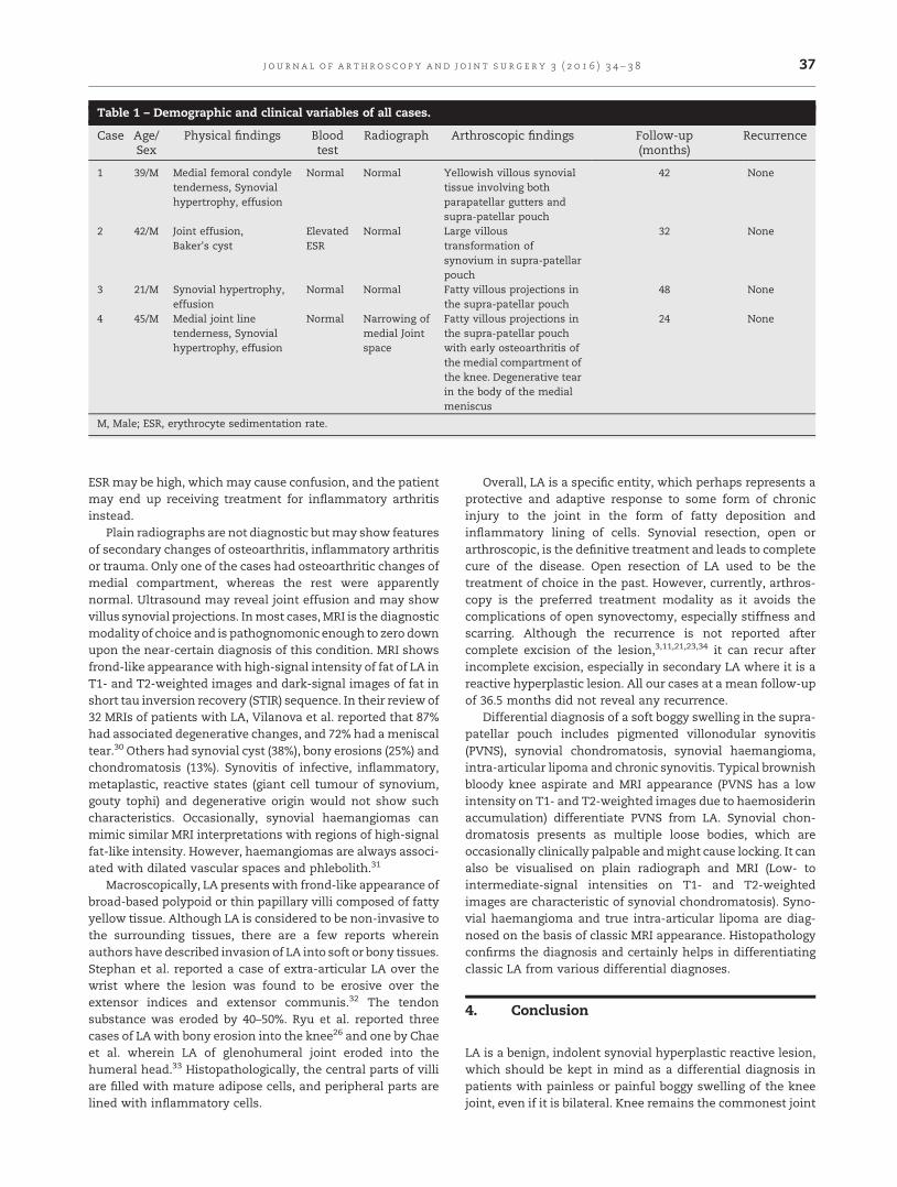
Table 1 – Demographic and clinical variables of all cases.
Case Age/Sex
Physical findings Bloodtest
Radiograph Arthroscopic findings Follow-up(months)
Recurrence
1 39/M Medial femoral condyletenderness, Synovialhypertrophy, effusion
Normal Normal Yellowish villous synovialtissue involving bothparapatellar gutters andsupra-patellar pouch
42 None
2 42/M Joint effusion,Baker's cyst
ElevatedESR
Normal Large villoustransformation ofsynovium in supra-patellarpouch
32 None
3 21/M Synovial hypertrophy,effusion
Normal Normal Fatty villous projections inthe supra-patellar pouch
48 None
4 45/M Medial joint linetenderness, Synovialhypertrophy, effusion
Normal Narrowing ofmedial Jointspace
Fatty villous projections inthe supra-patellar pouchwith early osteoarthritis ofthe medial compartment ofthe knee. Degenerative tearin the body of the medialmeniscus
24 None
M, Male; ESR, erythrocyte sedimentation rate.
j o u rn a l o f a r th r o s c o p y and j o i n t s u r g e r y 3 ( 2 0 1 6 ) 3 4 – 3 8 37
ESR may be high, which may cause confusion, and the patientmay end up receiving treatment for inflammatory arthritisinstead.
Plain radiographs are not diagnostic butmay show featuresof secondary changes of osteoarthritis, inflammatory arthritisor trauma. Only one of the cases had osteoarthritic changes ofmedial compartment, whereas the rest were apparentlynormal. Ultrasound may reveal joint effusion and may showvillus synovial projections. Inmost cases, MRI is the diagnosticmodality of choice and is pathognomonic enough to zero downupon the near-certain diagnosis of this condition. MRI showsfrond-like appearance with high-signal intensity of fat of LA inT1- and T2-weighted images and dark-signal images of fat inshort tau inversion recovery (STIR) sequence. In their review of32 MRIs of patients with LA, Vilanova et al. reported that 87%had associated degenerative changes, and 72% had ameniscaltear.30 Others had synovial cyst (38%), bony erosions (25%) andchondromatosis (13%). Synovitis of infective, inflammatory,metaplastic, reactive states (giant cell tumour of synovium,gouty tophi) and degenerative origin would not show suchcharacteristics. Occasionally, synovial haemangiomas canmimic similar MRI interpretations with regions of high-signalfat-like intensity. However, haemangiomas are always associ-ated with dilated vascular spaces and phlebolith.31
Macroscopically, LA presents with frond-like appearance ofbroad-based polypoid or thin papillary villi composed of fattyyellow tissue. Although LA is considered to be non-invasive tothe surrounding tissues, there are a few reports whereinauthors have described invasion of LA into soft or bony tissues.Stephan et al. reported a case of extra-articular LA over thewrist where the lesion was found to be erosive over theextensor indices and extensor communis.32 The tendonsubstance was eroded by 40–50%. Ryu et al. reported threecases of LA with bony erosion into the knee26 and one by Chaeet al. wherein LA of glenohumeral joint eroded into thehumeral head.33 Histopathologically, the central parts of villiare filled with mature adipose cells, and peripheral parts arelined with inflammatory cells.
Overall, LA is a specific entity, which perhaps represents aprotective and adaptive response to some form of chronicinjury to the joint in the form of fatty deposition andinflammatory lining of cells. Synovial resection, open orarthroscopic, is the definitive treatment and leads to completecure of the disease. Open resection of LA used to be thetreatment of choice in the past. However, currently, arthros-copy is the preferred treatment modality as it avoids thecomplications of open synovectomy, especially stiffness andscarring. Although the recurrence is not reported aftercomplete excision of the lesion,3,11,21,23,34 it can recur afterincomplete excision, especially in secondary LA where it is areactive hyperplastic lesion. All our cases at a mean follow-upof 36.5 months did not reveal any recurrence.
Differential diagnosis of a soft boggy swelling in the supra-patellar pouch includes pigmented villonodular synovitis(PVNS), synovial chondromatosis, synovial haemangioma,intra-articular lipoma and chronic synovitis. Typical brownishbloody knee aspirate and MRI appearance (PVNS has a lowintensity on T1- and T2-weighted images due to haemosiderinaccumulation) differentiate PVNS from LA. Synovial chon-dromatosis presents as multiple loose bodies, which areoccasionally clinically palpable andmight cause locking. It canalso be visualised on plain radiograph and MRI (Low- tointermediate-signal intensities on T1- and T2-weightedimages are characteristic of synovial chondromatosis). Syno-vial haemangioma and true intra-articular lipoma are diag-nosed on the basis of classic MRI appearance. Histopathologyconfirms the diagnosis and certainly helps in differentiatingclassic LA from various differential diagnoses.
4. Conclusion
LA is a benign, indolent synovial hyperplastic reactive lesion,which should be kept in mind as a differential diagnosis inpatients with painless or painful boggy swelling of the kneejoint, even if it is bilateral. Knee remains the commonest joint
j o u r n a l o f a r t h r o s c o p y and j o i n t s u r g e r y 3 ( 2 0 1 6 ) 3 4 – 3 838
to be involved, but other joints, as well as extra-articularlocations, can get involved. Secondary changes in the joint dueto degenerative or inflammatory arthritis often accompany adiagnosis of LA. MRI is the diagnostic modality and helps innarrowing the differential diagnosis. Arthroscopic resectionremains the treatment of choice,with no reported recurrences.
Conflicts of interest
All authors have none to declare.
r e f e r e n c e s
1. Adelani MA, Wupperman RM, Holt GE. Benign synovialdisorders. J Am Acad Orthop Surg. 2008;16:268–275.
2. Marengo MF, Suarez-Almazor ME, Lu H. Neoplastic andparaneoplastic synovitis. Rheum Dis Clin North Am.2011;37:551–572.
3. Al-Ismail K, Torreggiani WC, Al-Sheikh F, Keogh C, Munk PL.Bilateral lipoma arborescens associated with earlyosteoarthritis. Eur Radiol. 2002;12:2799–2802.
4. Coll JP, Ragsdale BD, Chow B, Daughters TC. Best cases fromthe AFIP: lipoma arborescens of the knees in a patient withrheumatoid arthritis. Radiographics. 2011;31:333–337.
5. D'Mello Z, Neogi DS, Punit AS, Sathe S. Lipoma arborescensof the knee joint after anterior cruciate ligament injury.Orthop Surg. 2013;5:142–145.
6. Fraser AR, Perry ME, Crilly A, Reilly JH, Hueber AJ, McInnesIB. Lipoma arborescens co-existing with psoriatic arthritisreleases tumour necrosis factor alpha and matrixmetalloproteinase 3. Ann Rheum Dis. 2010;69:776–777.
7. Hallel T, Lew S, Bansal M. Villous lipomatous proliferation ofthe synovial membrane (lipoma arborescens). J Bone JointSurg Am. 1988;70:264–270.
8. Howe BM, Wenger DE. Lipoma arborescens: comparison oftypical and atypical disease presentations. Clin Radiol.2013;68:1220–1226.
9. Weiss SWG. Benign Lipomatous Tumors. Enzinger and Weiss'sSoft Tissue Tumors. 4th ed. St. Louis: Mosby; 2001:571–639.
10. Erol B, Ozyurek S, Guler F, Kose O. Lipoma arborescens of theknee joint. BMJ Case Rep. 2013;2013:.
11. Haasbeek JF, Alvillar RE. Childhood lipoma arborescenspresenting as bilateral suprapatellar masses. J Rheumatol.1999;26:683–686.
12. Kamaci S, Doral MN, Ergen FB, Yucekul A, Cil A. Lipomaarborescens of the knee. Knee Surg Sports Traumatol Arthrosc.2014;23(8):2196–2201.
13. Huang GS, Lee HS, Hsu YC, Kao HW, Lee HH, Chen CY.Tenosynovial lipoma arborescens of the ankle in a child.Skeletal Radiol. 2006;35:244–247.
14. In Y, Chun KA, Chang ED, Lee SM. Lipoma arborescens of theglenohumeral joint: a possible cause of osteoarthritis. KneeSurg Sports Traumatol Arthrosc. 2008;16:794–796.
15. Ranganath K, Rao GB, Namitha. Lipoma arborescens of theelbow. Indian J Radiol Imaging. 2010;20:50–52.
16. Wolf RS, Zoys GN, Saldivar VA, Williams RP. Lipomaarborescens of the hip. Am J Orthop (Belle Mead NJ).2002;31:276–279.
17. Moukaddam H, Smitaman E, Haims AH. Lipoma arborescensof the peroneal tendon sheath. J Magn Reson Imaging.2011;33:221–224.
18. Teusink M, El-Khoury G, Buckwalter J. Lipoma arborescensof the subdeltoid bursa: a case report. Iowa Orthop J.2010;30:177–178.
19. White EA, Omid R, Matcuk GR, et al. Lipoma arborescens ofthe biceps tendon sheath. Skeletal Radiol. 2013;42:1461–1464.
20. Bejia I, Younes M, Moussa A, Said M, Touzi M, Bergaoui N.Lipoma arborescens affecting multiple joints. Skeletal Radiol.2005;34:536–538.
21. Cil A, Atay OA, Aydingoz U, Tetik O, Gedikoglu G, Doral MN.Bilateral lipoma arborescens of the knee in a child: a casereport. Knee Surg Sports Traumatol Arthrosc. 2005;13:463–467.
22. Silva L, Terroso G, Sampaio L, et al. Polyarticular lipomaarborescens—a clinical and aesthetical case. Rheumatol Int.2013;33:1601–1604.
23. Bansal M, Changulani M, Shukla R, Sampath J. Synoviallipomatosis of the knee in an adolescent girl. Orthopedics.2008;31:185.
24. Ikushima K, Ueda T, Kudawara I, Yoshikawa H. Lipomaarborescens of the knee as a possible cause ofosteoarthrosis. Orthopedics. 2001;24:603–605.
25. Natera L, Gelber PE, Erquicia JI, Monllau JC. Primary lipomaarborescens of the knee may involve the development ofearly osteoarthritis if prompt synovectomy is notperformed. J Orthop Traumatol. 2014;16:47–53.
26. Ryu KN, Jaovisidha S, Schweitzer M, Motta AO, Resnick D.MR imaging of lipoma arborescens of the knee joint. AJR AmJ Roentgenol. 1996;167:1229–1232.
27. Xiao J, Xu Y, Wang J, Feng J, Shi Z. Bilateral knee lipomaarborescens combined with osteoarthritis in elderlypatients. J Int Med Res. 2011;39:1563–1569.
28. Yacyshyn EA, Lambert RG. Lipoma arborescens: recurrentknee effusions with positive cyclic citrillunated peptide. JRheumatol. 2010;37:2188–2189.
29. Nisolle JF, Boutsen Y, Legaye J, Bodart E, Parmentier JM,Esselinckx W. Monoarticular chronic synovitis in a child. Br JRheumatol. 1998;37:1243–1246.
30. Vilanova JC, Barcelo J, Villalon M, Aldoma J, Delgado E,Zapater I. MR imaging of lipoma arborescens and theassociated lesions. Skeletal Radiol. 2003;32:504–509.
31. Greenspan A, Azouz EM, Matthews 2nd J, Decarie JC.Synovial hemangioma: imaging features in eighthistologically proven cases, review of the literature, anddifferential diagnosis. Skeletal Radiol. 1995;24:583–590.
32. Stepan JGG, Rubin DA, Osei DA. Extra-articular lipomaarborescens of the dorsal aspect of the wrist with invasionof the extensor tendons. A case report. JBJS Case Connect.2013;3:1–4.
33. Chae EY, Chung HW, Shin MJ, Lee SH. Lipoma arborescensof the glenohumeral joint causing bone erosion: MRIfeatures with gadolinium enhancement. Skeletal Radiol.2009;38:815–818.
34. Sola JB, Wright RW. Arthroscopic treatment for lipomaarborescens of the knee: a case report. J Bone Joint Surg Am.1998;80:99–103.
j o u rn a l o f a r t h r o s c o p y an d j o i n t s u r g e r y 3 ( 2 0 1 6 ) 3 9 – 4 1
Available online at www.sciencedirect.com
ScienceDirect
journal homepage: www.elsevier.com/locate/jajs
Case Report
Vertical intraarticular dislocation of patella
Amit Chauhan a,*, Shanmuga Maheswaran b, Sanjeev Anand b
aRegistrar, Department of Orthopaedics, University Hospital of North Tees, Hardwick Road,Stockton on Tees TS19 8PE, UKbConsultant, Department of Orthopaedics, University Hospital of North Tees, Hardwick Road,Stockton on Tees TS19 8PE, UK
a r t i c l e i n f o
Article history:
Received 3 July 2015
Accepted 23 July 2015
Available online 5 November 2015
Keywords:
Patella
Intraarticular dislocation
Vertical
a b s t r a c t
Dislocation of patella is a known orthopaedic emergency. It is more common in adolescents
who are involved in activities such as sports and dance. More commonly laterally, intraar-
ticular dislocation has also been described. We describe here a case of vertical intraarticular
dislocation of patella in a young male managed successfully by closed reduction. This rare
condition should be among the differential diagnosis of a locked knee.
# 2015 International Society for Knowledge for Surgeons on Arthroscopy and Arthro-
plasty. Published by Elsevier, a division of Reed Elsevier India, Pvt. Ltd. All rights reserved.
1. Case report
A 16-year-old male attended Accident and EmergencyDepartment with severe pain, deformity and inability tostand on right knee. He reported a twisting injury whileplaying football few hours before. No previous history of anyknee problems or family history of joint hyperlaxity wasinformed. During physical examination, there was visibleskin tenting anteriorly in the patellar region without signifi-cant joint effusion. Patella was felt standing abruptly in frontof his knee. One of the borders of patella was felt anteriorlywith articular surface facing laterally. Quadriceps tendonwasfound to be taut and intact. No retinacular tenderness or softtissue injurieswere felt onpalpation.No further tests could bedone in emergency roomdue to discomfort of the patient. Thedistal neurovascular examination of the affected leg wasnormal.
* Corresponding author. Tel.: +44 07474227060.E-mail address: [email protected] (A. Chauhan).
http://dx.doi.org/10.1016/j.jajs.2015.07.0022214-9635/# 2015 International Society for Knowledge for Surgeons onReed Elsevier India, Pvt. Ltd. All rights reserved.
2. What is your diagnosis?
Locked knee occurs commonly doe to meniscal or cartilageconditions. Radiographs of knee in our case (Fig. 1) confirmedintraarticular vertical dislocation of patella. Closed reductionunder conscious sedation was attempted with intravenousfentanyl and midazolam. Intraarticular injection of localanaesthetic and saline was also administered. Patient's legwas elevated, hyperextended and with slight manipulation,medial border of the patella was lifted off the intercondylargroove of the femur and reduction achieved. Patient immedi-ately recovered from discomfort and was able to activelyextend and flex his knee. Post reduction radiographs (Fig. 2)confirmed the normal alignment of patella. Knee wasimmobilized in extension brace with immediate full weightbearing for 4 weeks along with advice of elevation, rest andappropriate analgesics. Regular follow-up for 6 months with
Arthroscopy and Arthroplasty. Published by Elsevier, a division of
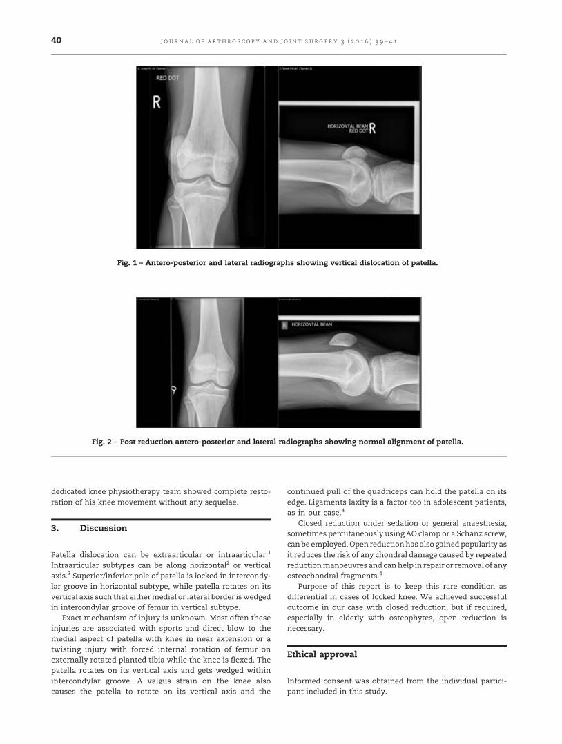
[(Fig._1)TD$FIG]
Fig. 1 – Antero-posterior and lateral radiographs showing vertical dislocation of patella.
[(Fig._2)TD$FIG]
Fig. 2 – Post reduction antero-posterior and lateral radiographs showing normal alignment of patella.
j o u r n a l o f a r t h r o s c o p y and j o i n t s u r g e r y 3 ( 2 0 1 6 ) 3 9 – 4 140
dedicated knee physiotherapy team showed complete resto-ration of his knee movement without any sequelae.
3. Discussion
Patella dislocation can be extraarticular or intraarticular.1
Intraarticular subtypes can be along horizontal2 or verticalaxis.3 Superior/inferior pole of patella is locked in intercondy-lar groove in horizontal subtype, while patella rotates on itsvertical axis such that eithermedial or lateral border is wedgedin intercondylar groove of femur in vertical subtype.
Exact mechanism of injury is unknown. Most often theseinjuries are associated with sports and direct blow to themedial aspect of patella with knee in near extension or atwisting injury with forced internal rotation of femur onexternally rotated planted tibia while the knee is flexed. Thepatella rotates on its vertical axis and gets wedged withinintercondylar groove. A valgus strain on the knee alsocauses the patella to rotate on its vertical axis and the
continued pull of the quadriceps can hold the patella on itsedge. Ligaments laxity is a factor too in adolescent patients,as in our case.4
Closed reduction under sedation or general anaesthesia,sometimes percutaneously using AO clamp or a Schanz screw,can be employed. Open reductionhas also gainedpopularity asit reduces the risk of any chondral damage caused by repeatedreductionmanoeuvres and canhelp in repair or removal of anyosteochondral fragments.4
Purpose of this report is to keep this rare condition asdifferential in cases of locked knee. We achieved successfuloutcome in our case with closed reduction, but if required,especially in elderly with osteophytes, open reduction isnecessary.
Ethical approval
Informed consent was obtained from the individual partici-pant included in this study.

j o u rn a l o f a r th r o s c o p y and j o i n t s u r g e r y 3 ( 2 0 1 6 ) 3 9 – 4 1 41
Conflicts of interest
All authors have none to declare.
r e f e r e n c e s
1. Ofluoglu O, Yasmin D, Donthineni R, Muzaffler Y.Superior dislocation of the patella with early onset
patellofemoral arthritis: a case report and literaturereview. Knee Surg Sports Traumatol Arthrosc. 2006;14:350–355.
2. Chauhan A, Maheswaran S, Anand S. Horizontalintraarticular dislocation of patella—a case reportand review of literature. Injury Extra. 2014;45(9):80–82.
3. Rollinson PD. Vertical intercondylar dislocation of the patella.Injury. 1988;19:281–282.
4. Shetty S, Ramesh B, Gul A, Madhusudan TR, Altayeb T.Vertical dislocation of the patella: report of 2 cases.Orthopedics. 2009;32:768–770.

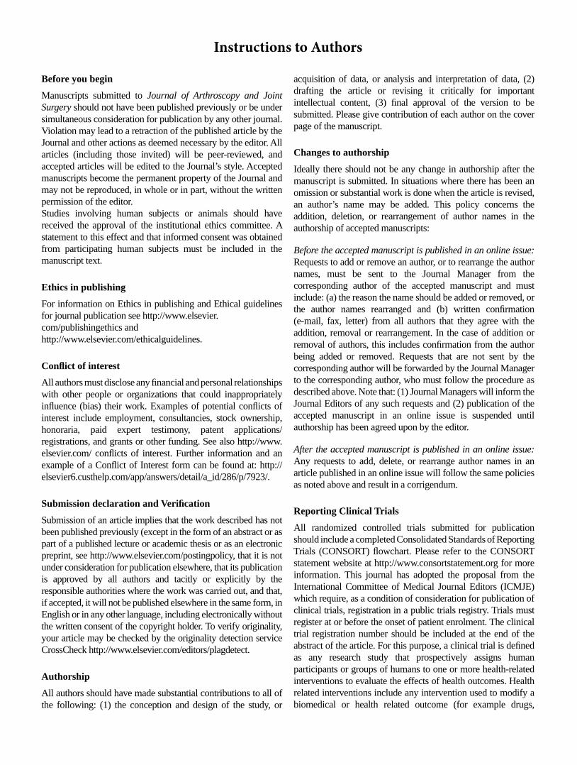
Before you begin
Manuscripts submitted to Journal of Arthroscopy and Joint Surgery should not have been published previously or be under simultaneous consideration for publication by any other journal. Violation may lead to a retraction of the published article by the Journal and other actions as deemed necessary by the editor. All articles (including those invited) will be peer-reviewed, and accepted articles will be edited to the Journal’s style. Accepted manuscripts become the permanent property of the Journal and may not be reproduced, in whole or in part, without the written permission of the editor.Studies involving human subjects or animals should have received the approval of the institutional ethics committee. A statement to this effect and that informed consent was obtained from participating human subjects must be included in the manuscript text.
Ethics in publishing
For information on Ethics in publishing and Ethical guidelines for journal publication see http://www.elsevier.com/publishingethics and http://www.elsevier.com/ethicalguidelines.
Confl ict of interest
All authors must disclose any fi nancial and personal relationships with other people or organizations that could inappropriately infl uence (bias) their work. Examples of potential confl icts of interest include employment, consultancies, stock ownership, honoraria, paid expert testimony, patent applications/registrations, and grants or other funding. See also http://www.elsevier.com/ confl icts of interest. Further information and an example of a Confl ict of Interest form can be found at: http://elsevier6.custhelp.com/app/answers/detail/a_id/286/p/7923/.
Submission declaration and Verifi cation
Submission of an article implies that the work described has not been published previously (except in the form of an abstract or as part of a published lecture or academic thesis or as an electronic preprint, see http://www.elsevier.com/postingpolicy, that it is not under consideration for publication elsewhere, that its publication is approved by all authors and tacitly or explicitly by the responsible authorities where the work was carried out, and that, if accepted, it will not be published elsewhere in the same form, in English or in any other language, including electronically without the written consent of the copyright holder. To verify originality, your article may be checked by the originality detection service CrossCheck http://www.elsevier.com/editors/plagdetect.
Authorship
All authors should have made substantial contributions to all of the following: (1) the conception and design of the study, or
acquisition of data, or analysis and interpretation of data, (2) drafting the article or revising it critically for important intellectual content, (3) fi nal approval of the version to be submitted. Please give contribution of each author on the cover page of the manuscript.
Changes to authorship
Ideally there should not be any change in authorship after the manuscript is submitted. In situations where there has been an omission or substantial work is done when the article is revised, an author’s name may be added. This policy concerns the addition, deletion, or rearrangement of author names in the authorship of accepted manuscripts:
Before the accepted manuscript is published in an online issue: Requests to add or remove an author, or to rearrange the author names, must be sent to the Journal Manager from the corresponding author of the accepted manuscript and must include: (a) the reason the name should be added or removed, or the author names rearranged and (b) written confi rmation (e-mail, fax, letter) from all authors that they agree with the addition, removal or rearrangement. In the case of addition or removal of authors, this includes confi rmation from the author being added or removed. Requests that are not sent by the corresponding author will be forwarded by the Journal Manager to the corresponding author, who must follow the procedure as described above. Note that: (1) Journal Managers will inform the Journal Editors of any such requests and (2) publication of the accepted manuscript in an online issue is suspended until authorship has been agreed upon by the editor.
After the accepted manuscript is published in an online issue: Any requests to add, delete, or rearrange author names in an article published in an online issue will follow the same policies as noted above and result in a corrigendum.
Reporting Clinical Trials
All randomized controlled trials submitted for publication should include a completed Consolidated Standards of Reporting Trials (CONSORT) fl owchart. Please refer to the CONSORT statement website at http://www.consortstatement.org for more information. This journal has adopted the proposal from the International Committee of Medical Journal Editors (ICMJE) which require, as a condition of consideration for publication of clinical trials, registration in a public trials registry. Trials must register at or before the onset of patient enrolment. The clinical trial registration number should be included at the end of the abstract of the article. For this purpose, a clinical trial is defi ned as any research study that prospectively assigns human participants or groups of humans to one or more health-related interventions to evaluate the effects of health outcomes. Health related interventions include any intervention used to modify a biomedical or health related outcome (for example drugs,
Instructions to Authors
surgical procedures, devices, behavioral treatments, dietary interventions, and process-of-care changes). Health outcomes include any biomedical or health-related measures obtained in patients or participants, including pharmacokinetic measures and adverse events. Purely observational studies (those in which the assignment of the medical intervention is not at the discretion of the investigator) will not require registration. Further information can be found at http://www.icmje.org.
Copyright
Upon acceptance of an article, authors will be asked to complete a ‘Journal Publishing Agreement’ (for more information on this and copyright see http://www.elsevier. com/copyright). Acceptance of the agreement will ensure the widest possible dissemination of information. An e-mail will be sent to the corresponding author confi rming receipt of the manuscript together with a ‘Journal Publishing Agreement’ form or a link to the online version of this agreement.
Role of the funding source
You are requested to identify who provided fi nancial support for the conduct of the research and/or preparation of the article and to briefl y describe the role of the sponsor(s), if any, in study design; in the collection, analysis and interpretation of data; in the writing of the report; and in the decision to submit the article for publication. If the funding source(s) had no such involvement then this should be stated. Please see http://www. elsevier.com/funding.
Submission of Manuscripts
The journal only accepts online submissions in electronic format. All new manuscripts must be submitted through Journal of Arthroscopy and Joint Surgery online and review website (http://ees.elsevier.com/jajs). Authors are requested to submit the text, tables, and fi gures in electronic form to this address. Please follow the following steps to submit your manuscript:
1. Open the homepage of the journal’s website (http://ees.elsevier.com/jajs).2. Register yourself for free by clicking on “Register” on the top and create a user profi le with a desired username and mandatory details. On submission of the information, you will receive an E-mail confi rming your registration along with the “Password”.3. Click “Log In” on the main navigation menu at the top of the journal screen to open the login page.4. Enter your username and password in the appropriate fi elds (E-mailed to you at the time of registration). Click “Author Log in”, this takes you to the “Author Main Menu”.
Note: Please note that the username and password combination required for Elsevier Editorial System is different from the username and password combination used to “Track your paper” on the Elsevier “Authors’ Home” website.
By submitting a manuscript, the author agrees to the following:1. The work is original and free from plagiarism.
2. It has neither been published, nor is it not under consideration for publication at another journal.3. All authors are aware of the authorship order. The corresponding author shall be responsible in case of dispute.4. Once published, copyright of manuscript shall stand transferred to the Journal.5. ‘Confl ict of interest’ if any, must be explicitly stated at the end of the manuscript.Manuscripts must conform to the instructions given below:
General: Type the manuscript using ‘Times New Roman’ font, size 12 in double space throughout. Please arrange the manuscript as follows: Title page, Abstract, Introduction, Methods, Results, Discussion, and References. Number all pages consecutively, beginning with the title page. All fi gures and Tables must be referred to in the manuscript. Consult a recent issue of the Journal for details. Only the Title page should bear the names and addresses of the author(s). Editorials, perspective and review articles are generally by invitation. However if you are interested in writing a review/perspective, you can send an email to the editor with the topic and a short summary of contents to be included. The editor will convey his decision in 7-10 days’ time.
Length of articles: Text of original articles should be between 2000 and 3500 words. The article should not ordinarily contain more than 3 tables, 2 fi gures and 25 references. Case Reports are accepted only if they can be converted into ‘What is your diagnosis?’ format (please consult a recent issue of the Journal). Briefl y, the format consists of case report of about 500 words, a diagnostic image followed by the actual diagnosis/answer and discussion (250 words) and upto 5 references. Letters discussing or criticizing material published recently in the Journal, brief presentations of data, or those pertaining to issues of relevance to health policy, practice of medicine, or the like, are welcome. These should not exceed 500 words, 1 table and 5 references.
Title page: In animal studies, the title should state the species; all other titles will refer to human studies. State names of authors (including fi rst names), the departments and the institution where the work was done. Please do not add your academic qualifi cations, designation etc. State contribution of each author clearly. A short, running title, not exceeding 40 characters, should be provided. Please provide the name, postal address with PIN code, facsimile number and E-mail address of the author to whom communications and proofs are to be sent. Acknowledgements, if any, may be mentioned on this page.
Acknowledgements: These should appear at the end of the manuscript. The source of funding as well as a disclosure statement mentioning confl ict of interest, if any, should appear under this heading.
References: Number the references in the order in which they fi rst appear in the text and identify the reference numbers in the text in superscript. References must be placed at the end of the manuscript. Please use recent references as much as possible. The responsibility for accuracy of
references lies with the respective authors. The Journal is in agreement with the International Committee of Medical Journal Editors (www. icmje.org). The general arrangement, abbreviations of Journal names and punctuations followed are as per the Uniform Requirements for Manuscripts submitted to Biomedical Journals (www.icmje.org). Please pay attention to the style of references and punctuations as follows:
Journal articleList all authors when six or less as shown in the example below: Tallon D, Chard J, Dieppe P. Exploring the priorities of patients with osteoarthritis of the knee. Arthritis Care and Res 2000;13:312–9.
When there are seven or more authors, list only the fi rst six and add et al.
Book or monographFollowing is an example: Cassidy JT. Juvenile rheumatoid arthritis. In: Textbook of Rheumatology 6th ed, Kelly et al (eds) Philadelphia Saunders 2000; pp. 1297–313.
Tables: Each Table should be typed on a separate page and numbered consecutively in Arabic numerals. Each table should have a title and all abbreviations should be explained in the footnote. Necessary explanatory notes, if any, may be given below the Table.
Figures/Illustrations/Photographs: Photographs of 300 dpi or higher resolution may be submitted as ‘jpeg’, or ‘tiff’ fi les in a zipped folder. In clinical photographs, identity of the subjects should be suitably masked; in case this is not
possible, a written permission from the concerned person should accompany the manuscript.
Legends to Figures: The Figure number (numbered consecutively in Arabic numerals), title and explanations of the Figures should appear in the legend (not on the Figure). Type the legends on a separate page. Enough information should be included to interpret the Figure without reference to the text.
Units: All measurements must be in metric units, preferably with corresponding SI units in parentheses.
Editorial Process: All articles submitted to the Journal undergo initial review by the Editor/associate editor and articles that are outside the scope of Journal or are not in the journal format are excluded. Later each article is reviewed by at least two reviewers. The time to fi rst decision is usually less than 6 weeks.
As per the policy of the Journal, an Editor, who is either author of a manuscript or belongs to the same institution as any of the authors, is not assigned that manuscript and is not involved in decision-making regarding its publication.Reviewers/Editorial Board members should decline the invitation to review a manuscript which is submitted by authors from their institution.
Reprints: Reprints may be requested and are provided on payment.
Address all correspondence to: Prof. Ravi Gupta or Mr. Sanjeev Anand, Chief Editors, Journal of Arthroscopy and Joint Surgery at [email protected].


![&$5%21 ',2;,'( 87,/,=$7,21 6800,7 - Whitmore Group | A ... · 7+ &$5%21 ',2;,'(87,/,=$7,21 6800,7 0ruh ,qir 5hjlvwudwlrq 3pibo #bszbi ] 5 ] & scbszbi!bdjfv ofu &wfou 4qpotps .(< 723,&6](https://img.pdfslide.us/doc/110x75/5f22d2cbc5cfc77a0a760e30/521-2-87721-68007-whitmore-group-a-7-521-287721.jpg)


![Reproduction 6.6 ;]](https://img.pdfslide.us/doc/110x75/568165e5550346895dd904fc/reproduction-66-.jpg)