Embed Size (px)
DESCRIPTION
Citation preview
Nondirective meditation activates default mode network and areasassociated with memory retrieval and emotional processing
Jian Xu, Alexandra Vik, Inge Rasmus Groote, Jim Lagopoulos, Are Holen, Øyvind Ellingsen and Svend Davanger
Journal Name: Frontiers in Human Neuroscience
ISSN: 1662-5161
Article type: Original Research Article
Received on: 26 Jun 2013
Accepted on: 04 Feb 2014
Provisional PDF published on: 04 Feb 2014
Frontiers website link: www.frontiersin.org
Citation: Xu J, Vik A, Groote IR, Lagopoulos J, Holen A, Ellingsen Ø andDavanger S(2014) Nondirective meditation activates default modenetwork and areas associated with memory retrieval andemotional processing. Front. Hum. Neurosci. 8:86.doi:10.3389/fnhum.2014.00086
Article URL: http://www.frontiersin.org/Journal/Abstract.aspx?s=537&name=human%20neuroscience&ART_DOI=10.3389/fnhum.2014.00086
(If clicking on the link doesn't work, try copying and pasting it into your browser.)
Copyright statement: © 2014 Xu, Vik, Groote, Lagopoulos, Holen, Ellingsen andDavanger. This is an open-access article distributed under theterms of the Creative Commons Attribution License (CC BY). Theuse, distribution or reproduction in other forums is permitted,provided the original author(s) or licensor are credited and thatthe original publication in this journal is cited, in accordance withaccepted academic practice. No use, distribution or reproductionis permitted which does not comply with these terms.
This Provisional PDF corresponds to the article as it appeared upon acceptance, after rigorous
peer-review. Fully formatted PDF and full text (HTML) versions will be made available soon.
1
Nondirective meditation activates default mode network 1
and areas associated with memory retrieval and emotional 2
processing 3 4 Running title: Brain activation in nondirective meditation 5 6 7 Jian Xu1, Alexandra Vik2, Inge Rasmus Groote3, Jim Lagopoulos4, Are Holen5,6, 8 Øyvind Ellingsen7,8, Asta K Håberg1,5, Svend Davanger9* 9 10 1. Department of Medical Imaging, St. Olavs Hospital, Trondheim, Norway 11 2. Department of Biological and Medical Psychology, University of Bergen, Bergen, 12 Norway 13 3. Department of Psychology, University of Oslo, Oslo, Norway 14 4. Brain & Mind Research Institute, University of Sydney, Sydney, Australia 15 5. Department of Neuroscience, Faculty of Medicine, Norwegian University of 16 Science and Technology, Trondheim, Norway 17 6. Centre for Pain and Complex Disorders, St. Olavs Hospital, Trondheim, Norway 18 7. Department of Circulation and Medical Imaging, Faculty of Medicine, Norwegian 19 University of Science and Technology, Trondheim, Norway 20 8. Department of Cardiology, St. Olavs Hospital, Trondheim, Norway 21 9. Institute of Basic Medical Science, University of Oslo, Oslo, Norway 22 23 24 *Correspondence: 25 Svend Davanger 26 Institute of Basic Medical Science 27 University of Oslo 28 P.O.Box 1105 Blindern 29 0317 Oslo 30 Norway 31 [email protected] 32 33 34 35
36
2
ABSTRACT 36 37 Nondirective meditation techniques are practiced with a relaxed focus of attention 38 that permits spontaneously occurring thoughts, images, sensations, memories and 39 emotions to emerge and pass freely, without any expectation that mind wandering 40 should abate. These techniques are thought to facilitate mental processing of 41 emotional experiences, thereby contributing to wellness and stress management. 42 The present study assessed brain activity by functional magnetic resonance 43 imaging in 14 experienced practitioners of Acem meditation in two experimental 44 conditions. In the first, nondirective meditation was compared to rest. Significantly 45 increased activity was detected in areas associated with attention, mind wandering, 46 retrieval of episodic memories and emotional processing. In the second condition, 47 participants carried out concentrative practicing of the same meditation technique, 48 actively trying to avoid mind wandering. The contrast nondirective meditation > 49 concentrative practicing was characterized by higher activity in the right medial 50 temporal lobe (parahippocampal gyrus and amygdala). In conclusion, the present 51 results support the notion that nondirective meditation, which permits mind 52 wandering, involves more extensive activation of brain areas associated with 53 episodic memories and emotional processing, than during concentrative practicing 54 or regular rest. 55 56 Keywords: fMRI, meditation, attention, nondirective, brain, default mode 57 network, mind wandering 58
59
3
INTRODUCTION 59 60 Volitional and spontaneous activities in meditation 61 Many types of meditation used for stress management and health can be described as 62 a cycle of volitional and spontaneous cognitive processes (Cardoso et al., 2004). 63 Attention is intentionally focused on a suitable meditation object, such as mental 64 repetition of a non-semantic meditation sound, sensations associated with breath or 65 specific regions of the body, a physical or mental visual image, or by simply being 66 aware of the shifting flow of inner experiences (Cardoso et al., 2004;Ospina et al., 67 2007). Focusing on the meditation object is typically interspersed with periods of 68 mind wandering (Cardoso et al., 2004;Ospina et al., 2007;Hasenkamp et al., 2012), 69 which has been defined as being absorbed in spontaneously occurring thoughts, 70 images, sensations, memories and emotions unrelated to current volitional activity, 71 more or less without really being aware of it (Mason et al., 2007;Christoff et al., 72 2009). An example of this cognitive cycle is given in a detailed temporal study of 73 meditation with focused attention on the breath (Hasenkamp et al., 2012). Functional 74 magnetic resonance imaging (fMRI) was used to correlate brain activation with 75 cognitive processes that describes the shifting between focusing on the meditation 76 object and spontaneously occurring thought. Mind wandering was associated with 77 activation of the default mode network as well as sensory and motor cortices and 78 posterior insula. Becoming aware that the breath was completely out of the focus of 79 attention was associated with activation of the salience network. Shifting back to the 80 breath and sustaining the focus on it were associated with elements of the executive 81 network (Hasenkamp et al., 2012). 82 83 Different perceptions of mind wandering 84 The function of spontaneous mental processes in meditation is controversial. How 85 they are dealt with, depends on the type of practice (Box 1-3). In most mindfulness 86 practices and many other techniques associated with Buddhist traditions, mind 87 wandering is considered a distraction and a gateway to rumination, anxiety and 88 depression (Sood and Jones, 2013). An ultimate goal of these methods is therefore to 89 reduce mind wandering and its purported negative consequences (Brewer et al., 90 2011;Sood and Jones, 2013;Taylor et al., 2013). In contrast, some practices consider 91 the spontaneous flow of inner experiences as part of the meditation process. 92 Accepting mind wandering while practicing is a core element in the Relaxation 93 Response, Transcendental Meditation, Clinically Standardized Meditation, and Acem 94 Meditation (Carrington, 1998)(Benson et al., 1975;Carrington et al., 1980;Ospina et 95 al., 2007;Davanger et al., 2010;Travis and Shear, 2010). As described below, these 96 techniques may be classified as nondirective, indicating less control of the process 97 while practicing (Box 3). It has been proposed that types of meditation that allow 98 spontaneous thoughts, images, sensations, memories and emotions to emerge and pass 99 freely without actively controlling or pursuing them, over time may reduce stress by 100 increasing awareness and acceptance of emotionally charged experiences (Ellingsen 101 and Holen, 2008;Lutz et al., 2008a;Davidson, 2010). This notion concurs with recent 102 articles suggesting that mind wandering and activation of the default mode network in 103 general may serve introspective and adaptive functions beyond rumination and 104 daydreaming (Ottaviani et al., 2013). Potentially useful functions would include 105 mental simulations, using autobiographical memory retrieval to envision the future 106 and conceiving the perspective of others (Buckner et al., 2008;Andrews-Hanna, 107 2012). An interesting question is therefore whether type of meditation and mode of 108 practicing might affect the extent of mind wandering and the pattern of default mode 109 activation during meditation. 110
4
111 Box 1: Focused Attention 112 Focused attention practices usually entail paying attention to the physical sensation of 113 the breath wherever it is felt most strongly in the body, without trying to change it in 114 any way. Whenever attention has wandered to something else, the meditator gently 115 but firmly brings it back to the physical sensation of the breath (Brewer et al., 2011). 116 Important aims of the practice are to quickly detect mind wandering and maintain 117 attention more stably on the breath, eventually needing less effort in the task, and over 118 time reducing emotional reactivity (Lutz et al., 2008b). Focused attention practices 119 typically involve a relatively narrow field of focus. As a result, the ability to identify 120 stimuli outside that field of focus may be reduced (Lutz et al., 2008b). 121 122 Box 2: Open Monitoring 123 Open monitoring practices (sometimes called choiceless awareness) are described as 124 paying attention to whatever comes into ones awareness - whether it is a thought, 125 emotion, or body sensation - just following it until something else emerges without 126 trying to hold onto it or change it in any way (Brewer et al., 2011). Even though 127 “effortful selection” or “grasping” of an object as primary focus is gradually replaced 128 by “effortless sustaining of awareness without explicit selection,” the core activity of 129 the practice is to sustain attention with the shifting flow of experiences, sometimes 130 detecting emotional tone as a background feature (Lutz et al., 2008b). 131 132 Box 3: Nondirective meditation 133 In nondirective meditation practices, a relaxed focus of attention is established by 134 effortless, mental repetition of a short sequence of syllables, which may either be a 135 traditional mantra or a non-semantic meditation sound (Benson et al., 1975;Carrington 136 et al., 1980;Ospina et al., 2007;Davanger et al., 2010;Travis and Shear, 2010). 137 Whenever the meditator becomes aware that the focus of attention has shifted to 138 mainly being occupied with spontaneously occurring thoughts, images, sensations, 139 memories or emotions, attention is gently and nonjudgmentally redirected to 140 repetition of the meditation sound. The aim of the practice is to increase the ability to 141 accept and tolerate stressful and emotional experiences as a normal part of meditation 142 as well as everyday life (Davanger et al., 2010). Attention is neither directed towards 143 staying with the meditation object like in focused attention techniques nor directed 144 towards observing the spontaneous flow of experiences like in open monitoring 145 meditation (Lutz et al., 2008b). Consequently, such methods comprise a distinct style 146 of practicing (Cahn and Polich, 2006;Ellingsen and Holen, 2008;Travis and Shear, 147 2010), that has previously been termed nondirective meditation, as the presence of 148 spontaneously occurring thoughts, images, sensations, memories and emotions is 149 accepted without actively directing attention towards them or away from them 150 (Ellingsen and Holen, 2008;Lagopoulos et al., 2009;Nesvold et al., 2011). Further 151 details on Acem meditation and its background are provided in previous publications 152 (Ellingsen and Holen, 2008;Davanger et al., 2010). 153 154 Extent of mind wandering 155 It is often assumed that mind wandering is reduced during meditation, and more so in 156 practitioners with many years of experience. The evidence comes from a relatively 157 small number of studies in which the extent of mind wandering was assessed by 158 questionnaire. Self-reported mind wandering during meditation was less abundant in 159 participants with long-term experience in “concentration” (focused attention on 160 breath), “loving-kindness meditation” (exercise oriented toward enhancing 161 unconditional, positive emotional states of kindness and compassion), and “choiceless 162
5
awareness” (open monitoring of mind wandering) compared to inexperienced controls 163 (Brewer et al., 2011;Hofmann et al., 2011). Self-reported time on task during 164 “mindfulness of breathing” was higher in experienced than in inexperienced 165 participants, indicating less mind wandering with training (Holzel et al., 2007). In 166 contrast, there was no correlation between the number of button presses indicating 167 epochs of mind wandering during focused attention on the breath with years of 168 practice or with high versus low practice groups (Hasenkamp et al., 2012). In this 169 study, participants recorded an average of one mind wandering per 80 seconds over a 170 20-minute fMRI session, by pressing a button whenever they realized that their mind 171 had wandered completely away from the breath. 172 173 Default mode network activation 174 Many concepts of how meditation affects mind wandering derive from its association 175 with the default mode network. A number of imaging studies have shown that a 176 system of cortical areas increase their activation when the brain is not engaged in an 177 externally defined task, and that the magnitude of increase correlates with the extent 178 of mind wandering (Mason et al., 2007;Buckner et al., 2008). Although some 179 variation occurs, the default network mostly includes medial brain structures, i.e., the 180 ventral medial prefrontal cortex, the posterior cingulate/retrosplenial cortex, the 181 inferior parietal lobe, the lateral temporal cortex, the dorsal medial prefrontal cortex, 182 and the hippocampal formation (Buckner et al., 2008). 183 184 A majority of the studies on meditation and mind wandering have measured how 185 fMRI activation and functional connectivity of the default mode network are related 186 to mind wandering. Most of these describe trait differences in brain activation patterns 187 arising from meditation, often showing decreased default mode network activation in 188 experienced meditators compared to novices (Brewer et al., 2011;Sood and Jones, 189 2013). 190 191 Only a few studies have reported state changes, contrasting meditation with various 192 control tasks in the same practitioners, but with varying results. Using rest as a 193 control, Brefczynski-Lewis and coworkers showed activation of a large overlapping 194 network of attention-related cortical regions during “concentration meditation” 195 (focused attention with a simple visual stimulus), including frontal, parietal regions, 196 lateral occipital cortex, and insula (Brefczynski-Lewis et al., 2007). Lazar and 197 coworkers showed activation of dorsolateral prefrontal and parietal cortices, 198 hippocampus/parahippocampus, temporal lobe, pregenual anterior cingulate cortex, 199 striatum, and pre- and post-central gyri during mantra meditation coordinated with 200 breath (Lazar et al., 2000). Generating a list of animals was used as control task. 201 Engström and coworkers compared mantra meditation with silent repetition of a short 202 semantic phrase as control and detected activation in bilateral 203 hippocampus/parahippocampal formations, as well as bilateral middle cingulate 204 cortex and bilateral precentral cortex (Engstrom et al., 2010). Interestingly, Manna 205 and coworkers (Manna et al., 2010) described reduced activation of precuneus (a core 206 default mode network area) compared to rest during meditation with focused attention 207 on the breath, and increased activation during meditation with open monitoring of 208 “any experiential or mental content” (Manna et al., 2010). None of the 209 aforementioned studies assessed the extent of mind wandering. 210 211 Aim and hypothesis 212 The aim of the present study was to determine whether nondirective meditation is 213 conducive to default mode network activation. We hypothesized that accepting the 214
6
spontaneous flow of thoughts, images, sensations, memories and emotions as part of 215 meditation, without any emphasis on reducing, monitoring, evaluating or directly 216 relating to it, would increase mind wandering and activation of the default mode 217 network, compared to practicing with more emphasis on control and a concentrative 218 focus of attention. We therefore assessed whether practicing the same technique 219 (Acem meditation) with different types of attentional focus would affect the 220 subjective experience and the pattern of brain activation during meditation assessed 221 by fMRI. 222 223 224 METHODS 225 226 Ethics statement 227 The National Committee for Medical Research Ethics in Norway approved the study. 228 Informed written consent was obtained from all participants before inclusion. 229 230 Participants 231 Twenty-seven experienced practitioners of Acem meditation (18 men and 9 women) 232 were recruited. All participants were regular practitioners (2x30 minutes daily) and 233 had extensive experience with longer meditation periods, including participation in at 234 least one three-week long retreat. Twenty-four were right handed, ascertained by the 235 Edinburg Handedness Inventory (Oldfield, 1971). Thirteen participants were excluded 236 from final data analysis due to rigorous quality control; only participants with 237 acceptable recordings from both fMRI sessions were included. Three were excluded 238 because of reported sleep during the recording, two because of significant head 239 motion (>1mm), one because of error in scanning protocol, and seven because of 240 technical problems that lead to corruption of the fMRI images. Even though the head 241 was securely fixed inside the headcoil according to standard procedure (using 242 triangular shaped foam pads), minor involuntary movements were difficult to avoid 243 during two 20-minute recordings in a relaxed reclining condition. Thus, fourteen 244 practitioners (8 men and 6 women, 13 right handed), aged 28-61 years (mean 49, SD 245 9) with 9-38 years of meditation practice (mean 27, SD 9) were included in final data 246 analysis. We included only experienced meditators in our study, since it takes 247 extensive training to reliably distinguish between nondirective and concentrative 248 practicing. 249 250 fMRI meditation instructions 251 Details on nondirective meditation has been provided above (Box 3) and in previous 252 publications (Ellingsen and Holen, 2008;Davanger et al., 2010). Participants were 253 asked to perform Acem meditation in two separate runs of fMRI acquisition. In 254 nondirective meditation the participants were instructed to repeat the meditation 255 sound in a relaxed and effortless manner, in the same way as during home practice. 256 Spontaneous mind wandering was neither prevented nor encouraged. In contrast, 257 during concentrative practicing, the meditation sound was repeated in a more forceful 258 manner, with strict regularity, in order to maintain the focus of attention on the sound, 259 attempting to avoid mind wandering. As expected, mind wandering was not avoided 260 completely, although more of the participants reported decreased mind wandering 261 during concentrative practicing than in nondirective meditation. During data 262 acquisition in the resting blocks (see below) participants were instructed to rest 263 without repeating the meditation sound, allowing mind wandering where 264 spontaneously occurring thoughts, images, sensations, memories and emotions could 265 emerge and pass freely. 266
7
267 Experimental design 268 In order to establish a stable, relaxed resting control state, all participants meditated 269 for 45-60 minutes before experimental recordings. Each practitioner was scanned in 270 one session with one run of nondirective meditation and one of concentrative 271 practicing (block design), presented in randomized order. In each run the practitioners 272 performed a sequence of four meditation blocks lasting 3, 5, 4 and 3 minutes 273 respectively, interspersed with five resting blocks lasting one minute each. Block 274 length was varied in order to avoid “false” fMRI activation induced by expectation. 275 All subjects were scanned with eyes closed. Concentrative practicing and rest were 276 used as contrasts for nondirective meditation. This would minimize the possible effect 277 of underlying traits in the subjects, each subject serving as his or her own control. 278 Immediately following each scanning run, all participants were asked to complete a 279 questionnaire assessing their meditation experiences: extent of mind wandering 280 compared to regular home practice, whether they became drowsy or briefly fell 281 asleep, and to what extent the sound from the MRI scanner was disturbing. They also 282 confirmed whether they had been able to carry out the meditation tasks. 283 284 Data acquisition 285 Structural and functional scanning was performed using a 3T Philips Intera scanner 286 (Philips Medical, Best, The Netherlands) with an 8-channel SENSitivity Encoding 287 (SENSE) head-coil (InVivo, Gainsville, FL, USA). Using BOLD-sensitive imaging, a 288 total of 400 volumes was acquired for each run with a gradient-echo echo-planar-289 imaging pulse sequence. Each volume consisted of 44 contiguous axial slices, with 290 the following scan parameters: SENSE-reduction factor = 2.2, TR = 3000ms; flip 291 angle = 90°; TE = 35ms; FOV = 230mm; slice thickness = 2.5mm; matrix = 64x64 292 giving an in-plane resolution of 3.6×3.6mm2. Also a high-resolution T1-weighted 293 image series was collected using a three-dimensional magnetization-prepared rapid 294 gradient echo sequence (MP-RAGE) consisting of 182 contiguous sagittal slices of 295 1.2-mm thickness with an in-plane resolution of 1x1 mm. For analysis, all images 296 were reconstructed to 1 mm3. 297 298 Data analysis 299 Imaging data were analyzed using FSL 4.0 (Analysis Group, FMRIB, Oxford, UK; 300 www.fmrib.ox.ac.uk/fsl/). First, non-brain tissue was removed from the T1-weighted 301 anatomical image-series using the Brain Extraction Tool (Smith, 2002). The resulting 302 images were transformed non-linearly to the MNI152 1x1x1 mm template (Montreal 303 Neurological Institute, Montreal, QC, Canada), and motion corrected with the median 304 volume of each run as reference using the FNIRT algorithm (Andersson et al., 2007). 305 Then each functional run was co-registered to the corresponding anatomical T1-306 weighted image-series and transformed into MNI152 space by the transformation 307 matrix obtained from the T1-weighted images. The functional data was smoothed by a 308 6 mm full-width at half-maximum (FWHM) Gaussian filter, and a temporal high-pass 309 filter with a cut-off time of 350 seconds. 310
The two-level random effects statistical analysis of the fMRI data was carried out 311 using Bayesian estimation techniques with FEAT (Smith et al., 2004). Conditions 312 were modeled according to a boxcar stimulus function convolved with a two-gamma 313 hemodynamic response function (Boynton et al., 1996). The first minute of each 314 meditation block was excluded from the analysis by modeling it as non-effect, as 315 meditation activations take time to build up (Davanger et al., 2010). The effect of 316 each condition was estimated according to a general linear model (Friston et al., 317
8
1995). A whole-brain analysis was performed using mixed effects FLAME-1 318 algorithms (Beckmann et al., 2003). Statistical thresholds for contrasts nondirective 319 meditation > rest, and concentrative practicing > rest were set to p < 0.05, family wise 320 error rate was corrected using cluster-level interference by setting cluster forming 321 threshold at z > 3.0 (p < 0.0027). For the contrast nondirective meditation > 322 concentrative practicing it was set to p < 0.05 and cluster forming z > 2.3 (p < 323 0.0214). To increase sensitivity, the threshold was set less stringently for the latter 324 comparison, because the expected difference between two similar conditions is 325 usually smaller and the variability greater than for respective comparisons with rest. 326 For all three contrasts, correlation analysis with years of experience as independent 327 variable was performed in FEAT using FLAME-1 algorithm. Years of experience was 328 defined as an extra environmental variable for all three contrasts. Brain areas were 329 identified by FSL atlases and other relevant sources for functional data as referenced. 330 331 Statistical analysis of questionnaire data 332 A post-scan behavior questionnaire comprised three questions (translated from 333 Norwegian): 1) How disturbing was the scanner sound in the background: 0 = not at 334 all, 1 = some, 2 = much. 2) What was the extent of mind wandering compared to 335 regular meditation outside the scanner: 0 = less, 1 = similar, 2 = more. 3) Did you 336 become drowsy or fall asleep: 0 = wakeful, 1 = drowsy, 2 = fell asleep. The 337 questionnaire data were analyzed in Microsoft Excel (Microsoft Corporation, 338 Redmond, Washington, US) (Fig. 2). Fisher’s exact test was performed to assess 339 whether mind wandering, drowsiness and disturbance by scanner depended on the 340 mode of practicing (nondirective versus concentrative) in 2x2 tables, excluding table 341 lines with zero-cells. As described below, participants who fell asleep during 342 scanning, were excluded from further analyses. 343 344 345 RESULTS 346 347 Behavioral data 348
Data from a brief questionnaire administered immediately after each fMRI recording 349 indicated a trend for less mind wandering with concentrative practicing compared to 350 regular meditation. Even though the meditation blocks were short and the number of 351 participants small, a larger number experienced less mind wandering during 352 concentrative practicing than during nondirective meditation, whereas the numbers of 353 participants who were wakeful/drowsy and disturbed some/much by noise were 354 similar during nondirective and concentrative practicing, respectively (Table 1). A 355 majority spontaneously remarked that concentrative practicing was effortful and 356 tiring, although it was not an item in the questionnaire. 357
358
359
360
361
362
363
364
365
366
367
9
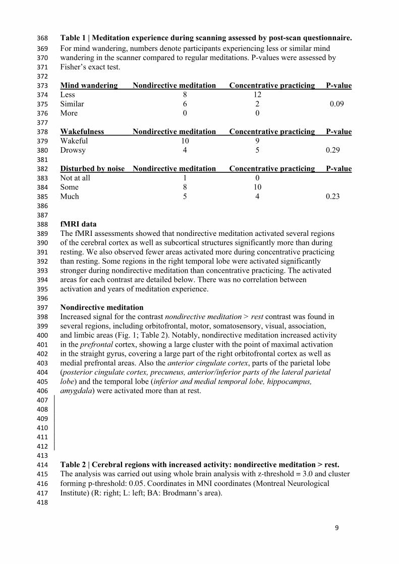
Table 1 | Meditation experience during scanning assessed by post-scan questionnaire. 368
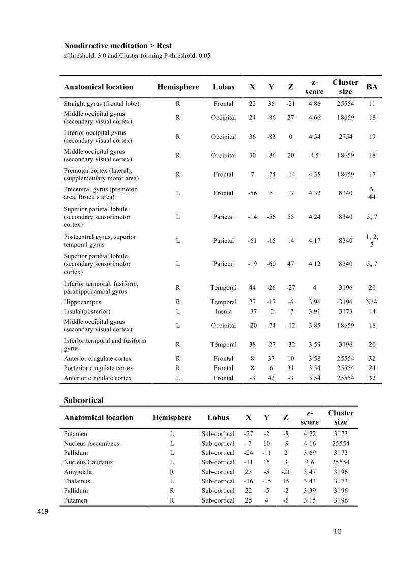
For mind wandering, numbers denote participants experiencing less or similar mind 369 wandering in the scanner compared to regular meditations. P-values were assessed by 370 Fisher’s exact test. 371 372 Mind wandering Nondirective meditation Concentrative practicing P-value 373 Less 8 12 374 Similar 6 2 0.09 375 More 0 0 376 377 Wakefulness Nondirective meditation Concentrative practicing P-value 378 Wakeful 10 9 379 Drowsy 4 5 0.29 380 381 Disturbed by noise Nondirective meditation Concentrative practicing P-value 382 Not at all 1 0 383 Some 8 10 384 Much 5 4 0.23 385 386 387 fMRI data 388 The fMRI assessments showed that nondirective meditation activated several regions 389 of the cerebral cortex as well as subcortical structures significantly more than during 390 resting. We also observed fewer areas activated more during concentrative practicing 391 than resting. Some regions in the right temporal lobe were activated significantly 392 stronger during nondirective meditation than concentrative practicing. The activated 393 areas for each contrast are detailed below. There was no correlation between 394 activation and years of meditation experience. 395 396 Nondirective meditation 397 Increased signal for the contrast nondirective meditation > rest contrast was found in 398 several regions, including orbitofrontal, motor, somatosensory, visual, association, 399 and limbic areas (Fig. 1; Table 2). Notably, nondirective meditation increased activity 400 in the prefrontal cortex, showing a large cluster with the point of maximal activation 401 in the straight gyrus, covering a large part of the right orbitofrontal cortex as well as 402 medial prefrontal areas. Also the anterior cingulate cortex, parts of the parietal lobe 403 (posterior cingulate cortex, precuneus, anterior/inferior parts of the lateral parietal 404 lobe) and the temporal lobe (inferior and medial temporal lobe, hippocampus, 405 amygdala) were activated more than at rest. 406 407 408 409 410 411 412 413 Table 2 | Cerebral regions with increased activity: nondirective meditation > rest. 414 The analysis was carried out using whole brain analysis with z-threshold = 3.0 and cluster 415 forming p-threshold: 0.05. Coordinates in MNI coordinates (Montreal Neurological 416 Institute) (R: right; L: left; BA: Brodmann’s area). 417 418
10
Nondirective meditation > Rest z-threshold: 3.0 and Cluster forming P-threshold: 0.05
Anatomical location Hemisphere Lobus X Y Z z-
score Cluster
size BA
Straight gyrus (frontal lobe) R Frontal 22 36 -21 4.86 25554 11 Middle occipital gyrus (secondary visual cortex) R Occipital 24 -86 27 4.66 18659 18
Inferior occipital gyrus (secondary visual cortex) R Occipital 36 -83 0 4.54 2754 19
Middle occipital gyrus (secondary visual cortex) R Occipital 30 -86 20 4.5 18659 18
Premotor cortex (lateral), (supplementary motor area) R Frontal 7 -74 -14 4.35 18659 17
Precentral gyrus (premotor area, Broca’s area) L Frontal -56 5 17 4.32 8340 6,
44
Superior parietal lobule (secondary sensorimotor cortex)
L Parietal -14 -56 55 4.24 8340 5, 7
Postcentral gyrus, superior temporal gyrus L Parietal -61 -15 14 4.17 8340 1, 2,
3
Superior parietal lobule (secondary sensorimotor cortex)
L Parietal -19 -60 47 4.12 8340 5, 7
Inferior temporal, fusiform, parahippocampal gyrus R Temporal 44 -26 -27 4 3196 20
Hippocampus R Temporal 27 -17 -6 3.96 3196 N/A Insula (posterior) L Insula -37 -2 -7 3.91 3173 14 Middle occipital gyrus (secondary visual cortex) L Occipital -20 -74 -12 3.85 18659 18
Inferior temporal and fusiform gyrus R Temporal 38 -27 -32 3.59 3196 20
Anterior cingulate cortex R Frontal 8 37 10 3.58 25554 32 Posterior cingulate cortex R Frontal 8 6 31 3.54 25554 24 Anterior cingulate cortex L Frontal -3 42 -3 3.54 25554 32
Subcortical
Anatomical location Hemisphere Lobus X Y Z z-score
Cluster size
Putamen L Sub-cortical -27 -2 -8 4.22 3173 Nucleus Accumbens L Sub-cortical -7 10 -9 4.16 25554 Pallidum L Sub-cortical -24 -11 2 3.69 3173 Nucleus Caudatus L Sub-cortical -11 15 3 3.6 25554 Amygdala R Sub-cortical 23 -5 -21 3.47 3196 Thalamus L Sub-cortical -16 -15 15 3.43 3173 Pallidum R Sub-cortical 22 -5 -2 3.39 3196 Putamen R Sub-cortical 25 4 -5 3.15 3196
419
11
420 Large clusters were also detected in the occipital lobe covering vision areas in the 421 middle occipital gyrus and striate cortex. In the posterior part of the frontal lobe, 422 activation occurred in primary and supplementary motor areas of the left hemisphere, 423 extending into Broca’s area. 424 425 In the left parietal lobe, sensorimotor and secondary sensory regions including part of 426 the precuneus were activated. There was no change in Wernicke’s receptive speech 427 area. 428 429 In the right temporal lobe, three clusters were found: the fusiform cortex/inferior 430 temporal gyrus/parahippocampal gyrus including the visual processing and facial 431 areas, the hippocampus, and the amygdala. 432 433 In the cingulate cortex, separate clusters in the right and left anterior regions were 434 activated, as well as in the right posterior regions. Activated clusters were also seen in 435 two non-cortical regions: In the left basal ganglia (putamen, globus pallidus, and the 436 nucleus accumbens), and in a right and a left cerebellar region. 437 438 The opposite contrast, nondirective meditation < rest, showed no positive activation. 439 440 Concentrative practicing 441 The contrast concentrative practicing >rest revealed significant activation in 442 three regions (Figure 1; Table 3). Motor area activation was present in the 443 posterior part of the middle frontal gyrus/premotor cortex, precentral gyrus, 444 the primary motor cortex, and the supplementary motor area/pre-motor 445 cortex. In visual areas, we observed activation of the middle and inferior 446 occipital gyrus/lateral occipital cortex, the occipital fusiform gyrus, and the 447 intracalcarine/visual and the occipital pole/visual cortices. Lastly, one cluster 448 was activated in the dorsal aspect of the anterior cingulate cortex, bilaterally. 449 No parietal or temporal clusters were seen during concentrative practicing. 450 451 The opposite contrast, concentrative practicing < rest, showed no positive 452 activation. 453 454 455 456 457 458 459 460 461 462 463 464 Table 3 | Cerebral regions with increased activity: concentrative practicing > 465 rest. 466 The analysis was carried out using whole brain analysis with z-threshold = 3.0 and 467 cluster forming p-threshold: 0.05. Coordinates in MNI coordinates (Montreal 468 Neurological Institute) (R: right; L: left; BA: Brodmann’s area). 469 470
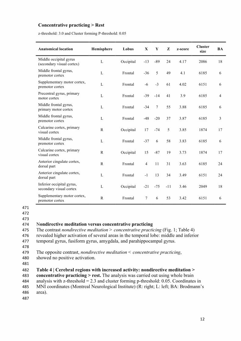
12
Concentrative practicing > Rest
z-threshold: 3.0 and Cluster forming P-threshold: 0.05
Anatomical location Hemisphere Lobus X Y Z z-score Cluster size BA
Middle occipital gyrus (secondary visual cortex) L Occipital -13 -89 24 4.17 2086 18
Middle frontal gyrus, premotor cortex L Frontal -36 5 49 4.1 6185 6
Supplementary motor cortex, premotor cortex L Frontal -6 -3 61 4.02 6151 6
Precentral gyrus, primary motor cortex L Frontal -39 -14 41 3.9 6185 4
Middle frontal gyrus, primary motor cortex L Frontal -34 7 55 3.88 6185 6
Middle frontal gyrus, premotor cortex L Frontal -48 -20 37 3.87 6185 3
Calcarine cortex, primary visual cortex R Occipital 17 -74 5 3.85 1874 17
Middle frontal gyrus, premotor cortex L Frontal -37 6 58 3.83 6185 6
Calcarine cortex, primary visual cortex R Occipital 15 -87 19 3.73 1874 17
Anterior cingulate cortex, dorsal part R Frontal 4 11 31 3.63 6185 24
Anterior cingulate cortex, dorsal part L Frontal -1 13 34 3.49 6151 24
Inferior occipital gyrus, secondary visual cortex L Occipital -21 -75 -11 3.46 2049 18
Supplementary motor cortex, premotor cortex R Frontal 7 6 53 3.42 6151 6
471 472 473 Nondirective meditation versus concentrative practicing 474 The contrast nondirective meditation > concentrative practicing (Fig. 1; Table 4) 475 revealed higher activation of several areas in the temporal lobe: middle and inferior 476 temporal gyrus, fusiform gyrus, amygdala, and parahippocampal gyrus. 477 478 The opposite contrast, nondirective meditation < concentrative practicing, 479 showed no positive activation. 480 481
Table 4 | Cerebral regions with increased activity: nondirective meditation > 482 concentrative practicing > rest. The analysis was carried out using whole brain 483 analysis with z-threshold = 2.3 and cluster forming p-threshold: 0.05. Coordinates in 484 MNI coordinates (Montreal Neurological Institute) (R: right; L: left; BA: Brodmann’s 485 area). 486 487
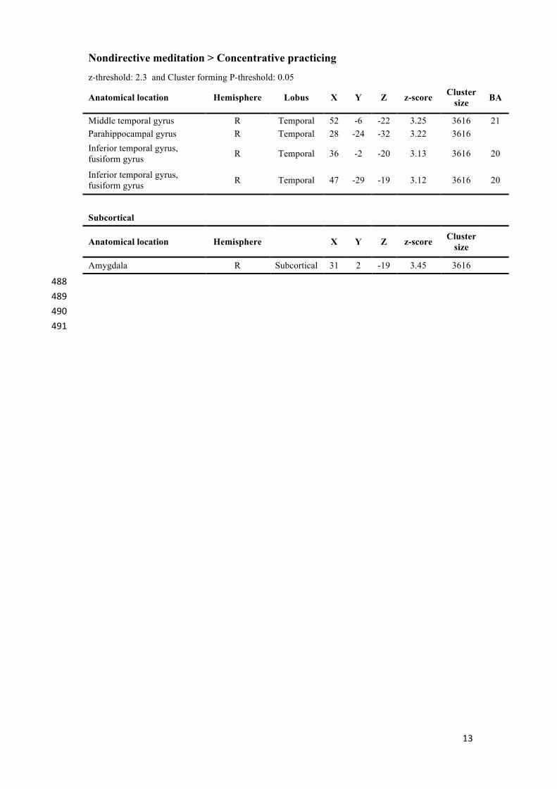
13
Nondirective meditation > Concentrative practicing
z-threshold: 2.3 and Cluster forming P-threshold: 0.05
Anatomical location Hemisphere Lobus X Y Z z-score Cluster size BA
Middle temporal gyrus R Temporal 52 -6 -22 3.25 3616 21 Parahippocampal gyrus R Temporal 28 -24 -32 3.22 3616 Inferior temporal gyrus, fusiform gyrus R Temporal 36 -2 -20 3.13 3616 20
Inferior temporal gyrus, fusiform gyrus R Temporal 47 -29 -19 3.12 3616 20
Subcortical
Anatomical location Hemisphere X Y Z z-score Cluster size
Amygdala R Subcortical 31 2 -19 3.45 3616 488
489
490
491
14
DISCUSSION 491 492 The present study sought to investigate state effects of nondirective meditation either 493 compared to rest or to concentrative practicing in participants with long-term 494 experience of Acem meditation. Results are consistent with the notion that 495 nondirective meditation involves more extensive activation of the default mode 496 network, including brain areas associated with episodic memories and emotional 497 processing. 498 499 Default mode network activation 500 Compared to rest, nondirective meditation increased activation within all cortical 501 areas defining the default mode network (Buckner et al., 2008), including the ventral 502 medial prefrontal cortex, the posterior cingulate/retrosplenial cortex, the inferior 503 parietal lobe, the lateral temporal cortex, the dorsal medial prefrontal cortex, and the 504 hippocampal formation (Figure 1, Table 2). The pattern of activations was similar to 505 that associated with mind wandering in a recent study of meditation with focused 506 attention on breath, including posterior cingulate cortex, medial prefrontal cortex, 507 posterior parietal and temporal cortex and the hippocampus (Hasenkamp et al., 2012). 508 In contrast, the control task of concentrative practicing in the present study seemed to 509 have little effect on default mode network activation, including only the anterior 510 cingulate cortex when compared to rest (Figure 1, Table 3). However, direct 511 comparison of nondirective meditation with concentrative practicing gave only 512 temporal clusters, including parahippocampal areas and amygdala. These observations 513 indicate that the extent of default mode network activation during concentrative 514 practicing probably lies somewhere between nondirective meditation and rest: slightly 515 more than in rest, but evidently not enough to yield significant clusters in most default 516 mode areas. This interpretation is consistent with the trend of less mind wandering 517 reported in concentrative practicing compared to nondirective meditation (Table 1). 518 519 Our results corroborate previous findings that suggest increased default mode network 520 activation during meditation. Experienced Vipassana meditators (focused attention on 521 breath) showed stronger activation of the anterior cingulate cortex and the dorsal 522 medial prefrontal cortex than control subjects (Holzel et al., 2007). During resting 523 state, practitioners of “brain-wave vibration meditation” (meditative movement) had 524 greater functional connectivity within the default mode network in the medial 525 prefrontal cortex than controls (Jang et al., 2011). Performing Transcendental 526 Meditation (another form of nondirective meditation) gave higher alpha1 EEG 527 activity in midline cortical regions that overlapped with the default mode network 528 (Travis et al., 2010). 529 530 Our findings regarding default mode network activation are in contrast with the 531 prevailing view of practices with reference to mindfulness or Buddhist traditions, as 532 recently reviewed (Sood and Jones, 2013). For example, experienced practitioners of 533 “concentration” (focused attention), “loving-kindness” (exercise oriented toward 534 enhancing unconditional, positive emotional states of kindness and compassion), and 535 “choiceless awareness” (open monitoring of mind wandering) showed decreased 536 default mode network activation compared to inexperienced controls (Brewer et al., 537 2011;Hofmann et al., 2011), and experienced Zen meditators had weaker connectivity 538 between the medial prefrontal cortex and several other default mode network nodes 539 (Taylor et al., 2013). These practices are described as “a training of attention away 540 from self-reference and mind-wandering, and potentially away from default-mode 541 processing” (Brewer et al., 2011). Reduced activation of a core default mode network 542
15
component (precuneus) has been described in experienced Buddhist monks during 543 focused attention on the breath, whereas the same area had larger activation than rest 544 during open monitoring of “any experiential or mental content” (Manna et al., 2010). 545 546 Altogether, present and previous results suggest that the relationship with type of 547 practice and years of experience is more complex than the presumption that 548 “meditation reduces mind wandering and default mode network activation”. Our 549 observations indicate a differential effect related to the relaxed focus of attention in 550 nondirective meditation versus concentrative practicing, actively trying to avoid mind 551 wandering. 552 553 Prefrontal and temporal functions: attention and emotional processing 554 Across several forms of meditation, regulation of attention has consistently been 555 linked to increased activity within the anterior cingulate cortex and the prefrontal 556 cortex (Lazar et al., 2000;Kubota et al., 2001;Cahn and Polich, 2006;Holzel et al., 557 2008;Chiesa and Serretti, 2010;Davanger et al., 2010;Engstrom and Soderfeldt, 558 2010;Manna et al., 2010;Hasenkamp et al., 2012). Some studies have indicated that in 559 meditation, the dorsal anterior cingulate cortex is most probably involved in attention 560 and in discriminating between relevant and distracting thoughts, whereas the ventral 561 aspect may serve as a link between emotional processing and autonomic regulation in 562 the hypothalamus (Ongur et al., 1998;Johansen-Berg et al., 2008). 563 564 In the present study, the prefrontal cortex was activated in a large orbitofrontal and 565 medial cortex cluster (included in the straight gyrus cluster, frontal lobe) and in an 566 anterior cingulate cluster during nondirective meditation (Figure 1, Table 2). In 567 contrast, orbitofrontal and medial areas of the prefrontal cortex (excluding anterior 568 cingulate cortex) were not activated during the control task of concentrative practicing 569 (Figure 1, Table 3). As suggested by observations from other contexts (Etkin et al., 570 2011), we speculate that part of the activation in these areas might be associated with 571 emotional processing related to mind wandering, which would be an interesting topic 572 for future research. A significant difference between nondirective meditation versus 573 the control conditions of either rest or concentrative practicing was activation of the 574 anterior hippocampus and amygdala (Figure 1, Tables 2-4). In addition to spatial 575 orientation, these areas have been associated with memory and emotional processing 576 (Fanselow and Dong, 2010). 577 578 Hippocampus activation has been associated with mind wandering by detailed 579 temporal analysis of meditation with focused attention on breath (Hasenkamp et al., 580 2012); as noted above, it is a core component of the default mode network (Buckner 581 et al., 2008). Concomitant activation of hippocampus and amygdala has been reported 582 in two previous studies of silent nondirective mantra meditation and relaxation 583 response (Lazar et al., 2000;Engstrom et al., 2010). In contrast, amygdala activation 584 was reduced in a study of mindfulness meditation (a breath-focused attention task) 585 (Goldin and Gross, 2010), and in loving-kindness meditation (Brewer et al., 2011). 586 Whereas isolated amygdala activation may indicate psychological strain in post-587 traumatic stress disorder (Hughes and Shin, 2011), concomitant activation with the 588 dorsolateral prefrontal cortex, anterior cingulate cortex, and the hippocampus may 589 possibly serve to modify stressful emotional memories (Phillips et al., 2003;Shin et 590 al., 2006). On the other hand, activation of amygdala has been correlated with 591 subjective effort (Dyck et al., 2011). Further investigations are needed to determine 592 the function of concomitant activation of hippocampus and amygdala in meditation. 593 594
16
595 LIMITATIONS 596 597 Some of the present experimental conditions differ significantly from actual 598 meditation and may limit generalizability of the results. A major issue was that the 599 participants meditated lying supine in the scanner (as opposed to sitting). As 600 emphasized in a recent source of mindfulness-based cognitive therapy (Segal et al., 601 2013), reclining with eyes closed predisposes for relaxation, drowsiness and even 602 brief episodes of sleep, e.g., during body scan (page 156). A consequence of this was 603 a tendency of subtle, involuntary movement during the two 20-minute fMRI 604 recordings, despite fixing the head according to standard procedure. Thirteen out of 605 54 original scans (24%) were excluded, a similar rate as observed in a previous study 606 of mantra meditation (Engstrom et al., 2010). Since data from nondirective meditation 607 and concentrative practicing was analyzed by pair-wise comparison, the whole data 608 set of a participant was removed if one of the recordings was excluded. Thus, 609 exclusion rate seems twice as high as actual problems with recordings. Nevertheless, 610 the number of exclusions was unusually high, and may limit the generalizability of the 611 findings. The low number included in final analyses is a limitation per se. 612 613 A factor that may have influenced activation patterns during meditation was noise 614 from the scanner, which might explain less mind wandering than “in usual 615 meditation” in more than 50% of the participants (Table 1). However, there was a 616 strong trend for less mind wandering during concentrative practicing than during 617 nondirective meditation, indicating their effort to maintain attention with the 618 meditation sound. This suggests that the meditation tasks were largely performed 619 according to instructions. It is also possible that the participants could have been 620 biased towards rating mind wandering more frequently during nondirective 621 meditation, as this was their regular practice. In summary, data from the questionnaire 622 suggest that results from the included participants may be relevant for understanding 623 mechanisms related to mind wandering, although external study conditions varied 624 significantly from actual meditation outside the scanner. 625 626 627 CONCLUSION 628 629 The present study demonstrates that nondirective meditation induces more extensive 630 default mode network activation than rest. Even though a core characteristic of the 631 practice is a relaxed focus of attention that accepts mind wandering as part of the 632 process, it is a paradox that the active task of effortless mental repetition of a 633 meditation sound yields larger default mode network activation than the passive task 634 of simply resting. This observation suggests that the nondirective meditation task 635 involves a minimal level of cognitive effort, which is often emphasized as an 636 important characteristic of successful practicing across different types of techniques 637 used for health and wellness, including focused attention, open monitoring and 638 nondirective meditation. The study also shows that the control task of concentrative 639 practicing of the same technique (Acem meditation), performed with an effort to 640 reduce mind wandering, reduced the extent of default mode network activation 641 compared to nondirective meditation, but not below the level of resting. 642 643 Altogether, our findings support the notion that nondirective meditation is conducive 644 for default mode network activation. They also indicate that this activation is related 645 to the relaxed focus of attention, which allows spontaneous thoughts, images, 646
17
sensations, memories and emotions to emerge and pass freely, accepting them as part 647 of the meditation process. Since the relaxed focus of attention is a core component of 648 several practices, we speculate that mental activities associated with default mode 649 network activation, may be essential for state and trait effects. Further research is 650 needed to determine whether this activation is associated with retrieval of episodic 651 memories and emotional processing during nondirective meditation. 652 653 654 Conflict of interest statement 655 Svend Davanger, Øyvind Ellingsen, and Are Holen are affiliated with Acem School 656 of Meditation, an international not for profit organization. 657 658 659 LEGENDS 660 661 Figure 1: 662 Areas with increased cerebral activation 663 Color-coded regions show activation above threshold in the following contrasts: 664 Nondirective meditation > rest (red-yellow), concentrative practicing > rest (dark 665 blue-light blue), and nondirective meditation > concentrative practicing (dark green-666 light green). Activations are superimposed on MNI template (Montreal Neurological 667 Institute). 668 669 670 671 REFERENCES 672
673
Andersson, J.L.R., Jenkinson, M., and Smith, S. (2007). "Non-linear registration, aka 674 Spatial normalisation", in: FMRIB technical report.). 675
Andrews-Hanna, J.R. (2012). The brain's default network and its adaptive role in 676 internal mentation. Neuroscientist 18, 251-270. 677
Beckmann, C.F., Jenkinson, M., and Smith, S.M. (2003). General multilevel linear 678 modeling for group analysis in FMRI. Neuroimage 20, 1052-1063. 679
Benson, H., Greenwood, M.M., and Klemchuk, H. (1975). The relaxation response: 680 psychophysiologic aspects and clinical applications. Int J Psychiatry Med 6, 87-98. 681
Boynton, G.M., Engel, S.A., Glover, G.H., and Heeger, D.J. (1996). Linear systems 682 analysis of functional magnetic resonance imaging in human V1. J Neurosci 16, 683 4207-4221. 684
Brefczynski-Lewis, J.A., Lutz, A., Schaefer, H.S., Levinson, D.B., and Davidson, R.J. 685 (2007). Neural correlates of attentional expertise in long-term meditation 686 practitioners. Proc Natl Acad Sci U S A 104, 11483-11488. 687
Brewer, J.A., Worhunsky, P.D., Gray, J.R., Tang, Y.Y., Weber, J., and Kober, H. 688 (2011). Meditation experience is associated with differences in default mode 689 network activity and connectivity. Proc Natl Acad Sci U S A 108, 20254-20259. 690
Buckner, R.L., Andrews-Hanna, J.R., and Schacter, D.L. (2008). The brain's default 691 network: anatomy, function, and relevance to disease. Ann N Y Acad Sci 1124, 1-692 38. 693
Cahn, B.R., and Polich, J. (2006). Meditation states and traits: EEG, ERP, and 694 neuroimaging studies. Psychol Bull 132, 180-211. 695
18
Cardoso, R., De Souza, E., Camano, L., and Leite, J.R. (2004). Meditation in health: 696 an operational definition. Brain Res Brain Res Protoc 14, 58-60. 697
Carrington, P. (1998). Learn to meditate: the complete course in mmodern meditation. 698 Shaftesbury: Element Books Ltd. 699
Carrington, P., Collings, G.H., Jr., Benson, H., Robinson, H., Wood, L.W., Lehrer, 700 P.M., Woolfolk, R.L., and Cole, J.W. (1980). The use of meditation--relaxation 701 techniques for the management of stress in a working population. J Occup Med 22, 702 221-231. 703
Chiesa, A., and Serretti, A. (2010). A systematic review of neurobiological and 704 clinical features of mindfulness meditations. Psychol Med 40, 1239-1252. 705
Christoff, K., Gordon, A.M., Smallwood, J., Smith, R., and Schooler, J.W. (2009). 706 Experience sampling during fMRI reveals default network and executive system 707 contributions to mind wandering. Proc Natl Acad Sci U S A 106, 8719-8724. 708
Davanger, S., Ellingsen, O., Holen, A., and Hugdahl, K. (2010). Meditation-specific 709 prefrontal cortical activation during acem meditation: an fMRI study. Percept Mot 710 Skills 111, 291-306. 711
Davidson, R.J. (2010). Empirical explorations of mindfulness: conceptual and 712 methodological conundrums. Emotion 10, 8-11. 713
Dyck, M., Loughead, J., Kellermann, T., Boers, F., Gur, R.C., and Mathiak, K. 714 (2011). Cognitive versus automatic mechanisms of mood induction differentially 715 activate left and right amygdala. Neuroimage 54, 2503-2513. 716
Ellingsen, O., and Holen, A. (2008). "Meditation: a scientific perspective," in 717 Fighting Stress, eds. S. Davanger, H. Eifring & A.G. Hersoug. (Oslo: Acem), 11-718 35. 719
Engstrom, M., Pihlsgard, J., Lundberg, P., and Soderfeldt, B. (2010). Functional 720 magnetic resonance imaging of hippocampal activation during silent mantra 721 meditation. J Altern Complement Med 16, 1253-1258. 722
Engstrom, M., and Soderfeldt, B. (2010). Brain activation during compassion 723 meditation: a case study. J Altern Complement Med 16, 597-599. 724
Etkin, A., Egner, T., and Kalisch, R. (2011). Emotional processing in anterior 725 cingulate and medial prefrontal cortex. Trends Cogn Sci 15, 85-93. 726
Fanselow, M.S., and Dong, H.W. (2010). Are the dorsal and ventral hippocampus 727 functionally distinct structures? Neuron 65, 7-19. 728
Friston, K.J., Holmes, A.P., Poline, J.B., Grasby, P.J., Williams, S.C., Frackowiak, 729 R.S., and Turner, R. (1995). Analysis of fMRI time-series revisited. Neuroimage 2, 730 45-53. 731
Goldin, P.R., and Gross, J.J. (2010). Effects of mindfulness-based stress reduction 732 (MBSR) on emotion regulation in social anxiety disorder. Emotion 10, 83-91. 733
Hasenkamp, W., Wilson-Mendenhall, C.D., Duncan, E., and Barsalou, L.W. (2012). 734 Mind wandering and attention during focused meditation: a fine-grained temporal 735 analysis of fluctuating cognitive states. Neuroimage 59, 750-760. 736
Hofmann, S.G., Grossman, P., and Hinton, D.E. (2011). Loving-kindness and 737 compassion meditation: potential for psychological interventions. Clin Psychol Rev 738 31, 1126-1132. 739
Holzel, B.K., Ott, U., Gard, T., Hempel, H., Weygandt, M., Morgen, K., and Vaitl, D. 740 (2008). Investigation of mindfulness meditation practitioners with voxel-based 741 morphometry. Soc Cogn Affect Neurosci 3, 55-61. 742
Holzel, B.K., Ott, U., Hempel, H., Hackl, A., Wolf, K., Stark, R., and Vaitl, D. 743 (2007). Differential engagement of anterior cingulate and adjacent medial frontal 744 cortex in adept meditators and non-meditators. Neurosci Lett 421, 16-21. 745
Hughes, K.C., and Shin, L.M. (2011). Functional neuroimaging studies of post-746 traumatic stress disorder. Expert Rev Neurother 11, 275-285. 747
19
Jang, J.H., Jung, W.H., Kang, D.H., Byun, M.S., Kwon, S.J., Choi, C.H., and Kwon, 748 J.S. (2011). Increased default mode network connectivity associated with 749 meditation. Neurosci Lett 487, 358-362. 750
Johansen-Berg, H., Gutman, D.A., Behrens, T.E., Matthews, P.M., Rushworth, M.F., 751 Katz, E., Lozano, A.M., and Mayberg, H.S. (2008). Anatomical connectivity of the 752 subgenual cingulate region targeted with deep brain stimulation for treatment-753 resistant depression. Cereb Cortex 18, 1374-1383. 754
Kubota, Y., Sato, W., Toichi, M., Murai, T., Okada, T., Hayashi, A., and Sengoku, A. 755 (2001). Frontal midline theta rhythm is correlated with cardiac autonomic activities 756 during the performance of an attention demanding meditation procedure. Brain Res 757 Cogn Brain Res 11, 281-287. 758
Lagopoulos, J., Xu, J., Rasmussen, I., Vik, A., Malhi, G.S., Eliassen, C.F., Arntsen, 759 I.E., Saether, J.G., Hollup, S., Holen, A., Davanger, S., and Ellingsen, O. (2009). 760 Increased theta and alpha EEG activity during nondirective meditation. J Altern 761 Complement Med 15, 1187-1192. 762
Lazar, S.W., Bush, G., Gollub, R.L., Fricchione, G.L., Khalsa, G., and Benson, H. 763 (2000). Functional brain mapping of the relaxation response and meditation. 764 Neuroreport 11, 1581-1585. 765
Lutz, A., Brefczynski-Lewis, J., Johnstone, T., and Davidson, R.J. (2008a). 766 Regulation of the neural circuitry of emotion by compassion meditation: effects of 767 meditative expertise. PLoS One 3, e1897. 768
Lutz, A., Slagter, H.A., Dunne, J.D., and Davidson, R.J. (2008b). Attention regulation 769 and monitoring in meditation. Trends Cogn Sci 12, 163-169. 770
Manna, A., Raffone, A., Perrucci, M.G., Nardo, D., Ferretti, A., Tartaro, A., Londei, 771 A., Del Gratta, C., Belardinelli, M.O., and Romani, G.L. (2010). Neural correlates 772 of focused attention and cognitive monitoring in meditation. Brain Res Bull 82, 46-773 56. 774
Mason, M.F., Norton, M.I., Van Horn, J.D., Wegner, D.M., Grafton, S.T., and 775 Macrae, C.N. (2007). Wandering minds: the default network and stimulus-776 independent thought. Science 315, 393-395. 777
Nesvold, A., Fagerland, M.W., Davanger, S., Ellingsen, O., Solberg, E.E., Holen, A., 778 Sevre, K., and Atar, D. (2011). Increased heart rate variability during nondirective 779 meditation. Eur J Cardiovasc Prev Rehabil. 780
Oldfield, R.C. (1971). Assessment and Analysis of Handedness - Edinburgh 781 Inventory. Neuropsychologia 9, 97-&. 782
Ongur, D., An, X., and Price, J.L. (1998). Prefrontal cortical projections to the 783 hypothalamus in macaque monkeys. J Comp Neurol 401, 480-505. 784
Ospina, M.B., Bond, K., Karkhaneh, M., Tjosvold, L., Vandermeer, B., Liang, Y., 785 Bialy, L., Hooton, N., Buscemi, N., Dryden, D.M., and Klassen, T.P. (2007). 786 Meditation practices for health: state of the research. Evid Rep Technol Assess 787 (Full Rep), 1-263. 788
Ottaviani, C., Shapiro, D., and Couyoumdjian, A. (2013). Flexibility as the key for 789 somatic health: From mind wandering to perseverative cognition. Biol Psychol 94, 790 38-43. 791
Phillips, M.L., Drevets, W.C., Rauch, S.L., and Lane, R. (2003). Neurobiology of 792 emotion perception I: The neural basis of normal emotion perception. Biol 793 Psychiatry 54, 504-514. 794
Segal, Z.V., Williams, J.M.G., and Teasdale, J.D. (2013). Mindfulness-Based 795 Cognitive Therapy for Depression. New York: Guilford Press. 796
Shin, L.M., Rauch, S.L., and Pitman, R.K. (2006). Amygdala, medial prefrontal 797 cortex, and hippocampal function in PTSD. Ann N Y Acad Sci 1071, 67-79. 798
20
Smith, S.M. (2002). Fast robust automated brain extraction. Human Brain Mapping 799 17, 143-155. 800
Smith, S.M., Jenkinson, M., Woolrich, M.W., Beckmann, C.F., Behrens, T.E., 801 Johansen-Berg, H., Bannister, P.R., De Luca, M., Drobnjak, I., Flitney, D.E., 802 Niazy, R.K., Saunders, J., Vickers, J., Zhang, Y., De Stefano, N., Brady, J.M., and 803 Matthews, P.M. (2004). Advances in functional and structural MR image analysis 804 and implementation as FSL. Neuroimage 23 Suppl 1, S208-219. 805
Sood, A., and Jones, D.T. (2013). On mind wandering, attention, brain networks, and 806 meditation. Explore (NY) 9, 136-141. 807
Taylor, V.A., Daneault, V., Grant, J., Scavone, G., Breton, E., Roffe-Vidal, S., 808 Courtemanche, J., Lavarenne, A.S., Marrelec, G., Benali, H., and Beauregard, M. 809 (2013). Impact of meditation training on the default mode network during a restful 810 state. Soc Cogn Affect Neurosci 8, 4-14. 811
Travis, F., Haaga, D.A., Hagelin, J., Tanner, M., Arenander, A., Nidich, S., Gaylord-812 King, C., Grosswald, S., Rainforth, M., and Schneider, R.H. (2010). A self-813 referential default brain state: patterns of coherence, power, and eLORETA sources 814 during eyes-closed rest and Transcendental Meditation practice. Cogn Process 11, 815 21-30. 816
Travis, F., and Shear, J. (2010). Focused attention, open monitoring and automatic 817 self-transcending: Categories to organize meditations from Vedic, Buddhist and 818 Chinese traditions. Conscious Cogn 19, 1110-1118. 819
820 821
822
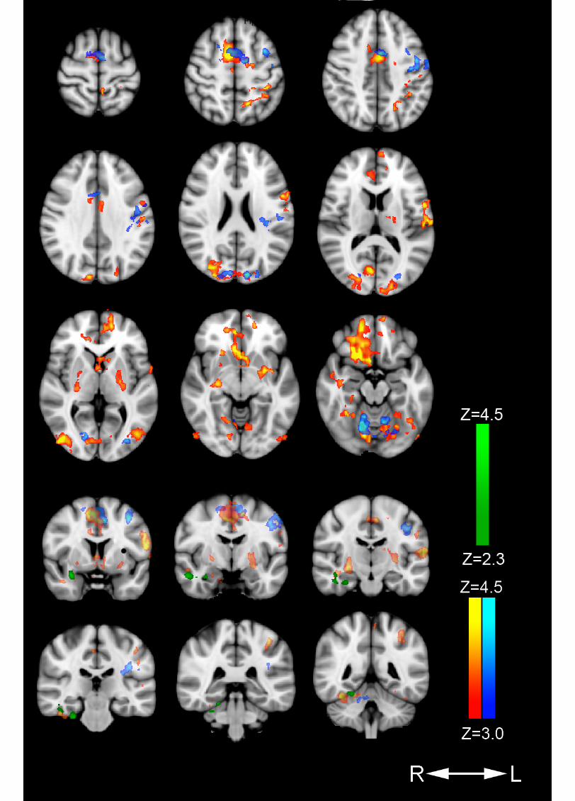
Figure 1.TIF


