Embed Size (px)
Citation preview
7/17/2019 58_613.pdf
http://slidepdf.com/reader/full/58613pdf 1/12
Endocrine Journal 2011, 58 (8), 613-624
Bone metabolism and fracture risk in type 2 diabetes mellitus
Toru Yamaguchi and Toshitsugu Sugimoto
Internal Medicine 1, Shimane University Faculty of Medicine, Shimane 693-8501, Japan
Abstract. Osteoporosis and type 2 diabetes mellitus (T2DM) are now prevalent in aging and westernized societies, and
adversely affect the health of the elderly people by causing fractures and vascular complications, respectively. Recent
experimental and clinical studies show that both disorders are etiologically related to each other through the actions of
osteocalcin and adiponectin. Meta-analyses of multiple clinical studies show that hip fracture risk of T2DM patients is
increased to 1.4 to 1.7-folds, although BMD of the patients is not diminished. Vertebral fracture risk of T2DM patients is
also increased, and BMD is not useful for assessing its risk. These ndings suggest that bone fragility in T2DM depends on
bone quality deterioration rather than bone mass reduction. Thus, surrogate markers are needed to replace the insensitivity
of BMD in assessing fracture risks of T2DM patients. Markers related to advanced glycation end products as well as insulin-
like growth factor-I may be such candidates, because these substances were experimentally shown to modulate bone qualityin DM. In practice, it is important for physicians to assess fracture risk in T2DM patients by evaluating prior VFs and
fracture histories using spine X-ray and interview, respectively, until the usefulness of surrogate markers is established.
Key words: Type 2 diabetes mellitus, Bone fragility, Fracture risk, Osteocalcin, Adiponectin
THE NUMBERS of patients with osteoporosis or
type 2 diabetes mellitus (T2DM) are increasing in
aging and westernized societies. Both disorders pre-
dispose the elderly people to disabled conditions bycausing fractures and vascular complications, respec-
tively, which eventually raise their mortality. Although
osteoporosis and T2DM are traditionally viewed as
separate disease entities, accumulating evidence indi-
cates that there are similar pathophysiological mecha-
nisms underlying them.
I. Interaction between bone metabolismand glucose/fat metabolism through
osteocalcin and Wnt signaling
Osteocalcin (OC), one of the osteoblast-specicsecreted proteins, has several hormonal features and is
secreted in the general circulation from osteoblasts [1,
2]. Recent animal studies have shown that OC action
is related to not only bone metabolism but also glu-
cose metabolism and fat mass [3, 4]. Lee et al. showed
that OC functions as a hormone that improves glucose
metabolism and reduces fat mass, because OC-decient
mouse aggravated these processes [3]. Moreover,Ferron et al. showed that recombinant uncarboxylated
OC administration to wild-type mice fed by high fat
diet regulated gene expression in pancreatic β cells
and adipocytes (including adiponectin expression), and
prevented the development of metabolic diseases, obe-
sity, and hyperglycemia [4]. Several clinical studies
have also conrmed the relationship between OC and
glucose/fat metabolism in humans [5-9]. We were the
rst to show that serum OC level was negatively cor -
related with plasma glucose level and atherosclero-
sis parameters in T2DM patients [5]. We also found
that undercarboxylated OC was negatively associatedwith plasma glucose level and fat mass, and positively
with adiponectin in the population [6]. In non-DM
subjects, it is documented that OC level was inversely
related to plasma glucose level and fat mass [7], and
was positively associated with insulin sensitivity [8].
Pittas et al. have shown that serum OC concentration
was inversely associated with fasting plasma glucose
(FPG), fasting insulin, homeostasis model assessment
for insulin resistance, high-sensitivity C-reactive pro-
tein, interleukin-6, body mass index (BMI), and body
Received Jun. 6, 2011; Accepted Jun. 21, 2011 as EJ11-0063
Released online in J-STAGE as advance publication Jul. 20, 2011
Correspondence to: Toru Yamaguchi, M.D., Internal Medicine 1,
Shimane University Faculty of Medicine, 89-1, Enya-cho, Izumo,
Shimane 693-8501, Japan.
E-mail: [email protected]
REVIEW
©The Japan Endocrine Society
7/17/2019 58_613.pdf
http://slidepdf.com/reader/full/58613pdf 2/12
614 Yamaguchi et al.
between bone mineral density (BMD) and fat massincluding visceral adipose tissue [20-22]. Adiponectin
is specically and highly expressed in visceral, subcu-
taneous and bone marrow fat depots. It is also abun-
dantly present in plasma and has been proposed to play
important roles in the regulation of energy homeosta-
sis and insulin sensitivity. We and other researchers
have shown that osteoblasts have an adiponectin recep-
tor and that the proliferation, differentiation and min-
eralization of osteoblastic cells were enhanced by adi-
ponectin [23, 24]. We also clinically found that serum
adiponectin was associated with BMD, bone turnover,and the presence of vertebral fractures (VFs) in T2DM
patients [25]. Moreover, high glucose was experimen-
tally shown to impair OC expression and secretion
from osteoblastic cells [26], and treatments for hyperg-
lycemia in T2DM patients were found to enhance their
serum OC level [27]. We found that serum adiponectin
level before starting to compensate poorly controlled
T2DM could predict the subsequent increase in serum
OC level during glycemic control [28]. These ndings
suggest that adiponectin as well as OC, secreted from
fat tissue and osteoblasts, respectively, are involved in
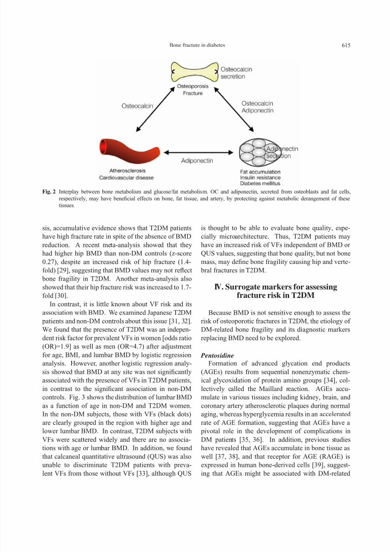
the interplay between glucose/fat metabolism and bonemetabolism (Fig. 2).
III. Fracture risk in T2DM is not refected
by BMD
Many clinical studies have investigated the associa-
tion between T2DM and osteoporosis, given that these
disorders affect a large proportion of the elderly pop-
ulation. Although BMD is considered as a gold stan-
dard for evaluating fracture risk in non-DM osteoporo-
fat in cross-sectional analyses on the subjects includ-ing 5% with T2DM [9]. They also found that OC lev-
els were associated with change in FPG in prospective
analyses on the same population [9]. These experimen-
tal and clinical ndings suggest that bone metabolism
and glucose/fat metabolism are etiologically related to
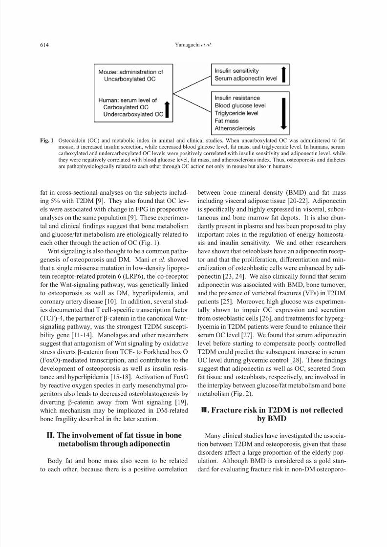
each other through the action of OC (Fig. 1).
Wnt signaling is also thought to be a common patho-
genesis of osteoporosis and DM. Mani et al. showed
that a single missense mutation in low-density lipopro-
tein receptor-related protein 6 (LRP6), the co-receptor
for the Wnt-signaling pathway, was genetically linkedto osteoporosis as well as DM, hyperlipidemia, and
coronary artery disease [10]. In addition, several stud-
ies documented that T cell-specic transcription factor
(TCF)-4, the partner of β-catenin in the canonical Wnt-
signaling pathway, was the strongest T2DM suscepti-
bility gene [11-14]. Manolagas and other researchers
suggest that antagonism of Wnt signaling by oxidative
stress diverts β-catenin from TCF- to Forkhead box O
(FoxO)-mediated transcription, and contributes to the
development of osteoporosis as well as insulin resis-
tance and hyperlipidemia [15-18]. Activation of FoxO
by reactive oxygen species in early mesenchymal pro-genitors also leads to decreased osteoblastogenesis by
diverting β-catenin away from Wnt signaling [19],
which mechanism may be implicated in DM-related
bone fragility described in the later section.
II. The involvement of fat tissue in bonemetabolism through adiponectin
Body fat and bone mass also seem to be related
to each other, because there is a positive correlation
Fig. 1 Osteocalcin (OC) and metabolic index in animal and clinical studies. When uncarboxylated OC was administered to fatmouse, it increased insulin secretion, while decreased blood glucose level, fat mass, and triglyceride level. In humans, serumcarboxylated and undercarboxylated OC levels were positively correlated with insulin sensitivity and adiponectin level, whilethey were negatively correlated with blood glucose level, fat mass, and atherosclerosis index. Thus, osteoporosis and diabetesare pathophysiologically related to each other through OC action not only in mouse but also in humans.
7/17/2019 58_613.pdf
http://slidepdf.com/reader/full/58613pdf 3/12
615Bone fracture in diabetes
sis, accumulative evidence shows that T2DM patients
have high fracture rate in spite of the absence of BMD
reduction. A recent meta-analysis showed that they
had higher hip BMD than non-DM controls (z-score
0.27), despite an increased risk of hip fracture (1.4-
fold) [29], suggesting that BMD values may not reect
bone fragility in T2DM. Another meta-analysis also
showed that their hip fracture risk was increased to 1.7-fold [30].
In contrast, it is little known about VF risk and its
association with BMD. We examined Japanese T2DM
patients and non-DM controls about this issue [31, 32].
We found that the presence of T2DM was an indepen-
dent risk factor for prevalent VFs in women [odds ratio
(OR)=1.9] as well as men (OR=4.7) after adjustment
for age, BMI, and lumbar BMD by logistic regression
analysis. However, another logistic regression analy-
sis showed that BMD at any site was not signicantly
associated with the presence of VFs in T2DM patients,
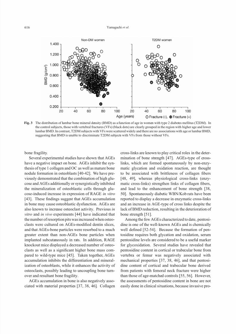
in contrast to the signicant association in non-DMcontrols. Fig. 3 shows the distribution of lumbar BMD
as a function of age in non-DM and T2DM women.
In the non-DM subjects, those with VFs (black dots)
are clearly grouped in the region with higher age and
lower lumbar BMD. In contrast, T2DM subjects with
VFs were scattered widely and there are no associa-
tions with age or lumbar BMD. In addition, we found
that calcaneal quantitative ultrasound (QUS) was also
unable to discriminate T2DM patients with preva-
lent VFs from those without VFs [33], although QUS
is thought to be able to evaluate bone quality, espe-
cially microarchitecture. Thus, T2DM patients may
have an increased risk of VFs independent of BMD or
QUS values, suggesting that bone quality, but not bone
mass, may dene bone fragility causing hip and verte-
bral fractures in T2DM.
IV. Surrogate markers for assessingfracture risk in T2DM
Because BMD is not sensitive enough to assess the
risk of osteoporotic fractures in T2DM, the etiology of
DM-related bone fragility and its diagnostic markers
replacing BMD need to be explored.
Pentosidine
Formation of advanced glycation end products
(AGEs) results from sequential nonenzymatic chem-
ical glycoxidation of protein amino groups [34], col-
lectively called the Maillard reaction. AGEs accu-mulate in various tissues including kidney, brain, and
coronary artery atherosclerotic plaques during normal
aging, whereas hyperglycemia results in an accelerated
rate of AGE formation, suggesting that AGEs have a
pivotal role in the development of complications in
DM patients [35, 36]. In addition, previous studies
have revealed that AGEs accumulate in bone tissue as
well [37, 38], and that receptor for AGE (RAGE) is
expressed in human bone-derived cells [39], suggest-
ing that AGEs might be associated with DM-related
Fig. 2 Interplay between bone metabolism and glucose/fat metabolism. OC and adiponectin, secreted from osteoblasts and fat cells,respectively, may have benecial effects on bone, fat tissue, and artery, by protecting against metabolic derangement of these
tissues.
7/17/2019 58_613.pdf
http://slidepdf.com/reader/full/58613pdf 4/12
616 Yamaguchi et al.
cross-links are known to play critical roles in the deter-
mination of bone strength [47]. AGEs-type of cross-
links, which are formed spontaneously by non-enzy-
matic glycation and oxidation reaction, are thought
to be associated with brittleness of collagen bers
[48, 49], whereas physiological cross-links (enzy-
matic cross-links) strengthen links of collagen bers,
and lead to the enhancement of bone strength [38,
50]. Spontaneously diabetic WBN/Kob rats have been
reported to display a decrease in enzymatic cross-links
and an increase in AGE-type of cross links despite the
lack of BMD reduction, resulting in the deterioration of
bone strength [51].
Among the few AGEs characterized to date, pentosi-
dine is one of the well-known AGEs and is chemically
well dened [52-54]. Because the formation of pen-
tosidine requires both glycation and oxidation, serum pentosidine levels are considered to be a useful marker
for glycoxidation. Several studies have revealed that
pentosidine content in cortical or trabecular bone from
vertebra or femur was negatively associated with
mechanical properties [37, 38, 46], and that pentosi-
dine content of cortical and trabecular bone derived
from patients with femoral neck fracture were higher
than those of age-matched controls [55, 56]. However,
the assessments of pentosidine content in bone are not
easily done in clinical situations, because invasive pro-
bone fragility.
Several experimental studies have shown that AGEs
have a negative impact on bone. AGEs inhibit the syn-
thesis of type 1 collagen and OC as well as mature bone
nodule formation in osteoblasts [40-42]. We have pre-
viously demonstrated that the combination of high glu-
cose and AGEs additionally or synergistically inhibited
the mineralization of osteoblastic cells through glu-
cose-induced increase in expression of RAGE in vitro
[43]. These ndings suggest that AGEs accumulation
in bone may cause osteoblastic dysfunction. AGEs are
also known to increase osteoclast activity. Previous in
vitro and in vivo experiments [44] have indicated that
the number of resorption pits was increased when osteo-
clasts were cultured on AGEs-modied dentin slices,
and that AGEs-bone particles were resorbed to a much
greater extent than non-AGEs bone particles whenimplanted subcutaneously in rats. In addition, RAGE
knockout mice displayed a decreased number of osteo-
clasts as well as a signicant higher bone mass com-
pared to wild-type mice [45]. Taken together, AGEs
accumulation inhibits the differentiation and mineral-
ization of osteoblasts, while it enhances the activity of
osteoclasts, possibly leading to uncoupling bone turn-
over and resultant bone fragility.
AGEs accumulation in bone is also negatively asso-
ciated with material properties [37, 38, 46]. Collagen
Fig. 3 The distribution of lumbar bone mineral density (BMD) as a function of age in women with type 2 diabetes mellitus (T2DM). In
the control subjects, those with vertebral fractures (VFs) (black dots) are clearly grouped in the region with higher age and lower
lumbar BMD. In contrast, T2DM subjects with VFs were scattered widely and there are no associations with age or lumbar BMD,
suggesting that BMD is unable to discriminate T2DM subjects with VFs from those without VFs.
7/17/2019 58_613.pdf
http://slidepdf.com/reader/full/58613pdf 5/12
617Bone fracture in diabetes
that the ratio of serum esRAGE to AGE levels could
be linked to clinical bone problems, such as fractures,
more prominently than either parameter alone.
We found that the esRAGE/pentosidine ratio in
T2DM patients with VFs was signicantly lower than
in those without VFs. Multivariate logistic regression
analysis adjusted for age, height, weight, HbA1c, serum
creatinine, DM duration, therapeutic agents, DM com-
plications, osteoporotic risk factors, and lumbar BMD
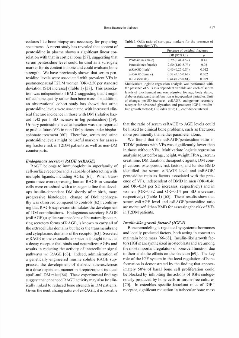
identied the serum esRAGE level and esRAGE/
pentosidine ratio as factors associated with the pres-ence of VFs, independent of BMD in men (OR=0.46
and OR=0.34 per SD increases, respectively) and in
women (OR=0.32 and OR=0.14 per SD increases,
respectively) (Table 1) [65]. These results show that
serum esRAGE level and esRAGE/pentosidine ratio
are more useful than BMD for assessing the risk of VFs
in T2DM patients.
Insulin-like growth factor-I (IGF-I)
Bone remodeling is regulated by systemic hormones
and locally produced factors, both acting in concert to
maintain bone mass [66-68]. Insulin-like growth fac-tors (IGFs) are synthesized in osteoblasts and are among
the most important regulators of bone cell function due
to their anabolic effects on the skeleton [69]. The key
role of the IGF system in the local regulation of bone
formation is demonstrated by the nding that approx-
imately 50% of basal bone cell proliferation could
be blocked by inhibiting the actions of IGFs endoge-
nously produced by bone cells in serum-free cultures
[70]. In osteoblast-specic knockout mice of IGF-I
receptor, signicant reduction in trabecular bone mass
cedures like bone biopsy are necessary for preparing
specimens. A recent study has revealed that content of
pentosidine in plasma shows a signicant linear cor -
relation with that in cortical bone [57], suggesting thatserum pentosidine level could be used as a surrogate
marker for its content in bone and could evaluate bone
strength. We have previously shown that serum pen-
tosidine levels were associated with prevalent VFs in
postmenopausal T2DM women [OR=2.50 per standard
deviation (SD) increase] (Table 1) [58]. This associa-
tion was independent of BMD, suggesting that it might
reect bone quality rather than bone mass. In addition,
an observational cohort study has shown that urine
pentosidine levels were associated with increased clin-
ical fracture incidence in those with DM (relative haz-
ard 1.42 per 1 SD increase in log pentosidine) [59].Urinary pentosidine level at baseline was also reported
to predict future VFs in non-DM patients under bispho-
sphonate treatment [60]. Therefore, serum and urine
pentosidine levels might be useful markers for assess-
ing fracture risk in T2DM patients as well as non-DM
counterparts.
Endogenous secretory RAGE (esRAGE)
RAGE belongs to immunoglobulin superfamily of
cell-surface receptors and is capable of interacting with
multiple ligands, including AGEs [61]. When trans-genic mice overexpressing human RAGE in vascular
cells were crossbred with a transgenic line that devel-
ops insulin-dependent DM shortly after birth, more
progressive histological change of DM nephropa-
thy was observed compared to controls [62], conrm-
ing that RAGE expression stimulates the development
of DM complications. Endogenous secretory RAGE
(esRAGE), a splice variant of one of the naturally occur-
ring secretory forms of RAGE, is known to carry all of
the extracellular domains but lacks the transmembrane
and cytoplasmic domains of the receptor [63]. Secreted
esRAGE in the extracellular space is thought to act as
a decoy receptor that binds and neutralizes AGEs and
results in reducing the activity of intercellular signal
pathways via RAGE [63]. Indeed, administration of
a genetically engineered murine soluble RAGE sup-
pressed the development of diabetic atherosclerosis
in a dose-dependent manner in streptozotocin-induced
apoE-null DM mice [64]. These experimental ndings
suggest that enhanced RAGE activity may also be clin-
ically linked to reduced bone strength in DM patients.
Given the neutralizing nature of esRAGE, it is possible
Table 1 Odds ratio of surrogate markers for the presence of
prevalent VFs.
Presence of vertebral fractures
OR (95% CI) p
Pentosidine (male) 0.79 (0.41-1.52) 0.47
Pentosidine (female) 2.50 (1.09-5.73) 0.03
esRAGE (male) 0.46 (0.25-0.84) 0.012
esRAGE (female) 0.32 (0.16-0.67) 0.002
IGF-I (female) 0.44 (0.23-0.81) 0.009
Multivariate logistic regression analysis was performed with
the presence of VFs as a dependent variable and each of serum
levels of biochemical markers adjusted for age, body statue,
diabetes status, and renal function as independent variables. Unit
of change: per SD increase. esRAGE, endogenous secretary
receptor for advanced glycation end products; IGF-I, insulin-
like growth factor-I; OR, odds ratio; CI, condence interval.
7/17/2019 58_613.pdf
http://slidepdf.com/reader/full/58613pdf 6/12
618 Yamaguchi et al.
to bone quality but not to bone mass in postmenopausal
T2DM women.
V. The effects of antidiabetic drugs onbone metabolism in T2DM
It is well documented that some antidiabetic drugs
affect bone metabolism in T2DM. Thiazolidinediones
(TZDs) such as pioglitazone and rosiglitazone are per-
oxisome proliferator-activated receptor-γ (PPAR-γ)
agonists, and are widely used for treatment of T2DM
patients. PPAR-γ is also expressed in bone marrow
cells, and it acts as a molecular switch that regulates
the fate of pluripotent mesenchymal stem cells, which
are able to differentiate into adipocytes or osteoblasts.
Previous in vitro studies have shown that TZDs stim-ulate the differentiation into adipocytes in preference
over osteoblasts [84, 85]. Haploinsufciency of the
PPAR-γ gene in mice induces a high bone density phe-
notype characterized by increased bone formation [86,
87], whereas treatment of rodents with PPAR-γ agonists
induces bone loss characterized by decient osteoblast
function [88-89].
Accumulating evidence also indicates that TZDs
negatively impact on bone metabolism in humans. Grey
et al. have shown that 14-week rosiglitazone treatment
decreased bone formation markers, OC, procollagentype I N-terminal propeptide, and femoral neck BMD
in healthy postmenopausal women [90]. Schwartzet al.
have reported that long-term use of TZDs caused reduc-
tion of whole body BMD and lumbar BMD in older
T2DM women by a 4-year observational cohort study
[91]. We also found that serum OC level, femoral neck
BMD, and radial BMD were signicantly decreased at
6 months in T2DM patients treated with pioglitazone
[92]. Recently, a meta-analysis has revealed that lum-
bar and femoral neck BMD was signicantly reduced,
and that risk of fractures was signicantly increased
in women exposed to TZDs (OR=2.23), but not in
men [93]. Moreover, a previous clinical trial showed
that risk of fractures in the bones of the extremities
(foot, hand, and proximal humerus) was signicantly
increased, while there was no increased risk identied
for clinical vertebral or hip fractures [94,95], suggest-
ing a negative impact on cortical bone. In contrast,
little is known whether morphometric fracture rate in
vertebra, which contains a relatively higher proportion
of trabecular bone, is increased or not. We found that
treatment with pioglitazone was signicantly and posi-
and decient mineralization has been observed [71].
On the other hand, circulating IGF-I, mainly produced
in the liver via regulation by growth hormone and diet,
acts in an endocrine manner, which also activates boneremodeling and exerts anabolic effects on bone tissues
[72-74]. Liver-specic IGF-I gene-null mice reveals a
marked reduction in bone volume, periosteal circum-
ference, and medial lateral width, suggesting that cir-
culating levels of IGF-I also directly regulate bone
growth and density [75]. Indeed, our clinical studies
showed that serum IGF-I levels were positively asso-
ciated with BMD and inversely with the risk of VFs in
postmenopausal non-DM women [76, 77]. These nd-
ings suggest that serum IGF-I levels could be clinically
useful for assessing bone mass and the risk of VFs in
the non-DM population.It is also possible that IGFs are linked to the patho-
genesis of DM-related complications [78]. Impaired
production of IGFs could cause bone complication in
DM by diminishing bone cell function [69]. An in vivo
study has demonstrated that IGF-I levels in serum and
cortical bone were signicantly reduced in spontane-
ously diabetic Goto-Kakizaki rats, which displayed a
signicant decrease in BMD at long bone metaphy-
ses and vertebrae [79]. On the other hand, several in
vitro studies have shown that the stimulatory actions
of IGF-I on osteoblasts were blunted by high glucoseconcentrations or AGEs. High glucose concentrations
signicantly impaired the proliferative and functional
responses of osteoblastic MG-63 cells to IGF-I [80].
AGEs also signicantly decreased IGF-I secretion in
osteoblastic MC3T3-E1 cells [81]. Thus, high glucose
concentrations or AGEs may cause the resistance of
osteoblasts to IGF-I actions in local environment.
In T2DM patients, however, the relationship
between serum IGF-I levels and bone metabolism has
little been documented. We indicated that serum IGF-I
levels were signicantly and inversely associated with
the presence of VFs in postmenopausal T2DM women
(OR=0.44 per SD increase) in a fashion independent
of age, body statue, DM status, renal function, insu-
lin secretion, or lumbar BMD (Table 1) [82]. We also
found that serum IGF-I level was inversely associ-
ated with the number of prevalent VFs independent of
lumbar BMD in postmenopausal T2DM women, sug-
gesting that serum IGF-I could be clinically useful for
assessing the severity of multiple VFs in the population
[83]. Accordingly, circulating IGF-I may have a pro-
tective effect on VFs, and this effect might be related
7/17/2019 58_613.pdf
http://slidepdf.com/reader/full/58613pdf 7/12
619Bone fracture in diabetes
Recently, the fracture risk assessment (FRAX) algo-
rithm has been developed by the WHO, which could
assess the fracture risk of an individual even if BMD
is not measured [99, 100]. Since this algorithm inte-grates the inuence of several well validated risk fac-
tors for fracture that are independent of BMD, it might
be useful for the case-nding strategy picking up
T2DM patients at high risk for fracture. Only recently,
Schwartz et al. have indicated that femoral neck BMD
T score and FRAX score were associated with hip and
nonspine fracture risk among older adults with T2DM;
however, in these patients compared with participants
without DM, the fracture risk was higher for a given T
score and age or for a given FRAX score [101]. These
ndings suggest that femoral neck BMD and FRAX
score may be partially effective for assessing hip andnonspine fracture risk in T2DM patients, although they
are not as sensitive as in non-DM counterparts.
VII. Conclusion
The fact that BMD is not useful for assessing frac-
ture risks in T2DM seems problematic, because T2DM
populations are expanding in every country. T2DM
patients may drop out from fracture prevention if
doctors diagnose osteoporosis based on BMD values
alone. Practitioners should be aware of the importanceof evaluating prior VFs and fracture histories by spine
X-ray and interview, respectively. These procedures
would broaden the spectrum of osteoporosis treatments
into T2DM population. Simultaneously, further stud-
ies are needed to clarify whether or not surrogate bio-
chemical markers such as pentosidine, esRAGE, and
IGF-I, as well as the WHO FRAX algorithm, would be
useful for predicting the occurrence of new fractures
of T2DM patients prospectively, with sensitivity and
specicity comparative to those of BMD in non-DM
osteoporosis.
tively associated with the presence of prevalent VFs
in postmenopausal women (OR=3.38), but not in men,
when VFs were evaluated by X-ray lms [96]. This
nding suggests that postmenopausal women treatedwith pioglitazone have high risks of VFs as well as the
bones of the extremities.
VI. Fracture risk assessment of T2DMpatients in clinical practice
In face of the ineffectiveness of BMD in assessing
fracture risks in T2DM, the major clinical problems
are how to assess the risks and when to start therapies
for preventing fractures in daily practice for patients.
Although the markers related to AGEs as well as IGF-I
are potential candidates for fracture risk assessments, itis unclear whether or not they could predict the occur-
rence of new fractures of T2DM patients in a prospec-
tive fashion.
On the other hand, the presence of prevalent VFs
could be used for the assessment of bone quality in
individual patients, because a large study on the inci-
dence of VFs in postmenopausal osteoporosis showed
that patients with previous VFs were more likely to
suffer from new VFs [97, 98] and hip fractures [97]
independent of BMD than those without VFs during
several-year study periods. Patient histories of non-VFs that previously happened are also established risk
factors for additional fractures [99]. We found that
38% of T2DM men and 31% of T2DM women had
prevalent VFs by X-ray lms, and that 16% each of
T2DM men and women had histories of previous non-
VFs [32]. Thus, if T2DM patients undergo spine X-ray
examination or are questioned about their fracture his-
tories, it is likely that about half of them are picked up
as those who have bone fragility and need osteoporo-
sis treatment for fracture prevention. These procedures
are simple and are recommended for all of physicians
engaged in T2DM treatments.
References
1. Hauschka PV, Lian JB, Cole DE, Gundberg CM (1989)
Osteocalcin and matrix protein: vitamin K-dependent
proteins in bone. Physiol Rev 69:990-1047.
2. Price PA (1989) Gla-containing proteins of bone.
Connect Tissue Res 21:51-57.
3. Lee NK, Sowa H, Hinoi E, Ferron M, Ahn JD, Confavreux
C, Dacquin R, Mee PJ, McKee MD, Jung DY, Zhang Z,
Kim JK, Mauvais-Jarvis F, Ducy P, Karsenty G (2007)
Endocrine regulation of energy metabolism by the skel-
eton. Cell 130:456-469.
4. Ferron M, Hinoi E, Karsenty G, Ducy P (2008)
Osteocalcin differentially regulates beta cell and adi-
7/17/2019 58_613.pdf
http://slidepdf.com/reader/full/58613pdf 8/12
620 Yamaguchi et al.
L, Xiang F, Saramies J, Buchanan TA, Watanabe RM,
Valle TT, Kinnunen L, Abecasis GR, Pugh EW, Doheny
KF, Bergman RN, Tuomilehto J, Collins FS, Boehnke
M (2007) A genome-wide association study of type 2
diabetes in Finns detects multiple susceptibility vari-ants. Science 316:1341–1345.
15. Manolagas SC, Almeida M (2007) Gone with the Wnts:
β-catenin, T-cell factor, forkhead box O, and oxidative
stress in age-dependent diseases of bone, lipid, and glu-
cose metabolism. Mol Endocrinol 21:2605–2614.
16. Hoogeboom D, Burgering BM (2009) Should I stay or
should I go: β-catenin decides under stress. Biochim
Biophys Acta 1796:63–74.
17. DeCarolis NA, Wharton Jr KA, Eisch AJ (2008) Which
way does theWntblow? Exploring the duality of canoni-
calWnt signaling on cellular aging. BioEssays 30:102–
106.
18. Jin T (2008) The WNT signalling pathway and diabetesmellitus. Diabetologia 51:1771–1780.
19. Manolagas SC (2010) From estrogen-centric to aging
and oxidative stress: A revised perspective of the patho-
genesis of osteoporosis. Endocr Rev 31:266-300.
20. Felson DT, Zhang Y, Hannan MT, Anderson JJ (1993)
Effects of weight, and body mass index on bone mineral
density in men and women. J Bone Miner Res 8: 567-
573.
21. Glauber HS, Vollmer WM, Nevitt MC, Ensrud KE,
Orwoll ES (1995) Body weight versus body fat distri-
bution, adiposity, and frame size as predictors of bone
density. J Clin Endocrinol Metab 80: 1118-1123.
22. Yamaguchi T, Kanazawa I, Yamamoto M, Kurioka S,
Yamauchi M, Yano S, Sugimoto T (2009) Associations
between components of the metabolic syndrome versus
bone mineral density and vertebral fractures in patients
with type 2 diabetes. Bone 45:174-179.
23. Kanazawa I, Yamaguchi T, Yano S, Yamauchi M,
Yamamoto M, Sugimoto T (2007) Adiponectin and
AMP kinase activator stimulate proliferation, differen-
tiation, and mineralization of osteoblastic MC3T3-E1
cells. BMC Cell Biol 8: 51-62.
24. Berner HS, Lyngstadaas SP, Spahr A, Monjo M,
Thommesen L, Drevon CA, Syversen U, Reseland JE
(2004) Adiponectin and its receptors are expressed in
bone-forming cells. Bone 35: 842-849.
25. Kanazawa I, Yamaguchi T, Yamamoto M, Yamauchi
M, Yano S, Sugimoto T (2009) Relationships between
serum adiponectin levels versus bone mineral density,
bone metabolic markers, and vertebral fractures in type
2 diabetes mellitus. Eur J Endocrinol 160: 265–273.
26. Inaba M, Terada M, Koyama H, Yoshida O, Ishimura E,
Kawagishi T, Okuno Y, Nishizawa Y, Otani S, Morii H
(1995) Inuence of high glucose on 1,25-dihydroxyvi-
tamin D3-induced effect on human osteoblast-like
MG-63 cells. J Bone Miner Res 10:1050-1056.
27. Okazaki R, Totsuka Y, Hamano K, Ajima M, Miura M,
pocyte gene expression and affects the development of
metabolic diseases in wild-type mice. Proc Natl Acad
Sci USA 105:5266-5270.
5. Kanazawa I, Yamaguchi T, Yamamoto M, Yamauchi M,
Kurioka S, Yano S, Sugimoto T (2009) Serum osteo-calcin level is associated with glucose metabolism and
atherosclerosis parameters in type 2 diabetes mellitus. J
Clin Endocrinol Metab 94: 45-49.
6. Kanazawa I, Yamaguchi T, Yamamoto M, Yamauchi M,
Kurioka S, Yano S, Sugimoto T (2011) Serum under-
carboxylated osteocalcin was inversely associated with
plasma glucose level and fat mass in type 2 diabetes
mellitus. Osteoporos Int 22:187-194.
7. Kindblom JM, Ohlsson C, Ljunggren O, Karlsson
MK, Tivesten A, Smith U, Mellstrom D (2009) Plasma
osteocalcin is inversely related to fat mass and plasma
glucose in elderly Swedish men. J Bone Miner Res 24:
785-791. 8. Fernandez-Real JM, Izquierdo M, Ortega F, Gorostiaga
E, Gomez-Ambrosi J, Moreno-Navarrete JM, Fruhbeck
G, Martinez C, Idoate F, Salvador J, Forga L, Ricart W,
Ibanez J (2009) The relationship of serum osteocalcin
concentration to insulin secretion, sensitivity, and dis-
posal with hypocaloric diet and resistance training. J
Clin Endocrinol Metab 94: 237-245.
9. Pitass AG, Harris SS, Eliades M, Stark P, Dawson-
Hughes B (2009) Association between serum osteo-
calcin and markers of metabolic phenotype. J Clin
Endocrinol Metab 94: 827-832.
10. Mani A, Radhakrishnan J, Wang H, Mani A, Mani MA,
Nelson-Williams C, Carew KS,ManeS, Najmabadi H,
Wu D, Lifton RP (2007) LRP6 mutation in a family
with early coronary disease and metabolic risk factors.
Science 315:1278–1282.
11. Smith U (2007) TCF7L2 and type 2 diabetes—we WNT
to know. Diabetologia 50:5–7.
12. Grant SF, Thorleifsson G, Reynisdottir I, Benediktsson
R, Manolescu A, Sainz J, Helgason A, Stefansson
H, Emilsson V, Helgadottir A, Styrkarsdottir U,
Magnusson KP, Walters GB, Palsdottir E, Jonsdottir
T, Gudmundsdottir T, Gylfason A, Saemundsdottir
J, Wilensky RL, Reilly MP, Rader DJ, Bagger
Y, Christiansen C, Gudnason V, Sigurdsson G,
Thorsteinsdottir U, Gulcher JR, Kong A, Stefansson
K Variant of transcription factor 7-like 2 (TCF7L2)
gene confers risk of type 2 diabetes. (2006) Nat Genet
38:320–323.
13. Owen KR, McCarthy MI (2007) Genetics of type 2 dia-
betes. Curr Opin Genet Dev 17:239–244.
14. Scott LJ, Mohlke KL, Bonnycastle LL, Willer CJ, Li
Y, Duren WL,Erdos MR, Stringham HM, Chines PS,
Jackson AU, Prokunina-Olsson L, Ding CJ, Swift AJ,
Narisu N,Hu T, Pruim R, Xiao R, Li XY, Conneely KN,
Riebow NL, Sprau AG, Tong M, White PP, Hetrick
KN, Barnhart MW, Bark CW, Goldstein JL, Watkins
7/17/2019 58_613.pdf
http://slidepdf.com/reader/full/58613pdf 9/12
621Bone fracture in diabetes
Kojima Y, Shima M, Okada S (2001) Role of advanced
glycation end products in adynamic bone disease in
patients with diabetic nephropathy. Am J Kidney Dis 38:
S161-S164.
41. Katayama Y, Akatsu T, Yamamoto M, Kugai N, Nagata N (1996) Role of nonenzymatic glycosylation of type 1
collagen in diabetic osteopenia. J Bone Miner Res 11:
931-937.
42. Sanguineti R, Storace D, Monacelli F, Federici A, Odetti
P (2008) Pentosidine effects on human osteoblasts in
vitro. Ann N Y Acad Sci 1126: 166-172.
43. Ogawa N, Yamaguchi T, Yano S, Yamauchi M,
Yamamoto M, Sugimoto T (2007) The combination
of high glucose and advanced glycation end-prod-
ucts (AGEs) inhibits the mineralization of osteoblastic
MC3T3-E1 cells through glucose-induced increase in
the receptor for AGEs. Horm Metab Res 39: 871-875.
44. Miyata T, Notoya K, Yoshida K, Horie K, Maeda K,Kurokawa K, Taketomi S (1997) Advanced glycation
end products enhance osteoclast-induced bone resorp-
tion in cultured mouse unfractionated bone cells and in
rats implanted subcutaneously with devitalized bone
particles. J Am Soc Nephrol 8: 260-270.
45. Ding KH, Wang ZZ, Hamrick MW, Deng ZB, Zhou
L, Kang B, Yan SL, She JX, Stern DM, Isales CM, Mi
QS (2006) Disordered osteoclast formation in RAGE-
decient mouse establishes an essential role for RAGE
in diabetes related bone loss. Biochem Biophys Res
Commun 340: 1091-1097.
46. Viguet-Carrin S, Roux JP, Arlot ME, Merabet Z,
Leeming DJ, Byrjalsen I, Delmas PD, Bouxsein ML
(2006) Contribution of the advanced glycation end
product pentosidine and of maturation of type I colla-
gen to compressive biomechanical properties of human
lumbar vertebrae. Bone 39: 1073-1079.
47. Saito M, Marumo K (2010) Collagen cross-links as a
determinant of bone quality: a possible explanation for
bone fragility in aging, osteoporosis, and diabetes mel-
litus. Osteoporos Int 21:195-214.
48. Garnero P, Borel O, Gineyts E, Duboeuf F, Solberg
H, Bouxsein ML, Christiansen C, Delmas PD (2006)
Extracellular post-translational modications of colla-
gen are major determinants of biomechanical properties
of fetal bovine cortical bone. Bone 38: 300-309.
49. Vashishth D (2007) The role of the collagen matrix in
skeletal fragility. Curr Osteoporos Rep 5: 62-66.
50. Banse X, Sims TJ, Bailey AJ (2002) Mechanical prop-
erties of adult vertebral cancellous bone: correlation
with collagen intermolecular cross-links. J Bone Miner
Res 17: 1621-1628.
51. Saito M, Fujii K, Mori Y, Marumo K (2006) Role of col-
lagen enzymatic and glycation induced cross-links as a
determinant of bone quality in spontaneously diabetic
WBN/Kob rats. Osteoporos Int 17: 1514-1523.
52. Odetti P, Fogarty J, Sell DR, Monnier VM (1992)
Hirota Y, Hata K, Fukumoto S, Matsumoto T. (1997)
Metabolic improvement of poorly controlled noninsu-
lin-dependent diabetes mellitus decreases bone turn-
over. J Clin Endocrinol Metab 82:2915-2920.
28. Kanazawa I, Yamaguchi T, Yamamoto M, Yamauchi M,Yano S, Sugimoto T (2009) Adiponectin is associated
with changes in bone markers during glycemic control
in type 2 diabetes mellitus. J Clin Endocrinol Metab
94:3031-3037.
29. Vestergaard P (2007) Discrepancies in bone mineral
density and fracture risk in patients with type 1 and type
2 diabetes--a meta-analysis. Osteoporos Int 18:427-
444.
30. Janghorbani M, Van Dam RM, Willett WC, Hu FB
(2007) Systematic review of type 1 and type 2 diabetes
mellitus and risk of fracture. Am J Epidemiol 166:495-
505.
31. Yamamoto M, Yamaguchi T, Yamauchi M, Kaji H,Sugimoto T (2007) Bone mineral density is not sensi-
tive enough to assess the risk of vertebral fractures in
type 2 diabetic women. Calcif Tissue Int 80:353-358.
32. Yamamoto M, Yamaguchi T, Yamauchi M, Kaji H,
Sugimoto T (2009) Diabetic patients have an increased
risk of vertebral fractures independent of bone mineral
density or diabetic complications. J Bone Miner Res
24:702-709.
33. Yamaguchi T, Yamamoto M, Kanazawa I, Yamauchi M,
Yano S, Tanaka N, Nitta E, Fukuma A, Uno S, Sho-no
T, Sugimoto T (2011) Quantitative ultrasound and ver-
tebral fractures in patients with type 2 diabetes. J Bone
Miner Metab (Epub ahead of print).
34. Brownlee M (1995) Advanced protein glycosylation in
diabetes and aging. Annu Rev Med 46: 223-234.
35. Brownlee M, Cerami A, Vlassara H (1988) Advanced
glycosylation end products in tissue and the biochemi-
cal basis of diabetic complications. N Engl J Med 318:
1315-1321.
36. Goldin A, Beckman JA, Schmidt AM, Creager MA
(2006) Advanced glycation end products sparking the
development of diabetic vascular injury. Circulation
114: 597-605.
37 Hernandez CJ, Tang SY, Baumbach BM, Hwu PB,
Sakkee AN, van der Ham F, DeGroot J, Bank RA,
Keaveny TM (2005) Trabecular microfracture and the
inuence of pyridinium and non-enzymatic glycation-
mediated collagen cross-links. Bone 37: 825-832.
38. Wang X, Shen X, Li X, Agrawal CM (2002) Age-related
changes in the collagen network and toughness of bone.
Bone 31: 1-7.
39. Takagi M, Kasayama S, Yamamoto T, Motomura
T, Hashimoto K, Yamamoto H, Sato B, Okada S,
Kishimoto T (1997) Advanced glycation end-products
stimulate interleukin-6 production by human bone-de-
rived cells. J Bone Miner Res 12: 439-446.
40. Yamamoto T, Ozono K, Miyauchi A, Kasayama S,
7/17/2019 58_613.pdf
http://slidepdf.com/reader/full/58613pdf 10/12
622 Yamaguchi et al.
63. Yonekura H, Yamamoto Y, Sakurai S, Petrova RG,
Abedin MJ, Li H, Yasui K, Takeuchi M, Makita Z,
Takasawa S, Okamoto H, Watanabe T, Yamamoto
H (2003) Novel splice variants of the receptor for
advanced glycation end-products expressed in humanvascular endothelial cells and pericytes, and their puta-
tive roles in diabetes-induced vascular injury. Biochem
J 370:1097-1109.
64. Park L, Raman KG, Lee KJ, Lu Y, Ferran LJ, Jr., Chow
WS, Stern D, Schmidt AM (1998) Suppression of accel-
erated diabetic atherosclerosis by the soluble receptor
for advanced glycation endproducts. Nat Med 4:1025-
1031.
65. Yamamoto M, Yamaguchi T, Yamauchi M, Sugimoto
T (2009) Low serum level of the endogenous secretory
receptor for advanced glycation end-products (esRAGE)
is a risk factor for prevalent vertebral fractures indepen-
dent of bone mineral density in patients with type 2 dia- betes. Diabetes Care 32:2263-2268.
66. Canalis E (1983) The hormonal and local regulation of
bone formation. Endocr Rev 4: 62-77.
67. Mohan S, Baylink DJ (1991) Bone growth factors. Clin
Orthop Relat Res 263: 30-48.
68. Ueland T (2004) Bone metabolism in relation to altera-
tions in systemic growth hormone. Growth Horm IGF
Res 14: 404-417.
69. McCarthy TL, Centrella M, Canalis E (1989) Insulin-
like growth factor (IGF) and bone. Connect Tissue Res
20: 277-282.
70. Mohan S (1993) Insulin-like growth factor binding pro-
teins in bone cell regulation. Growth Regul 3: 67-70.
71. Zhang M, Xuan S, Bouxsein ML, von Stechow D,
Akeno N, Faugere MC, Malluche H, Zhao G, Rosen CJ,
Efstratiadis A, Clemens TL (2002) Osteoblast-specic
knockout of the insulin-like growth factor (IGF) recep-
tor gene reveals and essential role of IGF signaling in
bone matrix mineralization. J Biol Chem 277: 44005-
44012.
72. Johansson AG, Lindh E, Ljunghall S (1992) Insulin-like
growth factor I stimulates bone turnover in osteoporo-
sis. Lancet 339: 1619.
73. Schwander JC, Hauri C, Zapf J, Froesch ER (1983)
Synthesis and secretion of insulin-like growth factor
and its binding protein by the perfused rat liver: depen-
dence on growth hormone status. Endocrinology 113:
297-305.
74. Spencer EM, Liu CC, Si EC, Howard GA (1991) In
vivo actions of insulin-like growth factor-I (IGF-I) on
bone formation and resorption in rats. Bone 12: 21-26.
75. Yakar S, Rosen CJ, Beamer WG, Ackert-Bicknell CL,
Wu Y, Liu JL, Ooi GT, Setser J, Frystyk J, Boisclair YR,
LeRoith D (2002) Circulating levels of IGF-I directly
regulate bone growth and density. J Clin Invest 110:
771-781.
76. Sugimoto T, Nishiyama K, Kuribayashi F, Chihara K
Chromatographic quantitation of plasma and eryth-
rocyte pentosidine in diabetic and uremic subjects.
Diabetes 41: 153-159.
53. Monnier VM, Sell DR, Nagaraj RH, Miyata S, Grandhee
S, Odetti P, Ibrahim SA (1992) Maillard reaction-medi-ated molecular damage to extracellular matrix and other
tissue proteins in diabetes, aging, and uremia. Diabetes
41: 36-41.
54. Grandhee SK, Monnier VM (1991) Mechanism of for-
mation of the Maillard protein cross-link pentosidine.
Glucose, fructose, and ascorbate as pentosidine precur-
sors. J Biol Chem 266: 11649-11653.
55. Saito M, Fujii K, Soshi S, Tanaka T (2006) Reductions
in degree of mineralization and enzymatic collagen
cross-links and increases in glycation-induced pentosi-
dine in the femoral neck cortex in cases of femoral neck
fracture. Osteoporos Int 17: 986-995.
56. Saito M, Fujii K, Marumo K (2006) Degree of miner-alization-related collagen crosslingking in the femoral
neck cancellous bone in cases of hip fracture and con-
trols. Calcif Tissue Int 79: 160-168.
57. Odetti P, Rossi S, Monacelli F, Poggi A, Cirnigliaro M,
Federici M, Federici A (2005) Advanced glycation end
products and bone loss during aging. Ann NY Acad Sci
1043: 710-717.
58. Yamamoto M, Yamaguchi T, Yamauchi M, Yano S,
Sugimoto T (2008) Serum pentosidine levels are posi-
tively associated with the presence of vertebral fractures
in postmenopausal women with type 2 diabetes. J Clin
Endocrinol Metab 93:1013-1019.
59. Schwartz AV, Garnero P, Hillier TA, Sellmeyer DE,
Strotmeyer ES, Feingold KR, Resnick HE, Tylavsky
FA, Black DM, Cummings SR, Harris TB, Bauer DC;
for the Health, Aging, and Body Composition Study
(2009) Pentosidine and increased fracture risk in older
adults with type 2 diabetes. J Clin Endocrinol Metab
94: 2380-2386.
60. Shiraki M, Kuroda T, Shiraki Y, Tanaka S, Higuchi T,
Saito M (2011) Urinary pentosidine and plasma homo-
cysteine levels at baseline predict future fractures in
osteoporosis patients under bisphosphonate treatment.
J Bone Miner Metab 29:62-70.
61. Kislinger T, Fu C, Huber B, Qu W, Taguchi A, Du
Yan S, Hofmann M, Yan SF, Pischetsrieder M, Stern
D, Schmidt AM (1999) N(epsilon)-(carboxymethyl)
lysine adducts of proteins are ligands for receptor for
advanced glycation end products that activate cell sig-
naling pathways and modulate gene expression. J Biol
Chem 274:31740-31749.
62. Yamamoto Y, Kato I, Doi T, Yonekura H, Ohashi
S, Takeuchi M, Watanabe T, Yamagishi S, Sakurai
S, Takasawa S, Okamoto H, Yamamoto H (2001)
Development and prevention of advanced diabetic
nephropathy in RAGE-overexpressing mice. J Clin
Invest 108:261-268.
7/17/2019 58_613.pdf
http://slidepdf.com/reader/full/58613pdf 11/12
623Bone fracture in diabetes
88. Soroceau MA, Miao D, Bai X-Y, Su H, Goltzman D,
Karaplis AC (2004) Rosiglitazone impacts negatively
on bone by promoting osteoblast/osteocyte apoptosis. J
Endocrinol 183: 203-216.
89. Rzonca SO, Suva LJ, Gaddy D, Montague DC, Lecka-Czernik B (2004) Bone is a target for the antidiabetic
compound rosiglitazone. Endocrinology 145: 401-406.
90. Grey A, Bolland M, Gamble G, Wattie D, Horne A,
Davidson J, Reid IR (2007) The peroxisome prolifera-
tor-activated receptor-γ agonist rosiglitazone decreases
bone formation and bone mineral density in healthy
postmenopausal women: a randomized, controlled trial.
J Clin Endocrinol Metab 92: 1305-1310.
91. Schwartz AV, Sellmeyer DE, Vittinghoff E, Palermo L,
Lecka-Czernik B, Feingold KR, Strotmeyer ES, Resnick
HE, Carbone L, Beamer BA, Park SW, Lane NE, Harris
TB, Cummings SR (2006) Thiazolidinedione use and
bone loss in older diabetic adults. J Clin Endocrinol Metab 91: 3349-3355.
92. Kanazawa I, Yamaguchi T, Yano S, Yamamoto M,
Yamauchi M, Kurioka S, Sugimoto T (2010) Baseline
atherosclerosis parameters could assess the risk of bone
loss in type 2 diabetes mellitus during pioglitazone
treatment. Osteoporos Int 21:2013-2018.
93. Loke YK, Singh S, Furberg CD (2009) Long-term use
of thiazolidinediones and fractures in type 2 diabetes: a
meta-analysis. CMAJ 180: 32-39.
94. Kahn SE, Zinman B, Lachin JM, Haffner SM, Herman
WH, Holman RR, Kravitz BG, Yu D, Heise MA,
Aftring RP, Viberti G; Diabetes Outcome Progression
Trial (ADOPT) Study Group (2008) Rosiglitazone-
associated fractures in type 2 diabetes: an Analysis
from A Diabetes Outcome Progression Trial (ADOPT).
Diabetes Care 31: 845-851.
95. Schwartz AV (2006) TZDs and bone: a review of the
recent clinical evidence. PPAR Res 2006: 24502.
96. Kanazawa I, Yamaguchi T, Yamamoto M, Sugimoto
T (2010) Relationships between treatments with insu-
lin and oral hypoglycemic agents versus the presence
of vertebral fractures in type 2 diabetes mellitus. J Bone
Miner Metab 28:554-560.
97. Black DM, Arden NK, Palermo L, Pearson J, Cummings
SR (1999) Prevalent vertebral deformities predict hip
fractures and new vertebral deformities but not wrist
fractures. Study of Osteoporotic Fractures Research
Group. J Bone Miner Res 14: 821-828.
98. Liberman UA, Weiss SR, Broll J, Minne HW, Quan H,
Bell NH, Rodriguez-Portales J, Downs RW Jr, Dequeker
J, Favus M (1995) Effect of oral adendronate on bone
mineral density and the incidence of fractures in post-
menopausal osteoporosis. The Alendronate Phase III
Osteoporosis Treatment Study Group. N Engl J Med
333: 1437-1443.
99. Kanis JA, Borgstrom F, De Laet C, Johansson H, Johnell
O, Jonsson B, Oden A, Zethraeus N, Peger B, Khaltaev
(1997) Serum levels of insulin-like growth factor (IGF)
I, IGF-binding protein (IGFBP)-2, and IGFBP-3 in
osteoporotic patients with and without spinal fractures.
J Bone Miner Res 12 1272-1279.
77. Yamaguchi T, Kanatani M, Yamauchi M, Kaji H,Sugishita T, Baylink DJ, Mohan S, Chihara K, Sugimoto
T (2006) Serum levels of insulin-like growth factor
(IGF); IGF-binding proteins-3, -4, and -5; and their rela-
tionships to bone mineral density and the risk of verte-
bral fractures in postmenopausal women. Calcif Tissue
Int 78: 18-24.
78. Thrailkill KM (2002) Insulin-like growth factor-I in
diabetes mellitus: its physiology, metabolic effects,
and potential clinical utility. Diabetes Technol Ther 2:
69-80.
79. Ahmad T, Ugarph-Morawski A, Lewitt MS, Li J, Saaf
M, Brismar K (2008) Diabetic osteopathy and the IGF
system in the Goto-Kakizaki rat. Growth Horm IGF Res 18: 404-411.
80. Terada M, Inaba M, Yano Y, Hasuma Y, Nishizawa Y,
Morii H, Otani S (1998) Growth-inhibitory effect of
a high glucose concentration on osteoblast-like cells.
Bone 22: 17-23.
81. McCarthy AD, Etcheverry SB, Cortizo AM (2001)
Effect of advanced glycation endproducts on the secre-
tion of insulin-like growth factor-I and its binding pro-
teins: role in osteoblast development. Acta Diabetol 38:
113-122.
82. Kanazawa I, Yamaguchi T, Yamamoto M, Yamauchi M,
Yano S, Sugimoto T (2007) Serum insulin-like growth
factor-I level is associated with the presence of vertebral
fractures in postmenopausal women with type 2 diabe-
tes mellitus. Osteoporos Int 18:1675-1681.
83. Kanazawa I, Yamaguchi T, Sugimoto T (2011) Serum
Insulin-like growth factor-I is a maker for assessing
the severity of vertebral fractures in postmenopausal
women with type 2 diabetes mellitus. Osteoporos Int
22:1191–1198.
84. Ali AA, Weinstein RS, Stewart SA, Partt AM,
Manolagas SC, Jilka RI (2005) Rosiglitazone causes
bone loss in mice by suppressing osteoblast differen-
tiation and bone formation. Endocrinology 146: 1226-
1235.
85. Tornvig L, Mosekilde LI, Justesen J, Falk E, Kassem
M (2001) Troglitazone treatment increases bone mar-
row adipose tissue volume but dose not affect trabecular
bone volume in mice. Calcif Tissue Int 69: 46-50.
86. Akune T, Ohba S, Kamekura S, Yamaguchi M,
Chung U-i, Kubota N, Terauchi Y, Harada Y, Azuma
Y, Nakamura K, Kadowaki T, Kawaguchi H (2004)
PPAR-γ insufciency enhances osteogenesis through
osteoblast formation from bone marrow progenitors. J
Clin Invest 113: 846-855.
87. Pei L, Tontonoz P (2004) Fat’s loss is bone’s gain. J Clin
Invest 113: 805-806.

7/17/2019 58_613.pdf
http://slidepdf.com/reader/full/58613pdf 12/12
624 Yamaguchi et al.
Harris TB, Koster A, Womack CR, Palermo L, Black
DM; Study of Osteoporotic Fractures (SOF) Research
Group; Osteoporotic Fractures in Men (MrOS) Research
Group; Health, Aging, and Body Composition (Health
ABC) Research Group (2011) Association of BMD andFRAX score with risk of fracture in older adults with
type 2 diabetes. JAMA 305: 2184-2192.
N (2005) Assessment of fracture risk. Osteoporosis Int
16: 581-589.
100. Kanis JA, Oden A, Johansson H, Borgström F, Ström
O, McCloskey E (2009) FRAX and its applications to
clinical practice. Bone 44: 734-743.101. Schwartz AV, Vittinghoff E, Bauer DC, Hillier TA,
Strotmeyer ES, Ensrud KE, Donaldson MG, Cauley JA,