Embed Size (px)
Citation preview
. - * ; ? ; ' 5--'"-' • - ' .
IMMUNODIAGNOSiS OF HUMAN FILARIASIS BASED ON ANTIGEN DETECTION
Thesis Submitted iw the award of the Degree of
Boctot of $i)tlos(opt)p •••-••• I ,.. ^ IN ^ X ' k A %
BI0TECHN0L0Qy4\'
» ?m .\ BY
HUM A MUSTAFA,/ U Zc.. M Phil. (Biotech.) 4 A*'
Dated:.... Approved:
Saad Tayyab (Supervisor)
INTERDISCIPLINARY BIOTECHNOLOGY UNIT ALI6ARH MUSLIM UNIVERSITY
ALIGARH (INDIA)
2000
CiSECl^^ -2.00^
X . ..,••
r r }-f
\ I A c e . i - J . - —••/••u;-
•^^f^j • : ' ^ > '
\^ ^'^\%^M'^^^'^f^'^^S^^^ .,
V 5 . ^ ; - -V ' 'V^AO
i»v' t»i
V«
IsvV; * « » • * - *
T5543
:b»)
.\'-,.. :,atc:

TO MY DEAREST SON

s m # r ^ , " t e ^RH Ho 173, ?ra^T^ - 226 001 {'TTOT)
Central Drug Research Institute Chattar Manzil, P.O. Box 173, Lucknow-226 001 (India)
Phone: 212411-18 (PABX) Fax: 091-(522J-223405/ 223938/ 229504 Gram: CENDRUG
Email: [email protected], [email protected] Web: http://www.cdriindia.org
CERTIFICATE
This is to certify fhat the work embodied in this thesis entitled
"Immuoodiagnosis of liuman filariasis based on antigen detection^^ has been
carried out by Ms. Huina Mjustafa, M.Sc, M.Phil. (Biotechnology) under my
supervision.
She has fulfilled the requirements of the Aligarh Muslim University,
Aligarh regarding the prescribed period of investigational work for the award of
Ph. D. degree in Biotechnology.
The work including in this thesis is original unless stated otherwise, and
has not been submitted for any other degree.
(Nmh^t A. Kaushal)
Scientist E-I
INTERDISCIPLINARY BIOTECHNOLOGY UNIT ALIGARH MUSLIM UNIVERSITY. ALIGARH-202 002 (INDIA)
Dr.SaadTayyab Reader and Member Academic Council
CERTIFICATE
This is to certify that the work embodied in this thesis entitled
"IMMUNODIAGNOSIS OF HUMAN FILARIASIS BASED ON ANTIGEN
DETECTION", is an original work carried out by Ms. Huma Mustafa, under the
guidance of Dr.(Mrs.) Nuzhat A. Kaushal, Assitant Director, Central Drug Research
Institute, Lucknow, for the award of the Degree of Doctor of Philosophy in
Biotechnology.
Ms. Huma Mustafa was registered for her Ph.D. Degree in
Biotechnology in Interdisciplinary Biotechnology Unit, Aligarh Muslim University,
Aligarh for the award of the Degree of Doctor of Philosophy in Biotechnology.
SAXDTWYAB Dr. SAAD T Reader
Tel (O) 0571-401718. (R) 0571-501587, Fax : 0571-401081, E-mail: [email protected] in
Acknowledgement Abbreviations Preface
CHAPTER I :
CHAPTER II :
CHAPTER III:
CHAPTER IV:
CONTENTS
Introduction and Review of Literature
Materials and Methods
Results and Discussions
Page N<
(i-ii) (iii-iv) (v-vii)
1-43
44-62
63-126
Summary and Conclusions 127-135
Bibliography 136-174
I express a deep sense of gratitude to my esteemed guide, Dr. (Mrs.) Nuzhat A. Kamhal, Assistant Director, Division of Biochemistry, Central Drug Research Institute (CDRI), Lucknow under whose proficient supervision this work has been accomplished. I also take the previledge to extend my thanks to Dr. Deep C. Kaushal, Assistant Director, Division of Microbiohgy, CDRI, Luchiow, for his aesthetic suggestions, untiring guidance and fruitful criticism through which this paramount task could acquire the present shape.
I also take this opportunity to express my sincere thanks to my respected teacher. Dr. Saad Tayyab, Lecturer, Interdisciplinary Unit of Biotechnology, Aligarh Muslim University (AMU), Aligarh, for his kind cooperation and helpful advice.
My greatfulness is also to Dr. C. M. Gupta, Director, CDRI, Lucknow and to Dr. A.K. Rastogi, Deputy Director and Head, Division of Biochemistry, CDRI, Lucknow for providing necessary laboratory facilities during the course of the present study.
Thanks are due to my lab colleagues. Dr. M.M. Khan, Neelu, Ranjana, Namita andNeetufor their friendly cooperation and help. I acknowledge the technical assistance of Mr. A.K. Chqata, Mr. Ravi Kumar Shukla, Mr. Rampal Rawat andJagdish Prasad.
No words could express my deepest gratitute, profound regards and immense love to my husband, parents, in-laws and my wonderful son for their forebearance and affectionate blessings enabled me to accomplish this dessertation.
Financial stq^portfrom Department of Biotechnology, New Delhi and Council of Scientific and Industrial Research, New Delhi, is greatly acknowledged.
(HUMA MUSTAFA)
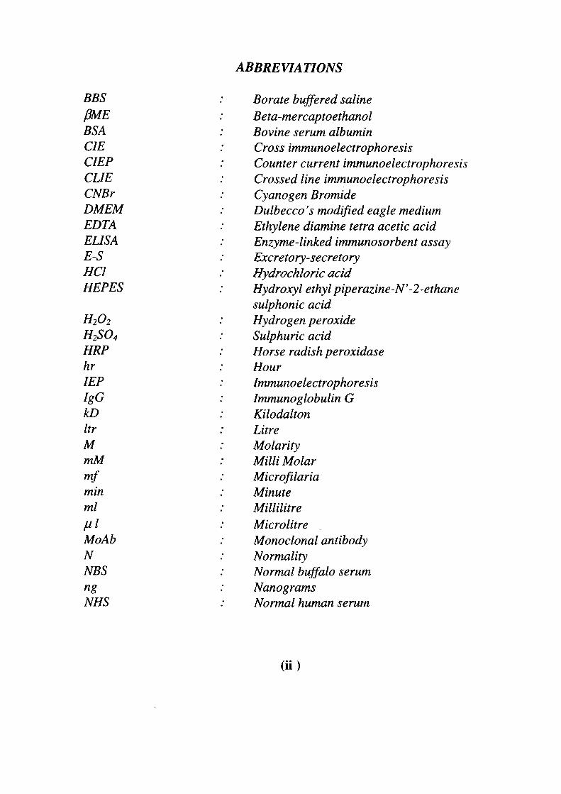
ABBREVIATIONS
BBS PME BSA CIE CIEP CUE CNBr DMEM EDTA ELISA E-S HCl HEPES
H2O2
H2SO4 HRP hr lEP IgG kD Itr M mM mf min ml Hi MoAb N NBS ng NHS
Borate buffered saline Beta-mercaptoethanol Bovine serum albumin Cross Immunoelectrophoresis Counter current Immunoelectrophoresis Crossed line Immunoelectrophoresis Cyanogen Bromide Dulbecco 's modified eagle medium Ethylene diamine tetra acetic acid Enzyme-linked immunosorbent assay Excretory-secretory Hydrochloric acid Hydroxyl ethyl piperazine-N' -2-ethane sulphonic acid Hydrogen peroxide Sulphuric acid Horse radish peroxidase Hour Immunoelectrophoresis Immunoglobulin G Kilodalton Litre Molarity Milli Molar Microfilaria Minute Millilitre Microlitre Monoclonal antibody Normality Normal buffalo serum Nanograms Normal human serum
(ii)
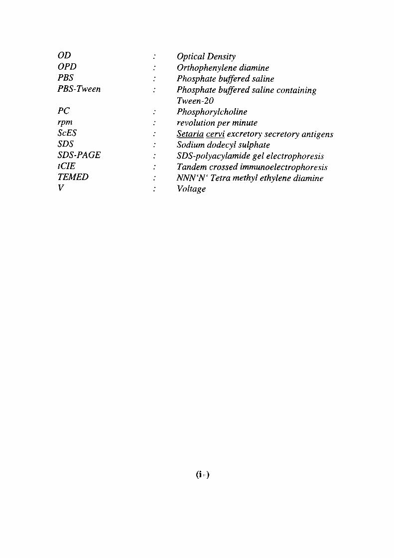
OD OPD PBS PBS-Tween
PC rpm ScES SDS SDS-PAGE tCIE TEMED V
Optical Density Orthophenylene diamine Phosphate buffered saline Phosphate buffered saline containing Tween-20 Phosphorylcholine revolution per minute Setaria cervi excretory secretory antigens Sodium dodecyl sulphate SDS-polyacylamide gel electrophoresis Tandem crossed Immunoelectrophoresis NNN'N' Tetra methyl ethylene diamine Voltage
(i-)
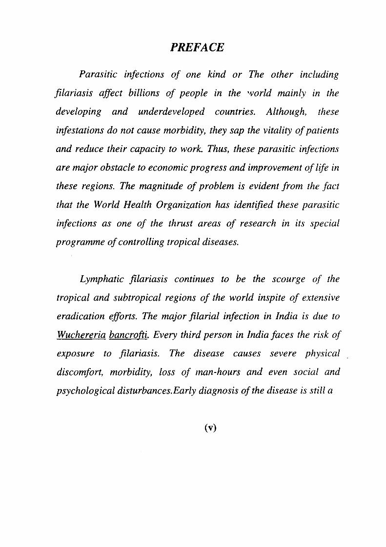
PREFACE
Parasitic infections of one kind or The other including
fdariasis affect billions of people in the 'vorld mainly in the
developing and underdeveloped countries. Although, these
infestations do not cause morbidity, they sap the vitality of patients
and reduce their capacity to work. Thus, these parasitic infections
are major obstacle to economic progress and improvement of life in
these regions. The magnitude of problem is evident from the fact
that the World Health Organization has identified these parasitic
infections as one of the thrust areas of research in its special
programme of controlling tropical diseases.
Lymphatic filariasis continues to be the scourge of the
tropical and subtropical regions of the world inspite of extensive
eradication efforts. The major filarial infection in India is due to
Wuchereria bancrofti. Every third person in India faces the risk of
exposure to fdariasis. The disease causes severe physical
discomfort, morbidity, loss of man-hours and even social and
psychological disturbances. Early diagnosis of the disease is still a
(V)
major problem and ejfective chemical or immunological remedies
against filariasis are not available. An unequivocal diagnosis of
filarial infection is still based on the detection of the circulating
microfilariae in night blood smears However, this method fails to
detect the disease when microfilariae are spares or sequestered in
tissues. This led to the use of immunological methods for the
diagnosis of filariasis. The immunodiagnostic methods based on
antibody detection showed extensive cross-reactivity with other
helminth infections and are unable to distinguish between present
and the past infection. In the recent past, more emphasis has been
given to the immunodiagnosis based on detection of circulating
antigen.
The studies reported in the present dissertation are directed
towards the immunochemical characterization of the excretory-
secretory (E-S) products ofSetaria cervi (a bovine filarial parasite)
which has been shown to have antigens common to the human
filarial parasites. The study deals with the preparation,
characterization and fractionation of 5 cervi E-S products,
production of polyclonal and monoclonal antibodies against the
E-S products and evaluation of the anti-E-S antibodies (polyclonal
and monoclonal antibodies) for the detection of circulating antigen
in filarial patient sera. These studies provide useful information
about the protein and antigenic make-up of S^ cervi E-S products,
presence of antigens, equivalent to filarial circulating antigen, as
well as the potential of anti-E-S antibodies in diagnosis of human
filariasis.
(vi)
CHAPTER I
INTRODUCTION AND
REVIEW OF LITERATURE
Lymphatic filariasis is a major public health problem in the
tropical and subtropical countries. About 1.1 billion people are
exposed to infection worldwide, accounting for about 20% of the total
world population (WHO, 1997). India alone contributes about 47% of
global prevalence of chronic patients and 39% of the population is at
the risk of filarial infection (NFCP, 1995; WHO, 1997). The disease
results in severe morbidity and globally is one of the most important
cause of permanent disability (WHO, 1995). While acute episodic
adenolymphangitis causes severe physical suffering, chronic disease
such as lymphoedema and hydrocoele causes permanent disfigurement
and psychosocial problems (Pani et al, 1995; Gyapong et al. ,1996;
Ramaiah et al, 1996a), which ultimately results in loss of work,
productivity, direct and indirect economic loss and functional
impairment (Pani et al., 1995; Ramaiah et al., 1996b; Ramu et al,
1996).
The most prevalent filarial parasites that infect man and
parasitise the lymphatics are Wuchereria bancrofti, Brugia malayi,
and B. trimori (Sasa, 1976). In India about 90% of the infection is
caused by W. bancrofti. Besides these, the filarial species infecting
animals and birds are Brugia pahangi (cats), Dirofilaria immitis
(dogs), Setaria cervi, S. digitata (catties), S. equina (horse).
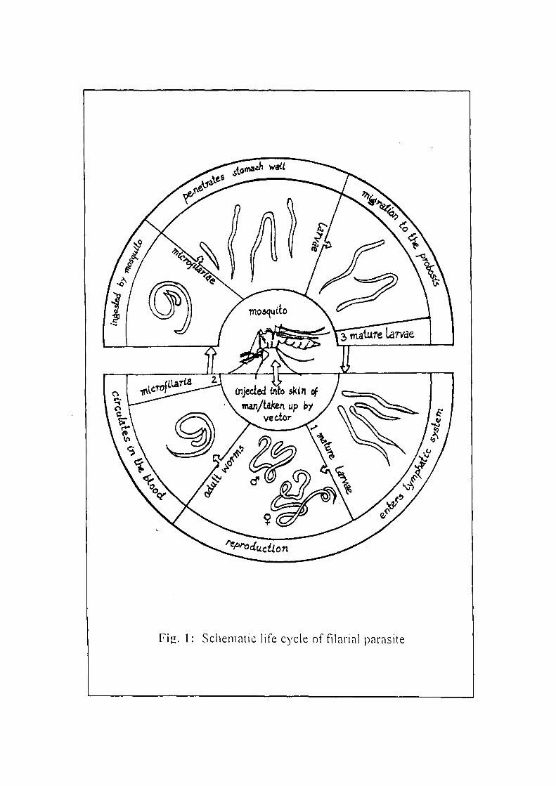
rijl. 1: Scliematic life cycle of filarial parasite
Acanthocheilonema vitae (rodents), Litomosoides carinii (cotton rat)
and Chandlerella hawkingii (Jungle crow).
The mosquito vectors that transmits lymphatic filariasis are
Culex, Aedes and Anopheles, including Mansoni. These are also
vectors of animal filariae, which are often sympatric with filariae of
man (Sasa, 1976; Denham and McGreevy, 1977). Filarial nematodes
have a biphasic life cycle comprising a period of larval development in
a blood sucking arthropod vector and the maturation and sexual
reproduction within a vertebrate host (Fig. 1). The adult filarial
worms are fine filiform found in the lymphatic vessels and
glands. The females (6.5-10 cm x 0.2-2.8 mm) are longer than the
males (4 cm x 0.1 mm). The female has a tapering anterior end with a
slight rounded swelling. The reproductive activity of the adults leads
to the release of microfilariae (mf) which are characteristically
present in circulation.
The microfilariae exhibits nocturnal periodicity, i.e. they display
circadian rhythm with regards to the number of mf in circulation,
which means that they are present in the peripheral blood in greater
numbers during the night as compared to the day. The peak
parasitemia is reached between 22.00 to 02.00 hour (hr). When the
mosquito bites an infected individual, the salivary secretion causes
the concentration of microfilariae near the site of bite and the
mosquito picks up the mf during the bite (Strong et al., 1934). In the
vector, the microfilariae undergoes metamorphosis and reaches the
mature infective larval stage. Now again, when this infected
mosquito bites another healthy individual, the larvae breaks free from
the labium and penetrates through the skin thus, causing infection. The
infective larvae passes through the peripheral blood vessels to the
lymphatics where they undergo development process and becomes
mature in an estimated period of three months.
In most cases of filarial infection the parasites does not exercise
any manifestation, therefore, a high but undefined proportion of
infection, in endemic areas, remains undetectable until clinical
pathology (lymphangitis and elephantiasis) develops. There are 70%
microfilareamic cases which do not show any clinical or pathological
sign of filarial infection. The pathological changes in W.
bancrofti and B. malayi are related to the developmental and adult
stages of the worms. The pathological changes in the lymphatics and
lymph glands are the result of an immunological reaction by the host.
The manifestations of the disease are the swelling of limbs, hydrocoele,
elephantiasis, tropical eosinophilia, chyluria, etc or other subclinical
abnormalities.
The effective control of filariasis depends on early and specific
diagnosis of filariasis. The definitive diagnosis of filarial infections in
humans is dependent solely on microscopic confirmation of the
presence of microfilariae in the peripheral blood in addition to the
pafients' clinical signs and symptoms (Sasa, 1978). But the
phenomenon of nocturnal periodicity demonstrated by many strains
dictates that collection of samples should be carried out between 22.00
and 02.00 hr when the peripheral microfilarial counts are at their
highest. However, parasitological diagnosis cannot detect the
prepatent phase of infection, obstructive lymphatic disease
(elephantiasis) or the tropical pulmonary eosinophilia syndrome
(Ottesen, 1980; Kagan, 1980; Piessens and Mackenzie, 1982). This
method is also very inconvenient for both the pateint as well as the
investigator. Furthermore, the distribution of microfilariae in the
bloodstream may be non-random (Eberhard et al, 1988) and thus,
making this procedure is relatively insensitive (Das et al, 1990).
Similarly, mf are not always detectable in the blood of asymptomatic
individuals, living in endemic areas, showing specific immunological
reactivity to filarial antigens (Ottesen et al, 1982).
For these reasons there has been considerable efforts focussed
towards developing non-parasitological tests, in particular
immunoassays for measuring antibody and circulating antigen (Kagan,
1981). Application of immunologic methods to the diagnosis of
lymphatic filariasis has earlier been focussed mainly on the detection
of the host antibody response to the parasite antigens (Kagan, 1963;
Ambroise-Thomas, 1974; Grover etal., 1977; Ottesen, 1980). Filaria
specific antibody can be detected in the blood of individuals who have
no apparent filarial infection as defined by clinical and parasitologic
criteria. Furthermore, filaria-specific antibody can persist long after the
clinically defined cure has been achieved, a fact that renders the
measurement of antibody levels an unreliable method for
discriminating between past and present infections. The anti-filarial
antibodies are extensively cross-reactive (Ambroise-Thomas, 1974;
Marcoullis and Grasbeck, 1976; Almond and Parkhouse, 1985; Cabrera
and Parkhouse, 1987) with different filarial parasites and other
helminths, thus the problems of specificity has always occurred with
serologic assays employing the whole worm extracts.
The detection of soluble circulating parasite material has been
suggested as the most likely approach for identifying the presence of
active infection (Frank, 1946; Harinath, 1984). Several investigations
of individuals suffering with either onchocerciasis (Steward et al,
1982; Des Moutis et al., 1983) or lymphatic filariasis (Au et al., 1981;
Dissanayake et al, 1982; Kaliraj et al., 1981a) and jirds with B.
pahangi infection (Karavodin and Ash, 1981) have demonstrated the
presence of circulating antigen and immune complexes during the
patent and post patent stages of infection. The development of
immunodiagnostic tests based on the detection of circulating filarial
antigens would be particulariy useful in epidemiological surveys of
lymphatic filariasis as well as for monitoring the mass scale drug trails.
Several groups of investigators have developed such assay by using
polyclonal antibodies (Harinath, 1984; Kaliraj et al., 1979a; 1981b;
Auetal, 1981; Oumssi et ai, 1981; Hamilton etal., 1984; Dasgupta
et al., 1984; Weil et al., 1984a; Weil, 1990) or monoclonal antibodies
(Des Moutis et al., 1983; Dissanayake et al., 1984; Forsyth et al., 1985;
Weil et al., 1984b; 1985) to detect filarial antigen in the sera and urine
of the infected individuals and animals, but the problem of sensitivity
and specificity of these assays still remains.
Specific antibodies against defined antigens are required for
developing antigen detection assays. In earlier studies, the antibodies
against the somatic antigens/extracts were employed for the detection
of circulating antigen in filarial patient sera (Weil, 1990; Ouaissi et al.,
1981; Hamilton et al., 1984; Weil et al., 1986). The excretory-secretory
(E-S) products, released by the living parasites in the host, are found to
be less complex in nature and more specific in defining infection as
compared to the somatic antigens/extracts. The fractionation and
characterization of E-S products is of immense interest for exploration
of their utility in immunodiagnosis and for the development of specific
antibodies and diagnostic reagent.
REVIEW OF LITERATURE
The accurate diagnosis of filarial infection is a major
requirement in the management of this disease. The identification and
characterization of parasite-derived antigenic components appears to be
especially important for understanding the functional immunity to the
parasite (Thorson, 1961; Poulain et al, 1976; Ogilvie et ai, 1973) and
for establishing specific diagnostic techniques in filarial infection.
Various filarial antigenic preparations such as somatic extracts, surface
antigens and excretory-secretory products, from both human as well as
animals, have been analysed and characterized by a number of
workers (Kwan-Lim et al, 1989; Kaushal et ai, 1982; Morgan et al,
1986; Egwang and Kazura, 1987; Malhotra et al., 1987; Devaney,
1988; Srivastava er a/., 1995).
I. CHARACTERIZATION OF ANTIGENS
The antigens of greater practical importance in filariasis are
those related to immunodiagnosis, immunopathology and protective
immunity. Since these are the molecules that are defined functionally
(by eliciting an immune response), their analysis should also be
closely integrated with functional studies. The characterization of
parasitic-derived antigen appears to be especially important for
identifying the antigenic molecules having protective and diagnostic
potential (Thorson, 1951, Poulain e? a/., 1976) Filarial antigens
such as somatic extracts, surface antigens along with the excretory-
secretory (E-S) antigens were analysed by different workers, using a
number of biochemical, immunochemical and radiolabelling
techniques (Forsyth et ah, 1981a; Maizels ef a/., 1982; Kaushal e/
a/., 1982; Malhotra ef a/., 1987).
Several investigators have analysed the crude somatic
extracts/antigens of the adult filarial parasites with the aim of
identifying and characterizing these antigens (Neilson 1975;
Dissanayake and Ismail, 1980b; 1983; Ho et al., 1986; Morgan et al.,
1986; Das et al, 1987; Kaushal et al, 1987; Maizels et al, 1987; Lai
and Ottesen, 1988; Lammie et al., 1990; Lobos et al, 1992; Bradley
et al., 1993a; b). In view of the non-availability of human filarial
parasites in sufficient quantities, antigens from related animal filarial
parasites including Setaria digitata (Dissanayake and Ismail,
1980b), Onchocerca gibsoni (¥orsyth et al., 19%\a; Ca.tmu\\ et al.,
1994), Brugia pahangi (Maizels et al., 1982), Dirofilaria immitis
(Weil et al, 1984; 1985; 1987) and S. cervi (Malhotra et al, 1986;
Kaushal e? a/., 1987; Srivastava et al, 1996) have been employed
in different studies for the characterization of somatic
extracts/antigens.
Several techniques were employed for the characterization of
somatic antigens such as, gel diffusion, immunoprecipitation, SDS-
polyacrylamide gel electrophoresis (SDS-PAGE), enzyme linked
immunosorbent assay (ELISA), western blotting/immunoblotting. The
Immunoelectrophoresis (lEP) and crossed Immunoelectrophoresis
(CIE) were used to analyse the antigenic pattern of somatic extracts of
bovine filarial parasites, S. cervi (Malhotra et al., 1987; Srivastava et
al., 1996) and Onchocerca species (Lobos and Weiss, 1986) were
analysed by immunoelectrophoretic techniques. The
immunoelectrophoretic analysis of the S. cervi somatic antigens
revealed the presence of 9-10 antigenic components, whereas, 22-24
antigens were observed on further analysis of the S. cervi somatic
antigen by crossed Immunoelectrophoresis (CIE) showed 22-24
antigens (Malhotra et al., 1986; Srivastava et ah, 1996). Lobos and
Wiess (1986) have also employed the lEP technique for analysing the
antigenic components of O. volvulus. The IE? analysis revealed 10-11
antigenic proteins in O. volvulus.
The SDS-polyacrylamide gel electrophoresis (SDS-PAGE), has
been employed by a number of workers to analyse the protein pattern
of the somatic antigens/extracts. The SDS-PAGE analysis of somatic
extracts of the B. malayi adult worms showed 30-35 protein bands, in
the molecular weight range of 10-200 kD (Kaushal etal, 1982). The
same technique was used to analyse the protein pattern of adult and mf
10
stages of bovine filarial parasite, S. cervi and ( S. cervi adult somatic
extracts showed the presence of 35-40 protein bands molecular weight
range of 10-200 kD), while 25-29 protein bands in the micro filarial
antigenic extracts (molecular weight range of 10-70 kD) (Malhotra et
al, 1986; Kaushal et al, 1987; Srivastava et al, 1996). Farrar and
others (1991) showed the presence of 42 protein bands in the
molecular weight range of 12-160 kD by the western blot analysis of
the B. pahangi adult somatic extracts.
Considerable efforts have been made to analyse the surface
antigens of filarial parasites as they appear to play a role in
protective immunity as well as in immunodiagnosis (Maizels et al.,
1982; Sutanto et al, 1985; Selkirk et al, 1986; Philipp and Davis,
1986; Theodore and Kaliraj, 1990; Devaney era/., 1990; Kwan-Lim
and Maizels, 1990; Petralanda and Piessens, 1991). Analysis of the
surface proteins by radiolabelling has suggested them to be
antigenically simple and distinct nature of nematodes surface (Philipp
et al, 1984; Storey and Philipp, 1990). The surface radioiodination and
immunoprecipitation studies of O. volvulus revealed a 22 kD antigen
which showed reactivity with sera from W. bancrofti infected
patients (Philipp et al, 1984). Morgan and others (1986) have analysed
the surface antigens of adult W. bancrofti using lodogen radiolabelling
techniques and Bolton-Hunter procedures, which revealed molecules in
the molecular weight range of 15-67 kD on the surface of W. bancrofti.
11
The 15, 20 and 29 kD protein bands showed resemblence with the
pattern obtained by lodogen labelHng of both B. pahangi (Sutanto et
al., 1985) and B. malayi (Maizels et al, 1985a). A 30 kD surface
antigen of B. pahangi was studied by radioiodination method
(Devaney, 1987; 1988).
The presence of carbohydrate and glycoprotein moieties on the
surfaces of filarial parasites was analysed by lectin binding studies. No
appreciable carbohydrate moieties exposed on the surfaces of either
adult or third stage larval (L3) forms of B. malayi could be detected
(Kaushale^a/., 1984; 1989). These studies have indicated that there is
stage specificity of the carbohydrate-containing antigens on the
parasite surface. The dynamic change of the antigens on surface
molecules may be either masked by host proteins or actual loss of
surface antigens, as microfilariae matures in vivo (Kaushal et al., 1984;
Ottesen, 1984). A major surface antigen of 35 KD was observed by 2-
dimensional gel electrophoresis on the surface of D. immitis L3
(Philipp and Davis, 1986).
The filarial parasites reside in lymphatics where they eat,
excrete and procreate therefore, the parasite products are detectable in
the blood of infected individuals. Such materials have been
collectively termed as "excretory-secretory (E-S) products".
Consequently, the E-S products come in close contact with the host
12
immune system, tending to provoke an immune response. The nature
and properties of the E-S products is one of the most intriguing and
highly speculated area of research on parasitic nematodes (Kaushal
et al., 1984; Piessens et al., 1982; Maizels et al, 1986; 1987b;
Sugunan and Raj, 1990). In filariasis, these materials have been
reported to play a significant role in the survival of the parasites,
immunopathological reactions in the host as well as in
immunodiagnosis (Sasa, 1976; Kaushal et al, 1984; Maizels et al,
1986; 1987a; Piessens et al, 1982; Mak et al, 1989; Parkhe et al,
1990).
The excretory-secretory (E-S) antigens of the filarial parasites
were found to be less complex in nature and more specific in
defining infection as compared to the somatic antigens (Desavigney
and Tizard, 1977; Kaushal et al, 1982; 1984; Malhotra et al,
1987). The importance of filarial excretory-secretory antigens to
diagnosis and immunizationn has also been shown in onchocerciasis
(Schiller et al, 1980) and bancroftian filariasis (Kharat et al, 1982;
Malhotra et al, 1982; Reddy et al, 1984c; Kaushal et al, 1984;
Harinath et al, 1984; Kaushal and Ottesen, 1987; Malhotra et al,
1987; Dumenigo etal, 1993; Espino and Finlay, 1994). However, not
much efforts were made to identify and characterize the excretory-
secretory products of human filarial parasites. The main reason is the
practical difficulties in obtaining sufficient quantities of parasitic
13
materials for characterizing the antigens and their use in the available
diagnostic procedures. First studies in this direction were done with
that of 5. malayi E-S products (Kaushal et al., 1982). Subsequently,
E-S products from a number of human (Maizels et al, 1986; 1987;
Kwan-Lim et al., 1989) and animal (Parkhouse et al., 1985; Maizels
et al., 1985a; Malhotra et al, 1987; Harnett et al., 1989;
Thilagavathy et al., 1990; Kaneko et al., 1990) filarial parasites were
analysed and characterized by immunochemical techniques.
The characterization of the antigenic components of the E-S
products of filarial parasites is essential not only to identify suitable
antigen target but for the development of sensitive and specific
immunodiagnostic tests (Morgan et al., 1986; Ottesen, 1984;
Selkirk et al., 1986; Harinath, 1984; 1986). A variety of techniques
have be?n used by several workers for the characterization of E-S
antigens of the adult filarial parasites (Kaushal et al., 1982; Malhotra
et al, 1987;Reddy et al., 1984c; Parkhouse etal, 1985; Maizels
et al., 1985a; 1987b; Thilagavathy et al., 1990; Wisrewshi et al.,
1990; Mizuno et al., 1991; Zamowska and Jastrzebka, 1994). The in
vitro released E-S products from Brugia (Kaushal et al, 1982;
Maizels etal., 1985a; Weil, 1988b) and Wuchereria (Kaushal et al,
1982) have been assayed for diagnostic specificities as have those for
both adults (Daveau and Ambroise-Thomas, 1982) and
14
microfilariae (Kharat et al., 1980) and also of O. volvulus (Schiller et
al, 1980; Ngue/^/., 1981).
The E-S products from adult B. malayi have been identified and
characterized by using ' I radiolabelling method (Kaushal et al,
1982; Kaushal and Ottesen, 1987). The autoradiography revealed the
presence of 11 radioactive protein bands in the B. malayi adult E-S
products in the molecular weight range of 10 000 to 70 000 D. Studies
of Kaushal et al. (1982; !984) on B. malayi E-S products as well as
studies by various workers with different filarial parasites have
indicated that the E-S products mainly comprised of low mol.wt.
proteins ranging between 10 to 200 kD (Kaushal et al., 1982; Maizels
etal, 1986; Weil, 1988; Kwan-Lim et al, 1989; Parkheefa/., 1990;
Lobos et al, 1992). Subsequent studies have also demonstrated major
protein bands in low molecular weight regions and a few high
molecular weight protein bands in E-S products of filarial parasites.
Low molecular weighjt (15-82 kD) protein bands were observed in L.
carina E-S products (Rajasekhariah et al, 1986a). Egwang and
Kazura, (1987) have analysed the B. malayi E-S products by lodobead
radioiodination method and have identified major protein bands of the
mol. wt. 30, 55 and 150 kD. Mainly low mol. wt antigens of 10-22 kD
along with few high molecular weight proteins of upto 120 kD in B.
malayi E-S products have also been identified by the studies conducted
by Kwan-Lim et al (1989) Kwan-Lim et al. (1989). The 29 kD
15
protein band identified as one of the major surface antigen in
B. pahangi (Lai and Ottesen, 1988; Devaney, 1987; 1988; Maizels et
al., 1985; Flecher and Wu, 1992). The E-S products of adult
D. immitis male and female worms revealed the presence of 16 and 21
protein bands respectively, while 7 and 10 protein bands were found in
both male and female D. immitis E-S products when
immunoblotting was employed (Kaneko et ah, 1990).
The E-S antigens are known to be formed in the uterus during
embryonic development (Decruize and Raj, 1988) and are released
during moulting as secreted or excreted products (Maizels and Selkirk,
1988a; b; Kaushal et al, 1982; Sugunan and Raj, 1986; Dhas et al,
1993). Sugunan and Raj (1986) have also shown to have a direct
relationship between E-S materials and the number of mf released.
Besides, the E-S products are amongst the first parasite molecules to
encounter the host immune system and continue to do so during the
course of parasite's life. They may therefore, influence parasite
survival and indeed, having a role in protective immunity against
filariasis (Mimori et al, 1987; Ey, 1988). The E-S products may
contribute to parasite pathology (Sisley et al, 1987), including that
arising during drug treatment (Greene et al, 1983).
16
Presence of host serum proteins in the filarial E-S products
The filarial parasites reside in the lymphatics of the host. To
protect themselves from the immunologically hostile environment of
the host and to evoke the immune response of the host, the filarial
parasites are known to adsorb some of the host serum proteins on
their surfaces. Several workers have reported the presence of
albumin on the surface of the parasites such as the infective larvae of
Trichenella spiralis (Parkhouse et al., 1981), O. gibsoni (Mitchell et
al., 1982), mf of W. bancrofti (Maizels et al., 1984a; b; Kar et al.,
1993; Mania and Kar, 1994), infective larvae of W. bancrofti
(Maizels er a/., 1986) and L. cam« (Phillip e/a/., 1984a). The
evidence for the presence ofhost serum proteins has also been shown
in the E-S preparation ofB. malayi (Kaushal et al., 1982), B. pahangi
(Parkhouse et al., 1985) and a bovine filarial parasite S. cervi
(Malhotra e/a/., 1987).
FRACTIONATION OF THE EXCRETORY-SECRETORY
PRODUCTS
The fractionation of antigenic preparations may lead to the
identification of the relevent antigen of diagnostic significance and
thus, for developing immunodiagnostic measures for human filariasis.
Relatively, little efforts has been made for the fractionation of E-S
17
products of the filarial parasites. The non-availability of a convenient
and suitable animal model for W. bancrofti makes it difficult to obtain
sufficient parasite material for immunodiagnosis of human filariasis.
Hence, antigens shared by different filarial species have been explored
by several workers for isolating the diagnostic important antigens
(Hamilton et al., 1984; Ottesen et al., 1985; Weil et al., 1987;
Cheirmaraj et al., 1990). W. bancrofti microfilaria! E-S products has
been found to be identical to some of the active circulating filarial
antigen fractions (CFA2-1, 9, 11 and 12) of microfilareamic cases
(Parkhe et al, 1990). Chenthamarakshan and colleagues (1996b)
fractionated the B. malayi microfilaria! E-S products using DEAE
cellulose and obtained two fractions BmE DEI and BmE DE2. The
BmE DEI was more active in binding to the immunoglobulin G
fraction of filarial serum immunoglobulin. This fraction has a
sensitivity and specificity of 83%. Similarly, E-S proteins of S.
digitata was fractionated on Sephadex G150 and resolved into three
protein components of mol. wt. 70 kD (ES FI), 16.5 kD (ES F2) and 11
kD (ES F3) (Sugunan and Raj, 1990). The 16.5 kD (ES F2) fraction
was found to be the major components. Later these fractions (ES F2
and ES F3) showed 100% sensitivity in detecting human filarial
antibodies in patients' sera.
Homologous filarial parasite antigen isolated from blood (Reddy
et al, 1986), hydrocoele fluids (Malhofra et al, 1985; Ramaprasad and
Harinath, 1989) and urine (Ramaprasad and Harinath, 1987) have been
fractionated and used as diagnostic reagent by Harinath and collegues.
The albumin absorbed UFA-C2 fraction has shown high sensitivity in
detecting filarial antibody (Ramaprasad and Harinath, 1987). The
same group of workers showed positive correlation between UFA C2-
A and mfES antigen in detection of filarial antibody. Ramaprasad and
Harinath (1995)have found that large scale isolation of UFA C2-A
fraction from filarial urine samples or production of monoclonal
antibodies to UFA C2-A overcomes the lack of parasite material and
removes the difficulty in collection of mfES antigen (.
Chenthamarakshan and others (1995) fractionated the B. malayi adult
SDS soluble antigen and evaluated the diagnostic utility of active
antigen fraction in bancroftian filariasis. The antigenic analysis of
BmA SDS S Ag revealed two fi-actions BmA-6 (37-45 kD) and BmA-
9 (20-25 kD) with high antigenic activity. Antibody were produced
against these antigen fi-actions in order to evaluate their utility in
detection of circulating filarial antigen in bancroftian filariasis.
III. IMMUNODIAGNOSIS
Diagnosis of parasite infectious forms an important element is
the identification of infected individuals and for studies on
19
epidemiology, protective immunity, immunopathology and control of
disease. Highly specific tests based on detection of parasites/ parasite
antigens and/or antibodiic requirement for specific and accurate
diagnosis of infection. Ideally diagnostic test should be simple,
sensitive, quantifiable and consistently reproducible. In addition, such
test should be inexpensive and applicable for large scale field use.
Immunodiagnosis of filariasis is one of the important area in
filarial immunology (Kagan, 1963; 1981; Ottesen, 1984; Taylor and
Denham, 1986). A number of immunodiagnostic tests based on
antibody and antigen detection have been developed for human and
animal filarial infections (Ambroise-Thomas, 1974).
Immunodiagnostic techniques with 'low or moderate sensitivity'
(Kagan, 1974) such as complement fixation, gel diffusion, latex
agglutination, indirect heameagglutination and indirect
immunoflorescence have largely been replaced by those of 'high
sensitivity', such as radio immunoassay, immunoflouresence assays,
enzyme linked immunosorbent assay, luminescence immunoassay,
immunoradiometric assay etc., have been explored for the diagnosis of
filariasis. A practical and sensitive assay would improve
understanding of the epidemology of the disease and could be useful
for monitoring the success of vector control efforts and drug trials
(WHO, 1984).
20
A. Parasitological Diagnosis
The diagnosis of filarial infections in humans still largely depends
on an assessment of the patients' clinical signs and symptoms in
addition to microscopic conformation for the presence of microfilariae
(mf) circulating in peripheral blood {W. bancrofti, B. malayi) or
dwelling in the skin {Onchocerca volvulus) (Sasa, 1978). However,
there are technical, practical and biological factors that limit the
usefulness of microfilaria detections as a diagnostic test for filarial
infection. The technical limitation is that the sensitivity of such test
depends on microfilariae counts, the volume of blood (or the number of
skin snips in case of onchocerciasis) examined, and the skill of the
microscopist. The microfilariae of W. bancrofti and B. malayi shows
nocturnal periodicity and this imposes a practical limitation on the
effective use of microfilarial detection. Moreover, this method is
inconvenient for both the investigator as well as the patient. The
biological limitation of microfilaria detection is that it fails to detect the
disease when mf are sparse or sequestered inside the tissues or in
inaccessible sites. This method is also insensitive because of
the well recognized phenomenon of filariasis without
microfilariae, i.e., amicrofilaremic stage (Beaver, 1970). The
parasitological detection of microfilariae is found to be relatively
insensitive, giving emphasis to the development of immunodiagnostic
techniques, based on antibody and antigen detection, for filariasis.
21
B. Antibody Detection
Application of immunological methods to the diagnosis of
lymphatic filariasis has earlier been focussed on the detection of the
antibody response to the parasite (Kagan, 1963; Ambroise-Thomas,
1974; Grover et ah, 1977). Antibodies have been detected readily in
sera of bancroftian filarial patient using hetrologous or homologous
antigens from adults, larvae and microfilariae (mf) stages, as well as
excretory-secretory products from filarial parasites (Ambroise-Thomas,
1974; 1980; Chandra et al, 1974; Gonsaga dos Santos et al, 1976;
Grover and Davies, 1978; Dissanayake and Ismail, 1981; Au et al,
1982; Kaushal et al., 1982). A number of immunological methods
such as gel diffusion (Petithory et a/., 1972; Khatoon e? a/., 1987),
immunoelectrophoresis (Capron et al, 1968; 1970), counter current
immunoelectrophoresis (Desowitz and Una, 1976; Weil et al, 1984;
1986; Dasgupta et al., 1980), indirect haemagglutination test
(Takahashi and Sato, 1976; Kaliraj et al, 1981a; b; Das et al, 1987),
indirect immunoflourescent antibody test (Diesfeld et al, 1973
Forsyth et al, 1985; Kaliraj, et al, 1981c; Das et al, 1987
Dissanayake et al, 1984) and the skin test (Swada et al, 1969; 1975
Swada and Sato, 1969; Gupta and Ansari, 1989) have been used for
detecting filarial antibodies employing crude antigenic extracts from
the homologous and heterologous filarial parasites (Ottesen et al,
1982).
22
The skin test used previously for the diagnosis of filarial
infection by detecting antibodies in filarial patients' sera. The test was
carried out by subcutaneous injection of the filarial antigen in the arm
of an individual, the response is of immediate type and can be read
usually within 15 to 30 min, but the delayed hypersensitivity reaction
i.e. after twenty four hour has also been noted. Wharton (1947) has
reported that delayed reaction, due to the use of antigen in very high
concentrations, was actually of immediate type skin reaction which
persisted and produced exacerbation of symptoms in the infected
individuals. The false positive results obtained with skin test may be
attributed to cross-reaction with intestinal helminths (Bozicevich and
Hutter, 1944; King, 1944; Hunter, 1958). The skin test showed cross-
reactivity with patients infected with hookworms and strongyloides
(Woodruff er ai, 1958). It has also been observed that in some cases
of elephantiasis, chyluria and hydrocoele skin test was found negative
(Frank et al, 1947). Chandra and coworkers (1974) developed a
filarial skin test kit using B. malayi infective larval antigen for the
detection of antibody in filarial patient sera. This test has been
evaluated for its sensitivity and specificity in the immunodiagnosis of
human filariasis covering over 2500 cases from filaria endemic and
non-endemic regions of India (Katiyar et al, 1985 and Sircar et al,
1990). The major limitation of the kit was that the crude L3 antigen
was used, which was prepared from the mosquito and it showed cross-
reactivity with other helminth parasites. Acton and Rao (1933) used
23

hydrocoele fluid as antigen in the skin test for the diagnosis of human
filariasis.
Another widely employed diagnostic test in sixties-seventies, the
complement fixation, was found to be more sensitive than skin test
for the diagnosis of human filariasis (Kagan, 1963). The complement
fixation test showed fairly high sensitivity with sera of patients
infected Loa loa whereas, fi-om W. bancrofti infected patients and
those having other filarial infections showed comparatively less
sensitivity (Scofield, 1957). This test also showed cross-reactivity
against anchylostoma, schistosomiasis and stronglyoidiasis and
infections with other intestinal parasites (Stemplen, 1944).
The presence of filarial antibody in the serum samples of
infected humans and animals has been reported by several
investigators using the precipitation reaction (Biguet et al., 1962;
Desowitz and Una, 1976; Khatoon et al., 1987). The precipitin test
has limited use in the diagnosis of filarial infection. The antigens used
in the test were the saline extracts of the whole worms and the results
varied considerably. The precipitin test was found to be less sensitive
than the skin test for the diagnosis of Mansonella ozzardi (Biagi,
1956). Ellsworth and Johnson (1973) used diffusion capillary tubes
for the precipitate formation using the sera of Dirqfilaria immitis.
infected dogs. The immunodiffusion method was used by Khatoon, et
al. (1987) for the diagnosis of Setaria cervi infection and this method
was found to be comparatively simple and reliable. The sensitivity and
reactivity of the precipitin test are so much lower than other
conventional serological tests that its use for routine diagnosis can be
hardly recommended.
Another precipitation technique i.e. immunoelectrophoresis
(lEP) has been used by Wheeling and Hutchinson (1971) for the
diagnosis of human filariasis employing antigens prepared from D.
immitis microfilariae and adults. Kaeuffer and coworkers (1974),
however, didnot show any false positive reactions when the sera of 14
Tahitians infected with W. bancrofti were tested in lEP using adult D.
immitis antigen. lEP has also successfully been employed for the
diagnosis of onchocerciasis with antigen extracted from the adult
worms of Onchocerca (Biguet et al, 1962; 1964; Capron et al,
1968; 1970). Using the above technique (i.e. IE?) Gentilini et al.
(1973) have found that 8 out of 9 patients, suffering from bancroftian
filariasis were positive for the infection, whereas, no precipitin bands
were observed when sera of 3 patients were tested against O. volvulus
adult antigen. The number of arcs produced have been shown to be
related to the clinical severity of the filarial infection (D'Haussy et al.,
1972). The degree of certainty for the specificity of these precipitin
test is not known. Wheeling and Hutchinson (1971) obtained positive
25
reaction with D. immitis antigen in case of human filariasis. On the
other hand Kauffer and others (1974) tested a number of W. bancrofti
microfilareamic patients with the same antigen {D. immitis aduh
antigen) and found entirely negative results. The investigation carried
out by Niel et al. (1972) have indicated the possibility of broad
spectrum of antigenic specificity, and have also found that the antigens
prepared from Setaria labiato-papillosa and Dipetalonema vitae
produced precipitin lines in immunodiffusion and lEP against the
sera of patients with loiasis, onchocerciasis, bancroftian filariasis and
dracunculiasis. The above workers also reported the cross-reactivity of
the sera of filarial patients with antigens of Ascaris suum using the
same technique. Coimter current Immunoelectrophoresis (CIEP) was
used by Desowitz and Una (1976) for the detection of filarial
antibodies using D. immitis adult antigen and was found to be fairly
sensitive. CIEP has the advantage of simplicity of performance,
rapidity and the capability of processing relatively large numbers of
serum samples at the same time.
The indirect haemagglutination test (THAT) has also been
employed for the diagnosis of human filariasis. The
heameagglutination test was used by Jung and Harris (1960) for the
detection of human filarial infection using somatic antigen from
D. immitis adult worms. The IHAT employing heterologus somatic
antigen extracts {D. immitis) showed apparent sensitivity in the
26
diagnosis of filariasis due to Achanthocheilonema perstans (Kagan et
al., 1963) and W. bancrofti (Fujita et al, 1970) and also gave false
positive reactions with sera from normal individuals as well as with
patient sera harbouring other helminth infections.
The indirect flourescent antibody test (IFAT) has been used for
immunodiagnosis of filariasis in limited number of studies. The adult
worm extracts of A. vitae (Ambroise-Thomas and Kein Trong, 1974),
S. digitata (Tan et al., 1988) or sonicated W. bancrofti microfilaria!
and larval antigens (Hedge and Ridley, 1977; Kaliraj et al, \979b;
1981c; Das et al, 1987; 1988c) have been used in IF AT for the
detection of filarial antibody. However, the use of
immunoflourescence test for large scale testing of patient samples is
tedious and requires special equipments.
The advancement of immunological techniques led to the
development of sensitive assays for detecting parasitic infections.The
enzyme linked immunosorbent assay (ELISA) was found to be
simple, sensitive and suitable for mass screening of most parasitic
infections (Voller et al, 1974; 1975a; b; 1976a; b; Kaliraj etal,
1981b; c). Since its development (Engvall and Pearlman, 1972)
ELISA has been successfully used for detecting antibodies and
antigens of a variety of organisms (Sever and Madden, 1977). In
recent years, ELISA had been found very useful in the diagnosis of
27
many parasitic diseases including filariasis (Voller et ai, 1976a;
Bartlett et al, 1975; Bartlett and Bidwell, 1976; Barakat e^a/., 1983;
Gueglio et ah, 1995). Both homologous and heterologous antigens
were used for measuring antibody response in filarial infection
(Ruitenberg et ah, 1975; Spencer et al., 1981). A microtitre plate
ELISA was used by Bartlett et al. (1975; 1976) for the detection of
antibody in sera oi Onchocerca volvulus infected patients' sera but the
use of homologous antigen was not possible as contaminants of host
origin reacted non-specifically with the secondary antibody conjugate.
No such problem was encountered when the same antigen was used
after purification (Marcoullis et al, 1978). The W. bancrofti mf
antigen and mf excretory-secretory (mf ES) antigen was used for the
detection of filariasis by a number of workers (Kaliraj etal., 1981 b; c;
Kharat ef a/., 1982; Malhotra er a/., 1982; Malhotra and Harinath,
1984; Harinath et al., 1984; 1986; Dissanayake and Ismail, 1980a). In
all these cases ELISA was found to have better sensitivity than night
blood examination. Isolation and characterization of filarial antigens
from urine and hydrocoele fluids have shown fair diagnostic potential
(Ramaprasad and Harinath, 1989). The purified antigens showed high
sensitivity and specificity when tested in sandwich ELISA (sELISA)
(Malhotra et al, 1985a; b; Singh et al, 1993; Ramaprasad and
Harinath, 1989; 1995) and stick enzyme immunoassay (Parkhe et al,
1988; Ramaprasad and Harinath, 1989) for the detection of filarial
antibodies in filarial patient sera. Besides, the use of homologous
28
antigens, heterologous antigens were also used by many workers
for the diagnosis of human filariasis, i.e., B. malayi (Spencer e/a/.,
1981; Cheirmaraj et al, 1990; Kumar and Santhanam, 1990;
Chanteau et al, 1991; Yuan et al, 1992; Li et al, 1993), D. immitis
(Weil et al, 1985; 1987) and S. digitata (John et al, 1995; Dhas and
Raj, 1995).
The serodiagnostic tests for filariasis based on antibody
detection were useful for testing sera from people visiting endemic
areas, but they were not very suitable for testing sera fi^om endemic
areas because of extensive antigenic cross-reactivity among different
nematode antigens (Oliver-Gonzales and Morales, 1945; Kagan, 1963;
Ambroise-Thomas, 1980). In addition, people who have been exposed
to filarial parasites, so called 'endemic normals', often have anti-
parasite antibody titres that are at least as high as those people with
proven infections (Ambroise-Thomas, 1980; Ottesen etal, 1982). The
next level of refinement in antibody testing improved specificity by
using subsets of antigens purified fi'om crude extracts (Weiss and
Karam, 1989) or by measuring antibodies of specific isotypes (Weiss et
al, 1982; Lai and Ottesen, 1988a; Kwan-Lim et al, 1990; Weil et al,
1990). The IgG4 is normally a minor component of total circulating
immunoglobulins in lymphatic filarial infection but shows prominent
response (Ottesen et al, 1985). Lai and Ottesen (1988a) demonstrated
that IgG4 antibody detection produce increased specificity for detecting
29
filarial infection, as compared to total IgG, even when crude parasite
antigen was used. These assays were more specific than assays those
measures total antibodies to crude antigen-mixture, but they did not
achieve full species-specificity and also do not correlate the active
infection. The lack of acceptable specificity and sensitivity of antibody
detection assays in the serodiagnosis of filarial infections was one of
the major reasons for the development of antigen detection assays
(Dissanayake and Ismail, 1987).
C. Antigen Detection
The parasite antigen detection is more sensitive and specific for
diagnosing active filarial infection than parasitological examination or
antibody detection (Harinath et al., 1984; Forsyth et al., 1985; Weil et
ai, 1985; 1987; Lai et al., 1987; Anon, 1989). Considerable emphasis
has been given to the detection of circulating antigen in the blood and
other body fluids of filarial patients (WHO, 1992; Weil, 1990). The
antibodies raised against the filarial antigen obtained from infected
patients have been employed by some investigators for the detection of
circulating filarial antigen inpatients' sera (Kaliraj et al., 1979; Forsyth
et al., 1985; Weil et al., 1986; 1988; Weil, 1990; Mustafa et al, 1996).
Several groups of investigators recently have developed antigen
30
detection assays using polyclonal (Harinath, l984;Kaliraj e?a/., 1979;
1981; Au ef ai, 1981; Ouaissi et ai, 1981; Hamilton et al., 1984;
Dasgupta et al., 1984; Weil et al., 1986; Mustafa et al., 1995; 1997)
and monoclonal (DesMoutis et al., 1983; Dissanayake et al., 1984;
Forsyth et al., 1985; Weil et al, 1985; More and Copeman, 1990;
Kaushal et al., 1994a; b; Weil et ai, 1997) antibodies (MoAb) to detect
filarial circulating antigen. The earlier studies were mainly focussed on
the antisera raised against somatic extracts of the heterologous filarial
parasites, i.e. L. carinii (Dasgupta and Bala, 1978; Kaliraj et al, 1981a)
andZ). /wm/ri5 (Tanabe, 1959; Weil e? a/., 1984a; 1986).
Circulating filarial antigen was first demonstrated in bancroftian
filariasis in 1946, using the technique of passive cutaneous anaphylaxis
(Frank, 1946). The soluble circulating parasite materials has been
detected by number of workers in W. bancrofti infected individuals
(Reddy et al, 1984a; b; Zheng et al, 1987b; Lutsch et al, 1987;
1988; Ramaprasad and Harinath, 1995) and also in patient
infected with onchocerciasis (Ouaissi et al, 1981; Des Moutis et al,
1983;Petralandaer al, 1988; Schlie-Guzman and Rivas Alcala, 1989;
Chandrashekhare/a/., 1990) and thus indicating the active infection of
filarial parasites.
31
Circulating parasite antigen in patients with bancroftian filariasis
has been demonstrated using techniques of 'moderate sensitivity' such
as counter current immunoelectrophoresis and indirect
heameagglutination test (Kaliraj et ai, 1981c; Au et al, 1981) but
positive results were obtained only in sera of microfilaraemic positive
individuals. The CIEP was used for the detection of circulating antigen
using rabbit anti-filarial sera (Kaliraj et al., 1979a; Dasgupta et al,
1980; 1984; Das et al, 1988c; Kumar et al, 1991;Dumenigoef a/.,
1993). The antibodies raised in rabbits against L. carinii somatic
antigen were used for the detection of filarial antigen in human
patients' sera and it was observed that 5.7% cases diagnosed by
parasitological examination can be increased to 62.8% by CIEP
(Dasgupta et al, 1980). The CIEP used to detect the D. immitis
antigens represents a significant improvement over previously
available diagnostic techniques because it is more sensitive than
microfilarial test, and can be related to the active infection and can
be used as practical diagnostic test for active W. bancrofti infection
((Kaliraj et al, 1981a; Weil era/., 1984; 1986). The CIEP is simple
and rapid as compared to IHAT, with increased sensitivity over
immunodiffusion for the detection of circulating antigen have been
established in various studies (Krupp, 1974; Dasgupta and Bala, 1978;
1980; Shariff and Parija, 1991). Hamilton et al, (1984) have used
antibodies specific for B. malayi adult worms to detect cross-reactive
antigens in bancroftian filarial pateint by immunoradiometric assay
32
(IRMA). Using polyclonal antibodies against adult S. digitata,
Dissanayake and others (1982) detected antigens in the immune
complex from bancroftian filariasis patients. The above workers
detected the antigens in the circulating immune complex
(CIC) reactive with polyclonal antibodies, but did not assay the
amount of each antigen. Zheng et al. (1987a), Using polyclonal and
monoclonal antibodies (E34 and HC 11), have shown filarial antigen
in 95% of sera from mf donors with bancroftian or brugian filariasis
and approximately 60% of sera from mf donors and 60% of clinical
cases by inhibition ELISA using E-S antigen conjugated to
penicillinase. However, filarial antigen was detected in all the 38 sera
from mf pateints by CIEP with rabbit antibodies to D. immitis and B.
malayi (Weil et al, 1986). Recently, 100% positive results were
reported with mf positive sera by Cheirmaraj et al. (1992) using
polyclonal antibodies raised in mouse ascites.
The non-availability of adult W. bancrofti for antigenic
material, demands the use of antigens from heterologous filarial
parasites to raise polyclonal antibodies in rabbits and monoclonal
antibodies for the detection of circulating antigens in filarial infections
has been employed by several workers (Dasgupta et al., 1980; 1984;
More and Copeman, 1990; 1991; Kumar et al, 1991; Jaoko, 1995;
Forsyth et al, 1985; Weil et al, 1984; 1985; 1986; 1987; Maizels et
al, 1983; 1985b; Kazura et al, 1986; Freedman et al, 1989; Parab et
33
al, 1990; Malhotra et al, 1987; Phillip et al., 1984a; Cabrera and
Parkhouse, 1986; Egwang and Kazura, 1987; Prasad and Harinath,
1988; Rajasekhariah et al., 1991; John et al., 1995). Besides these,
materials from the microfilariae and infective larvae of W. bancrofti
and of related filarial parasites were also used by a number of
researchers to produce antisera for the detection of filarial circulating
antigens in patient sera (Almeida f a/., 1990; Reddy et al., 1984b;
c; Lutsch et al., 1987; Cheirmaraj et al., 1990; 1992; More and
Copeman, 1991;Dumenigoe/fl/., 1993; Zheng etaL, 1987; Lai 1991;
Li et al., 1993; Chenthamarakshanera/., 1995; 1996).
Polyclonal antibodies were raised against somatic extracts of W.
bancrofti microfilariae, and could detect the filarial circulating antigen
in microfilaremic positive patient sera (Kaliraj et al., 1979a). Due to
the use of polyclonal antibodies raised against crude somatic antigens,
for the detection of circulating filarial antigen, problems of specificity
and sensitivity remains (Weil, 1990). Therefore, monoclonal
antibodies have been employed by several investigators for the
detection of circulating antigen in the filarial infected individuals
(Dissanayake et al, 1984; Weil et al., 1985; 1987; Forsyth et al., 1985;
Lai et al, 1987; Weil and Liftis, 1987; Zheng et al, 1987; Santhanam
et al., 1989; More and Copeman, 1990; Ramzy et al., 1991; Li et al.,
1993; Kaushal et al., 1994; Ramaprasad and Harinath, 1995; Gyapong
et al., 1998; Lalitha et al., 1998). Assays describing detection of
34
ciculating antigen in onchocerciasis patients were first reported in the
early 1980s (Ouaissi et al, 1981; Des Moutis et al, 1983) and was
found to be highly sensitive and specific (Cabrera and Parkhouse,
1987; More and Copeman, 1990; 1991). Phosphorylcholine (PC) was
the first truly defined molecules to be targeted in the serum of
onchocerciasis patients (Weiss, 1985; Maizels et al, 1990). An
improvement in sensitivity (92.3%) was obtained by Schlie-Guzman
and RivasAlcala (1989).
An immunoradiometric assay (IRMA) for the detection of
filarial antigen was developed using an IgM monoclonal antibody
(Gib-13) to O. gibsoni egg antigen (Dissanayake et al, 1984;
Forsyth et al., 1985). The assay detected the parasite circulating
antigen in 93% and 75% of the sera from amicrofilareamic subjects
with ^. Z a/icro/ z, however, it takes two days to perform the Gib-13
IRMA. Moreover, as it is an radioimmunoassay, it could not be
used in field conditions. The Gib-13 shows phosphorylcholine (PC)
specificity detecting molecules of 140, 52, 56 and 62 kD molecular
weight and recognizes a carbohydrate moieties. Zheng et al. (1987a; b;
1990) used monoclonal antibody based sandwich ELISA (sELISA)
for the detection of circulating filarial antigen using rabbit anti-
microfilarial immunoglobulin as first antibody and monoclonal
antibodies, against W. bancrofti mf E-S antigen and B. malayi L3
antigen as second antibody, the positivity rates were 94.5% and 89.0%
35
respectively in microfilareamic patients. Most of the earlier attempts
to produce monoclonal antibodies against filarial circulating antigen
have resulted in monoclonals showing reactivity with
phosphorylcholine epitope and to other, more specific determinants
(Forsyth et ai, 1985; Lai et al., 1987; Weil et ai, 1987; Zheng et ai,
1987a; More and Copeman, 1990). The sensitivity of PC assays for W.
bancrofti infection have ranged from 85 to 93% for sera from
microfilareamic patients depending upon the population surveyed. PC
antigenemia has also been detected in sera from patients infected with
B. malayi (Maizels et al., 1985a). However, the sensitivity of PC
antigen detection for human B. malayi infections has been very poor. In
addition, studies on PC antigenemia in filariasis have been reported
conflicting specificity results. The PC is a widely distributed
determinant, particularly among the nematode parasites and its
presence on the filarial parasite antigens may account for much of the
cross-reacivity observed with other helminth parasites (Maizels et al,
1987c; Pery et al, 1974; Zheng et al, 1987). Therefore,
assays detecting the PC determinant is not suitable for the
specific diagnosis of human filariasis (Forsyth et al, 1985; Lai et al,
1987; Sutanto et al, 1985). Lai and others (1987) detected a 200 KD
antigen in the sera of 93% of patients with microfilareamia, 46% of
those with lymphatic obstruction, and 56% of those with tropical
pulmonary eosinophilia syndrome using a monoclonal antibody (CA2
or CAioi) raised against circulating antigen from W. bancrofti. This
36
n;ionoclonal antibody could detect parasite antigen concentration as low
as 30 to 4000 ng/ml in patients living in an area endemic for
bancroftian filariasis and it also shows specificity for PC determinants.
The CAioi monoclonal antibody is an IgMk with the T15 idiotype.
There are few monoclonal antibodies which are directed against
non-phosphorylcholine epitopes and are also capable of detecting
filarial circulating antigen in bancroftian filariasis with minimal or no
cross-reactivity (Weil and Liftis, 1987; Weil et al, 1987; More and
Copeman, 1990; Zheng et al, 1990; Kaushal et al, 1994). These
assays have been successfully used for detecting the filarial circulating
antigen inpatients' sera (Weil etal., 1988; 1991). Another monoclonal
antibody (ES 34)) which is against W. bancrofti mf ES antigen (55-63
kD) has shown promising results in field studies conducted in India and
China (Zheng et al, 1987; Reddy et al, 1989). But this monoclonal
could not detect antigen in clinical cases and specificity data was
insufficient. A specific assay, which is currently 1 undergoing
commercial development, is based on the monoclonal antibody
AD12.1 (Weil et al, 1987) which recognises a 200 kD antigen in the
circulation of individuals infected with W. bancrofti (Weil and Liftis,
1987) which again is of ES products of adult worm (Weil et al, 1996)
and is specific for W. bancrofti only (Weil, 1990; Weil ef a/., 1987;
Ram2y et al, 1991). The monoclonal antibody is of IgGi isotype and
recognizes the carbohydrate epitopes and detects the absolute
37
amount of protein by a factor of at least 2.4. The sensitivity of
AD 12.1 assay for sera from microfilareamic patients was
approximately 95%. The filarial antigen was detected niether in sera
from non-endemic areas nor from patients with other parasitic
infections (Reddy et al, 1989). The ICT Filariasis card test is a new,
rapid-format filarial antigen test based on monoclonal antibody
AD12.1, was developed by ICT diagnostics (Balgowlah, New South
Wales, Australia). This assay takes only 5-15 min to complete, requires
no specialized equipment and gives comparable results (Weil et al,
1997).
Another assay that could successfully detect the circulating
antigen in ELISA is commercially available for detection of W.
bancrofti infection (Trop-Ag W. bancrofti ELISA kit, JCU Tropical
Biotechnology Pty Ltd, Queensland, Australia). The assay is based on a
monoclonal antibody, Og4C3, which curiously, inspite of its being
raised against antigens of the bovine parasite O. gibsoni, detects
circulating antigen in serum from W. bancrofti infected patients, but
not O. vovulus (More and Copeman, 1990) using sandwich ELISA.
Negative results were also obtained with sera from individuals
harbouring B. malayi, B. trimori and Loa loa infections. The antigen
detected in the circulation of bancroftian filariasis patients is of adult
origin (Chanteau et al, 1994). The monoclonal antibody Og4C3 has
IgM antibodies. Og4C3 binds to a range of filarial and non-filarial
38
nematodes (O. gibsoni. O. vovulus, D. immitis, A. caninum and
Toxocara canis) but not to phosphrylcholine. It recognizes antigens of
M W > 130 kD and 50-60 kD from Og male antigen, with epitopes
having both protein and carbohydrate moieties. The target antigens of
Og4C3 are located at the junction of the cuticle, hypodermis, in cells of
the gut, in intrauterine embryos and extra-uterine microfilariae of O.
gibsoni. Both assays have been evaluated as diagnostic tools in a
number of studies (Nicholas, 1997) and can detect circulating antigen
in virtually all (94-100%) microfilarial carriers (Weil et al., 1987;
1997; Turner et al, 1993; Chanteau et al, 1994a; b; Rocha et al, 1996;
Nicholas, 1997). However, most of these MoAbs are of IgM isotypes
and not very suitable for use in the field test. Kaushal and others
(1994a) produced MoAb against antigenic epitopes common between
the bovine (Setaria cervi) and human filarial parasites. The two
monoclonal antibodies (13B4 and 15D6) that showed reactivity with
filarial circulating antigen of IgGi isotype and are directed to the
protien epitope. The target antigen have been cloned using these
monoclonal antibodies (Kaushal et al, unpublished data). These
monoclonals showed high reactivity with filarial antigen while very
little or no reactivity was observed with the non-filarial antigen
(Kaushal et al, 1994; Kaushal and Kaushal, 1995).This assay has high
sensitivity but lacks sufficient sensitivity. Ramzy and others (1991)
evaluated the performance of antigen detection in the sera from an
endemic area of W. bancrofti in Egypt using MoAb raised against
39
Dirofilaria immitis. They reported that 97% of microfilareamic
subjects were antigen positive and antigen levels were significantly
correlated with mf counts. Those methods for detecting circulating
antigens using MoAbs were reported to have high sensitivities
correlating with mf counts.
40
SCOPE AND PLAN OF WORK
Improved methods for diagnosing active filarial infections are
needed to monitor control efforts and to evaluate new drugs. The most
commonly used method for the diagnosis of filariasis, is based on
demonstration of mf in the night blood smears of the patient. This
test is inconvenient for both the patients as well as the investigator
emd fails to detect the amicrofilareamic stage of the disease or when
mf are present in very small numbers or sequestered in the tissues. This
has put impetus for developing alternate means for filaria diagnosis
such as immunodiagnostic test. Earlier studies on immunodiagnosis
of filariasis were focussed mainly on the detection of antibodies, but
the major shortcoming is their inability to distinguish past exposure
from current infection. Therefore, in the recent past considerable
emphasis has been given to the detection of circulating antigen in
blood or other body fluids of the infected individuals. The
demonstration of the parasite antigen in circulation of the infected
individual appears to be an useful indicator of active filarial infection.
For establishing specific diagnostic techniques identification and
characterization of parasite-derived antigen is important. Due to the
complex nature and extensive cross-reactivity of the crude
somatic antigens with the other nematode parasites, antigens present
in the excretory-secretory (E-S) products (obtained by the in vitro
41
maintenance of filarial parasites) of these parasites were analysed
and evaluated for the immunodiagnosis of filariasis.
Due to the non-availability of human filarial parasite, antigens
from Setaria cervi (a bovine filarial parasite) have been used in the
present study. The E-S products were obtained by maintaining S.
cervi adult worm by short-term in vitro incubation in culture.An
effort was made to isolate the S. cervi E-S products, raise polyclonal
antibodies against E-S antigens and this anti E-S antibodies could
detect the circulating antigen in human filarial patient sera. Therefore,
the E-S products were fractionated in order to identify the relevent
antigen (equivalent to the filarial circulating antigen), produce
polyclonal and monoclonal antibodies and the potential of these
monoclonal antibodies were evaluated for the detection of circulating
antigen in human filariasis.
The present study deals with the following aspects:
I. Preparation and immunochemical characterization of excretory-
secretory (E-S) products from S. cervi adult worms.
42
II. Raising of polyvalent hyperimmune antibodies against S. cervi
E-S products in rabbits and its evaluation for the detection of
circulating antigen in filarial patient sera.
III. Fractionation of S. cervi E-S products and immunochemical
characterization of fractionated E-S products and identification
of antigen equivalent to filarial circulating antigen.
IV. Production and characterization of polyclonal/monoclonal
antibodies against the relevent E-S antigens.
V. Evaluation of the antibodies for the detection of circulating
antigen in filarial patient sera.
43

CHAPTER II
MATERIALS AND
METHODS
PARASITES
Setaria cervi, a bovine filarial parasite, was collected from the
peritoneal folds of freshly slaughtered Indian water buffaloes from the
local abbatoir and brought to the laboratory in normal saline. The
motile worms (both males and females) were washed thoroughly with
normal saline to remove all the adhering materials and were used
immediately for antigen preparation or stored at -20°C until used.
EXPERIMENT ANIMALS
Albino rabbits of either sex (weighing 1.5 to 2.0 Kg)and
BALB\c mice (6-8 weeks old) from the inbred colony of Central
Drug Research Institute's animal house, were used. All the animals
were kept in separate cages and fed on balanced diet and housed in an
air conditioned room.
CHEMICALS USED
Acrylamide, agarose, ammonium persulphate, anti-rabbit IgG
(whole molecule) horse radish peroxidase conjugate, 2-beta
44
mercaptoethanol (PME), bisacrylamide, bovine serum albumin (BSA),
bromophenol blue, coomassie brilliant blue (R~250), ethylene diamine
tetra acetic acid (EDTA), hypoxanthine aminopterin thymidine (HAT),
molecular weight markers, nitrocellulose paper (NCP), orthophenylene
diamine (OPD), polyethylene glycol (PEG), sodium dodecyl sulphate
(SDS), N N N'N' tetramethyl ethylene diamine (TEMED), Tris
(Hydroxymethyl) aminomethane (TRIS Buffer) were procurred from
Sigma Chemical Company, St. Louis, MO, USA, Freund's complete
and incomplete adjuvants were obtained from Difco Detroit, MI, USA.
The CNBr activated sepharose CL-4B was procurred from Pharmacia
Biotech AB, Uppsala, Sweden. The powdered Dulbecco's modified
eagle medium (DMEM) was obtained from Gibco Laboratories, NY,
USA. All other chemicals used were of analytical grade. Triple
distilled water was used in all the experiments.
PREPARATION OF ANTIGEN
The excretory-secretory (E-S) emtigens were prepared by short
term in vitro maintenance of bovine filarial parasites as described by
Malhotra et al. (1987). The medium used for the culture was prepared
by dissolving one packet of Dulbecco's modified eagle medium
(DMEM), 2.38g of N-2 hydroxylethyl piperazine-N'-2- ethane
45
sulphonic acid (HEPES), 3.8g sodium bicarbonate and 0.3g glutamine
to make 1 Itr with triple distilled water. This solution is filtered
through 0.22 |im millipore filter for sterilization and dispensed into
sterile screw capped flask.
The adult motile worms (70-75) of S. cervi (both males and
females) were maintained asceptically at 37°C for 32 hr in a protein
free defined medium i.e. DMEM containing 1000 U/ml, penicillin and
100 g/ml streptomycin. The medium was changed at regular intervals.
The spent medium was centrifuged at 1000 x g (3000 rpm) for 10 min
to remove the microfilariae released in the medium. The supernatant
was kept at -70°C until used. The E-S products were concentrated by
lyophilization. The lyophilized E-S materials were reconstituted in
minimal volume of normal saline and were dialysed against the
normal saline. This was again centrifuged at 1000 x g (3000 rpm) for
10 min and the E-S products finally prepared were stored at -70°C
until used.
PROTEIN ESTIMATION
The protein contents of the E-S products were determined by
the microassay procedure of Bradford (1976) while the procedure of
46
Lowry's et al. (1951) was used to measure the protein contents of
somatic antigen and antibodies.
Bradford's method: prepared several dilutions of 0.8 ml of
protein standard (BSA 1 mg/ml) from 1 to 25 |a.g/ml and 0.8 ml
of sample buffer for blank, then 0.2 ml of Biorad dye reagent
(Biorad, USA, Coomassie brilliant blue in phosphoric acid). It was
mixed gently and after 5 min optical density (O.D.) was measured at
595 nm in spectrophotometer.
Lowry's method: several dilutions of 0.4 ml of protein standard
(BSA, 500 |J.g/ml) from 10 to 50 |J,g/ml or above were made and 0.4
ml of sample buffer for blank was taken. To all the samples 2.0 ml
copper reagent was added (0.5 ml of Na, K, tartarate, 0.5 ml of 1%
copper sulphate. 5H2O and 50 ml of 2% sodium carbonate dissolved in
0.1 N sodium hydroxide), incubated for 10 mins 0.2 ml of Folin's
reagent (IN) was added and the mixture was mixed thoroughly and
optical density was measured at 625 nm after 30 min.
SDS-POLYACRYLAMIDE GEL ELECTROPHORESIS
The SDS-polyacrylamide gel electrophoresis (SDS-PAGE) of
S. cervi E-S antigen was done according to Laemmli (1970) using
47
Pharmacia slab gel electrophoresis apparatus. A separating gel of 10%
acrylamide was used. The composition of the mixture was 10 ml of
stock acrylamide (30%) and bisacrylamide (0.8%)), 7.5ml of 1.5M
Tris-HCl (pH 8.8), 300 |il of 10% sodium dodecyl sulphate (SDS),
100 |xl of 10% ammonium persulphate (APS), and 10 j l of N N N'N'
tetra methyl ethylene diamine (TEMED) in a total volume of 30 ml
made up with triple distilled water. The stacking gel (4.5%)
contained 1.49 ml of acrylamide stock solution (30% acrylamide and
1% bisacrylamide), 2.63 ml of 0.5M Tris-HCl (pH 6.8), 0.1 ml of
10% SDS, 75 |il of 10% APS and 10 |il of TEMED in a total volume
of 10 ml made up with distilled water. The samples were prepared by
mixing equal volumes of the E-S antigen with SDS-PAGE sample
buffer (containing 12.5 ml of IM Tris-HCl buffer, pH 6.8; 20 ml of
glycerol; 10 gm of SDS; 2 ml of 0.1% bromophenol blue; 10 ml of
2P-mercaptoethanol (PME) in a total volume of 100 ml) in 1:1 ratio
and keeping for 5 min in boiling water bath.
Electrophoresis was carried out at a constant current of 25 mA
per gel for about 4-5 hr till the tracking dye has reached 1 cm above
the bottom of the gel. After electrophoresis, the gel was stained with
0.25% coomassie brilliant blue R~250 (dissolved in 30% methanol
and 10% acetic acid) and destained with 30% methanol and 7%
acetic acid. The gel was also stained by silver staining method (Merril
48
et al, 1981). Briefly, the gel was fixed with 50% methanol and 12%
acetic acid, excess SDS was removed by washing three times with 200
ml of 10%) ethanol and 5% acetic acid. The gel was soaked for 10
min in 200 ml of 0.0034 M potassium dichromate and 0.0032N nitric
acid. The gel was then washed 4 times with 200 ml of distilled water
and placed in 0.2% silver nitrate solution for 30 min. The gel was
again washed twice with distilled water, followed by rapid staining
with two 300 ml of the image developing solution, containing 0.28 M
sodium carbonate and 0.5 ml formalin per Itr. Gel was gently
agitated in a third portion of the developing solution until the image
has reached the desired intensity. The reaction was stopped by
putting the gel in 100 ml of \% acetic acid.
IMMUNIZATIONS
Rabbits
The poljrvalent hyperimmune serum against the S. cervi E-S
antigens were raised in albino rabbits of either sex. The rabbits were
immunized intramuscularly with an emulsion of 0.5 mg of 5. cemE-
S product in Freund's complete adjuvant. One month after the first
injection, three subsequent injections were given at seven days
49
interval with E-S products emulsified in Freund's incomplete adjuvant.
The rabbits were bled one week after each injection starting from third
injection. The serum was obtained by centrifugation of blood at 900
X g (2500 rpm) for 10 min. The immunization and bleeding of
rabbits were continued and done alternatively at 15 days interval in
order to get the hyperimmune sera. The serum obtained was stored at -
20°C until used. Similarly rabbits were immunized with bovine serum
albumin (BSA, 2mg/rabbit) in order to raise the hyperimmune sera
against BSA.
ENZYME LINKED IMMUNOSORBENT ASSAY
Enzyme linked immunosorbent assay (ELISA) was performed
according to Voller et al. (1974), with few modifications, in a 96
well polystyrene plates. Briefly, the microtitre ELISA plate was
coated with different concentrations of S. cervi E-S antigens (7.5 ng-
1000 ng/well) diluted in phosphate buffered saline (PBS, 0.05M,
containing 0.04M disodium hydrogen phosphate, 9.5mM sodium
dihydrogen phosphate, and 0.15M sodium chloride, adjusted to pH
7.4) and incubating the plate for 18-20 hr at 37°C. The plate was
washed thrice with PBS to remove the unbound antigen. The
uncoated sites of the plate were blocked with 5% non-fat dry milk in
50
PBS for 2 hr at 37°C. The plate was washed thrice with PBS containing
0.05% Tween-20 (PBS-Tween). Then, the plate was incubated with
100 ml of fixed dilutions of antibody to each well and kept for 2 hr at
37°C. Again the plate was washed with PBS-Tween and then
incubated with peroxidase conjugated secondary antibody at 1:1000
dilution in 1% milk for 90 min at 37°C. Finally, the plate was washed
with PBS-Tween and was developed with substrate i.e.
orthophenylene diamine (OPD Img/ml) in 0.05M citrate buffer
(0.02M citric acid and 0.05M disodium hydrogen phosphate), pH
5.2, containing hydrogen peroxide (H2O2, 1 jil/ml). The reaction was
stopped after 10 min with 5N H2SO4 and the colour intensity was read
at 490 nm in an ELISA reader.
CROSSED IMMUNOELECTROPHORESIS
Crossed immunoelectrophoresis (CIE) was performed according
to Axelsen et al. (1973). The S. cervi E-S antigens were separated in
1.5 mm thick layer of 1% agarose in 0.02 M barbital buffer, pH 8.6 (20
mM diethylbarbituric acid, 80 mM Tris, 20 mM sodium azide, 0.8 mM
calcium lactate) for 1 hr at a constant voltage of 150 V. After
separation in the first dimension, 1x6 cm agarose strip containing the
separated antigens was transferred to another plate (6x9 cm). Anodic
51
(5x6x0.12 cm) and cathodic (2x6x0.12 cm) gel containing 5-10%
immune rabbit serum was poured onto the plate. An intermediate
gel (1x6x0.13 cm) containing no antibody, was poured between the
anodic gel and the first dimension gel containing the separated antigen
strip. The plate was electrophoresed at right angle to the first
dimension for 18-20 hr at 2-3 V/cm. The CIE plate was washed three
times with isotonic saline for 45 min each and finally with distilled
water for 15 min and alternate pressing of the plate was done under
several sheets of filter paper. The plate was air dried, stained with
0.25% w/v of commassie brilliant blue (R~250) made in 45% ethanol
and 10% acetic acid and destained with the same.
CROSSED LINE IMMUNOELECTROPHORESIS
Crossed line immunoelectrophoresis (CLIE) is the modification
of CIE and was carried out in the similar manner except that in
CLIE the test antigen (50-100 |j,g) was introduced into the
intermediate gel. After electrophoresis the plate was washed and
stained as described earlier for CIE. On staining, the common antigen
gave a line at the base of the antigenic peaks.
52
TANDEM CROSSED IMMUNOELECTROPHORESIS
Tandem crossed immunoelectrophoresis (tCIE) is almost similar
to that of CIE and is carried out in the same manner except that in
tCIE the test antigen was mixed with the antigen (1:1 ratio) and
electrophoresed togather in the first dimension. The second
dimension electrophoresis is similar to that of CIE. After
electrophoresis the plate was washed, dried and stained as described
earlier in CIE. On staining the common antigen showed the increase
in height of the precipitin peaks as compared with the control CIE.
ABSORPTION OF S. CERVIE-S PRODUCTS ON ANTI-BSA y-
GLOBULIN SEPHAROSE
The anti-BSA sera raised in rabbits were used to prepare y-
globulin fraction by ammonium sulphate fractionation method. The
anti-BSA y-globulin fraction was coupled to cyanogen bromide
activated sepharose CL-4B (Pharmacia) as follows: 100 mg anti-BSA
y-globulin fraction was mixed with 10 ml of the preswellon sepharose
CL-4Bin 45 ml coupling buffer (0.1 MNaHCOs and 0.5M NaCl, pH
8.3) by shaking end over end overnight at 4°C. The unbound
53
protein was removed by washing the beads with Tris-HCl buffer (pH
8.0) and the residual active groups were blocked with IM
diethanolamine (pH 8.0), overnight at 4°C. The sorbent was washed
alternatively with 0.1 M acetate buffer (IM NaCl, 0.012 M sodium
acetate.BHaO and 0.096M glacial acetic acid), pH 4.0, and 0.1 M
borate buffer (0.1 M boric acid, 0.025 M sodium tetraborate and 0.075
M NaCl), pH 7.9, with 10 min. minimum exposure to each buffer.
Finally, the sorbent was stored in 0.005M borate buffer containing
0.15 M NaCl, pH 8.0, having 0.01% sodium azide.
For the absorption, one ml of S. cervi E-S products were mixed
with 2 ml oftheanti-BSAsepharosebeadsand was rocked gently for
24 hr at 4°C, centrifiiged and taken out. Again the preabsorbed E-S
products were mixed with 1 ml anti-BSA sepharose beads and
rocked gently for 8 hrs at 4°C, centrifiiged and taken out. This
preabsorbed E-S products were stored at -70°C until used.
ABSORPTION OF RABBIT ANTI-5 . CERVI E-S SERUM WITH
BOVINE SERUM ALBUMIN-SEPHAROSE
The bovine serum albumin (BSA) was coupled to cyanogen
bromide activated Sepharose CL-4B (Pharmacia) as follows: 5 mg
54
BSA was conjugated to 1 ml moist gel in 45 ml coupling buffer (0.1 M
NaHCOa containing 0.5M NaCl, pH 8.3) by shaking end over end at
25±2°C for 2 hr. Unbound protein was removed by washing the
sorbent with Tris-HCl buffer (pH 8.0) and residual active groups
were blocked by incubation with IM diethanolamine (pH 8.0) for 2 hr
at 25±2°C. Excess diethanolamine was removed by alternate
washing of sorbent with 1% ammonium bicarbonate (0.1 M, pH 8.0)
and 0.1 M acetate buffer, pH 4, containing NaCl (0.5 M) with 10 min
minimum exposure to each buffer prior washing. One and a half ml of
cmti-S. ccm E-S serum was mixed with 1 ml of BSA-Sepharoseand
0.5 ml serum was taken out after centrifiigation (unabsorbed). The
beads were mixed, rocked gently for 1 hr at 25±2°C, centrifuged and
serum was again taken out (absorbed). The beads were washed three
times with borate buffered saline pH, 8.4, and bound antibodies were
eluted with 0.2 M glycine, pH 2.8, and immediately neutralized with
2.0 M Tris-HCl, pH 8.0. The eluted antibodies were dialysed and
concentrated to 1.0 ml (eluted). Both the absorbed and eluted
antibodies were stored at -20°C until used.
COUNTER CURRENT IMMUNOELECTROPHORESIS
The counter current Immunoelectrophoresis (CIEP) was
performed according to Siber and Skapriwsky (1978) by pouring 1%
1 ^
: r
55 •:^\-7^5"$'^2
agarose, w/v (1.5 mm thick) on a glass plate. Parallel rows of wells 2
mm in diameter and 5 mm apart were punched out on the glass plate
with the aid of a template. Each well was filled with five microlitre of
patient sera and 10 |al of rabbit anti-E-S serum. Wells containing
antisera were placed on the anodic side of the electrophoretic chamber,
those containing the antigen on the cathodic side. Electrophoresis
wasperformedataconstantvoltageof 100 volts for 30 min. The plate
was placed in a moist chamber at 37°C for 1 hr. The CIEP plate was
read unstained immediately after incubation with the use of oblique
lighting against a dark background. The plate was then placed in
normal saline overnight at room temperature. The plate was washed,
dried and stained similarly as described previously in CIE. Positive
reactions were defined by precipitation lines between antigen and the
sera. Positive and negative controls were included with each test.
FRACTIONATION OF S. cervi E-S PRODUCTS USING DEAE-
SEPHACEL COLUMN
A DEAE-Sephacel (Pharmacia) anion exchange column was
prepared having a bed dimension of 2x25 cm and was equilibrated
with 20mM Tris-HCl, pH 7.5. The S. cervi E-S products (5 mg
proteins) was applied to the DEAE-Sephacel column. The unabsorbed
56
E-S protem was removed by passing 20 ml of 20mM of Tris-HCl, pH
7.5. The absorbed E-S products were eluted by a linear chloride
gradient formed from 20 ml of starting buffer (20mM Tris-HCl, pH
7.5) and 20 ml of lOOmM sodium chloride, 250mM NaCl and 500mM
NaCl gradient.Fractions were collected of 3 ml volume at a flow rate of
1 ml/5 min and the absorbancy of the fractions was measured at 280
nm in a double beam spectrophotometer (Shimadzu). The fractions
were tested in ELISA against the anti-E-S rabbit antibodies and the
monoclonal antibodies.
HYBRIDOMA PRODUCTION
Immunization of mice
The BALB/c mice from the animal facility of Central Drug
Research Institute, Lucknow (India) were immunized by subcutaneous
(s.c.) injection of 20 |ig S. cervi E-S purified antigens emulsified in
Freund's complete adjuvant. Second injection was given at 15 days
interval after the first antigen. The mice were bled on 7** day from the
second injection and was tested for antibody to S. cervi E-S product by
enzyme linked immunosorbent assay (ELISA). The mice chosen for
57
fusion were boosted with 20 \ig of S. cervi E-S products and were
immunized intervenously 4 days before fusion.
Fusion
The fusion was performed according to Kohler and Milestein
(1975), Briefly, the mice were sacrificed and the spleen cells were
taken out. The spleen cells and the myeloma cells were washed twice
with DMEM before fusion and was centrifuged at 200xg for 5 min.
The erythrocytes present in the spleen cells were lysed with 0.9%
ammonium chloride. The viability of the cells was checked by trypan
blue exclusion and the cells were counted in haemocytometer. The
spleen cells from the immune mice were fused with sp2/0 myeloma
cells (in a ratio of 1:10) with the addition of 1 ml polyethylene glycol
(PEG, mol. wt. 1500) in 1 min and were cultured in a hypoxanthine
aminopterin thymidine (HAT) selection medium in 24 well culture
plates.
GROWTH IN HAT SELECTION MEDIUM
The pellat was suspende in hypoxanthine-aminopterin-thymidine
(HAT, 13.6 mg/ml hypoxanthine, 0.019 mg/ml aminopterin and 0.388
mg/ml thymidine) medium with 10% FCS-supplemented DMEM. The
suspension was diluted to 2x10^ cells/ml and immediately 1.0 ml of
58
this was aliquoted into the wells of costar, 24 well culture plates. The
plates were kept in humified atmosphere, 5% CO2 incubator at 37°C.
Next day half of the medium was replaced with fresh HAT selection
medium and the growing hybrids were identified by visible colonies in
an inverted phase contrast microscope.
HYBRIDOMA SCREENING PROCEDURE
The cell culture supematants were initially screened for anti-
filarial antibodies to S. cervi E-S products in ELISA. After few days of
regular screening, once the positivity of the wells was established, they
are subjected to growth in HAT selection medium. The positive wells
were cloned twice, by limiting dilution techniques in a 96 well
microtitre plate to ensure the monoclonality. The positive wells in each
cloning were screened by ELISA. Subsequently, the clones, secretory
anti-filarial antibodies were grown in DMEM cotaining 10% FCS, in a
25 cm^ tissue culture flasks.
MONOCLONAL ANTIBODY PRODUCTION AND
PURIFICATION
The hybridomas were maintained in cultures and the supernatant
collected was stored at -20°C. The Ig globulin fractions were isolated
59
from supernatant by 45% ammonium sulphate precipitatoin method. In
between the cells were detracted from the culture and were injected i.p.
to pristaned (Sigma) primed BALB/c mice for ascites production after
10 days the ascitic fluid was formed and this was collected by bleeding
the mice. The ascitic fluid was centrifuged at 800xg for 30 min to
remove the cellular materials and the supernatant collected was
aliquoted and stored at -70°C until used.
The Ig globulins were isolated from ascitic fluid by 45%
ammonium sulphate precipitation and was left overnight at 4°C. The
precipitate was collected after centrifiigation at 10 000 g for 20 min
the pellat was solubilised in 0.15M NaCl and was dialysed against it
and kept at -70°C until used. Further purification of IgG monoclonal
antibodies were achieved by batch wise freatment with protien-A
Sepharose (Pharmacia) column in 0.05M Tris-HCl with 0.05M NaCl,
pH 8.2. The bound IgGs were eluted at pH values between 6-3, which
is characteristic of their subclass (Ey et al., 1978). Briefly, to 100 ml
culture supernatant add 5ml of IM Tris-HCl, pH 8.2. At room
temperature pass the supernatant slowly over a column containing 1ml
protein-A Sepharose was swollen and washed in 0.05M Tris-HCL pH
8.2 containing 0.15M NaCl. The column was washed with 10 ml of the
previous buffer and elution was done with 4ml of 0.05 M sodium
citrate ,pH .5 containing 0.05 M NaCl into 2 ml of IM Tris-HCl, pH
60
8.2 to raise the pH. The eluate was dialysed against 0.05M Tris-HCl,
pH 8.2 and was stored in aliquotes at -70°C. The protein-A Sepharose
column was recovered by washing with 10 ml of 0.05M Tris-HCl
containing 0.15M NaCl, pH 8.3 and can be reused.
INfflBITION ELISA
The inhibition ELISA was done according to Stahli et al. (1983).
The microtitre polyvinyl plates were coated with 0.25 mg/well S. cervi
E-S purified antigen overnight at 37°C. The plate was washed with
PBS. The uncoated sites were blocked by 5% non-fat dry milk in PBS
(w/v) for 2 hr at 37°C. The plates were washed with PBS-Tween.
Wells were then exposed with two fold dilutions of purified antigen
and increasing concentration of monoclonal antibodies at 37°C for 2
hr. After washing the plate was incubated with anti-mouse horse
radish peroxidase for 1 Vi hr at 37°C and finally developed with OPD
as done in ELISA above and the reaction was stopped after 10 min
with 5N H2SO4 and OD was read at 490 nm in an ELISA reader.
SANDWICH ELISA
The sandwich ELISA was done as described by Zheng et al.
(1987) with few modifiations. Briefly, the wells of microtitre plates
61
were coated with 100ml of rabbit anti-S. cervi E-S IgG (20mg/ml) in
PBS, pH 7.4 by keeping at 37°C for 3 hr. The plate were blocked by
incubation with 5% non-fat dry milk in PBS (PBS-milk) for 2 hr at
37°C. After washing the plates with PBS-Tween (PBS containing
0.05% Tween 20). 100 ml of 1:1 diluted filarial patient sera or serum
pool (pool of 10 filarial patient sera positive for circulating antigen in
coimter immunoelectrophoresis with rabbt anti-E-S serum) or normal
human sera or serum pool were added to the wells and incubated at
37°C for 3 hr. The culture supematants (100 ml) from different
hybridoma clones were added to the wells and incubation was done
overnight at 4°C. The plates were washed with PBS-Tween, incubated
at 37°C for 90 min with peroxidase conjugated rabbit anti-mouse Ig
(1:2500 in PBS-milk Tween) and developed as described for ELISA.
62

CHAPTER III
RESULTS AND
DISCUSSIONS
RESULTS
Filariasis is a serious health problem in the tropical and sub
tropical countries. The only definite method for the detection of
filariasis is the presence of microfilariae in the blood of the infected
individuals. This method is inconvenient due to the nocturnal
periodicity of the microfilariae and often fails to detect the disease
when microfilariae are present in less numbers and/or hidden in
tissues. Therefore, immunodiagnosis based on antibody/antigen
detection has been found to be a major challenge for the diagnosis of
human filariasis. The antibody detection method, employing crude
somatic extracts, showed extensive cross-reactivity with antigens from
other helminth parasites and are not able to discriminate past and
present infections. Detection of circulating antigen in bancroftian
filariasis has been shown to be a useful tool for the conformation of an
active infection. The characterisation of parasite derived antigens
would be helpful for identifying the diagnostically important antigens
and also for developing specific diagnostic reagents for human
filariasis. The excretory-secretory antigens are comparatively less
complex in nature and more specific in defining infection than crude
somatic antigens. Therefore, the present study was undertaken on the
identification and characterisation of the excretory-secretory (E-S)
products, from the adult bovine filarial parasites, Setaria cervi. The
antibodies (polyclonals and monoclonals) against E-S products were
63
also evaluated for their potential in detecting the filarial circulating
antigen in patients' sera.
I. PREPARATION OF S. CERVI EXCRETORY-
SECRETORY PRODUCTS AND PRODUCTION OF
POLYCLONAL ANTIBODIES
I. Preparation and protein analysis of the S. cervi E-S products
The S. cervi E-S products were prepared by in vitro maintenance
of adult worms (both males and females) in a protein free defined
medium i.e. DMEM (Dulbecco's modified eagle medium) for 32 hr at
37°C imder asceptic conditions. The incubation medium was changed
at regular intervals and fresh medium was added. The spent medium
was centrifiiged at 3000 rpm (lOOOxg) for 10 min., to remove the
microfilariae released in the medium, and was kept at -70°C until
lyophilization. The medium was concentrated by lyophilization and
was reconstituted with minimum volume of normal saline and was
dialysed extensively against the normal saline. The dialysed E-S
products were kept frozen in small aliquotes until used. The protein
content of the S. cervi E-S products was found to be 12-15 mg/100
64
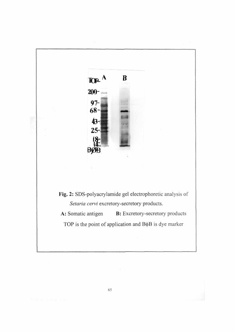
Fig. 2: SDS-polyacrylamide gel electrophoretic analysis of
Setaria cervi excretory-secretory products.
A: Somatic antigen B: Excretory-secretory products
TOP is the point of application and B(j)B is dye marker
65
adult worms The protein pattern of the S. cervi E-S antigen was
analysed on SDS-polyacrylamide gel electrophoresis (SDS-PAGE)
along with the S. cervi adult somatic antigens. Out of the different
concentrations of the gel tried, 10% of the polyacrylamide gave the
best resolution. The SDS-PAGE pattern of the E-S antigen is shown in
Fig. 2. The S. cervi E-S product showed the presence of 14-15
protein bands, whereas, the adult somatic antigen showed 35-40
protein bands. Standard molecular weight markers were used in order
to determine the molecular weights. The protein bands of the E-S
product lie in the molecular weight range of 10-200 kD with major
protein bands in low molecular region (10-70 kD).
II. Immunization of rabbits with S. cervi E-S products and
quantitation of parasite specific antibodies immune sera
The immunization ofthe rabbits with 5. cem E-S products was
done in order to raise hyperimmune rabbit sera. Three rabbits (R-1,
R-2 and R-3) were immunized intramuscularly for a period of 6-7
months. The immune sera were collected from these rabbits after
each injection and tested in enzyme linked immunosorbent assay
(ELISA) for parasite specific antibodies. ELISA was standardised in
terms of optimum antigen concentration, antibody conjugate and
66
dilutions of antiserum. Different concentrations of S. cervi E-S
products (1, 0.5, 0.25, 0.125, 0.062 and 0.03 i g per well) were
tried using hyperimmune sera at a fixed dilution. The immune sera
from 3 rabbits gave comparable antigen titration curve as shown in Fig.
3. Out of the different antigen concentrations, 0.25 \i% concentration
of S. cervi E-S product was found to be optimum (Fig. 3). The
optimum antibody conjugate was previously standardised in the
laboratory by using different dilutions of antibody enzyme conjugate
(1:250,1:500,1:1000 and 1:2000). The 1:1000 dilution of antibody
conjugate was found to be optimum. The other assay conditions such
as the optimum orthophenylene diamine (OPD) and hydrogen
peroxide (H2O2) were also previously standardised in our laboratory
and found to be Img/ml OPD and l|il/ml H2O2.
The optimum antigen concentration (0.25 j^g/well) and 1:1000
dilution of antibody conjugate were used further in all the
experiments for determining the antibody titres of the three immune
rabbit sera. The immune rabbit sera from the three rabbits (immunised
against ScES), collected after different injections (starting from Iir**
injection to the last injection) were tested in ELISA at different
dilutions. Though, the antibody titre was found to be increasing upto
the 8* injection in the sera of rabbits 1 and 3 and after that it became
constant, while the antibody titre of immune serum from rabbit 2
67
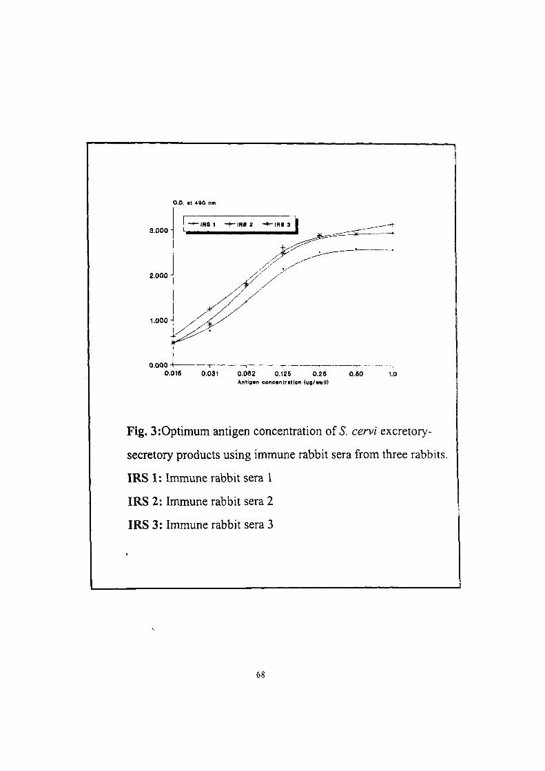
O.D. t t 490 nm
3.000
2,000 -
1.000
0.000 0.016 0.031 0.062 0.126 0.26
Antlgin concantratlon (up/mil) 0.60 1.0
Fig. 3:0ptimum antigen concentration of 5. cervi excretory-
secretory products using immune rabbit sera from three rabbits.
IRS 1: Immune rabbit sera I
IRS 2: Immune rabbit sera 2
IRS 3: Immune rabbit sera 3
68
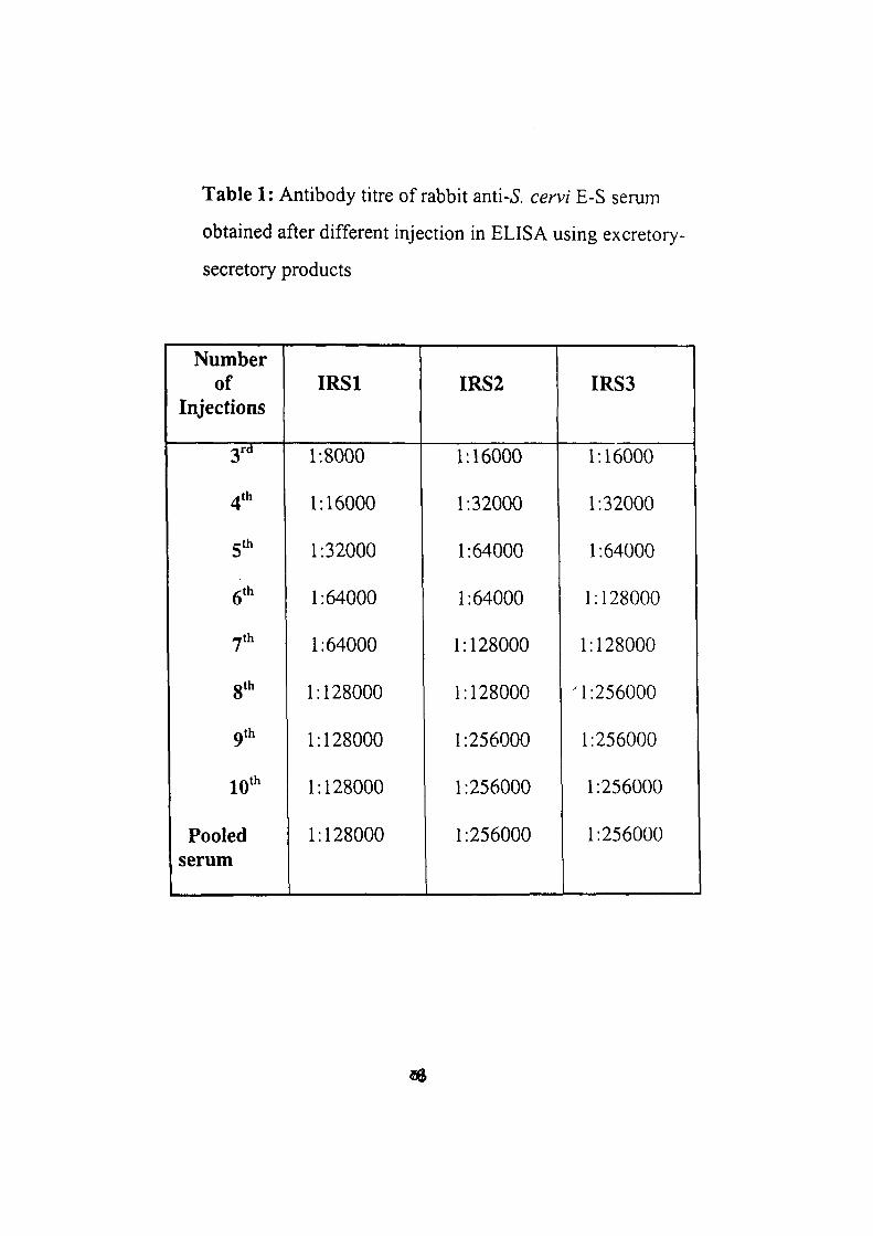
Table 1: Antibody titre of rabbit anti-5. cervi E-S serum
obtained after different injection in ELISA using excretory-
secretory products
Number of
Injections
3rd
4th
S'''
6'*'
/yth
gth
9th
10"'
Pooled serum
IRSl
1:8000
1:16000
1:32000
1:64000
1:64000
1:128000
1:128000
1:128000
1:128000
IRS2
1:16000
1:32000
1:64000
1:64000
1:128000
1:128000
1:256000
1:256000
1:256000
IRS3
1:16000
1:32000
1:64000
1:128000
1:128000
M:256000
1:256000
1:256000
1:256000
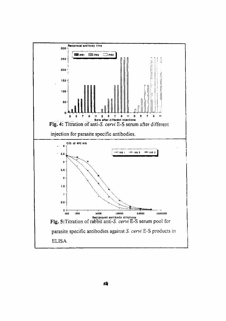
300-
260
200
150
100-
60
Reciprocal antibody titrs
linsi IRS2 CZDiRsa
1 i| " i " I " i " I
n n
- f4 3 6 7 9 11 3 5 7 9 11 3 6 7 9 11
8«ra after different injectlona
Fig. 4: Titration of anti-5. cervi E-S serum after different
injection for parasite specific antibodies.
O.D. at 490 nm
600 1000 l«000 138000 S13000 10000000 Reciprocal antibody dlltitloni
Fig. 5:Titration of rabbit anti-^. cem E-S serum pool for
parasite specific antibodies against S. cervi E-S products in
ELISA
showed increasing trends upto 9* injection. The immune rabbit serum
1 (IRS 1) showed lower antibody titres as compared to IRS 2 and IRS
3 (Fig. 4). The titres of parasite specific antibodies in sera of three
rabbits collected after different injections are given in Table 1.
The immune rabbit sera showing the same titre values were
pooled separetly for each rabbit. The pooled rabbit anti-ScE-S serum
from rabbit 1 (IRS 1) showed reciprocal antibody titre of 128 000
whereas, reciprocal antibody titre of 256 000 as obtained for sera
from rabbit 2 (IRS 2) and rabbit 3 (IRS 3). On titrating the immun
rabbit serum pools, parallel titration curves were obtained for the
three rabbits, however, IRS 1 gave lower ELISA values (Fig. 5).
II. IMMUNOCHEMICAL CHARACTERIZATION OF S.
CERVIE-S PRODUCTS
i. Crossed immunoelectrophoretic analysis ©f S..cervi E-S
products
The antigenic analysis of the S. cervi excretory-secretory products
were done by immunochemical techniques such as crossed
10
immunoelectrophoresis using hyperimmune rabbit anti-E-S sera. The
S. cervi E-S products were analysed by two dimensional gel
electrophoresis or crossed immunoelectrophoresis (CIE), in order to
get a better resolution of the antigenic components. Different
concentrations of the rabbit anti-E-S serum (2.5%, 5% and 10%) were
tried in CIE. The number of precipitin peaks remained same in all the
three concentrations but greater resolution ofthe peaks were found in
10% serum concentration in the sera from rabbit 1 while the sera from
rabbits 2 and 3 gave optimum results with 5% serum concentration. The
hyperimmune rabbit anti-ScES serum pools was also used to analyse
the S. cervi E-S products in CIE and gave 10 precipitin peaks with
immune rabbit serum pool 1 (R-1). However, 11-12 precipitin peaks
were seen with immune serum pool 2 (IRS 2) and 3 (IRS 3)
respectively (Fig. 6).
The rabbit anti-ScE-S serum was evaluated for the detection of
circulating antigen in filarial patients' sera using counter current
immunoelectrophoresis (CIEP). The rabbit anti-ScE-S serum detected
the circulating filarial antigen in patient serum pool (pools of the
microfilareamic positive patient sera), but also showed non-specific
reaction with normal human serum (Fig. 7). This may be due to the
presence of host serum proteins in the S. cervi E-S products. Therefore,
72
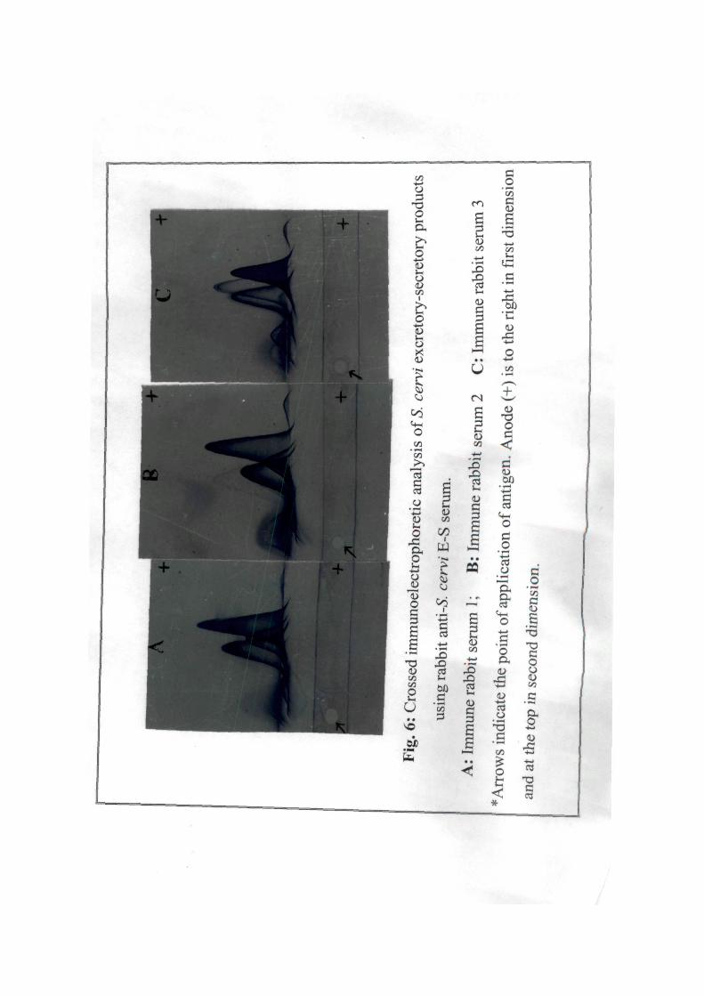
X
o CO
on
c 03 O
-* ;w O
O
y
o c 3
T3 1/3
O
u • •
I
E .ti
;3
m
s 2 (U
X)
c £
Cfi
5 S
X ) X i
IS c S S
C/3
0
a, C 0 -*—»
0 <u (/I
i 0
•4-<
0
e 2 00
IS X> cd l-H
g S
£ "B
•4—»
05
^3 l-J
' ^^ < * - ^
u.
XI
• • • < - >
O
e 2
w ^ .2
op
o
o 03 O
o
•X • « — t
c .2 c
2 ^ ^ T3 C O
00
03 0
t 3 C ' ' CD
0
h < «
e 0
4 0
OJ x: - K J
T3
§
/
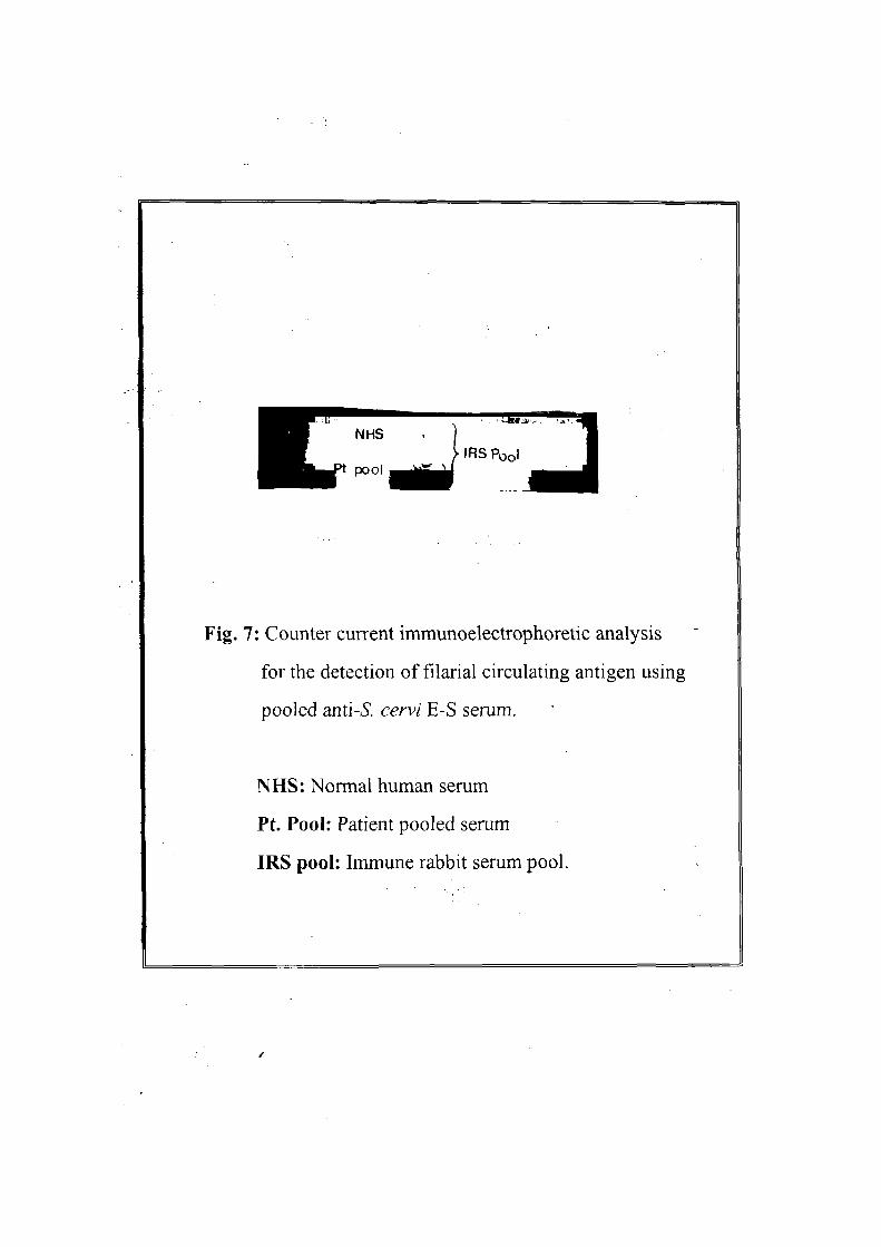
ENHS . 1 WL^-r' . ^A
IRS Pool
Fig. 7: Counter current immunoelectrophoretic analysis
for the detection of filarial circulating antigen using
pooled anti-iS". cervi E-S serum.
NHS: Normal human serum
Pt. Pool: Patient pooled serum
IRS pool: Immune rabbit serum pool.
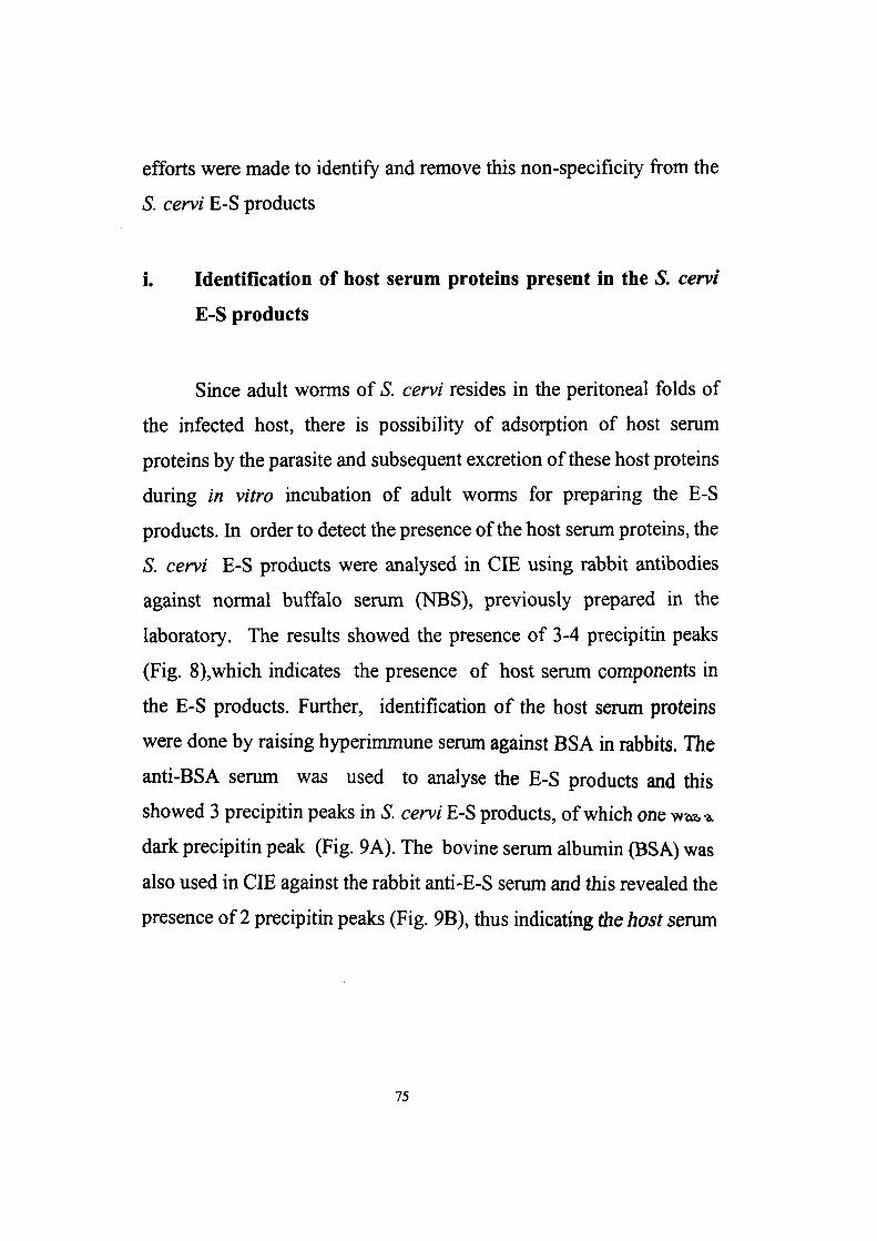
efforts were made to identify and remove this non-specificity from the
S. cervi E-S products
i. Identification of host serum proteins present in the S. cervi
E-S products
Since adult worms of S. cervi resides in the peritoneal folds of
the infected host, there is possibility of adsorption of host serum
proteins by the parasite and subsequent excretion of these host proteins
during in vitro incubation of adult worms for preparing the E-S
products. In order to detect the presence of the host serum proteins, the
S. cervi E-S products were analysed in CIE using rabbit antibodies
against normal buffalo serum (NBS), previously prepared in the
laboratory. The results showed the presence of 3-4 precipitin peaks
(Fig. 8),which indicates the presence of host serum components in
the E-S products. Further, identification of the host serum proteins
were done by raising hyperimmune serum against BSA in rabbits. The
anti-BSA serum was used to analyse the E-S products and this
showed 3 precipitin peaks in S. cervi E-S products, of which one^at,^
dark precipitin peak (Fig. 9A). The bovine serum albumin (BSA) was
also used in CIE against the rabbit anti-E-S serum and this revealed the
presence of 2 precipitin peaks (Fig. 9B), thus indicating the host serum
75
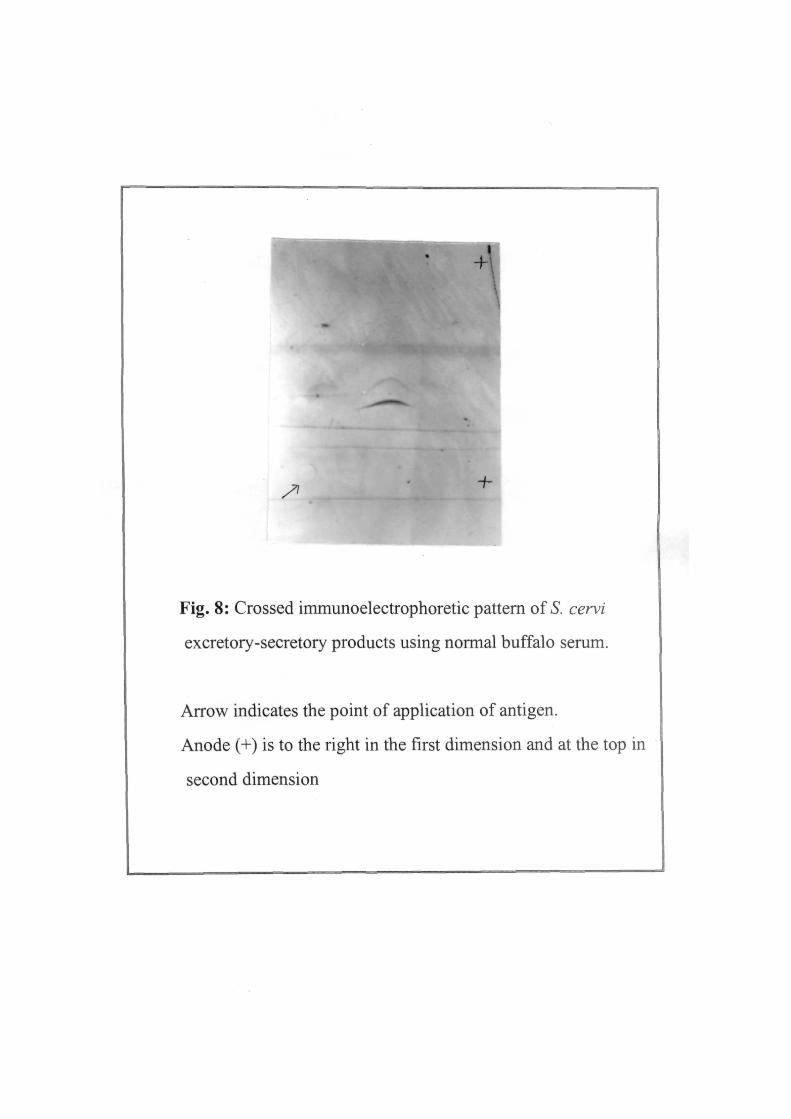
7^ -h
Fig. 8: Crossed immunoelectrophoretic pattern of 5. cervi
excretory-secretory products using normal buffalo serum.
Arrow indicates the point of application of antigen.
Anode (+) is to the right in the first dimension and at the top in
second dimension
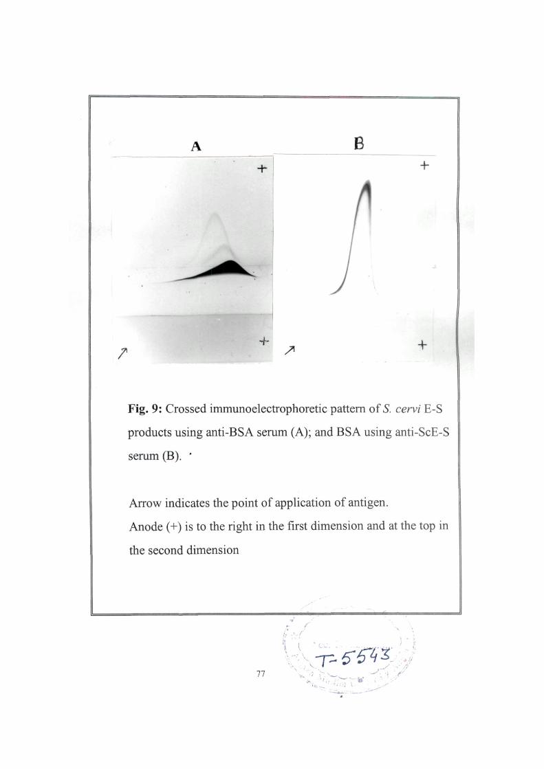
B
^ +
Fig. 9: Crossed immunoelectrophoretic pattern of S. cervi E-S
products using anti-BSA serum (A); and BSA using anti-ScE-S
serum (B). *
Arrow indicates the point of application of antigen.
Anode (+) is to the right in the first dimension and at the top in
the second dimension
.7 r
11
5-r42: '•A
-rfy
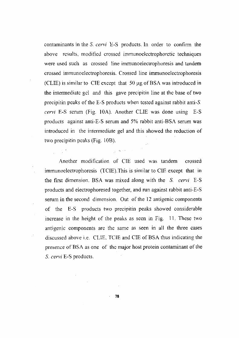
contaminants in the S. cervi B-S products. In order to confirm the
above results, modified crossed immunoelectrophoretic techniques
were used such as crossed hne immunoelectrophoresis and tandem
crossed immunoelectrophoresis. Crossed line immunoelectrophoresis
(CLIE) is similar to CIE except that 50 \ig of BSA was introduced in
the intermediate gel and this gave precipitin line at the base of two
precipitin peaks of the E-S products when tested against rabbit anti-S.
cervi E-S serum (Fig. 10A). Another CLIE was done using E-S
products against anti-E-S serum and 5% rabbit anti-BSA serum was
introduced in the intermediate gel and this showed the reduction of
two precipitin peaks (Fig. 1 OB).
Another modification of CIE used was tandem crossed
immunoelectrophoresis (TCIE).This is similar to CIE except that in
the first dimension. BSA was mixed along with the S. cervi E-S
products and electrophoresed together, and run against rabbit anti-E-S
serum in the second dimension. Out of the 12 antigenic components
of the E-S products two precipitin peaks showed considerable
increase in the height of the peaks as seen in Fig. 11. These two
antigenic components are the same as seen in all the three cases
discussed above i.e. CLIE, TCIE and CIE of BSA thus indicating the
presence of BSA as one of the major host protein contaminant of the
S. cervi E-S products.
78
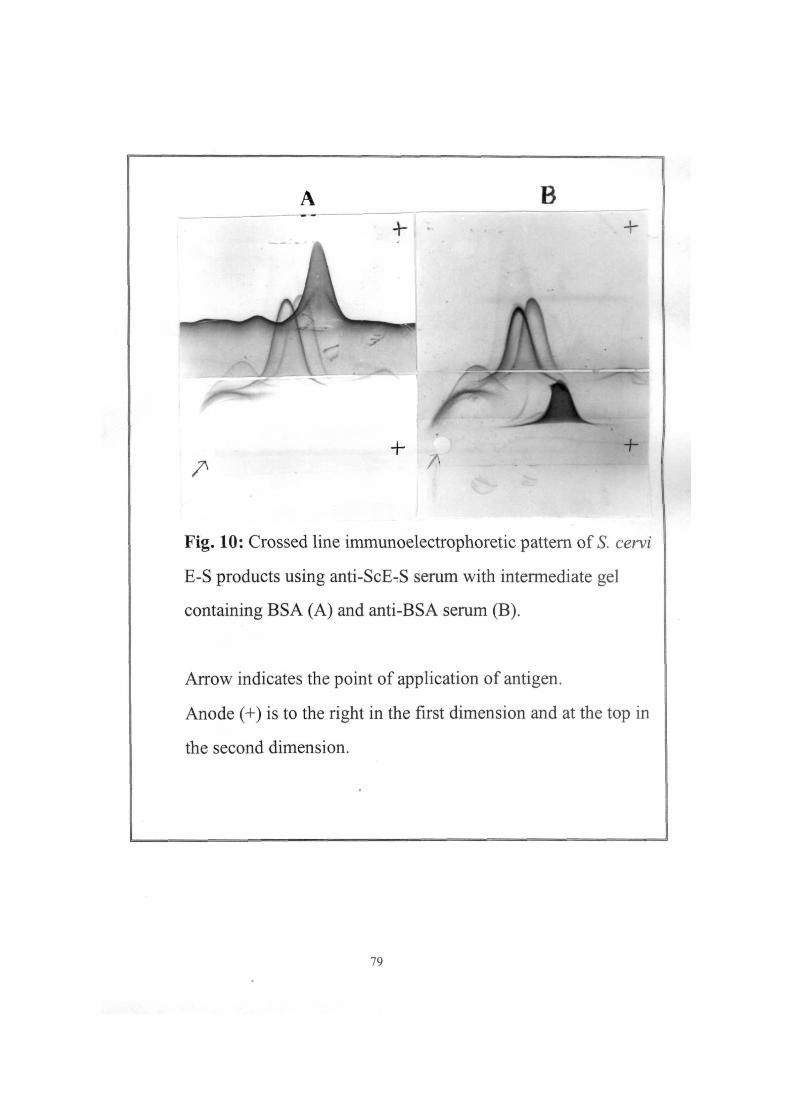
B -h
A r
-\-
/ ^ /^
-f-
Fig. 10: Crossed line immunoelectrophoretic pattern of 5. cervi
E-S products using anti-ScE-S serum with intermediate gel
containing BSA (A) and anti-BSA serum (B).
Arrow indicates the point of application of antigen.
Anode (+) is to the right in the first dimension and at the top in
the second dimension.
79
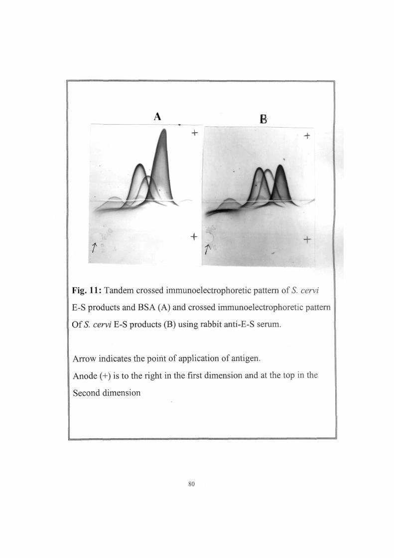
B 4- +
.M t
+ T
+
Fig. 11: Tandem crossed immunoelectrophoretic pattern of S. cervi
E-S products and BSA (A) and crossed immunoelectrophoretic pattern
Of 5. cervi E-S products (B) using rabbit anti-E-S serum.
Arrow indicates the point of application of antigen.
Anode (+) is to the right in the first dimension and at the top in the
Second dimension
80
iii. Removal of host serum proteins
t
An effort was made to remove the host serum components major
BSA present in the S. cervi E-S products, by using anti-BSA sepharose
sorbent. The preabsorbed ScE-S products were analysed for the
protein pattern in SDS-PAGE and this showed the removal of a major
protein band of 66 kD present in the S. cervi E-S products (data not
shown), which corresponds to the bovine albumin band of the
molecular weight markers.
Further, the preabsorbed ScE-S products were analysed in CIE
using anti-E-S serum. Two major antigenic peaks (detected as host
serum albumin earlier in Fig. 11) were found missing in the
preabsorbed E-S products. The preabsorbed ScE-S products were
found to contain 10 precipitin peaks, whereas, in the unabsorbed 12
precipitin peaks were present (Fig. 12 and Table 2).
The parasite specific antibodies in the anti-E-S serum was
quantitated by ELISA using preabsorbed S. cervi E-S products.
The preabsorbed E-S products gave a parallel curve with the
unabsorbed ScE-S products (Fig. 13), but the reciprocal antibody
titre of the absorbed ScE-S product was found to be 120 000 while the
81
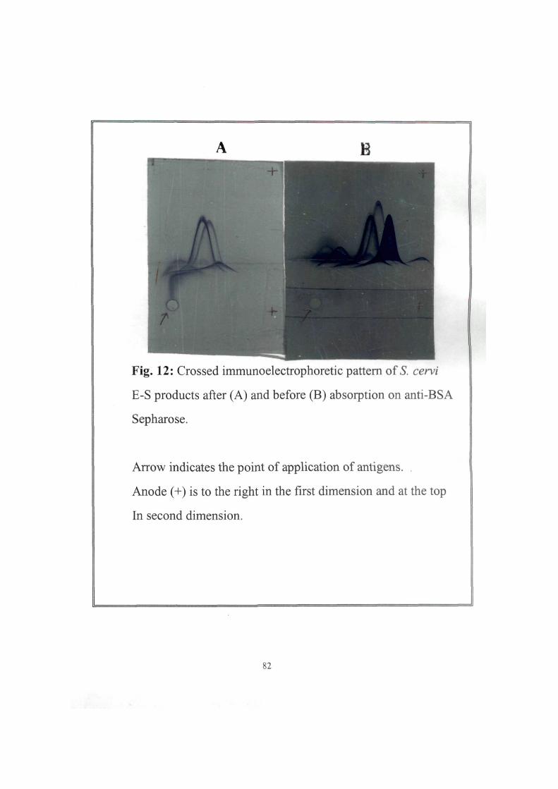
Fig. 12: Crossed immunoelectrophoretic pattern of S. cervi
E-S products after (A) and before (B) absorption on anti-BSA
Sepharose.
Arrow indicates the point of application of antigens. .
Anode (+) is to the right in the first dimension and at the top
In second dimension.
82
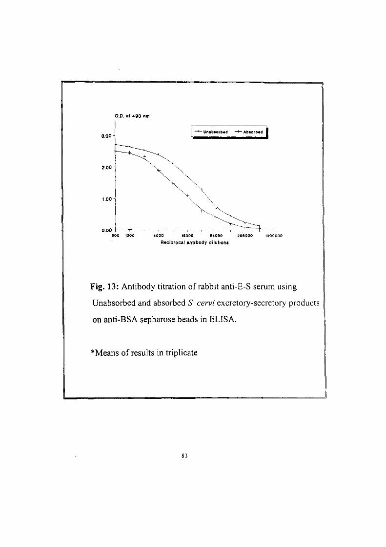
O.D. at 490 nm
3.00
2.00
1.00
0.00
' Unabtorbtd ~f~ Abtorbtd
SOO 1000
Reciprocal antibody dilutions
Fig. 13: Antibody titration of rabbit anti-E-S serum using
Unabsorbed and absorbed S. cervi excretory-secretory products
on anti-BSA sepharose beads in ELISA.
*Means of results in triplicate
83
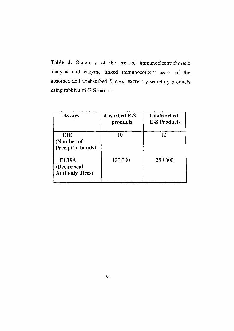
Table 2: Summary of the crossed immunoelectrophoretic
analysis and enzyme linked immunosorbent assay of the
absorbed and unabsorbed S. cervi excretory-secretory products
using rabbit anti-E-S serum.
Assays
CIE (Number of Precipitin bands)
ELISA (Reciprocal Antibody titres)
Absorbed E-S products
10
120 000
Unabsorbed E-S Products
12
250 000
84
iinabsorbed ScE-S products gave a reciprocal antibody titre of 256 000
summarised in Table 2.
Similarly, the anti-S. cervi E-S antibodies were absorbed onto
BSA-Sepharose in order to remove the antibodies against the host
serum components. The analysis of the preabsorbed anti-E-S serum in
CIE using ScE-S antigen showed the presence of 10 precipitin peaks,
(using 5% concentration in both absorbed and unabsorbed anti-E-S
serum) as compared to 12 precipitin peaks in the unabsorbed ScE-S
products (Fig. 14). Further quantitation of the preabsorbed anti-E-S
products was done by ELISA using S. cervi E-S products. Both the
sera gave parallel curves and the antibody titre of the absorbed anti-E-S
serum was found to be 1:120 000 and that of unabsorbed E-S products
gave antibody titre of 1:250 000 (Fig. 15).
iv. Potential of anti-E-S serum for detection of circulating
antigen in filarial patient sera using CIEP
The preabsorbed anti-E-S serum was used to detect the
circulating filarial antigen in the patient pooled serum (pools of the
microfilareamic positive patients' sera) by using CIEP. The
preabsorbed anti-E-S serum gave no cross-reactivity with the normal
human serum, whereas, successfully detected the circulating antigen
85
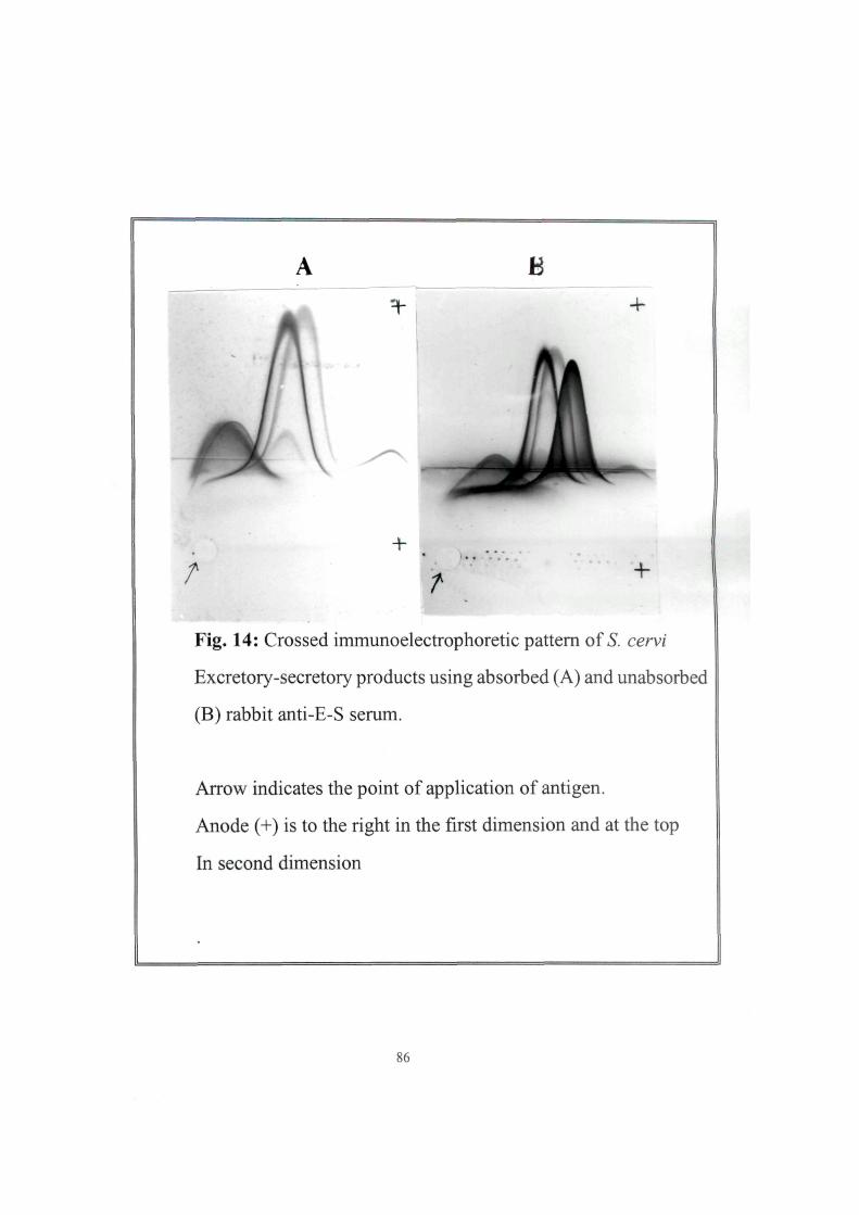
B
/ f ^-
Fig. 14: Crossed immunoelectrophoretic pattern of 5. cervi
Excretory-secretory products using absorbed (A) and unabsorbed
(B) rabbit anti-E-S serum.
Arrow indicates the point of application of antigen.
Anode (+) is to the right in the first dimension and at the top
In second dimension
86
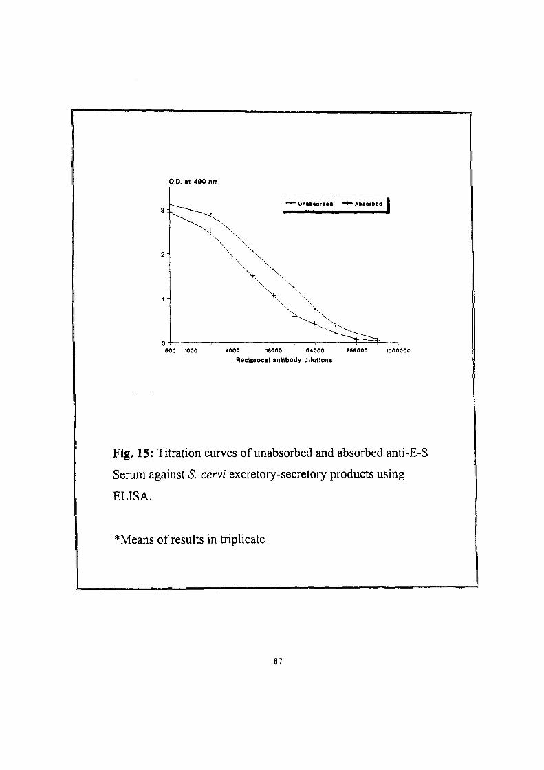
• B ^ H ^ ^ B ^
0.0. at 490 nm
600 1000 4000 16000 64000 296000 1000000
Reciprocal antibody dilutions
Fig. 15: Titration curves of unabsorbed and absorbed anti-E-S
Serum against .S. cervi excretory-secretory products using
ELISA.
•Means of results in triplicate
87
in the filarial patient pooled sera (Fig. 16). On testing small number
of individual human patient sera, the preabsorbed immune rabbit
anti-i". cervi E-S serum could detect the circulating filarial antigen in
all the 15 microfilarial positive patient sera but not in the 10 non-
endemic control human sera and also did not show any cross-reactivity
with any non-filarial (ascaris, malaria, amoebae) patients' sera (Table
3).
III. FRACTIONATION OF S. CER VI E-S PRODUCTS
The S. cervi E-S products were fractionated by ion-exchange
chromatographic technique using DEAE-Sephacel column
(Pharmacia). The elution was done by chloride gradient using different
molarity of sodium chloride (NaCl) to elute the antigenic components
of the S. cervi E-S products i.e. 100 mM, 250mM and 500mM NaCl.
The protein started eluting with 250mM sodium chloride gradient and
also continued with 500mM chloride gradient. Three ml of the
firactions were collected. Each fi-action was measured in an
spectrophotometer at 280 nm CD for the presence of proteins and this
showed the presence of 3 distinct peaks showed in Fig. 17. These
column fractions were further analysed in ELISA using rabbit anti-E-S
serum and rabbit anti-BSA serum. All fractions showed high reactivity
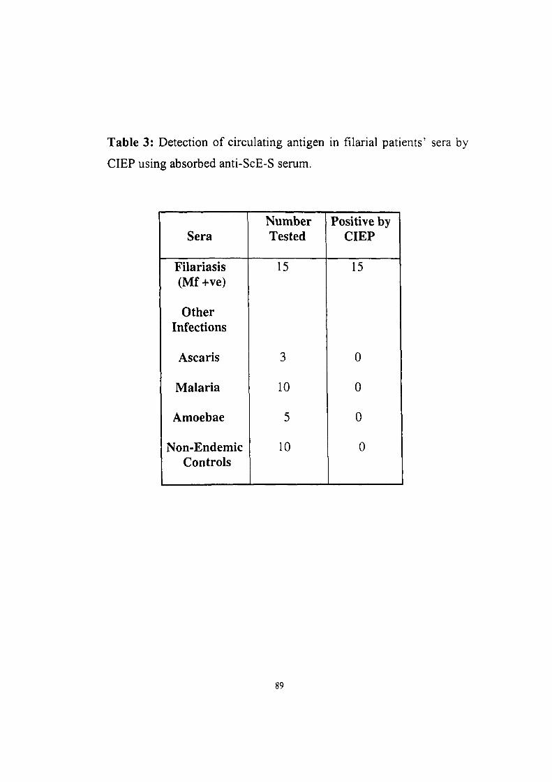
Table 3: Detection of circulating antigen in filarial patients' sera by
CIEP using absorbed anti-ScE-S serum.
Sera
Filariasis (Mf+ve)
Other Infections
Ascaris
Malaria
Amoebae
Non-Endemic Controls
Number Tested
15
3
10
5
10
Positive by CIEP
15
0
0
0
0
89
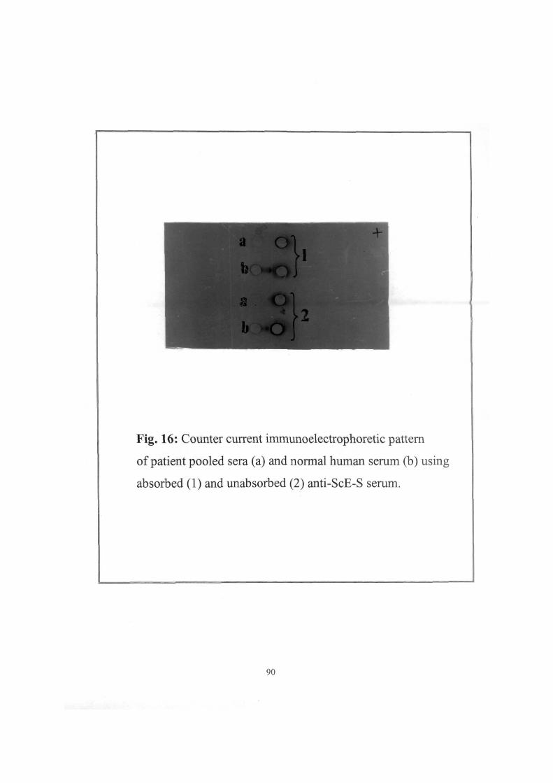
^^^^^^^^^|^3X?^^^^K«« .. .'^^^^^^B^^^^^^^l
^ ^ 1 a . on 1
Fig. 16: Counter current immunoelectrophoretic pattern
of patient pooled sera (a) and normal human serum (b) using
absorbed (1) and unabsorbed (2) anti-ScE-S serum.
90
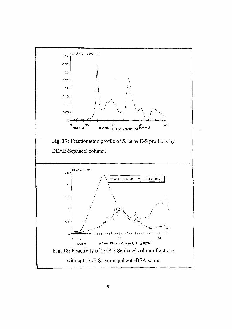
(O.D.) at 280 nm
3 30 75 120 204 100 mM 250 mM Elution Volume (mfl OO "" ^
Fig. 17: Fractionation profile of .S. cervi E-S products by
DEAE-Sephacel column.
OD at 490 nn
Ant i -z-S serum - + - A n l i - B S A ser t
lOOmM aeOmM Elution VoljjmeJmO. SPQmM
Fig. 18: Reactivity of DEAE-Sephacel column fractions
with anti-ScE-S serum and anti-BSA serum.
91
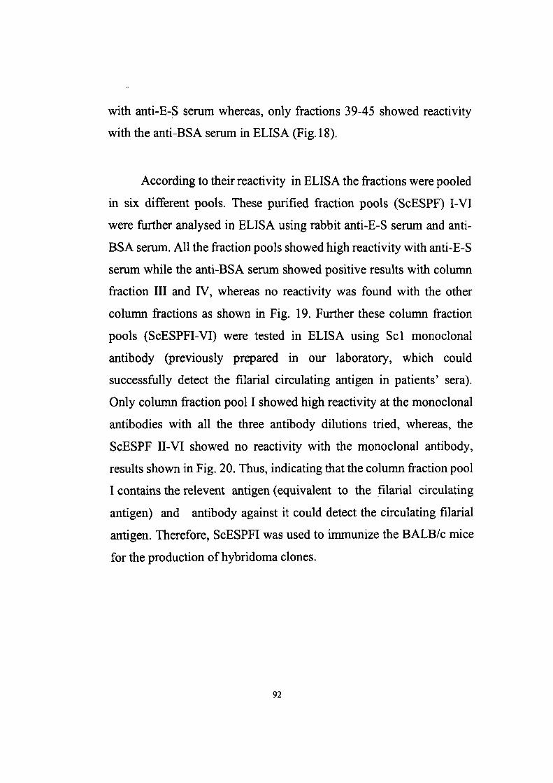
with anti-E-S serum whereas, only fractions 39-45 showed reactivity
with the anti-BSA serum in ELISA (Fig. 18).
According to their reactivity in ELISA the fractions were pooled
in six different pools. These purified fraction pools (ScESPF) I-VI
were further analysed in ELISA using rabbit anti-E-S serum and anti-
BSA serum. All the fraction pools showed high reactivity with anti-E-S
serum while the anti-BSA serum showed positive results with column
fraction III and IV, whereas no reactivity was found with the other
column fractions as shown in Fig. 19. Further these column fraction
pools (ScESPFI-VI) were tested in ELISA using Scl monoclonal
antibody (previously prepared in our laboratory, which could
successfully detect the filarial circulating antigen in patients' sera).
Only column fraction pool I showed high reactivity at the monoclonal
antibodies with all the three antibody dilutions tried, whereas, the
ScESPF II-VI showed no reactivity with the monoclonal antibody,
results shown in Fig. 20. Thus, indicating that the column fraction pool
I contains the relevent antigen (equivalent to the filarial circulating
antigen) and antibody against it could detect the circulating filarial
antigen. Therefore, ScESPFI was used to immunize the BALB/c mice
for the production of hybridoma clones.
92
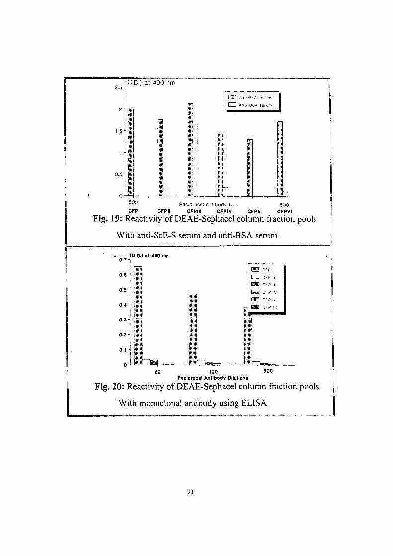
2 . 5 -
2 -
1,5
{0,D.; at 490 nm
0.5
-h
iiiiiU Ant'-E-S serum
I I Anli-BSA serurn
mn
-+-^'^'-' Reciprocal antlDoay titre 500 CFPI CFPII CFPIII CFPIV CFPV CFPVI
Fig. 19: Reactivity of DEAE-Sephacel column fraction pools
With anti-ScE-S serum and anti-BSA serum.
0.7
o.e
0.6
0.4
0.3
0.2
o.^-{
0
(OJ}.) at 490 nm
ms c-p I cm c-p II ^ C ^ P III
^ S C"P V
B9i c-p VI
^*y^-^
60 100 600 Reciprocal Antibody DMutlons
Fig. 20: Reactivity of DEAE-Sephacel column fraction pools
With monoclonal antibody using ELISA
93
IV. PRODUCTION OF MONOCLONAL ANTIBODIES
AGAINST PURIFIED E-S PRODUCTS
Five BALB/c mice were immunized subcutaneously with 20 )j,g
of ScES purified antigen fraction pool I (ScESFPI) emulsified in
Freund's complete adjuvant (FCA). The immune mice sera were
collected and analysed in ELISA and CIEP against ScESPFI The
immune mice sera of mice 1, 2 and 5 showed high antibody titre in the
range of 1:3200-1:6400 while immune sera of mice 3 and 4 showed
lower titre of 1:800-1:1600 (Table 4). Further, these immune mice sera
were also analysed in CIEP to detect the circulating antigen in filarial
patient sera. The immune sera of mice 1, 2 and 5 could successfully
detect the circulating antigen in patients' sera while immune mice sera
3 and 4 gave negative results also shown in Table 5. Out of the three
mice, having sera positive for circulating antigen, two were selected for
frision with Sp2/0 myeloma cell for hybridoma production.
Two mice chosen for fusion were boosted with 10 fig of
ScESFPI, by intervenous injection 4 days before fusion. The spleen
cells from the immune animals were fused with Sp2/0 myeloma cells
in polyethylene glycol and the cell suspension was plated in 24 well
culture plates and grown in the presence of a selective hypoxanthine-
aminopterin-thymidine (HAT). After, HAT selection, the hybridoma
94
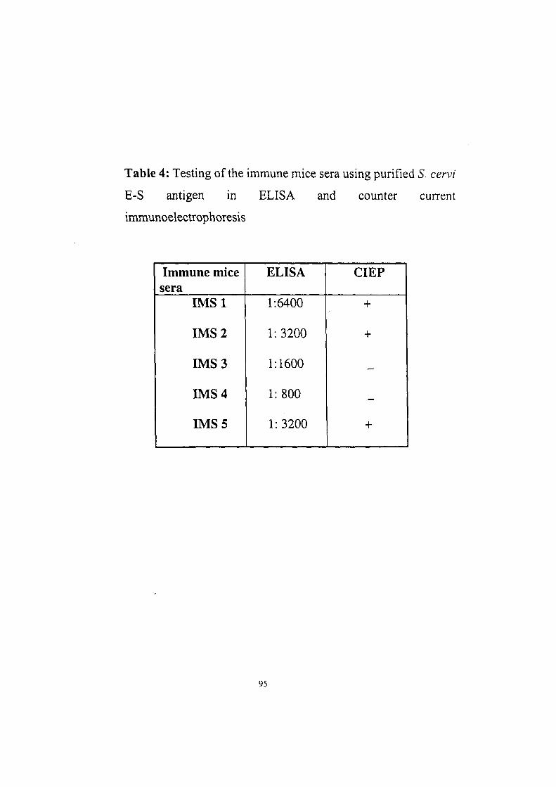
Table 4: Testing of the immune mice sera using purified 5. cervi
E-S antigen in ELISA and counter current
immunoelectrophoresis
Immune mice sera
IMSl
IMS 2
IMS 3
IMS 4
IMS 5
ELISA
1:6400
1:3200
1:1600
1:800
1:3200
CIEP
+
+
—
—
+
95
were selected on the basis of their reactivity with both bovine (S. cervi)
and human {B. malayi and W. bancrofti) filarial parasite antigens in
ELISA. The hybrids showing high reactivity with both the antigens
were selected and cloned twice by limiting dilution. The wells were
examined microscopically and out of 264 wells plated 251 wells were
found positive for the hybrid growth with a fusion efficiency of about
95%. The culture supematants from these wells were tested in ELISA
for the anti-filarial antibody against ScES and ScESPFI, B. malayi
antigen and W. bancrofti microfilarial antigen and 69 hybrids (27%)
were found to have anti-filarial antibody. Out of these 69 hybridomas,
27 showed reactivity with the purified E-S antigen. The reactivity of
the supematants were also tested in ELISA against {B. malayi and W.
bancrofti) filarial parasite antigens. The supernatant from 36
hybridomas showed reactivity with B. malayi adult antigen and the
supematants from 21 hybridoma clones were positive against W.
bancrofti microfilarial antigen. The reactivities of these hybridoma
clones were also tested with non-filarial antigens in ELISA and 12
hybridoma clones were found to be specific to filaria and did not show
cross-reactivity with non-filarial antigens (data not shown). The
monoclonal antibodies produced by four hybridoma clones (designated
as FES 2, FES 7, FES 9 and FES 11), showing high and consistent
reactivities with both bovine {S. cervi E-S and ScESPFI) and human
{B. malayi and W. bancrofti) filarial antigens were selected for further
studies. All the four monoclonal antibodies showed high reactivity with
96
the filarial circulating antigen present in filarial patient serum pool. The
reactivities of these 12 hybridoma clones with the filarial antigens (both
human, B. malayi and W. bancrofti, and bovine, S. cervi E-S and
purified antigen) are shown in Table 5. All the hybridoma clones
showed significantly high reactivity with the filarial antigens.
i. Characterization of the monoclonal antibodies
The isotyping analysis of the four monoclonal antibodies was
performed using subclass specific reagents. The monoclonals FES 7
and FES 9 were found to be IgG] antibody whereas, monoclonal
antibody FES 2 belongs to IgG2b and FES 11 belongs to IgM isotype.
Both positive and negative controls were used in each experiment. The
Scl, a monoclonal antibody produced earlier in our laboratory
(Kaushal et al, 1995) is an IgGi isotype was used as a positive control,
while a malarial monoclonal antibody (DNKIO), kindly supplied by
Dr. D. C. Kaushal, Division of Microbiology, Central Drug Research
Institute, Lucknow (India) was used as negative control (Table 6).
97
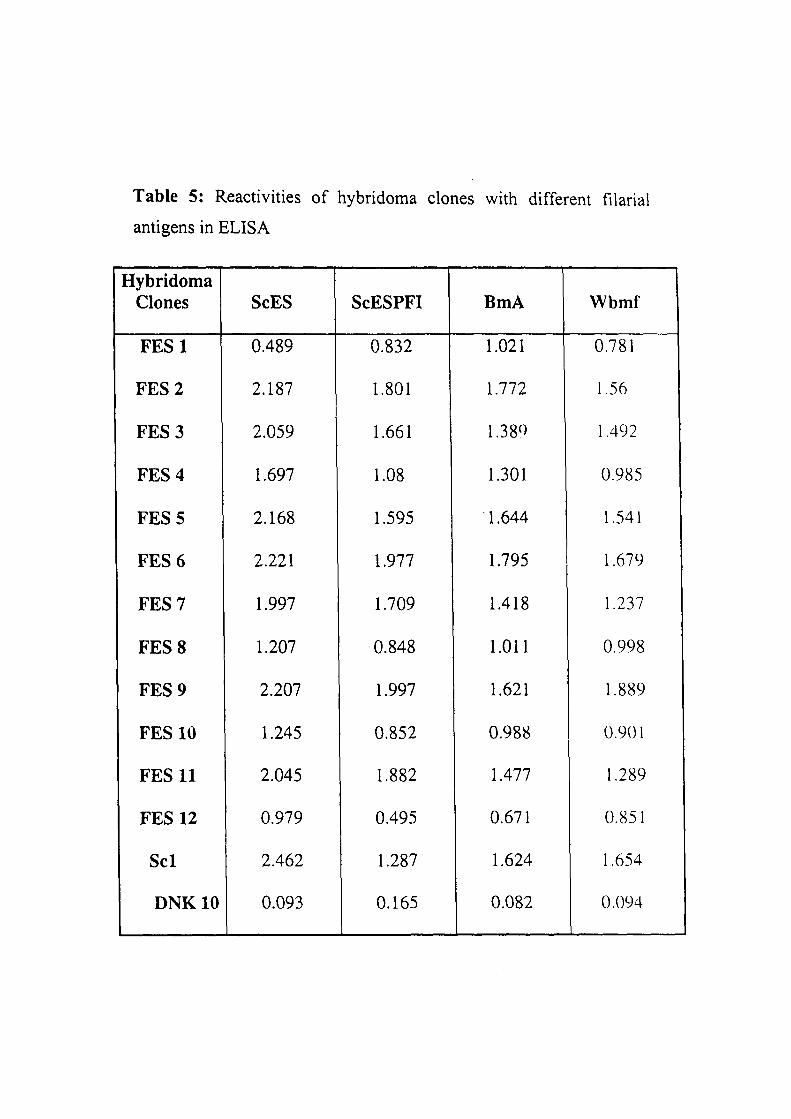
Table 5: Reactivities of hybridoma clones with different filarial
antigens in ELISA
Hybridoma Clones
FESl
FES 2
FES 3
FES 4
FES 5
FES 6
FES 7
FES 8
FES 9
FES 10
FES 11
FES 12
Scl
DNKIO
ScES
0.489
2.187
2.059
1.697
2.168
2.221
1.997
1.207
2.207
1.245
2.045
0.979
2.462
0.093
ScESPFI
0.832
1.801
1.661
1.08
1.595
1.977
1.709
0.848
1.997
0.852
1.882
0.495
1.287
0.165
BmA
1.021
1.772
1.380
1.301
1.644
1.795
1.418
1.011
1.621
0.988
1.477
0.671
1.624
0.082
Wbmf
0.781
1.56
1.492
0.985
1.541
1.679
1.237
0.998
1.889
0.901
1.289
0.851
1.654
0.094
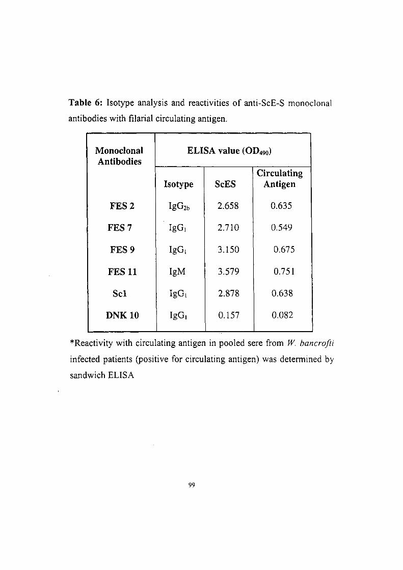
Table 6: Isotype analysis and reactivities of anti-ScE-S monoclonal
antibodies with filarial circulating antigen.
Monoclonal Antibodies
FES 2
FES 7
FES 9
FES 11
Scl
DNKIO
ELISA value (OD490)
Isotype
IgG2b
IgG,
IgG,
IgM
IgG,
IgG,
ScES
2.658
2.710
3.150
3.579
2.878
0.157
Circulating Antigen
0.635
0.549
0.675
0.751
0.638
0.082
*Reactivity with circulating antigen in pooled sere from W. bancrofti
infected patients (positive for circulating antigen) was determined by
sandwich ELISA
99
ii. Reactivity of monoclonal antibodies with different antigens
The specificity of the four monoclonal antibodies was examined
by testing their reactivity against a panel of non-filarial antigens such
as ascaris, hookworm, malaria, amoebae and bacteria. The filarial
antigens (5. malayi adult, W. bancrofti microfilariae, S. cervi adult,
microfilarial, excretory-secretory products and purified antigens) were
also used for comparison. The positive control used was S. cervi E-S
antigens and the negative control was normal human serum (NHS)
from the endemic area. All the four monoclonal antibodies (FES 2,
FES 7, FES 9 and FES 11) exhibited high reactivity with all the filarial
antigens {B. malayi, W. bancrofti, S. cervi adult, mf and purified
antigen fraction). The FES 9 showed highest reactivity with all the
filarial antigens (Table 7). With regards to the reactivities of four
monoclonal antibodies with non-filarial antigens, none of the
monoclonal showed any significant reactivity with any of the non-
filarial antigens (Table 8). These finding suggest the filaria specificity
all the four monoclonal antibodies.
iii. Analysis of the monoclonal antibodies in inhibition ELISA
The inhibition ELISA was done using Scl-HRP conjugate. Out
of the four monoclonal antibodies, FES 7, FES 9 and FES 11 strongly
100
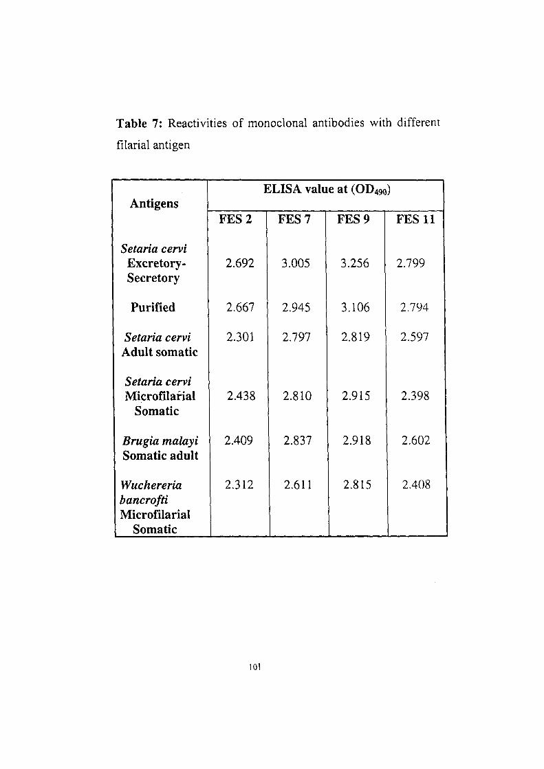
Table 7: Reactivities of monoclonal antibodies with different
filarial antigen
Antigens
Setaria cervi Excretory-Secretory
Purified
Setaria cervi Adult somatic
Setaria cervi Microfilaria!
Somatic
Brugia malayi Somatic adult
Wuchereria bancrofti Microfilaria!
Somatic
ELISA value at (OD490)
FES 2
2.692
2.667
2.301
2.438
2.409
2.312
FES 7
3.005
2.945
2.797
2.810
2.837
2.611
FES 9
3.256
3.106
2.819
2.915
2.918
2.815
FES 11
2.799
2.794
2.597
2.398
2.602
2.408
101
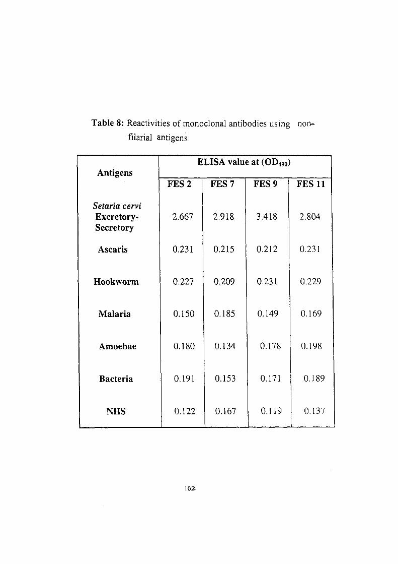
Table 8: Reactivities of monoclonal antibodies using non-
filarial antigens
Antigens
Setaria cervi Excretory-Secretory
Ascaris
Hookworm
Malaria
Amoebae
Bacteria
NHS
ELISA value at (OD490)
FES 2 FES 7 FES 9
2.667
0.231
0.150
0.180
0.191
0.122
2.918
0.215
0.227 0.209
0.134
0.153
0.167
3.418
0.212
0.231
0.185 0.149
0.178
0.171
0.119
FES 11
2.804
0.231
0.229
0.169
0.198
0.189
0.137
102
inhibited the binding of Scl-HRP to the antigen, while monoclonal
FES 2 did not exert any inhibitory effect on binding. These results
suggest that the three MoAbs (FES 7, FES 9 and FES 11) are directed
against the same epitope that of Scl monoclonal antibody and the
target epitope of FES 2 monoclonal appeared to be different. The
results are given in Table 9,
iv. Titration of monoclonal antibody-HRP conjugate in ELISA
In order to test these monoclonal antibodies in sandwich ELISA
for detection of circulating antigen, the monoclonal antibodies were
purified and conjugated to horse radish peroxidase. The monoclonal
antibodies FES 2, FES 7 and FES 9 were affinity purified using
protein-A Sepharose column while the FES 11 monoclonal antibody
was purified on Sephacryl S-200 column. The purified monoclonal
antibodies were conjugated with horse radish peroxidase and HRP
conjugates were tested at different dilutions against ScESPFI in
ELISA. Two fold dilutions starting from 1:250 to 1:8000 were used
and the titration curves are shown in Fig. 21. The two monoclonal
antibody conjugates (FES 7-HRP and FES 9-HRP) showed optimum
ELISA (OD) upto 1:1000 dilution while FES 2 gave optimum results
upto 1:500 dilution. The optimum dilution of FES 9-HRP conjugates
102?
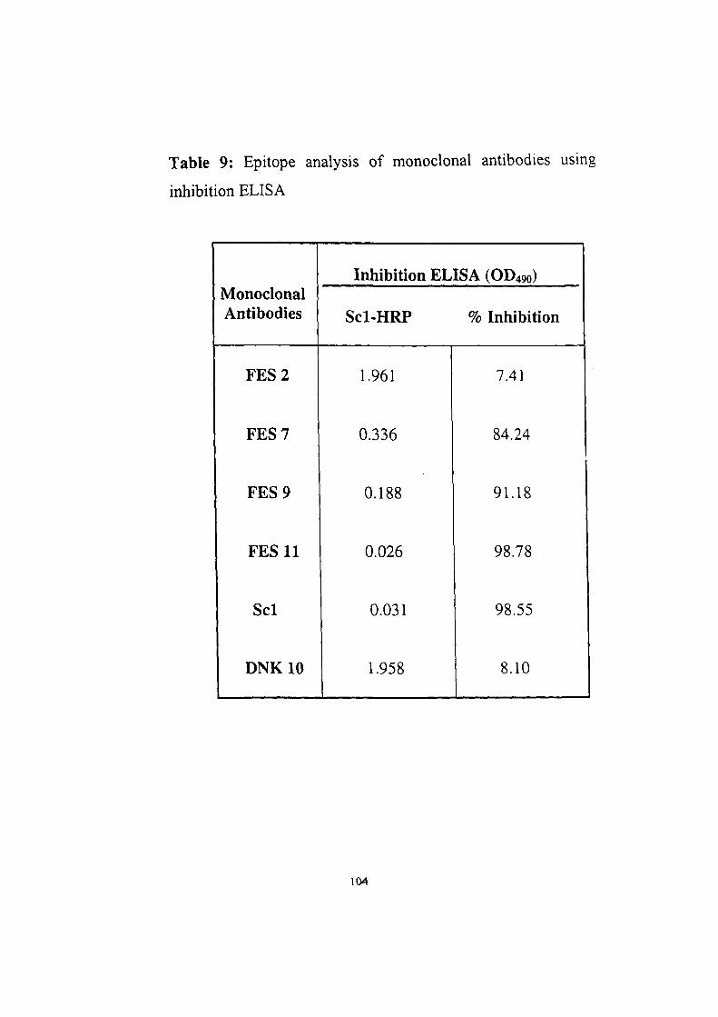
Table 9: Epitope analysis of monoclonal antibodies using
inhibition ELISA
Monoclonal Antibodies
Inhibition ELISA (OD490)
Scl-HRP % Inhibition
FES 2
FES 7
FES 9
FES 11
Scl
DNKIO
1.961
0.336
0.188
0.026
0.031
1.958
7.41
84.24
91.18
98.78
98.55
8.10
104
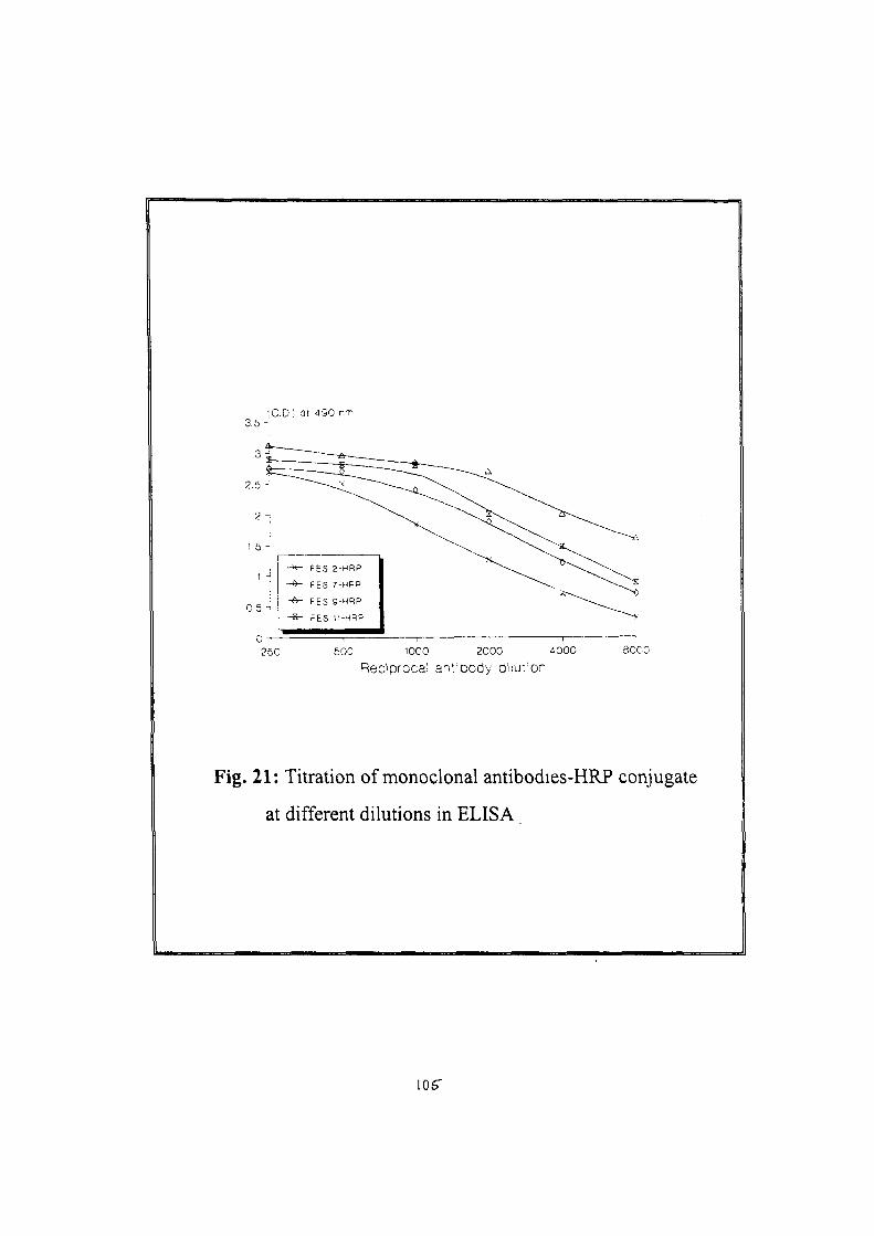
3.5
3
2.5
2
1.5
0 5 •
iO.D.; .at 4S0 nrf
- ^ -d-
- A -
-«-
FES 2
FES 7
FES S
FES 1
-HRP
HRP
-MRP
-HRP
250 500 1000 2000 4000
Reciprocal antibody diiu-ion BOO:
Fig. 21: Titration of monoclonal antibodies-HRP conjugate
at different dilutions in ELISA
105"
was found to be 1:2000. These HRP conjugates were used in sandwich
ELISA at respective dilutions.
V. DEVELOPMENT OF IMMUNODIAGNOSTIC TEST FOR
FILARIASIS USING ANTI-E-S ANTIBODIES
In order to evaluate the diagnostic potential of anti-ScE-S
monoclonal antibodies for the detection of filarial circulating antigen in
patients' sera, all the four monoclonal antibodies (FES 2, FES 7, FES 9
and FES 11) were used in sandwich ELISA in combination with Scl
monoclonal antibody (an IgGj isotype directed against a repetitive
protien epitope, detecting the filarial circulating antigen, previously
developed in our laboratory). The Scl monoclonal antibody was used
for the capturing of antigen. The sandwich ELISA was standardised In
terms of optimum concentration of antibodies to capture and reveal the
antigen. Out of different concentrations tried, 2 |ig/well concentration
of Scl monoclonal for capturing of antigen and 1:1000 dilution in case
of peroxidase conjugated FES 7, FES 9, FES 11 and !:500 dilution of
peroxidase' conjugate FES 2 for the revealing step showed better
results. In case of anti-ScE-S antibodies for the revealing step 1:2000
dilution was found to be optimum. Out of the four monoclonals the
combination of Scl-FES 9 showed higher sensitivity in detecting the
antigen as this could detect 2.5 ng of antigen, while other monoclonal
\oc
combination showed slightly lower sensitivity i.e detecting 5-10 ng of
antigen (Table 10). These monoclonal antibodies were further tried as
capturing antibody in combination with the anti-ScE-S polyclonal (for
revealing the antigen). The combination of FES 9-polyclonal resulted
in increasing the detection limit as it could detect 0.62 ng of antigen
(Table 11).
The sandwich ELISA employing the combination of FES 9
monoclonal-anti-ScE-S polyclonal was used for the detection of
antigen in filarial patient sera. The sera collected from the
microfilareamic positive filarial patient along with the endemic and
non-endemic controls and also from patient having non-filarial
infections were used in sandwich ELISA.The FES 9 -polyclonal based
sandwich ELISA could detect antigen in all the microfilariae positive
filarial patient sera as well as in one of non-endemic control serum
which was also found positive for microfilariae. None of the other
infection sera nor the non-endemic control sera were found positive by
the sandwich ELISA (Table 12).
Therefore in the present study we have been able to identify and
characterize the excretory-secretory products from adults of S. cervi, a
bovine filarial parasite. The hyperimmune rabbit serum raised against
the 5. cervi E-S products showed high antibody titre and could identify
10-12 antigenic components, out of which 2-3 antigens were of host
origin and one being identified as serum albumin. These host serum
107
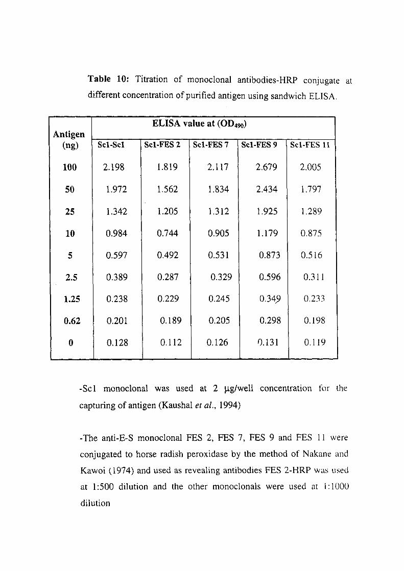
Table 10; Titration of monoclonal antibodies-HRP conjugate at
different concentration of purified antigen using sandwich ELISA.
Antigen
(ng)
100
50
25
10
5
2.5
1.25
0.62
0
ELISA value at (OD490)
Scl-Scl
2.198
1.972
1.342
0.984
0.597
0.389
0.238
0.201
0.128
Scl-FES2 Scl-FES7 Scl-FES9 Scl-FES 11
1.819
1.562
1.205
0.744
0.492
0.287
0.229
0.189
0.112
2.117
1.834
1.312
0.905
0.531
0.329
0.245
0.205
0.126
2.679
2.434
1.925
1.179
0.873
0.596
0.34^
0.298
0.131
2.005
1.797
1.289
0.875
0.516
0.311
0.233
0.198
0.119
-Scl monoclonal was used at 2 |j,g/well concentration for the
capturing of antigen (Kaushal et ai, 1994)
-The anti-E-S monoclonal FES 2, FES 7, FES 9 and FES 11 were
conjugated to horse radish peroxidase by the method of Nakane and
Kawoi (1974) and used as revealing antibodies FES 2-HRP was used
at 1:500 dilution and the other monoclonals were used at 1:1000
dilution
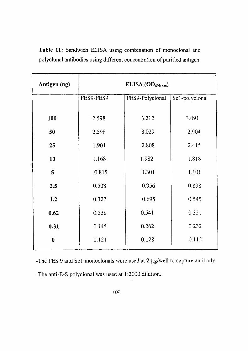
Table 11: Sandwich ELISA using combination of monoclonal and
polyclonal antibodies using different concentration of purified antigen.
Antigen (ng)
100
50
25
10
5
2.5
1.2
0.62
0.31
0
FES9-FES9
2.598
2.598
1.901
1.168
0.815
0.508
0.327
0.238
0.145
0.121
ELISA(OD490nJ
FES9-Polyclonal
3.212
3.029
2.808
1.982
1.301
0.956
0.695
0.541
0.262
0.128
Scl-polyclonal
3.091
2.904
2.415
1.818
1.101
0.898
0.545
0.321
0.232
0.112
-The FES 9 and Scl monoclonals were used at 2 |a,g/well to capture antibody
-The anti-E-S polyclonal was used at 1:2000 dilution.
loq
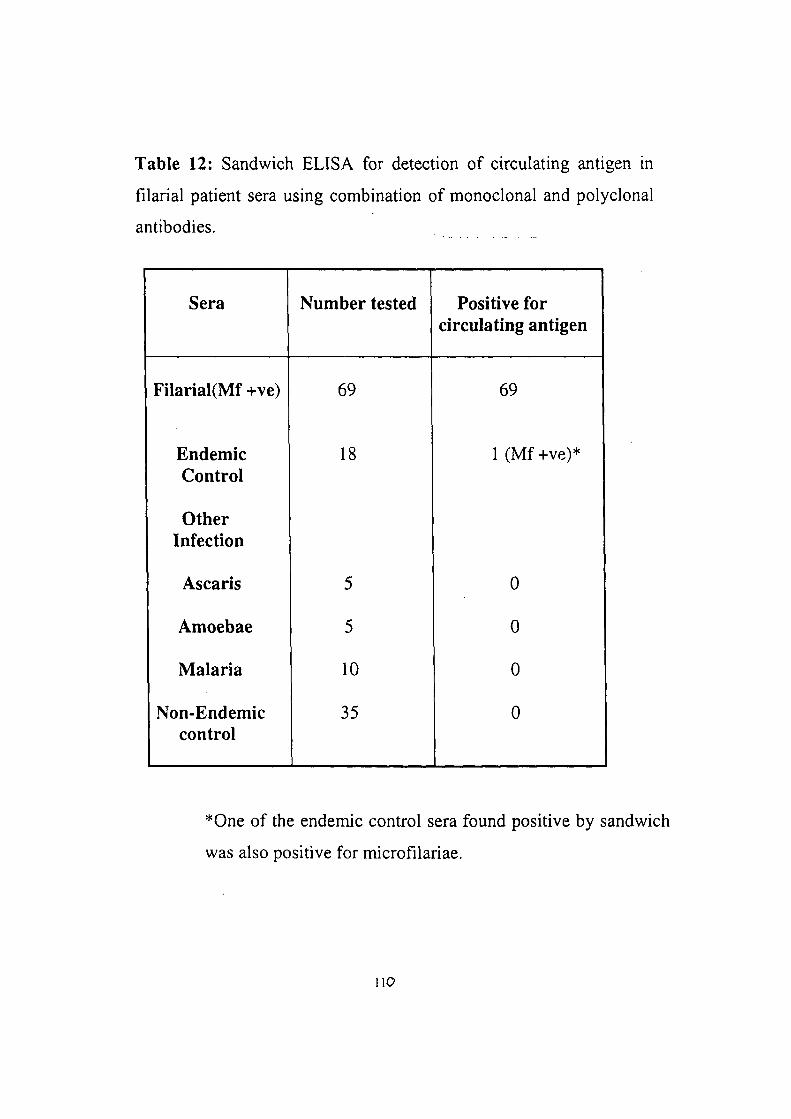
Table 12: Sandwich ELISA for detection of circulating antigen in
filarial patient sera using combination of monoclonal and polyclonal
antibodies.
Sera
FilariaKMf +ve)
Endemic Control
Other Infection
Ascaris
Amoebae
Malaria
Non-Endemic control
Number tested
69
18
5
5
10
35
Positive for circulating antigen
69
1 (Mf +ve)*
0
0
0
0
*One of the endemic control sera found positive by sandwich
was also positive for microfilariae.
no
albumin could be removed by absorption with anti-BSA antibodies.
The potential of anti-E-S serum was evaluated for the detection of
filarial circulating antigen and it successfully detected the circulating
antigen if filarial patient. The fi-actionation of the S. cervi E-S products
on DEAE-sephacel results in isolating a purified E-S fraction showing
reactivity with monoclonal (detecting circulating antigen). The
monoclonal antibodies produced against ScES purified antigen fraction
I and out of 4 monoclonals showing high reactivity, one showed
potential for developing antigen detection test for human filariasis.
II •
DISCUSSIONS
Filariasis is a serious disabilitating parasitic disease. The filarial
parasites W. bancrofti, B. malayi and B. trimori are the major cause of
human lymphatic filariasis in the tropical and sub-tropical regions of
the world with an estimated 120 million people infected and an
additional 900 million people at the risk of infection (Ottesen and
Ramachandran, 1995). One of the routine ways of diagnosing active
filarial infection is by detecting the presence of microfilaria in the
blood smears (Eberhard and Lammie, 1991). This method is less
sensitive because of the nocturnal periodicity and of missing
microfilaria in the blood smear if they are present in extremely low
numbers and thereby introducing an error in diagnosing the active
infection. The circulating filarial antigen was first demonstrated in
bancroftian filariasis by Frank in 1946, with the technique of passive
cutaneous anaphylaxis. The circulating antigen in the body fluids
(serum and urine) of microfilareamic and clinical filarial patients has
been suggested to be a better approach for the immunodiagnosis of
filariasis (Ottesen, 1980; 1984; Harinath, 1986) as it is much more
effective for assessing the status of an early and active infection.
The E-S products, released by the living parasites in the host and
these are the antigens which may probably be present in the circulation.
112
The demonstration of the E-S antigens in the circulation of the
infected host could provide a better indicator for active filarial
infection (Kaliraj et ah, 1979a). Inspite of the less complex nature of
the filarial E-S antigens, (Kaushal et al, 1982; 1984; 1989; Prasad et
ai, 1983; Kharat et al., 1983; Malhotra et al., 1987). not much efforts
have been made to critically characterize and identify the
diagnostically important antigen present in the E-S products. This may
probably be due to the limitations in obtaining sufficient amount of
E-S products from human filarial parasites (Kaushal et al., 1982;
1984; Kharat et al., 1983). In the present study, an effort was made to
analyze qualitatively and quantitatively the E-S products of S. cervi
adults and to fractionate the E-S products in order to identify and
isolate the antigen(s) equivalent to the filarial circulating antigen. The
antibodies produced against these antigens have been evaluated for
their potential to detect the circulating antigen in filarial patient sera.
Setaria cervi, a bovine filarial parasite, was used as an alternate source
for human filarial parasite antigens because the earlier studies from our
laboratory on the characterization of S. cervi antigens, has identified
some common antigenic epitopes between the bovine and human
filarial parasites (Kaushal et al., 1987) and the monoclonal antibodies
against the common epitope of high molecular weight antigen
showed reactivity with the filarial circulating antigen (Kaushal et al.,
1994b).
113
In the present study E-S products from S. cervi adult worms
were prepared by maintenance of parasites in vitro in serum free
culture medium, conditions for preparing the E-S products were
optimized and 32 hrs incubation period and DMEM appeared to be the
best for getting the better recovery of E-S products at 37°C. The
amount of protein released during the preparation of S. cervi E-S
products by the adult filarial parasites was far greater (2-15 mg of E-
S products per 100 S. cervi adult parasites) than that reported for the
other filarial parasites. Kaushal et al. (1982) obtained as low as 18
fxgs of protein after incubating 100 B. malayi adult in synthetic
medium for a duration of 48 hr. Rotmans et a/. (1981) have reported
the release of 100 |ig protein/100 Schistosoma mansoni worms by
maintaining them in medium 199 for 24 hrs. The release of large
amounts of E-S products by the S. cervi adult parasites, fascilitated
their characterization using conventional immunochemical methods
and use for developing immunodiagnostic reagents.
The protein pattern of S cervi E-S products was analyzed on SDS-
polyacrylamide gel elecfrophoresis which revealed the presence of
14-15 protein bands in the molecular weight range of 10-200 kD,
whereas, the crude somatic antigenic preparation contained 35-40
protein bands thus, indicating the less complex nature of the S. cervi
E-S products as compared to the somatic antigen whole worm
extract of 5. cem. The B. wa/ayz products were also found to be less
114
complex in nature having eleven protein bands in the molecular weight
range of 10 kD to 70 kD (Kaushal et al, 1982). Few protein bands
having low molecular weight were also reported for the E-S product of
L. carina (Rajasekhariah et al, 1986b) and O. volvulus mf (Ngu et al,
1981). Antigens of the molecular weight of 17 and 21 kD were
synthesized and release by the mf and L3 stages of W. bancrofti and the
17 kD antigen was located on the surface of these stages (Maizels et
al, 1986).
In order to produce polyclonal antibodies against S. cervi E-S
products, rabbits were immunized and the immune sera obtained
from these rabbits were tested in ELISA using ScE-S products. To
optimize the assay conditions, optimum antigen concentration and the
optimum dilution of the anti-ScE-S antibodies were determined. Out of
different concentrations of antigen (ScE-S) tried, optimum results were
obtained at 0.25 fig/well. The decrease in antigen concentrations
resulted in decrease ELISA values. The B. pahangi E-S products
showed the optimum antigen concentration as low as 10 ng/ml when
tested in ELISA (Maizels et al, 1985). Using this optimum antigen
concentration the immune rabbit sera from the three rabbits gave high
antibody titres in the range of 1:100 000 to 1:250 000. The increase in
tifre value with increasing number of immunization was observed in
all the three rabbits, which became constant after the 8**' injection.
The results indicate the highly immunogenic nature of S. cervi E-S
115-
products. The immunogenic nature of E-S products from filarial and
other nematode parasites have been shown by a number of studies. In
case of B. malayi E-S antigens 5 ng antigen/well showed optimum
results (Kaushal etal, 1982; Weil, 1988).
The antigenic pattern of iS. cervi E-S products were analyzed by
employing the technique of crossed Immunoelectrophoresis (CIE)
using the rabbit anti-5'. cervi E-S antibodies. The CIE is sophisticated
reasonably sensitive and semi-quantitative technique which gave
better resolution of the antigenic components of the S. cervi E-S
products. The immune rabbit sera obtained from the three rabbits
showed comparable antigenic pattern of S. cervi E-S antigen in CIE
except for the slight differences in the intensity and the heights of
certain peaks were observed. The number of peaks remained the same
in all the different concentrations of the serum tried,, but the optimum
resolution of the precipitin peaks was observed using 10% serum
concentration in case of immune rabbit serum 1 (IRS 1), which showed
comparatively lower antibody titre, whereas, in case of immune rabbit
serum 2 (IRS 2) and 3 (IRS 3) lower serum concentration (5% v/v) was
required to get a better resolution. A careful examination of CIE
pattern revealed the presence of 10-14 antigenic peaks in S. cervi E-
S product. Earlier the technique of CIE has been employed by various
workers to analyze the antigenic components of filarial and other
parasites (Kaushal et al., 1982; Malhotra et al, 1987; Mohan et al.
116
1988; Haider e? a/., 1990, Sugunan etal, 1990). Kaushale?al.(1982)
could resolve the B. malayi E-S antigens in 10-11 precipitin peaks
using CIE. However, the S. digitata E-S products showed the
presence of only 3-4 antigenic peaks by the same technique (Sugunan
and Raj, 1990).
The parasitic worms are known to adsorb host proteins on their
surfaces to get established in the host by protecting themselves from
the immunologically hostile environment of the host, (Smither et
ah, 1969). The S. cervi adult worm resides on the peritoneal folds of
the buffaloes and the adsorption of the host protein can subsequently be
released into the in vitro maintenance medium during the preparation
of E-S products. Host serum proteins have been shown to be present on
the surfaces and E-S products of other certain filarial and other helmith
parasites (Mania and Kar, 1994). The results of the present study
indicated the presence of 3-4 host proteins contamination in S. cervi
E-S products. One of the major components was found to be serum
albumin which was confirmed by absorbing the ScE-S with rabbit anti-
BSA antibodies and the anti- ". cervi E-S serum with BSA-sepharose
sorbent. Kaushal et al. (1982) have identified host serum protein
contaminants on the surface adult and microfilarial stages as well as in
the E-S products of human filarial parasites B. malayi. Albumin has
been shown to mark the surfaces of W. bancrofti microfilaria! antigen
(Kar et al, 1993; Mania and Kar 1994).
117
Various immunodiagnostic techniques based on the detection
of circulating antigen has been employed by a number of workers
(Au ef a/., 1981; Des Moutis et al, 1983; Kaliraj e/a/., 1981a;
Ouassiefa/., 1981; Phillip et al, 1982; Dissanayake et al, 1984;
Forsyth et al, 1985; Hamilton et al, 1984; Reddy et al, 1984b; c)
and the counter current Immunoelectrophoresis (CIEP) was one of
them. The counter current Immunoelectrophoresis (CIEP) has earlier
been used for the detection of filarial circulating antigen by a
number of workers (Kaliraj et al,\ 979; 1981 a; Dasgupta et al, 1984;
Weil et al, 1984a; 1986a).This method appears to be reasonably
sensitive and convenient to perform as compared to night blood
examination for the presence of microfilariae, but the problem of
specificity was encountered as antibodies used in these studies were
against the crude somatic extracts of filarial parasite (Weil et al,
1984a; 1986; Hamilton et al, 1984). In the present study, the
polyclonal antibodies raised in rabbits against S. cervi E-S antigens
were used for the detection of circulating antigen in filarial patient
using small number of serum samples. The hyperimmune serum from
all the three rabbits were found to have antibodies against filarial
circulating antigen. Initially, these immune rabbit sera showed non
specific reaction with normal human serum in CIEP. However, this
non-specificity was removed by preabsorbing the immune rabbit sera
with albumin-sepharose. The preabsorbed anti-E-S serum could detect
the circulating antigen in all the microfilariae positive patient sera but
l]S
not in the non-endemic control sera from the healthy individuals used
in the study.
Detection of circulating antigen, however, can be complicated
by the binding of antigen into immune complexes (Hamilton et ai,
1984). Such complexes are common in sera from patients with
filariasis (Au er a/., 1981; Steward et ai, 1982; Chandershekhar e/
ai, 1990; Das ef al., 1988a; Dissanayake et al., 1982; Lutsch et al,
1988; Thambiah et al, 1992). Variability in the amount of serum
antigen in free and bound state may make difficult the detection of
circulating antigen. Therefore, in the present study the filarial patient
sera were pretreated with EDTA and heated to release the antigen
from the immune complexes, thus, in turn increasing the sensitivity of
the assay. Earlier studies from our laboratory on identification and
characetrization of antigens of human {B. malayi) and bovine {S. cervi)
filarial parsites revealed common antigens between these two parasites.
In the present study, the fractionation and immunochemical
characterization of S. cervi E-S products was done in order to identify
and isolate the antigen(s) equivalent to filarial circulating antigen and
having potential for developing immunodiagnostic reagents for antigen
detection.
The S. cervi E-S products were fractionated using DEAE-
Sephacel column which resulted in the resolution of three distinct
protein peaks. All these fractions showed high immuno-reactivity with
119
rabbit anti-E-S serum while only few fractions were reactive with anti-
BSA serum in ELISA. Based on their reactivity in ELISA the fractions
were pooled in six different antigen pools fractions. Among the six
ScE-S purified fractions only fraction I (ScESPFI) showed
significantly high reactivity with monoclonal antibody Scl (prepared
previously in our laboratory detecting the circulating antigen in human
patient sera)Antigens from human filarial parasites have been antigens
isolated from blood (Reddy et al, 1986), hydrocoele fluid (Malhotra et
al, 1985; Ramaprasad and Harinath, 1987) of infected patients and
have been used as diagnostic reagents.
In the present study, the ScESPFI has been used to produce
monoclonal antibodies to evaluate its diagnostic potential. The
selection of hybridoma clones was done on the basis of their reactivity
with both bovine and human flleirial parasite in order to identify the
clones against common antigenic epitopes. In all the fusions done in
the present study high fusion efficiency (96%) was obtained. The
present studies resulted in the identification of 12 monoclonal
antibodies showing high and consistent reactivity with both bovine and
human filarial parasites thereby suggesting that these monoclonal
antibodies are against the shared antigens of these parasites. The
monoclonal antibodies against the common antigenic epitopes of
human and animal parasites (Dirofilariae immitis and Onchocerca
gibsoni) have earlier been produced (Dissanayake et al., 1984; Weil
120
and Liftis, 1987), Out of the 12 hybridoma clones, four monoclonal
antibodies (designated as FES 2, FES 7, FES 9and FES 11) were found
to show high affinity for the filarial circulating antigen present in
filarial patient sera. Scl, monoclonal antibody an IgGi isotype directed
against a non-phosphorylcholine repetitive epitope , (prepared
previously in our laboratory) and detecting the circulating filarial
antigen in human patient sera was used as positive control in all the
experiments.The target epitopes of these monoclonal antibodies
appears to be present not only in S. cervi antigenic preparations (ScA,
Scmf, ScES and ScESPFI) but also in the somatic extracts of the other
filarial parasites, as shown by high reactivity of these monoclonal
antibodies with B. malayi and W. bancrofti antigen. However, all the
four monoclonal antibodies did not cross-react with the antigens
obtained from the non-filarial parasites. This lack of cross-reactivity of
the monoclonals with non-filarial antigens and the high reactivity with
different filarial parasite antigens suggest filaria-specificity of these
monoclonal antibodies. The isotyping analysis of four monoclonals
revealed FES 2as IgGzb, FES 7 and FES 9 as IgG, and FES 1 las IgM
isotype.
These monoclonals were further characterized for their epitope
specificity.The epitope specificty of all the four monoclonals were
assessed by inhibition ELISA using Scl-HRP conjugate. The results of
the inhibition ELISA indicated that the three monoclonals (FES 7, FES
12(
9 and FES 11) are directed against the target epitope of Scl
monoclonal antibody as revealed by strong inhibition of binding of
Scl-HRP By all the three monoclonal antibodies. However, the FES 2
appeared to be directed against a different epitope as it did not have
effect on binding of Scl-HRP to filarial antigen. The potential of these
4 monoclonal antibodies (FES 2, FES 7, FES 9 and FES 11) was
evaluated for the detection of circulating antigen in filarial patient sera
by sandwich ELISA.
A number of antigen detection assays have been developed by
several investigators using monoclonal anti bodies against antigens
fi-om animal filarial parasites (Des Moutis et al., 1983; Dissanayake et
ah, 1984; Forsyth et ah, 1985). The Gib-13 was the first monoclonal
antibody (produced against O. gibsoni egg antigen) that was used to
develop an immunoradiometric assay (IRMA) for the detection of
antigen in bancroftian filariasis (Forsyth et al, 1985). Unfortunately
none of these assays could be integrated in filaria control programmes
as these test has either been insufficiently sensitive or technically
demanding for use outside the specialized laboratories. Few other
monoclonal antibodies, which were produced against the common
antigenic epitopes of human and animal filarial parasites, include the
monoclonals produced against D. immitis (Weil et al, 1987);
O. gibsoni adult antigen (More and Copeman, 1990). These
122
monoclonal antibodies have been used to develop antigen detection test
for bancroftian filariasis and these are under commercialisation.
In our laboratory we have produced a monoclonal antibody
(Scl) against an antigenic epitope common between the bovine and
human filarial parasite. The Scl monoclonal is an IgGi isotype,
directed against a non-phosphorylcholine, heat stable, repetitive
epitope (Kaushal et ai, 1994). The Scl based sandwich ELISA, though
highly specific for diagnosing active filarial infection, the sensitivity of
the test was not upto the mark. In the present study, we have evaluated
the diagnostic potential of anti-E-S monoclonal and polyclonal
antibodies in combination with Scl monoclonal antibody. All the four
anti-ScE-S monoclonal antibodies (FES 2, FES 7, FES 9 and FES 11)
were conjugated to HRP and evaluated in sandwich ELISA for antigen
detection. A sandwich ELISA was standardised in terms of optimum
concentration of antibody (developed earlier in our laboratory,
detecting circulating antigen) was used as capturing antibody and used
in combination with the peroxidase conjugated anti-E-S monoclonal
antibodies (for revealing step) in sandwich ELISA. Out of different
combinations (Scl-FES2, Scl-FES 7, Scl-FES 9 and Scl-FES 11)
tried, the combination of Scl-FES 9 showed better sensitivity for
antigen detection as compared to other monoclonal antibodies. When
these monoclonal antibodies (as capturing antibodies) were used in
combination with the peroxidase conjugated rabbit anti-ScE-S antibody
m
(as revealing antibody), the FES 9 monoclonal-polyclonal combination
showed better sensitivity for antigen detection. The better results
obtained with FES 9 monoclonals may be attributed to the high affinity
of this monoclonal antibody for the antigen. Differences in the
affinities of different monoclonal antibodies have been observed
(Kaushal et al., 1994; 1995). The FES 9-polyclonal sandwich ELISA
was evaluated for its immunodiagnostic potential using patients' sera
from filarial (microfilariae positive), non-filarial infections (ascaris,
amoebae, malaria) infections as well as endemic and non-endemic
control sera. The results of the present study revealed that the FES 9-
polyclonal system is highly sensitive as it could detect antigen in all the
microfilarial positive patients' sera. One of the endemic control sera
positive by the sandwich ELISA was found to be positive for
microfilariae. The specificity of the system (FES 9-polyclonal ELISA)
was evident by the negative results obtained in case of other infection
sera (ascaris, amoebae and malaria) and the non-endemic control sera.
A sandwich ELISA, employing monoclonal antibody (AD 12) against
dog filarial parasite antigen showed a sensitivity of 95% in filaria
diagnosis (Weil and Liftis, 1987; Weil et a/., 1989). However, the
problem with this monoclonal antibody is that it is directed against a
carbohydrate epitope and thus may not be very suitable for specific
diagnosis of human filariasis.
124
In conclusion, the excretory-secretory products from S. cervi
adults Ccin be obtained by short term in vitro maintenance of filarial
adult worms in protein free defined medium.The polyclonal antibodies
produced against .S". cervi E-S products showed high antibody titres and
revealed 10-14 antigenic proteins in the ScE-S products. The S. cervi
E-S products were found to contain host serum albumin which can be
removed by absorption with anti-BSA antibodies. The fractionation of
iS". cervi E-S products led to the isolation of an antigen fraction which
may contain antigens equivalent to filarial circulating antigen as the
antibodies against this fraction showed reactivity with filarial
circulating antigen. We have been able to produce monoclonal
antibodies against this purified S. cervi E-S purified fraction. Out of 4
monoclonal antibodies specific to filaria, one showed better sensitivity,
when used in combination with anti-E-S polyclonal, for detecting
circulating antigen in filarial patients' sera. A logical extension of these
studies to use this anti-ScE-S monoclonal-polyclonal combinations for
developing specific
12S"

CHAPTER IV
SUMMARY AND
CONCLUSIONS
Filariasis is a major health problem in tropical and subtropical
countries. It is caused by the parasites belonging to class filaroidea,
which have complex life cycle that alternates between an arthropod and
vertebrate host. Though, the disease is not fatal, but. these parasites
cause lymphatic damage that leads to elephantiasis and hydrocoele
which is responsible for considerable disability and social stigma.
Important pathogenic species infecting humans are Wuchereria
bancrofti, Brugia malayi and B. trimori. In India over 90% of the
disease is caused by Wuchereria bancrofti.
The unequivocal diagnosis of filarial infection still largely
depends on the detection of the microfilariae in the blood. Nocturnal
periodicity of microfilariae imposes a practical limitation on the
effective use of microfilaria detection of lymphatic filariasis because of
the need for night blood sampling. The parasitological method is
inconvenient for both the patient as well as the investigator and fails to
detect the amicrofilaraemic stage of the disease or when mf are
present in very small numbers. This has put forward an emphasis for
improved immunodiagnostic methods for species and accurte diagnosis
of active filarial infection.
Earlier studies on immunodiagnosis of filariasis was focussed
mainly on the demonstration of antibodies to filarial antigens. The
major disadvantage of the immunodiagnostic test based on antibody
126
detection is their inability to distinguish between the past exposure and
current infections as well as their cross-reactivity with other helminth
parasites. The detection of filarial circulating antigen in blood and
other body fluids of infected individuals has been shown to be a better
approach for diagnosing active filarial infections. Though a number of
antigen detection assays have been developed but the problem of
sensitivity and specificity of these assays still remains. There is still a
need to identify specific filarial antigen for developing better
immunodiagnostic reagents.
Due to the non-availability nof human filarial parasites in
sufficient quantities, heterologous antigens from animal filarial parasite
including S. cervi (bovine filarial parasite) have been used for
developing immunodiagnostics. However, the use of antibodies
prepared against crude antigenic preparation results in false positive
reactions.This may be due to complex nature of these antigens and
their cross-reactivity with other helminths parasites. Polyclonal
antibodies against purified or simple antigenic preparations or
monoclonal antibodies against filaria-specific epitopes are required for
developing specific immunodiagnostic methods. The excretory-
seretory antigens released by the living parasites in the host are known
to be less complex in nature and may contain antigen(s) equivalent to
filarial circulating antigen. The present study was undertaken on the
characterization of excretory-secretory products of S. cervi (bovine
127
filarial parasite) to identify the antien equivalent to filarial circulating
antigen, production of antibodies against these antigens.
The studies were initiated with the preparation of S. cervi E-S
products, by maintaining the adult parasites asceptically in protein free
medium (DMEM) for 32 hrs at 37°C. Substantial amount (12-15 mg
protein/100 worms) of E-S products were obtained fi-om S. cervi adult
by maintainin in DMEM and the worms remained motile for the
maximum period of time without change of medium. The release of
large amount of protein facilitated the fractionation and
characterization of .S. cervi E-S products. The SDS-polyacrylamide gel
electrophoretic analysis of S. cervi E-S products showed the presence
of 14-15 protein bands in molecular weight range of 10-200 kD.
Theseresults revealed less complex nature of ScE-S products as
compared to S. cervi somatic antigenic preparation which showed 30-
35 protein bands using hyperimmune rabbit serum. The ScE-S products
were used to immunized the rabbits for raising hyperimmune
polyvalent serum. Three rabbits (Rl, R2 & R3) were immunized over a
period of 5-6 months. The immune rabbit sera collected after each
immunization starting from the 3"* injection and were tested for the
parasite specific antibodies by employing enzyme linked-
immunosorbent assay (ELISA). An increase in the antibody titre, with
increasing number of immunization, was observed and the titre value
became constant after VIII* injection in all the three rabbits (IRS 1,
125
IRS 2 and IRS 3). The immune serum pool from IRS 1 showed the
reciprocal antibody titre of 128 000, while the reciprocal antibody titre
of 256 000 were observed for IRS 2and IRS 3 pools. The antigenic
components of 5. cervi E-S antigens were analysed by employing the
techniques of CIE using rabbit anti-iS. cervi E-S serum. The best
resolution in CIE was obtained by using 5-10% concentration of anti-5.
cervi E-S serum and it revealed the presence of 10-12 precipitin peaks
in E-S products.
Parasite worms are known to adsorb host serum proteins (mainly
albumin) on their surfaces in order to evade the immunologically
hostile environment of the host. The S. cervi adult worms resides on the
peritoneal folds of buffaloes and absorption of normal host proteins
and their subsequent release during in vitro maintenance is likely to
occur. In the present study, the S. cervi E-S products were also
analysed for the presence of host serum protein contaminents by
using rabbit anti-normal buffalo serum and it showed the presence of
3-4 host proteins. Further, efforts were made to characterize these host
protein contaminants by using the techniques such as crossed line
Immunoelectrophoresis (CLIE) and tandem crossed
Immunoelectrophoresis (TCIE). The CLIE was done by adopting two
ways, firstly the BSA was introduced in the intermediate gel and
secondly by introducing the rabbit anti-BSA serum in the intermediate
gel, and the rabbit anti-E-S serum was used in both the cases. In the
\29
former case, when BSA was used in the intermediate gel, two
precipitin peaks of the E-S products showed Hnes at their bases. The
reduction in the heights of the same precipitin peaks was observed in
the latter case. In case of TCIE, BSA was run along with the E-S
products in the first dimension, using the rabbit anti-E-S serum, and it
showed considerable increase in the heights of the same two
corresponding precipitin peaks as observed in case of CLIE. The above
results with CLEE and TCIE indicates that the major components of
host serum proteins is albumin.
The rabbit anti-E-S serum was evaluated for its potential to
detect circulating antigen in filarial patient serum. Initially, the rabbit
anti-ScE-S serum showed non-specific reaction with the normal
human serum. This may be due to the presence of host serum albumin
in the S. cervi E-S products. Therefore, efforts were made to absorb
the host serum proteins from the S. cervi E-S products using anti-
BSA sepharose beads. The preabsorbed .S. cervi E-S products were
analysed using SDS-PAGE, CIE and ELISA. The SDS-PAGE analysis
of the preabsorbed E-S products showed the removal of major 66 kD
protein band, which was present in the unabsorbed S. cervi E-S
products. The antigenic analysis by CIE, using rabbit anti-E-S serum.,
revealed the presence of 10 precipitin peaks in the preabsorbed E-
S products. On testing the preabsorbed S. cervi E-S products in ELISA
against rabbit anti-ScE-S serum, the antibody titre was of 1:120 000.
130
Similarly, the absorption of rabbit anti-S. cervi E-S serum on BSA-
sepharose beads, resulted in the removal of the antibodies against the
host protein components. The preabsorbed rabbit anti-ScE-S serum,
showed the removal of the antibodies against the two major host
protein contaminants of E-S products. This preabsorbed rabbit anti-
ScE-S serum, when tested in ELISA against S. cervi E-S products,
gave an antibody titre of 1:120 000 as compared to the reciprocal
antibody titre of 250 000 obtained for the unabsorbed anti-E-S serum.
The preabsorbed anti-E-S serum was evaluated for the detection of
circulating filarial antigen in patient sera by counter current
Immunoelectrophoresis. This absorbed rabbit anti-E-S serum showed
no cross-reactivity with the normal human serum, and could
successfully detect the circulating antigen in the filarial patient sera.
The fractionation of S. cervi E-S products on DEAE-Sephacel
column yeilded 3 major protein peaks. Six antigen fraction pools were
made according to their reactivity with rabbit anti-E-S serum and rabbit
anti-BSA serum. The reactivities of the antigen fraction pools were
tested against the monoclonal antibody (detecting the filarial
circulating antigen) in ELISA. The ScESPFI showed high reactivity
with the monoclonal antibody, whereas, all other antigen fraction pools
did not show any significant reactivity. Therefore, the ScESPFI which
may be having the antigen equivalent to the filarial circulating antigen,
was used to immunize the mice for hybridoma production.
131
The production of monoclonal antibodies against S. cervi E-S
products was done by the fusion of spleen cells from mice (immunized
against ScESPFI) with myeloma cells. A fusion efficiency of 96% was
obtained. The 69 hybridoma clones showed reactivity reactivity with S.
cervi antigens, 27 with ScESFPI, 36 with B. malayi adult antigen and
21 with W. bancrofti microfilarial antigens. A total of 12 monoclonal
antibodies having consistent reactivity with both bovine and human
parasites were selected and out of these four monoclonal antibodies
(FES 2, FES 7, FES 9and FES 11) were found to have high affinity for
the filarial circulating antigen. All the 4 monoclonals showed high
reactivity with human {B. malayi and W. bancrofti) and bovine filarial
{S. cervi E-S, ScESFPI, ScA and ScMf) antigen but the highest
reactivity in all the cases was observed with FES 9 monoclonal
antibody. None of these monoclonal antibodies showed reactivity with
any significant the non-filarial antigens (ascaris, hookworm, malaria,
amoebae, bacteria) indicate the specificity of the monoclonal
antibodies to filarial antigen.Isotyping analysis revealed that one (FES
2) out of four monoclonals, was found IgGib one (FESl 1) was IgM and
two (FES 7and FES 9) were IgGi isotype.The target epitope of the tree
monoclonal antibodies (FES 7, FES 9, FES 11) was found to be same
as that of Scl monoclonal antibody, while FES 2 monoclonal was
directed against a different epitope as shown by inhibition ELISA
studies
13Z
Therefore, in the present study we have been able to analyse and
characterize the excretory-secretory products ofS. cervi adult worms.
The hyperimmune sera raised against the S. cervi E-S product showed
high antibody titres in S. cervi E-S products and showed the presence
of 10-12 antigenic components. Out of these 3-4 antigens were
identified as host serum protein components and one of them being
albumin. The host serum proteins present in S. cervi E-S products
could be removed by absorbing with anti-BSA antibodies which
resulted in the removal of host serum protein bands.The absoption of
rabbit anti- ". cervi E-S serum with BSA removed the antibodies the
against host serum proteins. The preabsorbed rabbit anti-ScE-S serum
successfully detected the circulating antigen in filarial patients' sera.
Fractionation on DEAE-Sephacel column resulted in isolating a
purified antigen fi-action common with human filarial parasite and
showing high reactivity with the monoclonal, (detecting filarial
circulating antigen), the four monoclonal antibodies (FES 2, FES 7,
FES 9 and FES 11) produced against this purified antigen fraction
(ScESPFI) showed high reactivity with filarial antigen and did not
cross react with non-filarial antigens thereby, suggesting their filaria
specificity. Out of these four monoclonals, three (FES 7, FES 9 and
FES 11) were found to be directed against the same epitope as that of
Scl monoclonal antibody, while the fourth (FES 2) was against a
different epitope. The FES 9 monoclonal antibody in combination with
rabbit ScE-S polyclonal, showed higher sensitivity for the detection of
132-

circulating antigen in filarial patient sera. These findings suggest the
potential of anti-ScE-S antibodies for developing specific
immunodiagnostic test based on antigen detection for human filariasis.
134

BIBLIOGRAPHY
BIBLIOGRAPHY
Acton, H.W. and Rao, S.S. (1933). Ind. Med. Gaz. 68, 305.
Alikhan, A.; Parkhe, K.A.; Reddy, M.V.R. and Harinath, B.C. (1990).
Natl. Med. J. India. 3 ,265.
Almeida, A.J.;Bhopale, M.K. and Renapurkar, D.M. (1990). Folia
Parasitol. 37, 323.
Almond, N.M. and Parkhouse, R.M.E. (1985). In Current Topics in
Microbiology (ed. Parkhouse, R.M.E.) Heidelberg, Springer-Verlag. p
173.
Ambroise-Thomas, P. (1974). Acta Tropica. 31,108.
Ambroise-Thomas, P. (1980). Filariasis. In Practical Methods in
Clinical Immunology (ed. V. Houba) Churchill-Livingstone,
Edinburgh, p 2.
Ambroise-Thomas, P. and Kein Troung, T. (1974). Annals Trop. Med.
Parasitol. 68, 435.
135-
Anon (1933). Filariasis Immunology/ molecular Biology meetings.
TDR NEWS. UNDPAVorld Bank/ WHO Special programme for
Research and Training in Tropical Dieseases, Geneva. Pp 3.
Au. A.C.S.; Denham, D.A.; Steward, M.W.; Draper, C.C; Ismail.
M.M.; Rao, C.K.; and Mak, J.W. (1981). S.E. Asian J. Trop. Med. Pub.
Health. 12, 492.
Au, A.C.S.; Draper C.C; Denham, D.A.; Rao, C.K.; Ismail, M.M. and
Mak, J.W. (1982). S. E. Asian J. Trop. Med. Pub. Health. 13, 142.
Axelsen, N.H.; Kroll, J. and Weeke, B. (1973). Scan. J. Immunol. 2,
Supp. 1.
Barakat, R.M.; El-Gassim, E.E.; Awadalla, H.N.; El-Molla, A. and
Omer, E.A. (1983). Trans. Roy. Soc. Trop. Med. Hyg. 71, 109.
Bartlett, A.; Bidwell, D.E. and Voller, A. (1975). Trop. Med.
Parasitol. 26, 370.
Bartlett, A. and Bidwell, D.E. (1976). Trop. Med. Parasitol. 26, 370.
Beaver, P.C. (1970). Am. J. Trop. Med. Hyg. 19, 181.
\Z6
Biagi, F.F. (1956). Medicina. 761, 545.
Biguet, J.; d'Haussy, R.; Capron, A.; Tran Van Ky, and Aubry, M.
(1962). Bull, de la Societe de Pathol. Exotique. 55, 845.
Biguet, J.; d'Haussy, R.; Aubry, M. and Rose, F. (1964). Bull, de la
Societe de Pathol. Exotique. 57, 1098.
Bozicevich, J. and Hutter, A.M. (1944). Am. J. Trop. Med. 24, 203.
Bradford, M.M. (1976). Anal. Biochem. 72,248.
Bradley, J.E.; Taun, R.S.; Shepley, K.J.; Tree, T.I.M.; Maizels, R.M.;
Helm, R.; Gregory, W.F. and Unnasch, T.R. (1993). Exptal. Parasitol.
77.414.
Bright, J.J. and Raj, R.K. (1991). Ind. J. Exptal. Biol. 29, 725.
Cabrera, Z. and Parkhouse, R.M.E. (1986). Mol. Biochem. Parasitol.
20,225.
Cabrera, Z. and Parkhouse, R.M.E. (!987). Para. Immunol. 9, 39.
131
Capron, A.;Gentilini, M. and Vemes,I. (1968). Pathalog. Biol. 16,
1039.
Capron, A.; Gentilini, M. and Biguet, J. (1970). In Immunologic
Diagnosis of Filariasis by Immunoelectrophoresis. H.D. Srivastava
Commemorative volume. P 527.
Catmull, J.; Zhang, D.; Ruggiero, F.; Copeman, D.B. and Miller, D.J.
(1994). Mol. Biochem. Parasitol 63, 49.
Centre of Disease Control (1993). Morbid. Mortal. Weekely Rep. 42,.
Chandra, R.; Govilla, P.; Chandra, S.; Katiyar, J.C. and sen, A.B.
(1974). Ind. J. Med Res. 62, 1017.
Chandrashekhar, R.; Ogunrinade, A.F.; Alveraz, R.M.; Kale, O.O.
and Weil, G.J. (1990). J. Infect. Dis. 162, 1159.
Chanteau, S; Moulia-Pelat, J.P.; Glaziou, P.; Nguajen, N.L.;
Luquiaud, P; Plichart, C. Martin, P.M.V. and Cartel, J.L. (1994a). J.
Infec. Dis. 170, 247.
13.^
Chanteau, S.; Glazio, P.; Luquiared, P.; Plichart, C ; Moulia-Pelat, J.P.
and Cartel, J.L. (1994b). Trop. Med. Parasitol. 45, 255.
Chatterjee, R.K.; Tandon, A.; Saxena, K.C.; Ghatak, S. and Sen, A.B.
(1978). Ind. J. Med Res. 67, 34.
Cheirmaraj, K.; Reddy, M.V.R. and Harinath, B.C. (1990) J.
Immunoassay 11,429.
Cheirmaraj, K.; Reddy, M.V.R. and Harinath, B.C. (1992). S.E. Asian
J. Trop. Med. Pub. Health 23, 444.
Chenthamarakshan, V.; Cheirmaraj, K.; Reddy, M.V.R. and
Harinath, B.C. (1995). J. Trop. Med Hyg. 98, 233.
Chenthamarakshan, V.; Reddy, M.V.R. and Harinath, B.C. (1996a).
J. BioSci. 21,27.
Chenthamarakshan, V.; Reddy, M.V.R. and Harinath, B.C. (1996b).
Trans. Roy. Soc. Trop. Med. Hyg. 90, 252.
Culbertson, J.T.; Rose, H.M. and Demarest, C.R. (1944). Am. J.
Trop. Med Hyg. 39, 152.
13^
Das, M.K.; Subramanyam, V.R.; Ravindran, B. and Pattanaik, N.M.
(1987). J. Trop. Med. Hyg. 90, 135.
Das, M.K.; Subramanyam, V.R.; Ravindran, B. and Pattanaik, N.M.
(1988a). Med. Sci. Res. 16, 1055.
Das, M.K.; Beuria, M.K.; Dash, A.P.; and Pattnaik, N.M. (1988b). Ind.
J. Expt. Biol. 26, 1003.
Das, P.K.; Manoharan, A.; Srividya, A; Grenfel, B.T.; Bundy, D.A.P.
and Vanamail,P. (1990). Parasitol. 101,429.
Dasgupta, A. and Bala, S. (1978a). Prog. Fil. 1, 93.
Dasgupta, A. and Bala, S. (1978b). Ind. J. Med. Res. 67, 30.
Dasgupta, A.; Bala, S.; Banerjee, A. and Choudhary, A. (1980). J.
Helminthol. 54, 83.
Dasgupta, A.; Bala, S. andDutta, S.N. (1984). Para. Immunol. 6,341.
Daveau, C. and Ambroise-Thomas, P. (1982). Med. Trop. 42 513.
Decruize, S.W. and Raj, R. K. (1988). Ind J. Expt. Biol. 26, 781.
140
Denham, D.A. and McGreevy, P.B. (1977). Adv. Parasitol. 15, 243.
De Savigney, D.D. and Tizard, I.R. (!977). Trans. Roy. Soc. Trop.
Med. Hyg.ll, 501.
De Savigney, D.D.; Voller, A. and Woodruff, A.W. (1979). J. Clin.
Pathol. 32, 284.
Desowitz, R.S. and Una, S.R. (1976). J. Helminthol. 50, 53.
Des Moutis, I.; Ouaissi, A.; Grzych, J.M.; Yarzabal, L.; Haque, A. and
Capron, A. (1983). Am. J. Trop. Med. Hyg. 32, 533.
Devaney, E. (1987). Para. Immunol. 9,401.
Devaney, E. (1988). Mol. Biochem. Parasitol. 11, 83.
Devaney, E.; Betschart, B. and Rudin, W. (1990). Acta Tropica 47,
365.
Dhas, K.P.L.; Decruize, S.W. and Raj, R. K. (1993). J. Bio. Sci. 18,
311.
141
D'Haussy, R.; Capron, A.; Rolland, A. and Biguet, J. (1972). Rev.
Immunol. 36, 55.
Diesfeld. H.J.; Dutta, S.N.and Braun Munzinger, R. (1973). Zeits.
Trop. Med. Parasitol. 24,439.
Dissanayake, S. and Ismail, M.M. (1980a). Bull. WHO. 58, 655.
Dissanayake, S. and Ismail, M.M. (1980b). Bull. WHO. 58, 649.
Dissanayake, S. and Ismail, M.M. (1981). Bull. WHO. 59, 753.
Dissanayake, S.; Galahitiyawa, S.C. and Ismail, M.M. (1982). Bull.
WHO. 60,919.
Dissanayake, S. and Ismail, M.M. (1983). Bull. WHO. 61, 725.
Dissanayake, S. ; Forsyth, K.P.;Ismail, M.M. and Mitchell, G.F.
(1984). Am. J. Trop. Med. Hyg. 33, 1130.
Dissanayake, S. and Ismail, M.M. (1987). Filariasis. p 655.
Dumenigo, B.E.; Espino, A.M.; Danger, C. and Finlay, CM. (1993).
Res. Rev. Parasitol. 51, 47.
142.
Eberhard, M.L.; Roberts, J.M.; Lammie, P.J. and Lowrie, R.C. Jr.
(1988).rro/7. Med. Parasitol. 39, 295.
Eberhard, M.L. and Lammie, P.J. (1991). Clin. Lab. Med. 11, 977.
Egwang, T.G. and Kazura, J.W. (1987). Mol. Biochem. Parasitol. 22,
159.
Ellsworth, J.H. and Johnson, A.H. (1973). Am. J. Veterin. Res. 34,
682.
Engvall, E and Pearlman, P. (1972). J. Immunol. 109, 129.
Espino, A.M. and Finlay, CM. (1994). J. Clin. Microbiol. 32, 190.
Ey, P.L.; Prowse, S.J. and Jenkins, C.R. (1978). Immunochem. 15,429.
Ey, P.L. (1988). Exp. Parasitol. 65, 232.
Farrar, R.G.; Klei, T.R.; McVay, C.S. and Coleman, S. (1991). J.
Parasitol. 77,718.
143
Flecher, C. and Wu, C.C. (1992). Parasitol 105,425.
Forsyth, K.P.; Copeman, D.B.; Anders, R.F. and Mitchell, G.F.
(1981a). Acta Tropica. 38, 343.
Forsyth, K.P.; Copeman, D.B.; Abbot, A.P.; Anders, R.F. and
Mitchell, G.F. (1981b). Acta Tropica. 38, 329.
Forsyth, K.P.; Spark, R.; Kazura, J.; Brown, G.V.; Peters, P;
Heywood, P. Dissanayake, S. and Mitchell, G.F. (1985). J. Immunol.
134,1172.
Farris, R.; Ramzy, R.M.R.; Gad, A.M.; Weil, G.J. and Buck, A.A.
(1993). Trans. Roy. Soc. Trop. Med. Hyg. 87, 659.
Frank, B. (1946). J. Parasitol. 32, 400.
Frank, M.B.; ChenoWeth, B.M.Jr. and StoU, N.R. (1947). Am. J.
Trop. Med. 27, 617.
Freedman, D.O.; Nutman, T.B. and Ottesen, E.A. (1989). J.
Clin.Investig. 83, 14.
14^
Fujita, K.; Tanaka, H.; Sasa, M.; Shichinoha, K.; Asai, Y. and
KuroKawa, K. (1970). Jap. J. Exp. Med. 40, 67.
Gentilini, M.; Pinon, J.M.; Niel, G. and Rosin, G. (1973). Bull. Soc.
Pathologie. Exot. 66, 133.
Gonsaga dos Santos, L.; Santos, D.S. and Azevedu, R. (1976). Am. J.
Trop. Med. Patasitol. 70, 219.
Greene, B.M.; Taylor, H.R.; Brown, E.J. Humphery, R. J. and Lawley,
T.J. (1983). J. Inf. Dis. 141, 890.
Grove, D.I.; Cabrera, B.D.; Vleza, F.S.; Guinto, R.S. Ash, L.R. and
Warren, K.S. (1977). Am. J. Trop. Med Hyg. 26, 220.
Grove, D.I. and Davies, R.S. (1978). Am. J. trop. Med. Hyg.ll, 508.
Gueglio, B.; Border, C. and Marjolet, M. (1995). Bull. Soc. Fran.
Parasitol. 13, 215.
Gupta, S.C. and Ansari, J.A. (1989). Rev. Parasitol. 4, 165.
Gyapong, J.W.; Adjei, S. Gyapong, M and Asamoah, G (1996). Acta
Tropica. 61, 65.
146"
Gyapong, J.W.; Webber, R.H.; Morris, J. and Bennett, S (1998).
Trans. Roy. S.oc. Trop. Med. Hyg. 92, 40.
Haider, S. (1990). Ph. D. Thesis, Faculty of Science, University of
Kanpur.
Hamilton, R.G.; Hussain, R. and Ottesen,E.A. (1984). J. Immunol.
133, 2237.
Hamilton, R.G. (1985). Rev. Inf. Dis. 7, 837.
Harinatli, B.C. and Kaliraj, P. (1979). Immunodiagnosis of
bancroftian filariasis with microfilarial antigens: Problems and
progress. Paper presented at Tropical Immunology Seminar. Sydney,
Australia.
Harinath, B.C. (1984). J. Bio. Set 6, 691.
Harinath, B.C.; Malhotra, A; Ghimikar, S.N.; v^nnadate, S.D.; Isaacs,
V.P. and Bharti, M.S. (1984). Bull. WHO. 62, 941.
Harinatli, B.C. (1986). J. Comm. Dis. 18, 261.
146
Harnett, W.; Meghji, M.;Worms, M.J. and Parkhouse, R.M.E. (1986).
Parasitol. 93,317.
Harnett, W.; Grainer, M.; Worms, M.J. and Parkhouse, R.M.E. (1989.
Parasitol. Res. 76, 39.
Hedge, E.G. and Ridley, D.S. (1977). Trans. Roy. Soc. Trop. Med.
Hyg. 71, 304.
Ho, L.C.; Singh, M.; Yap, E.H.; Ho, B.C. and Thong, T.W. (1986).
Trop. Biomed. 32, 125.
Hunter, G.W. HI (1958). Skin testing for filariasis with an
homologous antigen. IV Int. Cong. Trop. Med. Malaria. P 479.
Jaoko, W.G. (1995). E. African Med. J. 12, 176.
John, L.; Bright, J.J. and Raj, R.K. (1995). J. Bio. Sci. 20, 69.
Jung, R.C. and Harries, F.H. (1960). Arch. Path. 69, 371.
147
Kagan, I.G. (1963). J. Parasitol. 49,773.
Kagan, I; Norman, L. and Allain, D.S. (1963). Am. J. Trop. Med. Hyg.
12, 548.
Kagan, I.G. (1974). Hosp. Prac. 9, 157.
Kagan, I.G. (1981). WHO. 6, 81.
Kaeuffer, H.; Merlin, M and Laigret, J. (1974). Reference paper.
Fourth Joint WHO/SPC Seminar on filariasis and vector control.
Apica, Western Sanooa,.
Kaliraj, P.; Ghimikar, S.N. and Harinath, B.C. (1979a). Ind. J. Exptal.
5/0.17,1148.
KaUraj, P.; Ghimikar, S.N. and Harinath, B.C. (1979b). Ind. J. Exptal.
Bio. 17, 332.
Kaliraj, P.; Kharat, I.; Ghimikar, S.N. and Harinath, B.C. (1981a). J.
Helminthol. 55, 133.
Kaliraj, P.; Ghimikar, S.N. and Harinath, B.C. (1981b). Trans. Roy.
Soc. Trop. Med. Hyg 75, 119.
14A
Kaliraj, P.; Ghimikar, S.N. and Harinath, B.C. (1981c). .Am. J. Trop.
Med.Hyg. 30, 982.
Kaneko H.; Hiyasaki, M and Ohishi, I (1990). Jap. J. Vet. Sci. 52,995.
Kar, S.K.; Mania, J.; Baldwin, C.I. and Denham, D.A. (1993). Para.
Imunol. 15, 297.
Karavodin, L.M. and Ash, L.R. (1980). Clin. Expt. Immunol. 17,312.
Katiyar, J.C.; Chandra, R.; Murthy, P.K.; Tyagi, K. and Sen, A.B.
(1985). Ind. J. Med.Res. 81, 465.
Kaushal, N.A.; Hussain, R.; Nash, T.E. and Ottesen, E.A. (1982). J.
Immunol. 129, 338.
Kaushal, N.A.; Hussain, R. and Ottesen, E.A. (1984).. C/m. Expt.
Immunol. 56, 567.
Kaushal, N.A. and Ottesen, E.A. (1987). J. Comm. Dis. 19, 412.
u^
Kaushal, N.A.; Kaushal, D.C. and Ghatak, S.N. (1987). Immunol.
Invest. 16, 139.
Kaushal, D.C; Kaushal, N.A.; Murthy, P.K.; Sen, A.B. and Ghatak,
S.N. (1989). Ind. J. Biochem. Biophy. 26, 52.
Kaushal, N.A.; Kaushal, D.C; Ghosh, S. and Talwar, G.P. (1994a).
Ind.J.Expt.Bio.'il,2>l\.
Kaushal, N.A.; Kaushal, D.C; Ghosh, S. and Talwar, G.P. (1994b).
In: Tropical Disease: Molecular Biology and Control Strategies edited
by S. Kumar, A.K. Sen, G.P. Dutta and R.N. Sharma (Publications and
Information Directorate, CSIR, New Delhi-110 012, India), p 28.
Kaushal, N.A. (1995). Curr. R&D Might. 17, 7.
Kazura, J.W.; Cicirello, H. and Forsyth, K.P. (1986). J. Clin. Invest.
11, 1985.
Khatoon, H.; WajihuUah and Ansari, J.A. (1987). Ind. J. Helminthol.
37,81.
Kharat, I.; Satyanarayana, U.; Ghimikar, S.N. and Harinath, B.C.
(1980). Ind J. Expt. Bio. 18, 1245.
150
Kharat, I.; Harinath, B.C. and Ghimikar, S.N. (1982). Ind. J. Expt.
Bio. 20, 378.
Kharat, I.; Ghimikar, S.N. and Harinath, B.C. (1983). Ind. J. Med.
Res. 78, 497.
King, B.G. (1944). Am. J. Trop. Med. 24, 285.
Kohler, G. and Milstein, C. (1975) Nature 56,495.
Kumar, H.; Baldwin, C; Birch, D.W.; Denham, D.A.; Medeiros,
De.F.; Midwinter, I.T.C. and Small, A. (1991). Para. Immunol. 13,
405.
Krupp, I.M. (1974). Am. J. Trop. Med Hyg. 23, 27.
Kwan-Lim, G.E.; Gregory, W. F.; Selkirk, M.E,; Partono, F. and
Maizels, R..M. (1989). Para. Immunol. 11, 629.
Kwan-Lim, G.E.; Forsyth, K.P. and Maizels, R.M. (1990). J.
Immunol. 145, 4298.
151
Kwan-Lim, G.E. and Maizels, R.M. (1990). J. Immunol 145, 1912.
Lai, R.B.; Lynch, J.J. and Nutman, T.B. (1987a). J. Immunol. 139,
1652.
Lai, R.B.; Paranjape, R.S.; Briles, D.E.; Nutman, T.B. and
Ottesen,E.A. (1987). J. Immunol. 138, 3454.
Lai, R.B. and Ottesen, E.A. (1988a). J. Inf. Dis. 158,1034.
Lai, R.B. and Ottesen, E.A. (1988b). J. Immunol. 138,1652.
Lal,R.B. (1991). Immunol. Cell Biol. 69, 127.
Lalitha, P.; Ravichandran, N.; Suba, S,; Kaliraj, P. Narayanan, P.B.
and Kunthala, J. (1998). Trop. Med. Inter. Health 3, 41.
Lambert, P.H.; Dixon, F.J.; Zubler, R.H.; Agnello, V.; Cambiaso, C;
Casali, P.; Clarke, J.; Chowdery, J.S.; Mcduffie, F.C. and Hay, F.C.
(1978). J. Clin. Lab. Immunol. 1,1.
Lammie, P. J.; Eberhard, M.L. and Lowrie, R.C.Jr. (1990). Trans.
Roy. Soc. Trop. Med. Hyg. 84, 407.
152
Lammie, P.J.; Hightower, A.W. and Eberhard, M.L. (1994). Am. J.
Trop. Med. Hyg. 57, 348.
Laemmli, U.K. (1970). Nature 111, 680.
Li, G. P.; Xu, F.Q.; Gao, J.T.; Wang, F.Y.; Liu, Y.B.; Wang, P.Y. and
Yin, K.Y. (1993). Chin. J. Para. Dis. Cont. 6, 167.
Lobos, E. and Weiss, N.(1986). Parasitol. 93, 389.
Lobos, E.; Ondo, A.; Ottesen, E.A. and Nutman, T.B. (1992). J.
Immunol.149,3029.
Lowry, O.H.; Rosebrough, N. and Fair, A. (1951). J. Bio. Chem. 103,
265.
Lutsch, C; Cesbron, J.Y.; Zahner, H. and Capron, A. (1987).
Parasitol. Res. 74, 191.
Lutsch, C ; Cesbron, J.Y.; Henry, D.; Dessaint, J.P.; Wandji, K.;
Ismail, A. and Capron, A. (1988). Clin. Expt. Immunol. 71, 253.
153
Maizels, R.M.; Phillip, M. and Ogilvie, B.M. (1982). Immunol. Rev.
61, 109.
Maizels, R.M.; Partono, F.; Oemijati, S. and Ogilvie, B.M. (1983a).
Clin. Expt. Immunol.51, 269.
Maizels, R.M.; Partono, F.; Oemijati, S.; Denham, D.A. and Ogilvie,
B.M. (1983b). Parasitol.Sl, 249.
Maizels, R.M.; Phillip, M.; Dasgupta, A and Partono, F. (1984a).
Para. Immunol.6, 185.
Maizels, R.M.; De Savigney, D. and Ogilvie, B.M. (1984b). Para.
Immunol.6,23.
Maizels, R.M.; Sutanto, I.; Gomez-Preigo, A.; Lillywhite, J. and
Denham, D.A. (1985a). Trop. Med. Parasitol.36,233.
Maizels, R.M.; Denham, D.A. and Sutanto, I. (1985b). Mol. Biochem.
Parasitol. 17, 277.
Maizels, R.M.; Burke, J.; Sutanto, I.; Pumamo and Partono, F. (1986).
Mol. Biochem. Parasitol.l9, 27.
154
Maizels, R.M.; Selkirk, M.E.; Sutanto, I. And Partono, F. (1987a).
Filariasis (CIBA Symposium 127), (John Wiley, Chicester) 189.
Maizels, R.M.; Selkirk, M.E.; Sutanto, I. And Partono, F. (1987b).
Filariasis (CIBA Symposium 127), (John Wiley, Chicester) 200.
Maizels, R.M.; Burke, J. and Denham, D.A. (1987c). Para. Immunol.
9,49.
Maizels, R.M. and Selkirk, M.E. (1988a). ISIAtlas Sci. Immunol 143.
Maizels, R.M. and Selkirk, M.E. (1988b). In Biology of Parasitism
MBL Lectures in Biology. Edited by PT Englund and F.A. Sher (Alan,
R Liss Inc., New York) 283.
Maizels, R.M.; Bradely, J.E.; Helm, R. and Karam, M. (1990). Acta
Leidensia 59,261.
Mak, J.W.; Chong, M.F. and Tan M.A.J.A. (1989). In Immunol. Mol.
Basis Path. Para. Dis. P 47.
Malhotra, A.; Reddy, M.V.R.; Naidu, J.N.; Ghimikar, S.N. and
Harinath, B.C.(1982). J. Bio. Sci. 4, 507.
\5S
Malhotra, A. and Harinath, B.C. (1984). Ind. J. Expt. Bio. 11, 520.
Malhotra, A.; Prasad, G.B.K.S. and Harinath, B.C. (1985a). Ind. J.
Expt. Bio. 23, 180.
Malhotra, A.; Reddy, M.V.R.; Naidu, J.N. and Harinath, B.C.
(1985b). Ind J. Med Res. 81,123.
Malhotra, A.; Kaushal, N.A. and Ghatak, S. (1986). Immunol. Invest.
15, 505.
Malhotra, A.; Kaushal, N.A.; Kaushal, D.C. and Ghatak, S. (1987).
Trop. Med. Parasitol. 38, 106.
Mania, J. and Kar, S.K. (1994). Ind J. Med Res. 99, 65.
MarcouUis, G. and Grasbeck, R. (1976). Trop. Med. Parasitol. 11,
314.
MarcouUis, G. Salonen, E.M. and Grasbeck, R. (1978). Trop. Med.
Parasitol. 29, 39.
156
McCarthy, J.; Zhong, M.; Gopinath, R.; Ottesen, E.A.; Williams, S.A.
and Nutman, T.B. (1996). J. Inf. Dis. 173, 1510.
Merril, C.R.; Goldman, D.; Sedman, S. A. and Ebert, M.H. (1981).
Sci. 211, 1437.
Mitchell, G.R.; Anders, R.F.; Brown, G.V.; Handman, E.; Roberts-
Thomas, I.e.; Chapman, C.B.; Forsyth, K.P.; Kahl, L.P. and Cruise,
K.M. (1982). Immunol. Invest. 61, 137.
Mimori, T.; Tanaka, M. and Tada, I. (1987). Exp. Parasitol. 64, 342.
Mizuno, N.; Takahashi, Y.; Una, T.; Suzuki, H. and Araki, T. (1991).
Jap. J. Parasitol. 40, 260.
Mohan, S.; Kaushal, N.A.; Misra, A.; Kaushal, D.C.; Katiyar, J.C. and
Ghatak, S. (1988). Immunol. Investig. 17, 295.
More, S.J. and Copeman, D.B. (1990). Trop. Med. Parasitol. 41, 403.
More, S.J. and Copeman, D.B. (1991). Trop. Med. Parasitol. 42, 91.
Morgan, T.M.; Sutanto, I.; Pumomo; Sukartono; Partono, F. and
Maizels, R.M. (1986). Parasitol. 93, 559.
157
Mustafa, H.; Srivastava, N.; Kaushal, D.C. and Kaushal, N.A. (1995).
J.Para.Dis. 19,41.
Mustafa, H.; Srivastava, N.; Kaushal, D.C. and Kaushal, N.A. (1996).
Ind. J.Expt. Biol. 34, 508.
Mustafa, H. (1997). M. Phil. Thesis Submitted To AMU, Aligarh.
Nakane, P.K. and Kawoi, A. (1974). J. Histochem. 11, 1084.
National Malaria Eradication Programme (1995). National Filarial
Control Programme Operation Manual Directorate, National Malaria
Eradication Programme, Delhi. Pp-1.
Nicholas, L. (1997). Parasitol Today 13, 370.
Neil, G.; Gidel, R.; Couture, J.; Pinon, J.M.; Brengues, J. and Gentilini,
M. (1972). Med Malad Infect. 2, 193.
Neilson, J.T.M. (1975). J. Parasitol. 61, 785.
Ngu, J.L.; Ndumbe, P.M.; Titanji, V. and Rose, Leke (1981).
Parasitol. 31, 165.
15S
Oliver-Gonzalez, J. and Morales, F.H. (1945). J. Inf. Dis. 77, 91.
Ottesen, E.A. (1980). Springer Sem. Immunopathol. 2, 373.
Ottesen, E.A.; Weller, P.F.; Lunde, M.N. and Hussain, R. (1982). Am.
J. Trop. Med. Hyg. 31, 953.
Ottesen, E.A. (1984). Trans. Roy. Soc. Trop. Med. Hyg 78, 9.
Ottesen, E.A.; Skvaril, F.; Tripathi, S.P.; Poindexter, R.W. and
Hussain, R. (1985). J. Immunol. 134, 2707.
Ottesen, E.A. and Ramachandran, C.P. (1995). Pra. Immunol. 11,129.
Ouaissi, A.; Kouemeni, L.E.; Haque, A.; Ridel, P.R.; Andre, P.S. and
Capron, A. (1981). Am. J. Trop. Med Hyg. 30, 1211.
Pani, S.P.; Yuvaraj, J.; Vanamail, P.; Dhanda, V.; Grenfell, B.T. and
Bimdy, D.A.P. (1995). Trans. Roy. Soc. Trop. Med Hyg. 89, 72.
Parab, P.B.; Rajasekhariah, G.R.; Carvalho, P.A. and Subramanyam,
D. (1990). Ind J. Med Res. 91, 138.
15<?
Paranjape, R.S.; Hussain, R.; Hussain, R.; Nutman, T.B.; Hamilton,
R. and Ottesen, E.A. (1986). Clin. Expt. Immunol. 63 508.
Parkhe, K.A.; Ramaprasad, P. and Harinath, B.C. (1988). J. Bio. Sci.
13, 229.
Parkhe, K.A.; Reddy, M.V.R.; Cheirmaraj, K.; Ramaprasad, P. and
Harinath, B.C. (1990). J. Bio. Sci. 15, 37.
Parkhouse, R.M.E.; Philipp, M. and Ogilvie, B.M. (1981). Para.
Immunol. 3, 339.
Parkhouse, R.M.E.; Clark, N.W.T.; Maizels, R.M. and Denhm, D.A.
(1985). Para. Immunol. 7, 665.
Petralanda, I.; Yarzabal, L. and Piessens, W.F. (1988). Am. J. Trop.
Med. Hyg. 38, 372.
Petralanda, I. and Piessens, W.F. (1991). Expt. Parasitol. 11, 164.
Petithory, J.; Brumpt, L.C.; Jaeger, G. and Soilleux, M. (1972). Bull.
Soc. Pathol. Exot.65, 859.
160
Pery, P.; Petit, A; Poulain, J and Luffau, G. (1974). Eurp. J. Immunol.
4, 637.
Phillip, M.; Worni, M.J.; Maizels, R.M.; Mclaren, D.J,; Parkhouse,
R.M.E.; Taylor, P.E. and Ogilvie, B.M. (1982). Parasitol. 84, 30.
Philipp, M.; Worms, M.J.; Mclaren, D.J.; Ogivie, B.M.; Parkhouse,
R..M.E. and Taylor, P.E. (1984a). Para. Immunol. 6, 63.
Philipp, M.; Gomez-Priego, A; Parkhouse, R.M.E.; Davis, M.W.;
Clark, N.W.T.; Ogilvie, B.M. and Beltran-Hemandez, F. (1984b).
Parasitol 89, 295.
Philipp, M. and Davis, T.B. (1986). J. Immunol. 136, 2621.
Piessens, W.F.; Felix, P.; Hoffman, S.L.; Sutanto, R, Piessens, P.W.
Palmieri, J.R.; Koiman, I.; Dennis, D.T. and Camay, P.W. (1982a). A .
Eng. J. Med. 307, 144.
Piessens, W.F. and Mackenzie, CD. (1982b). In Immunology of
Parasitic Infections Vol. 2. Cohen, S. and Warren, K.S. (Editors)
Oxford; Blackwell Scientific, pp-622.
161
Prasad, G.B.K.S.; Reddy, M.V.R. and Harinath, B.C. (1983). Ind. J.
Med. Res. 78, 780.
Prasad, G.B.K.S. and Harinath, B.C. (1988). Ind. J. Med. Res. 78,
780.
Prasad, G.B.K.S.; Ramaprasad, P. and Harinath, B.C. (1988). Dig.
Clin. Immunol. 5, 269.
Poulain, J.G.; Luffau and Pery, P. (1976). Ann. Immunol. 127c, 215.
Rajasekhariah, G.R.; Parab, P.B.; Deshpande, L.; Alkan, S.S.; Braun,
D. and Subramanyam, D. (1986a). J. Comm. Dis. 18, 314.
Kajasekhariah, G.R.; Parab, P.B.; Swamy, K.H.S. and Subramanyam,
D. (1986b). J. Comm. Dis. 18, 223.
Rajasekhariah, G.R.; Parab, P.B. and Subramanyam, D. (1991). Trop.
Med. Parasitol. 42, 103.
Ramaprasad, P. and Harinath, B.C. (1987). Asian Pacific J. Aller.
Immunol. 5, 173.
162
Ramaprasad, P. ; Prasad, G.B.K.S. and Harinath, B.C. (1988). Acta
Tropica. 45, 245.
Ramaprasad, P. and Harinath, B.C. (1989). Trans. Roy. Soc. Trop.
Med. Hyg. 83, 90.
Ramaprasad, P. and Harinath, B.C. (1995). J. Trop. Med. Hyg.. 98,
35.
Ramaiah, K.D.; Ramu, K.; Vijaya Kumar, K.N. and Guyatt, H.
(1996). Trans. Roy. Soc. Trop. Med Hyg 90, 639.
Ramaiah, K.D.; Vijaya Kumar, K.N. and Ramu, K. (1996b). Trop.
Med. Int. Health 1, 433.
Ramu, K.; Ramaiah, K.D.; Guyatt, H. and Evans, D.B. (1996). Trans.
Roy. Soc. Trop. Med Hyg 90, 669.
Ramzy, R.M.R.; Gad, A.M.; Paris, R. and Weil, G.J. (1991). Am. J.
Trop. Med. Hyg. 44,691.
Reddy, M.V.R.; Piessens, W.F. and Harinath, B.C. (1984a). J. BioSci.
6,1\1.
163
Reddy, M.V.R.; Malhotra, A. and Harinath, B.C. (1984b). J.
Helminthol. 58, 259.
Reddy, M.V.R.; Malhotra, A.; Prasad, G.B.K.S. and Harinath, B.C.
(1984c).y.5/o5d. 6, 165.
Reddy, M.V.R.; Prasad, G.B.K.S. and Harinath, B.C. (1986). Ind. J.
Pathol. Microbiol. 29, 179.
Reddy, M.V.R.; Parkhe, K.A.; Piessens, W.F. and Harinath, B.C.
(1989). J. Clin. Lab. Analysis 3, 277.
Rocha, A.; Addiss, D.; Ribeiro, M.E.; Noroes, J.; Baliza, M.;
Medeiros, Z. and Dreyer, G. (1996). Trop. Med. Int. Health 1, 859.
Rotmans, J.P.; Vander Woort, M.J.; Looze, M.; Mooij, G.W. and
Decider, A.M. (1981). Expt. Parasitol 52, 319.
Ruitenberg, E.J.; Sturenberg, P.A. and Brosi, B.J.M. (1975). Medicon
Nederland 4,30.
1&4
Santhanam, S.; Kumar, H.; Sethumadhavan, K.V.P.;
Chandrashekharan, A.; Jain, D.C.; Malhotra, A.; Ghosh, T.K. and
Weil, G.J. (1989). Trop. Med. Parasitol 40, 440.
Sasa, M. (1976). Human Filariasis: A Global Survey of Epidemiology
and Control (University Park Press, Baltimore). Pp-334.
Sasa, M. (1978). In Human Filariasis: Tokyo, University of Tokyo
Press. Pp-166, 687.
Schlie-Guzman, M.A. and Rivas-Alcala, A.R. (1989). Trop. Med.
Parasitol. 40, 47.
Schiller, EX.; D'Antonio, R. and Marroquin, H.F. (1980). Am. J.
Trop. Med. Hyg. 29, \2\5.
Scofeild, F.D. (1957). J. Trop. Med. Hyg. 60,170.
Selkirk, M.E.; Denham, D.A.; Partono, F.; Sutanto, I. And Maizels,
R.M. (1986). Parasitol. 92, S 15.
Sever, J.L. and Madden, D.L. eds (1977). J. Inf. Dis. 136 (supp), S
257.
\(>S
Shariff, G.M. and Parija, S.C. (1991). J. Microbiol. Methods 14, 71.
Siber, G.R. and Skapriwsky, P. (1978). J. Clin. Microbiol. 7, 392.
Sisley, B.M.; MacKenzie, CD.; Steward, M.w>; Williams, J.F.;
O'Duo, J; Luty, A.J.F.; Biaga, M. and El Sheikh, H. (1987). Para.
Immunol. 9, 447.
Sircar, A.R.; Katiyar, J.C; Murthy, P.K.; Narain, K.; Singh, A.K.;
Tyagi, K.; Srivastava, V.K.; Kapil, A.; Puri, A.; Sumati and Saxena,
K.C. (1990). Jap. J. Parasitol. 39, 521.
Singh, T.A.; Cheirmaraj, K.; Reddy, M.V.R.; Narang, R. and Harinath,
B.C. (1993). Ind. J. Expt. Biol. 31, 1.
Smithers, S.R.; Terry, R.J. and Hockleu, D.J. (1969). Proc. Roy. Soc.
171,483.
Stahli, C; Miggiano, V.; Stocker, J.; Staehelin, Th.; Haring, P. and
Takacs, B. (1983). Method Enzymol. 92, 242.
Steward, M.W.; Sisley, B.; MacKenzie, CD. and Sheikh, H.E. (1982).
Clin. Exp. Immunol. 48, 17.
166
Spencer, H.C.; Collins, W.E.; Stanfill, P.S.; Huong, A.Y.; Barber,
A.M. and Contacos, P.G. (1981). Am. J. Trop. Med. Hyg. 30, 358.
Stemplen, S.J. (1944), Abs. Ann. Bib. Fil. Elephant. 3, 12.
Strong, R.P.; Sandground, J.H.; Bequaert, J.C. and Ochoa, M.M.
(1934). Trop. Med. Inst. Trop. Biol. Med. Com. No. VI., Cambridge,
Mass.
Storey, N. and Philipp, M. (1992) Expt. Parasitol. 74, 57.
Srivastava, N.; Mustafa, H.; Kaushal, D.C. and Kaushal, N.A. (1995).
Helminthol. 32,21.
Sugunan, V.S. and Raj, R.K. (1986). Curr. Sci. 55, 331.
Sugunan, V.S. and Raj, R.K. (1990). Ind J. Expt. Biol. 28, 1124.
Sutanto, I.; Maizels, R.M. and Denham, D.A. (1985). Mol. Biochem.
Parasitol 15, 203.
Swada, T. and Sato, K. (1969). Jap. J. Expt. Med. 39 541.
16 7
Swada, T.; Sato, K. and Sato, S. (1969). Jap.J. Exp. Med. 39, 427.
Swada, T. ; Sato, K.; Takei, K. and Goil, M.M. (1975). In Progress in
Drug Research, (ed Herdusge Gebenvon) Bierkhanser Verlag Baset
Stuttgart. Pp 128.
Takahashi, J. and Sato, K. (1976). Jap. J. Exp. Med. 46, 7.
Tan, Q.F.; Yang, Y.L.; Xia, X. F.; Li, J.Q.; Wu, Z.Y. and Xu, J.Q.
(1988). Acta Univ. Med. Anhui. 23, 99.
Tanabe, N. (1959). End Dis. Bull. Nagasaki Univ. 1, 38.
Taylor, A.E.R. and Denham, D.A. (1986). Trop. Dis. Bull. 83, Rl.
Thambiah, G.; Whitworth, J.; Hommel, M. and Devaney, E. (1992).
Trop. Med Parasitol. 43, 271.
Theodore, J.G. and Kaliraj, P. (1990). J. Helminthol 64, 105.
Thilagavathy, A.H.P.; Prabha, B. and Raj, R.K. (1990). Ind. J. Exptal.
Biol. 2S, 291.
\6&

Thorson, R.E. (1951). J. Parasitol 37 (Supp), 18.
Towbin, H.; Staehelin, T. and Gordon, J. (1979). Proc. Natl Acad.
Scu 76,4350.
Turner, P.; Copeman, B.; Gerisi, D. and Speare, R. (1993). Trop. Med.
Parasitol. 44, 45.
Verroust, P.J.; Adam, C ; Smith, M.D.; Richard, L.D.; Kourilsk, O.
and Morelmar, I.J. (1979). Kidney Inter. 16, 9.
VoIIer, A.; Bidwell, D.; Huldt, G. and Engvall, E. (1974). Bull. WHO.
51, 209.
Voller, A.; Bidwell, D.E. and Bartlett, A. (1975a). Trop. Med.
Parasitol. 26, 247.
Voller, A.; Drapper, C.C; Bidwell, D.E. and Bartlett, A. (1975b).
Lancet i, 426.
Voller, A.; Bartlett, A. and Bidwell, D.E. (1976a). Trans. Roy. Soc.
Trop. Med. Hyg. 70, 98.
169
VoUer, A.; Bidwell, D.E.; Bartlett, A.D.G.; Perkins, M. and Oladehin,
B. (1976b). J. Clin. Pathol. 29, 150.
Weil, G.J.; Malane, M.S. and Powers, K.G. (1984a). Am. J. Trop. Med.
i/>;g. 33,425.
Weil, G.J.; Malane, M.S.; Powers, K.G. and Blair, L.S. (1984b). Fed.
Proc. 43, 1667.
Weil, G.J.; Malane, M.S.; Powers, K.G. and Blair, L.S. (1985). J.
Immunol. 134, 1185,
Weil, G.J.; Kumar, H.; Santhanam, S.; Sethumadhvan, K.V.P. and
Jain, D.C. (1986). Am. J. Trop. Med. Hyg. 35, 565.
Weil, G.J. and Liftis, F. (1987). J. Immunol. 138, 3035.
Weil, G.J.; Jain, D.C; Santhanam, S.; Malhotra, A.; Kumar, H.;
Sethumadhavan, K.V.P.; Liftis, F. and Ghosh, T.K. (1987). J. Inf. Dis.
156, 350.
Weil, G.J. (1988). Expt. Parasitol. 67, 54.
170
Weil, G.J.; Sethumadhavan, K.V.P.; Snthanam, S.; Jain, D.C. and
Ghosh, T.K. (1988). Am. J. Trop. Med. Hyg. 38, 589.
Weil, G.J. (1990). Expt. Parasitol 71, 353.
Weil, G.J.; Chandrashekhar, R.; Liftis, F,; McVay, C.S.; Bosshardt,
S.C. and Klei, T.R. (1990). J. Parasitol. 76, 78.
Weil, G.J.; Sethumadhavan, K.V.P.; Santhanam, S.; Jain, D.C. and
Ghosh, K. (1991). / Inf. Dis. 164, 814.
Weil, G.J.; Ramzy, R.M.R.; Ramaswamy; Chandrashekhar; Gad,
A.M.; Lowrie, R.C. jr. and Paris, R. (1996). Am. J. Trop. Med. Hyg. 55,
333.
Weil, G.J.; Lammie, P.J. and Weiss, N. (1997). Parasitol. Today 13,
401.
Weiss, N. ; Hussain, R. and Ottesen, E.A. (1982). Immunol. 45, 129.
Weiss, N. (1985). Rev. Infect. Dis. 7, 826.
Weiss, N. and Karam, M. (1989). Am. J. Trop. Med. Hyg 40, 261.
171
Wharton, D.R.A. (1947). J. Inf. Dis. 80, 117.
Wheeling, C.H. and Hutchinson, W.F. (1971). Jap. J Expt. Med. 41,
171.
Wisrewshi, N.; Saz, H.J.; Mossinger, J.; Biwyn, B.S.De. and
Weinstein, P.P. (1990). J. Parasitol. 76, 302.
Woodruff, A.W.; Bell, S.; Ridley, D.S. and Schofeild, F.D. (1958).
Trans. Roy. Soc. Trop. Med. Hyg. SI, 91.
World health Organization (1984). WHO. Tech. Rep. Ser. 702.
World Health Organization (1992). WHO Tech Rep. Ser. 821.
World Health Organization (1995). World Health Report, WHO,
Geneva.
World Health Organization (1997). WHO/CTD/FIL/97.4. 1.
Yang, J. and Kennedy, M. (1979). Chin. J. Pub. Health 70, 58.
172
Zheng, H.J.; Zhenghou, T.; Reddy, M.V.R.; Harinath, B.C. and
Piessens, W.F. (1987a). Am. J. Trop. Med. Hyg. 36, 554.
Zheng, H.J.; Tao, Z.H.; Reddy, M.V.R.; Harinath, B.C. and Piessens,
W.F. (1987b). Chin. J. Parasitol. Para. Dis. 5,1.
Zheng, H.J.; Fuhraian, J.A.; Xu, M.; Cheng, W.F.; Reddy, M.V.R. and
Piessens, W.F. (1990). Chin. Med. J. 103, 709.
Zarnowska, H. and Jastrzebska, M. (1994). Acta Parasitol. 39, 41.
173


![Filariasis by Cj[1]](https://img.pdfslide.us/doc/110x75/577cdbff1a28ab9e78a9973c/filariasis-by-cj1.jpg)