Embed Size (px)
Citation preview
107
5 HARNESSING THE PURSE STRING FOR
ACCELERATED WOUND CLOSURE
Abstract
Wound healing is essential in maintaining tissue integrity. Wounds can close via
lamellipodial crawling, which involves the rapid migration of cell sheets. A slower
mechanism also exists, which involves the assembly of an actomyosin cable and
subsequent contraction in a “purse string” manner to close the wound. Here we vary
wound geometry to generate conditions where both mechanisms act synergistically to
accelerate wound closure. In wounds that take the shape of zigzag patterns, cells at the
apical points develop into leader cells while the cell sheet undergoes purse string
contraction in concave regions. Especially strong purse string contraction was observed
in 45° zigzag wounds, resulting in nearly eight-fold faster wound healing rates compared
to wounds with straight edges.
108
5.1 Introduction
The process of collective migration has been well-studied for its importance in
wound healing, morphogenesis, and tumor metastasis. In tissue repair, wound closure can
occur via two distinct mechanisms (1). Using cell-ECM interactions, cells at the wound
edge can actively extend their lamellipodia as they migrate into the wound area (2-3).
These leader cells guide the cells behind them, forming “finger-like” protrusions. Wound
closure can also occur via a “purse string” mechanism using a continuous actomyosin
cable that develops along the periphery of the wound (4-5). As the cable contracts, the
mechanical force it generates can be transmitted through intercellular adhesions along the
wound edge (6-8).
There are numerous strategies to influence cell migration, which include
controlling surface adhesiveness (9), substrate stiffness (10), and micropatterning (11).
We present here a novel strategy to influence cell sheet migration in wound healing in the
absence of ECM. We pre-dispose wounds to a zigzag geometry and create conditions
where both the lamellipodial crawling and purse string mechanisms can act
synergistically to accelerate wound closure.
Although the selection of wound healing mechanism has been reported to be
independent of wound shape (12), we report here configurations that strongly bias the
selection of wound healing mechanisms based on local geometry. Whitesides and
coworkers demonstrate that cells confined to a square shape are likely to form
lamellipodia at corners (13). Likewise, in zigzag wounds, cells located at the apex have a
high propensity to develop into leader cells. Conversely, cells confined to concave
109
regions are likely to undergo purse string contractions. Local geometry at the apex
induced the formation of leader cells, but on glass surfaces, cells were unable to migrate
into the wound area. The traction forces generated by the leader cells however, generate
tension in the adjacent actomyosin cables. The tension reinforces purse string contraction
in the neighboring regions and drives the cell sheet forward. We further hypothesize that
the propensity for leader cell formation increases with decreasing angle θ at the apex, and
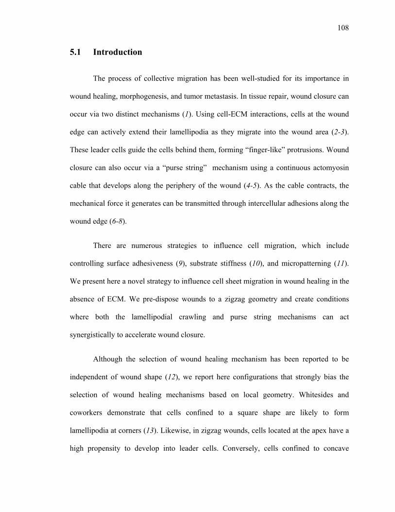
that the resulting contractile forces will increase with decreasing angle (Figure 5.1).
Figure 5.1 Schematic of wound closure in zigzag wounds and the effect of angles on purse
string contraction. Cells at the apex develop into leader cells and create traction forces (F, green
arrows) that are transmitted through the neighboring actomyosin cables (red arrows). The
increased tension, T strengthens the contraction forces (f, black arrows) in the actomyosin purse
string, drawing the cell sheet forward. The propensity for leader cell formation increases with
decreasing angle θ at the apex, and contraction forces are expected to increase with decreasing
angle.
110
5.2 Materials and methods
Cell culture
Madin-Darby Canine Kidney (MDCK) cells were cultured in Dulbecco’s
modified eagle medium (DMEM) containing 10% fetal bovine serum, 1%
penicillin/streptomycin, and phenol-red (growth medium). All experiments were
performed in DMEM lacking phenol-red, supplemented with 1% penicillin/streptomycin
(serum-free medium).
Wound healing with micropatterned PDMS blocks
The micropatterned PDMS blocks were fabricated as described in Chapter 4. The
wound features were designed using DesignCAD software and printed on transparencies
as positive photomasks (CAD/Art Services, Inc., Bandon, OR) as shown in Figure 5.2A.
Similarly, the wound healing assay used in this work was described in Chapter 4 (Figure
5.2A).
Fluorescence imaging
The MDCK cell monolayers were wounded and maintained in serum-free media
at 37 ºC for 4 h. Cells were fixed with pre-warmed 3.7% paraformaldehyde in PBS at pH
7.5 for 20 min at 37 ºC. After washing with PBS, cells were blocked in blocking solution
(10% FCS, 5% sucrose, 2% BSA in PBS) for 30 min at room temperature. To visualize
actin, cells were incubated with phalloidin-rhodamine (1:50 in PBS, Molecular Probes,
Inc., Eugene, OR) for 1 h 37 ºC. Glass coverslips were mounted with 1:1 glycerol in PBS
111
and imaged using an Zeiss Axiovert 200 M microscope with epifluorescence optics and
AxioVision LE software.
Data analysis
The wound areas demarcated by the red box (Figure 5.3B) were traced manually
using ImageJ v1.42 (NIH). The displacement of the cell sheet in the direction of the
wound was calculated by dividing the change in the wound area by the length of the
wound (l) at various time points. The point on the wound edge furthest from the apex was
followed for 24 h using MTrackJ, an ImageJ plugin developed by Meijering and
coworkers at the Biomedical Imaging Group Rotterdam. The displacement was averaged
for all videos and plotted as a function of time for each angle. The data was fitted to a
linear fit and the slopes resulting from the fit were reported as contractile speeds (µm/h).
Statistical Analysis
For all experimental data, the statistical significance of differences was estimated
by analysis of variance followed by the Tukey test. Differences were taken to be
significant at P ≤ 0.05.
112
5.3 Results and discussion
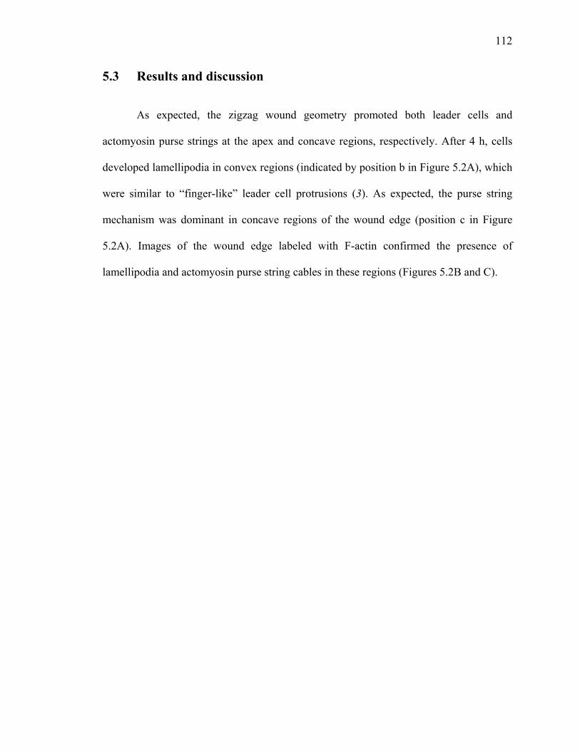
As expected, the zigzag wound geometry promoted both leader cells and
actomyosin purse strings at the apex and concave regions, respectively. After 4 h, cells
developed lamellipodia in convex regions (indicated by position b in Figure 5.2A), which
were similar to “finger-like” leader cell protrusions (3). As expected, the purse string
mechanism was dominant in concave regions of the wound edge (position c in Figure
5.2A). Images of the wound edge labeled with F-actin confirmed the presence of
lamellipodia and actomyosin purse string cables in these regions (Figures 5.2B and C).
113
A
Figure 5.2 Initial wound geometry determines mode of wound healing. (A) Schematic of
wound healing assay. The PDMS micropatterns were released and MDCK cell monolayers were
allowed to migrate on glass surfaces for 4 h in serum-free media. Cells were fixed and stained
with phalloidin-rhodamine. (B, C) Fluorescence images show the formation of leader cells (white
arrow) and purse string structures (white triangles) after 4 h, corresponding to positions b and c as
shown in A. Scale bars represent 20 µm.
B C
B
glass
Time lapse Microscopy
1% Pluronic
Fibronectin b
c
micropatterned PDMS
114
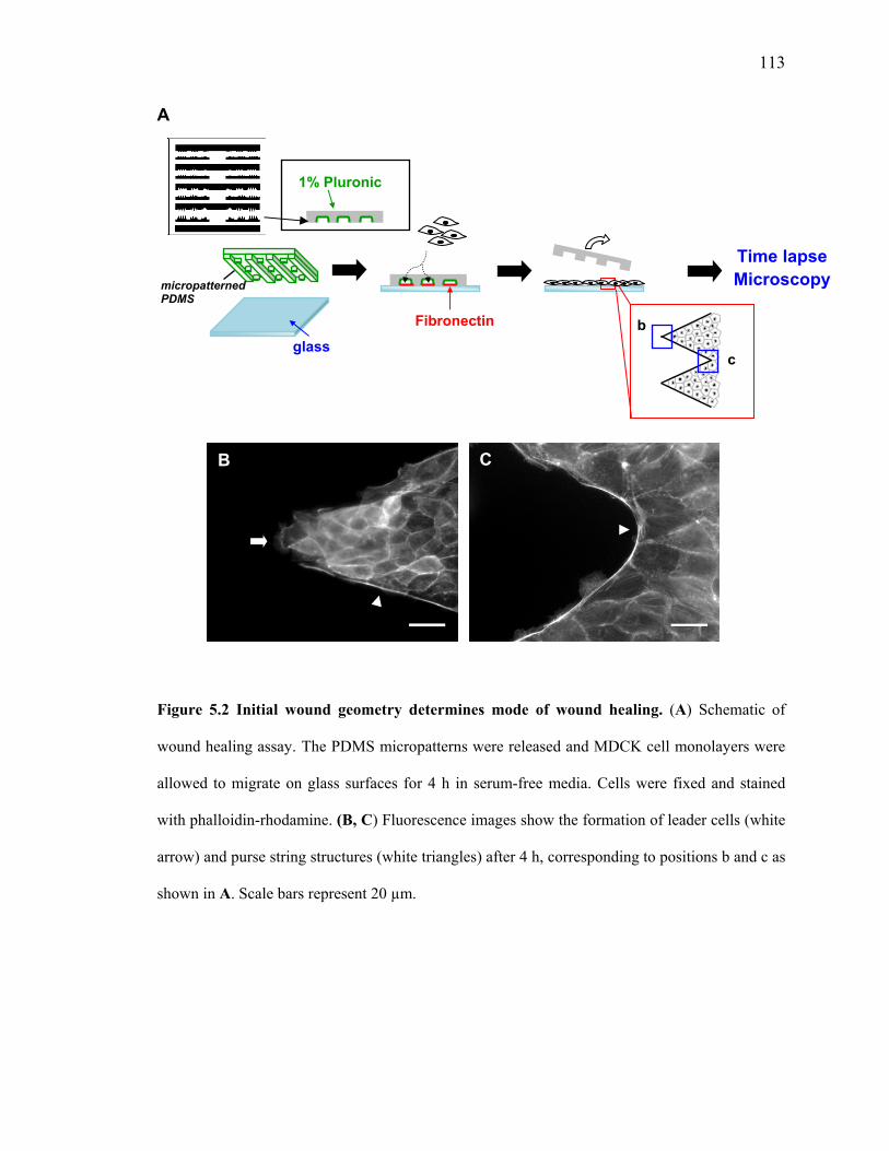
Figure 5.3A shows time-lapse images of zigzag wounds for varying angles 2θ.
For all zigzag wounds with 2θ = 45°, cells in the convex regions extended their
lamellipodia, but did not migrate into the wound area. Instead, cells often rearranged
themselves at the corners while preserving the “finger-like” patterns. In the concave
regions of the cell sheet, purse strings contracted in the direction of the wound, propelling
the cell sheet forward. At later times (> 20 h), the finger-like patterns also became motile.
Wounds with 2θ = 45° are most likely to promote the formation of leader cells.
Similar behavior was also observed in zigzag wounds with larger angles.
Although cells at the apex extend lamellipodia, these finger-like features were quickly re-
integrated into the cell sheet. Nonetheless, purse string contractions were also observed in
the concave regions, giving rise to net movement in the direction of the wound. In
comparison, minimal migration was observed on the straight-edged wounds (i.e., 2θ =
180°).
To quantify the wound closure rates, we manually tracked the change in the
wound area over time (Figure 5.3B). We represented the displacement of the cell sheet by
dividing the change in the wound area by the length of the cell sheet, l (Figure 5.3C). The
wound closure rates were obtained by fitting the data from time interval t = 12 h to 22 h
to a linear fit (Table 5.1).
115
A
2θ = 45° 2θ = 90° 2θ = 120° 2θ = 180° 0
h
10 h
24 h
B C
Figure 5.3 Time course of wound healing for zigzag wounds. (A) Time-lapse images of
various zigzag wounds with angles 2θ. Scale bar represents 100 µm for all images. (B)
Schematic of the wound closure analysis. The length of the wound (l) is highlighted in black. (C)
The displacement of the cell sheet over time. Cell sheet displacement for each time point was
0
50
100
150
200
0 4 8 12 16 20 24
Cel
l sh
eet
dis
pla
cem
ent
(µm
)
Time (h)
45°
90°
120°
180°
2θ l
116
calculated by measuring the change in wound area within the red box and divided by the length of
the wound (l). Data are means ± SEM for at least 10 independent experiments.
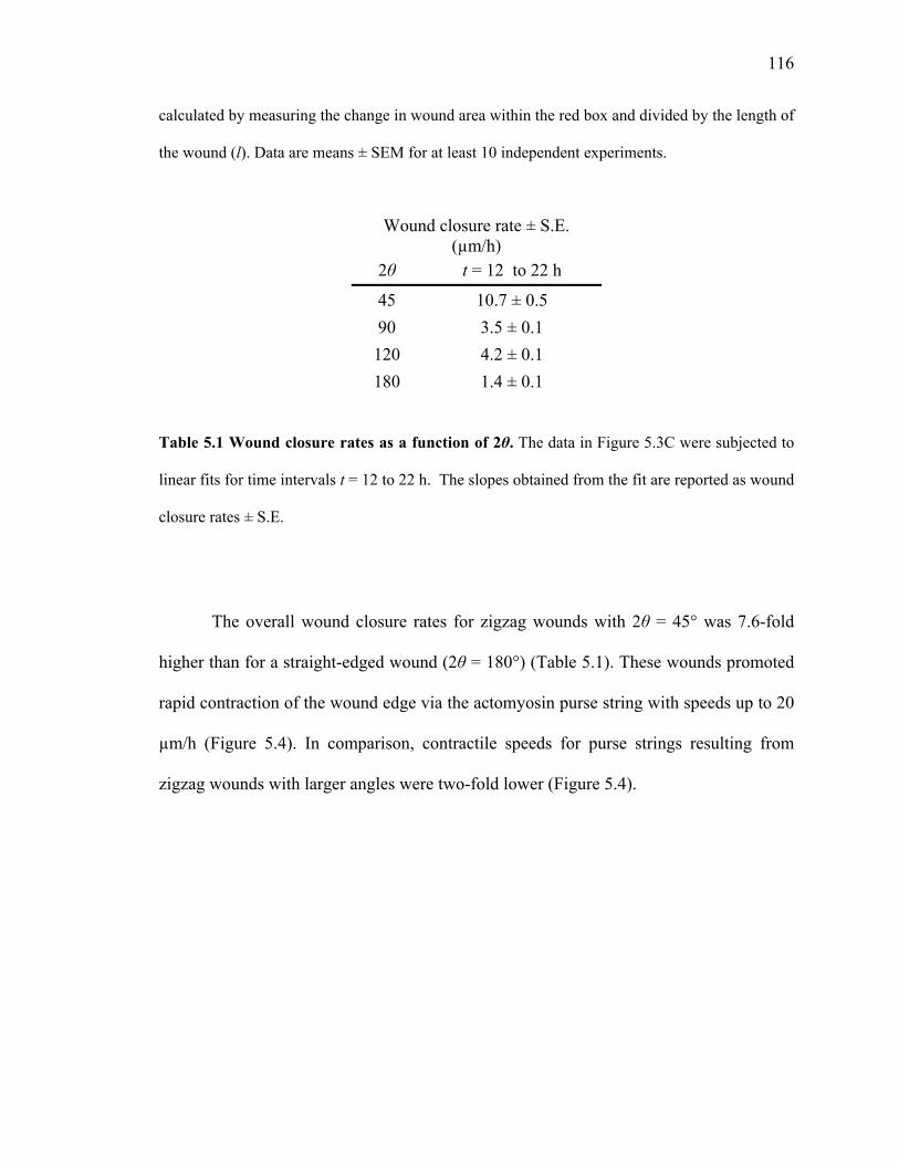
Table 5.1 Wound closure rates as a function of 2θ. The data in Figure 5.3C were subjected to
linear fits for time intervals t = 12 to 22 h. The slopes obtained from the fit are reported as wound
closure rates ± S.E.
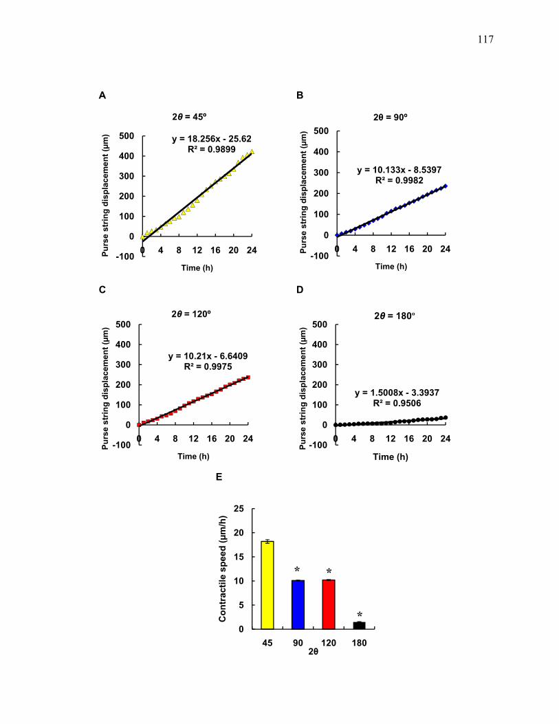
The overall wound closure rates for zigzag wounds with 2θ = 45° was 7.6-fold
higher than for a straight-edged wound (2θ = 180°) (Table 5.1). These wounds promoted
rapid contraction of the wound edge via the actomyosin purse string with speeds up to 20
µm/h (Figure 5.4). In comparison, contractile speeds for purse strings resulting from
zigzag wounds with larger angles were two-fold lower (Figure 5.4).
Wound closure rate ± S.E.
(µm/h) 2θ t = 12 to 22 h
45 10.7 ± 0.5
90 3.5 ± 0.1
120 4.2 ± 0.1
180 1.4 ± 0.1
117
A B
C D
E
y = 18.256x - 25.62R² = 0.9899
-100
0
100
200
300
400
500
0 4 8 12 16 20 24Pu
rse
stri
ng
dis
pla
cem
ent
(µm
)
Time (h)
2θ = 45º
y = 10.133x - 8.5397R² = 0.9982
-100
0
100
200
300
400
500
0 4 8 12 16 20 24Pu
rse
stri
ng
dis
pla
cem
ent
(µm
)
Time (h)
2θ = 90º
y = 10.21x - 6.6409R² = 0.9975
-100
0
100
200
300
400
500
0 4 8 12 16 20 24
Pu
rse
stri
ng
dis
pla
cem
ent
(µm
)
Time (h)
2θ = 120º
y = 1.5008x - 3.3937R² = 0.9506
-100
0
100
200
300
400
500
0 4 8 12 16 20 24
Pu
rse
stri
ng
dis
pla
cem
ent
(µm
)
Time (h)
2θ = 180º
0
5
10
15
20
25
45 90 120 180
Co
ntr
acti
le s
pee
d (
µm
/h)
2θ
**
*
118
Figure 5.4 Overall contractile speed of purse string as a function of 2θ. The point on
the wound edge furthest away from the apex was tracked manually for 24 h and the
displacement of the purse string was measured over time. (A to D) Average purse string
displacement over time of all the videos analyzed. (E) Contractile speed of purse string
for zigzag wounds of with angles 2θ. Data are slopes from fitted data shown in A to D.
Error bars are standard errors calculated from the fit. *, significant difference from 2θ =
45°.
5.4 Conclusions
In summary, we demonstrate the synergistic use of both wound healing modes to
accelerate wound closure. We also demonstrate that cells at the wound edge can be made
to adopt specific wound healing mechanisms based on the local geometry. By patterning
wound geometry in a zigzag shape, we show that cells at the apex develop into leader
cells and cells at concave regions activate the actomyosin purse string. Under these
conditions, leader cells generate traction forces that are transmitted through the
actomyosin purse strings. The increased tension results in higher contractile forces,
pulling the cell sheet forward. As a result of the strengthened purse string contraction,
wound closure rates were increased nearly eight-fold in zigzag wounds with the smallest
angles (i.e. 2θ = 45°), compared to wounds with straight edges.
119
5.5 Acknowledgements
I thank Dr. Ouyang Mingxing for help with producing the CAD drawing for the
micropatterns and Elowitz lab for the MDCK cells. This work is funded by the NIH.
5.6 References
1. P. Martin, J. Lewis, Nat. Lett. 360, 179 (1992).
2. G. Fenteany, P. A. Janmey, T. P. Stossel, Curr. Biol. 10, 831 (2000).
3. T. Omelchenko, J. M. Vasiliev, I. M. Gelfand, H. H. Feder, E. M. Bonder, Proc. Natl. Acad. Sci. USA 100, 10788 (2003).
4. W. M. Bement, P. Forscher, M. S. Mooseker, J. Cell Biol. 121, 565 (1993).
5. D. P. Kiehart, Curr. Biol. 9, R602 (1999).
6. M. Tamada, T. D. Perez, W. J. Nelson, M. P. Sheetz, J. Cell Biol. 176, 27 (2007).
7. Y. Danjo, I. K. Gipson, J. Cell Sci. 111, 3323 (1998).
8. O. M. Rossier et al., EMBO J. 29, 1055 (2010).
9. S. P. Palecek, J. C. Loftus, M. H. Ginsberg, D. A. Lauffenburger, Nature 385, 537 (1997).
10. C. M. Lo, H. B. Wang, M. Dembo, Y-L. Wang, Biophys. J. 79, 144 (2000).
11. X. Jiang, D. A. Bruzewicz., A. P. Wong, M. Piel, G. M. Whitesides, Proc. Natl. Acad. Sci. USA 102, 975 (2005).
12. S. Grasso, J. A. Hernandez, S. Chifflet, Am. J. Physiol. Cell Physiol. 293, C1327 (2007).
13. K. K. Parker et al., FASEB J. 16, 1195 (2002).
![[thesis.library.caltech.edu] - Welcome to …thesis.library.caltech.edu/7076/1/Frydman_Thesis2012.pdfTitle Microsoft Word - Frydman_Thesis2012.docx](https://img.pdfslide.us/doc/110x75/5b2f75657f8b9a55208ceae1/-welcome-to-microsoft-word-frydmanthesis2012docx.jpg)