Embed Size (px)
Citation preview
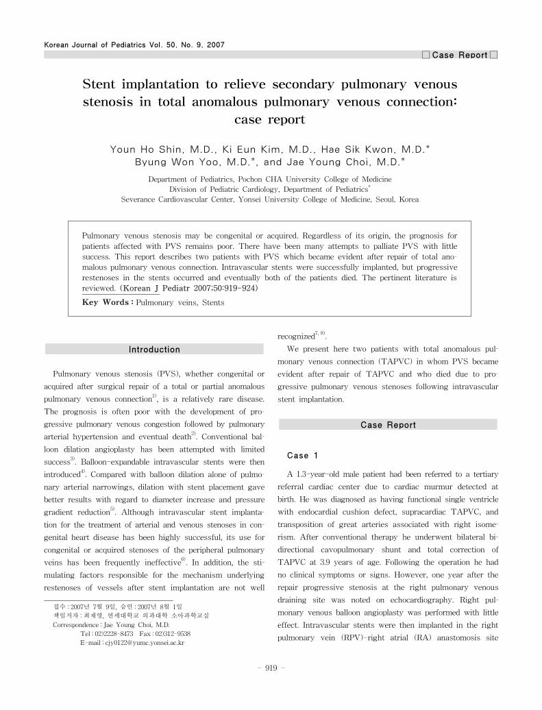
Korean Journal of Pediatrics Vol. 50, No. 9, 2007
□ Case Report□
1)
Introduction
Pulmonary venous stenosis (PVS), whether congenital or
acquired after surgical repair of a total or partial anomalous
pulmonary venous connection1), is a relatively rare disease.
The prognosis is often poor with the development of pro-
gressive pulmonary venous congestion followed by pulmonary
arterial hypertension and eventual death2). Conventional bal-
loon dilation angioplasty has been attempted with limited
success3). Balloon-expandable intravascular stents were then
introduced4). Compared with balloon dilation alone of pulmo-
nary arterial narrowings, dilation with stent placement gave
better results with regard to diameter increase and pressure
gradient reduction5). Although intravascular stent implanta-
tion for the treatment of arterial and venous stenoses in con-
genital heart disease has been highly successful, its use for
congenital or acquired stenoses of the peripheral pulmonary
veins has been frequently ineffective6). In addition, the sti-
mulating factors responsible for the mechanism underlying
restenoses of vessels after stent implantation are not well
접수 : 2007년 7월 9일, 승인 : 2007년 8월 1일
책임저자 :최재 , 연세대학교 의과대학 소아과학교실
Correspondence : Jae Young Choi, M.D.
Tel : 02)2228-8473 Fax : 02)312-9538
E-mail : [email protected]
recognized7, 8).
We present here two patients with total anomalous pul-
monary venous connection (TAPVC) in whom PVS became
evident after repair of TAPVC and who died due to pro-
gressive pulmonary venous stenoses following intravascular
stent implantation.
Case Report
Case 1
A 1.3-year-old male patient had been referred to a tertiary
referral cardiac center due to cardiac murmur detected at
birth. He was diagnosed as having functional single ventricle
with endocardial cushion defect, supracardiac TAPVC, and
transposition of great arteries associated with right isome-
rism. After conventional therapy he underwent bilateral bi-
directional cavopulmonary shunt and total correction of
TAPVC at 3.9 years of age. Following the operation he had
no clinical symptoms or signs. However, one year after the
repair progressive stenosis at the right pulmonary venous
draining site was noted on echocardiography. Right pul-
monary venous balloon angioplasty was performed with little
effect. Intravascular stents were then implanted in the right
pulmonary vein (RPV)-right atrial (RA) anastomosis site
Stent implantation to relieve secondary pulmonary venous
stenosis in total anomalous pulmonary venous connection:
case report
Youn Ho Shin, M.D., Ki Eun Kim, M.D., Hae Sik Kwon, M.D.*
Byung Won Yoo, M.D.*, and Jae Young Choi, M.D.*
Department of Pediatrics, Pochon CHA University College of Medicine
Division of Pediatric Cardiology, Department of Pediatrics*
Severance Cardiovascular Center, Yonsei University College of Medicine, Seoul, Korea
Pulmonary venous stenosis may be congenital or acquired. Regardless of its origin, the prognosis for
patients affected with PVS remains poor. There have been many attempts to palliate PVS with little
success. This report describes two patients with PVS which became evident after repair of total ano-
malous pulmonary venous connection. Intravascular stents were successfully implanted, but progressive
restenoses in the stents occurred and eventually both of the patients died. The pertinent literature is
reviewed. (Korean J Pediatr 2007;50:919-924)
Key Words : Pulmonary veins, Stents
- 919 -
Youn Ho Shin, et al. : Stent implantation for pulmonary venous stenosis
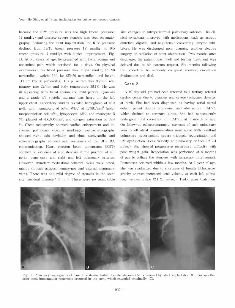
because the RPV pressure was too high (mean pressure:
17 mmHg) and discrete severe stenosis was seen on angio-
graphy. Following the stent implantation, the RPV pressure
declined from 19/15 (mean pressure 17: mmHg) to 9/5
(mean pressure: 7 mmHg) with clinical improvement (Fig.
1). At 5.5 years of age, he presented with facial edema and
abdominal pain which persisted for 4 days. On physical
examination, his blood pressure was 110/70 mmHg (75-90
percentiles), weight 16.1 kg (25-50 percentiles) and height
111 cm (25-50 percentiles). His pulse rate was 95/min, res-
piratory rate 22/min and body temperature 36.7℃. He was
ill appearing with facial edema and mild perioral cyanosis
and a grade 3/6 systolic murmur was heard on the left
upper chest. Laboratory studies revealed hemoglobin of 15.2
g/dL with hematocrit of 53%, WBC of 12,990/mm3(poly-
morphonuclear cell 46%, lymphocyte 43%, and monocyte 5
%), platelet of 466,000/mm3, and oxygen saturation of 78.4
%. Chest radiography showed cardiac enlargement and in-
creased pulmonary vascular markings, electrocardiography
showed right axis deviation and sinus tachycardia, and
echocardiography showed mild restenosis of the RPV-RA
communication. Heart electron beam tomograms (EBT)
showed no evidence of any stenosis at the junction of su-
perior vena cava and right and left pulmonary arteries.
However, abundant mediastinal collateral veins were noted,
mainly through azygos, hemiazygos and internal mammary
veins. There was still mild degree of stenosis in the stent
site (residual diameter: 3 mm). There were no remarkable
size changes in intrapericardial pulmonary arteries. His cli-
nical symptoms improved with medications, such as aspirin,
diuretics, digoxin, and angiotensin-converting enzyme inhi-
bitors. He was discharged upon planning another elective
surgery or redilation of stent obstruction. Two months after
discharge, the patient was, well and further treatment was
delayed due to his parents request. Six months following
the procedure, he suddenly collapsed showing circulation
dysfunction and died.
Case 2
A 10 day-old girl had been referred to a tertiary referral
cardiac center due to cyanosis and severe tachypnea detected
at birth. She had been diagnosed as having atrial septal
defect, patent ductus arteriosus, and obstructive TAPVC
which drained to coronary sinus. She had subsequently
undergone total correction of TAPVC at 1 month of age.
On follow-up echocardiography, stenoses of each pulmonary
vein to left atrial communication were noted with resultant
pulmonary hypertension, severe tricuspid regurgitation and
RV dysfunction (Peak velocity at pulmonary orifice: 2.2-2.4
m/sec). She showed progressive respiratory difficulty with
poor weight gain. Reoperation was performed at 9 months
of age to palliate the stenoses with temporary improvement.
Restenoses occurred within a few months. At 1 year of age,
she was readmitted due to shortness of breath. Echocardio-
graphy showed increased peak velocity at each left pulmo-
nary venous orifice (2.1-2.3 m/sec). Trido repair (patch en-
Fig. 1. Pulmonary angiograms of case 1 is shown. Initial discrete stenosis (A) is relieved by stent implantation (B). Six monthsafter stent implantation restenosis occurred in the stent which extended proximally (C).
- 920 -
Korean J Pediatr : 제 50 권 제 8 호 2007년
largement of left pulmonary veins) was performed and her
clinical symptoms improved with a decline of the peak
velocity at PV orifice to around 1.5 m/sec. However, PVS
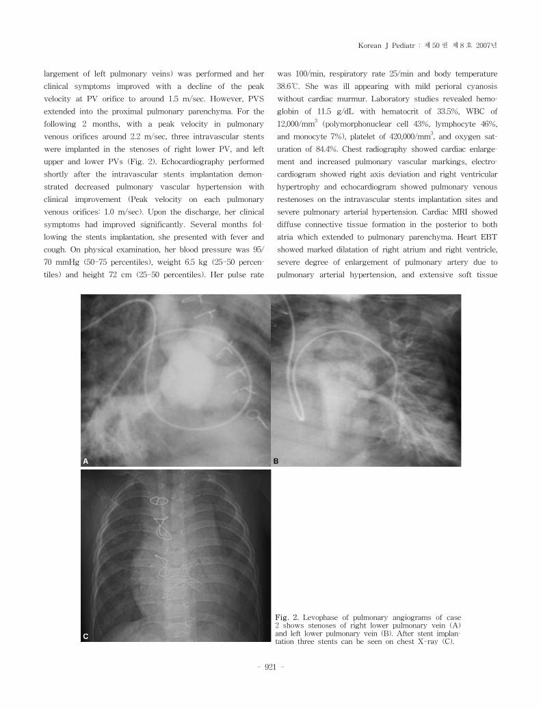
extended into the proximal pulmonary parenchyma. For the
following 2 months, with a peak velocity in pulmonary
venous orifices around 2.2 m/sec, three intravascular stents
were implanted in the stenoses of right lower PV, and left
upper and lower PVs (Fig. 2). Echocardiography performed
shortly after the intravascular stents implantation demon-
strated decreased pulmonary vascular hypertension with
clinical improvement (Peak velocity on each pulmonary
venous orifices: 1.0 m/sec). Upon the discharge, her clinical
symptoms had improved significantly. Several months fol-
lowing the stents implantation, she presented with fever and
cough. On physical examination, her blood pressure was 95/
70 mmHg (50-75 percentiles), weight 6.5 kg (25-50 percen-
tiles) and height 72 cm (25-50 percentiles). Her pulse rate
was 100/min, respiratory rate 25/min and body temperature
38.6℃. She was ill appearing with mild perioral cyanosis
without cardiac murmur. Laboratory studies revealed hemo-
globin of 11.5 g/dL with hematocrit of 33.5%, WBC of
12,000/mm3(polymorphonuclear cell 43%, lymphocyte 46%,
and monocyte 7%), platelet of 420,000/mm3, and oxygen sat-
uration of 84.4%. Chest radiography showed cardiac enlarge-
ment and increased pulmonary vascular markings, electro-
cardiogram showed right axis deviation and right ventricular
hypertrophy and echocardiogram showed pulmonary venous
restenoses on the intravascular stents implantation sites and
severe pulmonary arterial hypertension. Cardiac MRI showed
diffuse connective tissue formation in the posterior to both
atria which extended to pulmonary parenchyma. Heart EBT
showed marked dilatation of right atrium and right ventricle,
severe degree of enlargement of pulmonary artery due to
pulmonary arterial hypertension, and extensive soft tissue
Fig. 2. Levophase of pulmonary angiograms of case2 shows stenoses of right lower pulmonary vein (A)and left lower pulmonary vein (B). After stent implan-tation three stents can be seen on chest X-ray (C).
- 921 -
Youn Ho Shin, et al. : Stent implantation for pulmonary venous stenosis
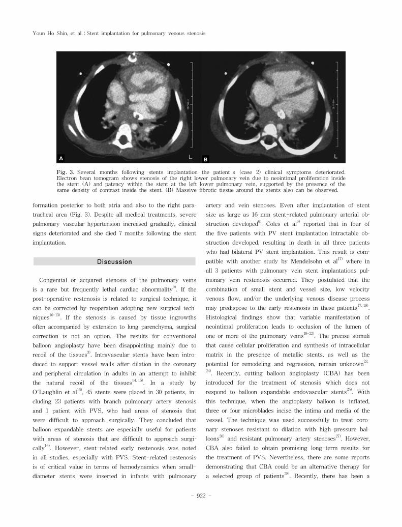
formation posterior to both atria and also to the right para-
tracheal area (Fig. 3). Despite all medical treatments, severe
pulmonary vascular hypertension increased gradually, clinical
signs deteriorated and she died 7 months following the stent
implantation.
Discussion
Congenital or acquired stenosis of the pulmonary veins
is a rare but frequently lethal cardiac abnormality9). If the
post-operative restenosis is related to surgical technique, it
can be corrected by reoperation adopting new surgical tech-
niques10-13). If the stenosis is caused by tissue ingrowths
often accompanied by extension to lung parenchyma, surgical
correction is not an option. The results for conventional
balloon angioplasty have been disappointing mainly due to
recoil of the tissues3). Intravascular stents have been intro-
duced to support vessel walls after dilation in the coronary
and peripheral circulation in adults in an attempt to inhibit
the natural recoil of the tissues14, 15). In a study by
O'Laughlin et al16), 45 stents were placed in 30 patients, in-
cluding 23 patients with branch pulmonary artery stenosis
and 1 patient with PVS, who had areas of stenosis that
were difficult to approach surgically. They concluded that
balloon expandable stents are especially useful for patients
with areas of stenosis that are difficult to approach surgi-
cally16). However, stent-related early restenosis was noted
in all studies, especially with PVS. Stent-related restenosis
is of critical value in terms of hemodynamics when small-
diameter stents were inserted in infants with pulmonary
artery and vein stenoses. Even after implantation of stent
size as large as 16 mm stent-related pulmonary arterial ob-
struction developed6). Coles et al
6)reported that in four of
the five patients with PV stent implantation intractable ob-
struction developed, resulting in death in all three patients
who had bilateral PV stent implantation. This result is com-
patible with another study by Mendelsohn et al17)where in
all 3 patients with pulmonary vein stent implantations pul-
monary vein restenosis occurred. They postulated that the
combination of small stent and vessel size, low velocity
venous flow, and/or the underlying venous disease process
may predispose to the early restenosis in these patients17, 18).
Histological findings show that variable manifestation of
neointimal proliferation leads to occlusion of the lumen of
one or more of the pulmonary veins19-22). The precise stimuli
that cause cellular proliferation and synthesis of intracellular
matrix in the presence of metallic stents, as well as the
potential for remodeling and regression, remain unknown23,
24). Recently, cutting balloon angioplasty (CBA) has been
introduced for the treatment of stenosis which does not
respond to balloon expandable endovascular stents25). With
this technique, when the angioplasty balloon is inflated,
three or four microblades incise the intima and media of the
vessel. The technique was used successfully to treat coro-
nary stenoses resistant to dilation with high-pressure bal-
loons26)and resistant pulmonary artery stenoses
27). However,
CBA also failed to obtain promising long-term results for
the treatment of PVS. Nevertheless, there are some reports
demonstrating that CBA could be an alternative therapy for
a selected group of patients28). Recently, there has been a
Fig. 3. Several months following stents implantation the patient s (case 2) clinical symptoms deteriorated.Electron bean tomogram shows stenosis of the right lower pulmonary vein due to neointimal proliferation insidethe stent (A) and patency within the stent at the left lower pulmonary vein, supported by the presence of thesame density of contrast inside the stent. (B) Massive fibrotic tissue around the stents also can be observed.
- 922 -
Korean J Pediatr : 제 50 권 제 8 호 2007년
report suggesting that a combined approach of using cutting
balloons followed by insertion of drug-eluting stents may,
in the future, provide the best treatment for PVS2).
These two cases showed disappointing results, consistent
with previous studies. We speculate that more research
and experience would be necessary to have long-lasting
stents patency for PVS.
한 요 약
총 폐정맥 환류 이상 완전교정술 후 이차적으로
발생한 폐정맥 협착에서 스텐트 삽입술 2례
포천중문 의과대학교 소아과학교실
연세대학교 의과대학 소아과학교실*
심장혈관병원 소아심장과
신윤호·김기은·권해식*·유병원
*·최재
*
폐정맥 협착은 선천적 또는 총 폐정맥 환류 이상 완전교정술
후에 이차적으로 발생한다. 선천적, 후천적 폐정맥 협착은 많은
연구에도 불구하고 예후가 대단히 불량한 것으로 알려져 있다.
저자들은 총 폐정맥 환류 이상 완전교정술 후에 이차적으로 발
생한 폐정맥 협착 환아 2명에게 풍선 혈관 성형술 후 협착부가
다시 좁아져서 스텐트 삽입술을 시행하여 증상이 호전되다가 다
시 진행성 협착이 발생하여 사망한 2례를 보고하는 바이다.
References
1) van Son JA, Danielson GK, Puga FJ, Edwards WD, Driscoll
DJ. Repair of congenital and acquired pulmonary vein ste-
nosis. Ann Thorac Surg 1995;60:144-50.
2) Seale AN, Daubeney PE, Magee AG, Rigby ML. Pulmonary
vein stenosis: initial experience with cutting balloon angio-
plasty. Heart 2006;92:815-20.
3) Lock JE, Bass JL, Castaneda-Zuniga W, Fuhrman BP,
Rashkind WJ, Lucas RV, Jr. Dilation angioplasty of con-
genital or operative narrowings of venous channels. Circu-
lation 1984;70:457-64.
4) Mullins CE, O'Laughlin MP, Vick GW, 3rd, Mayer DC,
Myers TJ, Kearney DL, et al. Implantation of balloon-ex-
pandable intravascular grafts by catheterization in pulmonary
arteries and systemic veins. Circulation 1988;77:188-99.
5) Rothman A, Perry SB, Keane JF, Lock JE. Early results
and follow-up of balloon angioplasty for branch pulmonary
artery stenoses. J Am Coll Cardiol 1990;15:1109-17.
6) Coles JG, Yemets I, Najm HK, Lukanich JM, Perron J,
Wilson GJ, et al. Experience with repair of congenital heart
defects using adjunctive endovascular devices. J Thorac
Cardiovasc Surg 1995;110:1513-9; discussion 9-20.
7) Gordon PC, Gibson CM, Cohen DJ, Carrozza JP, Kuntz RE,
Baim DS. Mechanisms of restenosis and redilation within
coronary stents--quantitative angiographic assessment. J Am
Coll Cardiol 1993;21:1166-74.
8) Palmaz JC. Intravascular stents: tissue-stent interactions
and design considerations. AJR Am J Roentgenol 1993;160
:613-8.
9) Driscoll DJ, Hesslein PS, Mullins CE. Congenital stenosis of
individual pulmonary veins: clinical spectrum and unsuccess-
ful treatment by transvenous balloon dilation. Am J Cardiol
1982;49:1767-72.
10) Caldarone CA, Najm HK, Kadletz M, Smallhorn JF, Freedom
RM, Williams WG, et al. Relentless pulmonary vein stenosis
after repair of total anomalous pulmonary venous drainage.
Ann Thorac Surg 1998;66:1514-20.
11) Najm HK, Caldarone CA, Smallhorn J, Coles JG. A suture-
less technique for the relief of pulmonary vein stenosis with
the use of in situ pericardium. J Thorac Cardiovasc Surg
1998;115:468-70.
12) Lacour-Gayet F, Zoghbi J, Serraf AE, Belli E, Piot D, Rey
C, et al. Surgical management of progressive pulmonary
venous obstruction after repair of total anomalous pulmonary
venous connection. J Thorac Cardiovasc Surg 1999;117:679-
87.
13) Hyde JA, Stumper O, Barth MJ, Wright JG, Silove ED, de
Giovanni JV, et al. Total anomalous pulmonary venous
connection: outcome of surgical correction and management
of recurrent venous obstruction. Eur J Cardiothorac Surg
1999;15:735-40; discussion 40-1.
14) Levine MJ, Leonard BM, Burke JA, Nash ID, Safian RD,
Diver DJ, et al. Clinical and angiographic results of balloon-
expandable intracoronary stents in right coronary artery
stenoses. J Am Coll Cardiol 1990;16:332-9.
15) Palmaz JC, Richter GM, Noeldge G, Schatz RA, Robison PD,
Gardiner GA, Jr., et al. Intraluminal stents in atherosclerotic
iliac artery stenosis: preliminary report of a multicenter
study. Radiology 1988;168:727-31.
16) O'Laughlin MP, Perry SB, Lock JE, Mullins CE. Use of
endovascular stents in congenital heart disease. Circulation
1991;83:1923-39.
17) Mendelsohn AM, Bove EL, Lupinetti FM, Crowley DC, Lloyd
TR, Fedderly RT, et al. Intraoperative and percutaneous
stenting of congenital pulmonary artery and vein stenosis.
Circulation 1993;88:II210-7.
18) Nakib A, Moller JH, Kanjuh VI, Edwards JE. Anomalies of
the pulmonary veins. Am J Cardiol 1967;20:77-90.
19) Bini RM, Cleveland DC, Ceballos R, Bargeron LM, Jr.,
Pacifico AD, Kirklin JW. Congenital pulmonary vein stenosis.
Am J Cardiol 1984;54:369-75.
20) Haworth SG. Total anomalous pulmonary venous return.
Prenatal damage to pulmonary vascular bed and extrapul-
monary veins. Br Heart J 1982;48:513-24.
21) Shone JD, Amplatz K, Anderson RC, Adams P, Jr., Edwards
JE. Congenital stenosis of individual pulmonary veins. Cir-
culation 1962;26:574-81.
22) Edwards JE. Congenital stenosis of pulmonary veins. Path-
ologic and developmental considerations. Lab Invest 1960;9:
46-66.
23) Roubin GS, King SB, 3rd, Douglas JS, Jr., Lembo NJ,
Robinson KA. Intracoronary stenting during percutaneous
transluminal coronary angioplasty. Circulation 1990;81:IV92-
- 923 -
Youn Ho Shin, et al. : Stent implantation for pulmonary venous stenosis
100.
24) White CJ, Ramee SR, Banks AK, Mesa JE, Chokshi S, Isner
JM. A new balloon-expandable tantalum coil stent: angio-
graphic patency and histologic findings in an atherogenic
swine model. J Am Coll Cardiol 1992;19:870-6.
25) Suda K, Matsumura M, Hayashi H, Nishimura K. Compari-
son of efficacy of medium-sized cutting balloons versus
standard balloons for dilation of peripheral pulmonary sten-
osis. Am J Cardiol 2006;97:1060-3.
26) Unterberg C, Buchwald AB, Barath P, Schmidt T, Kreuzer
H, Wiegand V. Cutting balloon coronary angioplasty--initial
clinical experience. Clin Cardiol 1993;16:660-4.
27) Sugiyama H, Veldtman GR, Norgard G, Lee KJ, Chaturvedi
R, Benson LN. Bladed balloon angioplasty for peripheral
pulmonary artery stenosis. Catheter Cardiovasc Interv 2004;
62:71-7.
28) Rotzsch C, Wiener M, Henning B, Daehnert I. Stents for
treatment of obstructed pulmonary venous return[abstract].
Catheter Cardiovasc Interv 2004;63:125.
- 924 -