Embed Size (px)
Citation preview
4D Cardiac Reconstruction Using High
Resolution CT Images
Mingchen Gao1, Junzhou Huang1, Shaoting Zhang1,Zhen Qian2, Szilard Voros2, Dimitris Metaxas1, and Leon Axel3
1 CBIM Center, Rutgers University, Piscataway, NJ, 08550, USA2 2 Piedmont Heart Institute, Atlanta, GA, 30309, USA
3 New York University, 660 First Avenue, New York, NY, 10016, USA
Abstract. Recent developments on the 320 multi-detector CT technolo-gies have made the volumetric acquisition of 4D high resolution cardiacimages in a single heart beat possible. In this paper, we present a frame-work that uses these data to reconstruct the 4D motion of the endocardialsurface of the left ventricle (LV) for a full cardiac cycle. This reconstruc-tion framework captures the motion of the full 3D surfaces of the complexanatomical features, such as the papillary muscles and the ventricular tra-beculae, for the first time, which allows us to quantitatively investigatetheir possible functional significance in health and disease.
1 Introduction
Most countries face high and increasing rates of cardiovascular diseases. There-fore, it is critical to detect and diagnose such diseases in early stages. Computedtomography (CT) and Magnetic resonance imaging (MRI), as non-invasive meth-ods to generate three dimensional images of an organ, have been widely used overthe decades. If the detailed cardiac shape features are accurately reconstructedfrom these images, we can potentially obtain additional clinically valuable in-formation besides the widely used wall thickness and global function features.Many cardiac reconstruction methods have been developed to build 3D modelsof the heart from cardiac MR images, such as, but not limited to registration-based propagation framework [12] and deformable model [8] [9]. However, due tothe sparsity of the data in usual MRI data sets, important anatomic structures,such as the papillary muscles, the ventricular trabeculae and the valves, are oftendifficult to capture in this modality.
There has also been work on the 3D cardiac reconstruction from CT im-ages [10,7,2]. However, despite the higher level of structural detail potentiallyavailable in CT data, most of the prior work has not sought to capture the finerdetail structures of the myocardium, such as papillary muscles and trabeculae.The conventional approach to reconstructing cardiac structures from 3D images(e.g., for generating generic or patient-specific models of the heart) is a model-based one that uses a smooth parametric model to guide the segmentation ofthe cardiac structures from the 3D images. Such parametric models capture the
D.N. Metaxas and L. Axel (Eds.): FIMH 2011, LNCS 6666, pp. 153–160, 2011.c© Springer-Verlag Berlin Heidelberg 2011

154 M. Gao et al.
overall shape of the heart wall, but are too coarse to capture or incorporatemany of the finer scale anatomical features. For example, Zheng et al. proposedan automatic method to segment the four-chamber heart [10]. Their method em-ploys Active Shape Model to exploit a large database of annotated CT volumes.It efficiently segments all the four chambers of heart. However, the inner wall ofthe chambers is simply modeled as a smooth surface.
Recent advances in CT technology have made the acquisition of higher res-olution cardiac images possible, which can capture previously unseen cardiacstructure details. However, these anatomical details are currently only visual-ized using methods such as volume rendering, which do not lend themselves tothe quantitative analysis of the 3D anatomical structures. Segmentation and re-construction of the endocardial surface of the ventricles with incorporation offiner details can potentially greatly assist doctors in diagnosis and functionalassessment. Chen et al. proposed a hybrid framework for 3D cardiac reconstruc-tion [1]. That method has provided high resolution segmentation results of thecomplex cardiac structure. Their results captured the papillary muscles and de-tail structure of the myocardium. However, CT data sets from different timeframes were segmented independently. Prior model knowledge from neighboringtime frames is not used in their framework. Topological consistency betweenconsecutive time frames is not guaranteed, either.
For the case of 4D cardiac images, two problems have to be considered simul-taneously: 1) a heart wall segmentation problem in each image and 2) a trackingproblem of the left ventricle motion given the data set. In [3], Mcinerney et al.proposed a method for 4D cardiac reconstruction, in which the output of theprevious time frame is used as the initial guess for the current time frame inorder to do a sequential segmentation. They used a finite element surface modelwhich may not be able to handle topology changes. Montagnat et al. proposedan extended deformable framework by introducing time-dependent constraints.Thus, in addition to computing an internal force to enforce the regularity of thedeformable model, prior motion knowledge is introduced in the deformation pro-cess through either temporal smoothing or trajectory constraints [4]. However,these 4D reconstruction methods do not capture detailed features.
In this paper, we present a framework for 4D left ventricle (LV) segmentationwith inclusion of small scale anatomical features. Semi-automatic segmentationis used to get the initial segmentation from high resolution CT data for an initial(3D) frame of data. This semi-automatic segmentation is time consuming andtedious, so it is not efficient to use it for segmentation of all the frames. Theinitial high resolution mesh model is generated as an isosurface of the segmenta-tion. Geometric processing is then applied to the initial model to get a smoothand regular mesh with an appropriate number of vertices. Based on the initialmodel from one time frame, our method deforms it towards the boundaries onthe other frames. During the formation, the topology of the model is kept un-changed. We can also get the one-to-one correspondence between frames, as anadditional benefit during the segmentation process. With the one-to-one corre-spondence, we can easily do interpolation among different time frames to get a
4D Cardiac Reconstruction Using High Resolution CT Images 155
smoother heart cycle animation. We have applied our framework on a whole car-diac cycle. The results have been validated based on the ground truth segmentedby multiple clinical experts. These novel and powerful methods can extract thefull 3D surfaces of these complex anatomical structures, which allows us for thefirst time to quantitatively investigate their possible functional significance.
2 Methodology
We propose a framework to reconstruct the cardiac model. This framework in-cludes: initial model construction, deformable model based segmentation, andinterpolation between time frames. The initial model is generated using 3Dsnake-based segmentation on one time frame of the CT image. The initial modelneeds geometry processing, such as decimating, detail-preserving smoothing andisotropic remeshing to get high-quality meshes. Based on the initial model, seg-mentation of the rest of the CT images is automatically performed using thedeformable model. The segmentation of a sequence of CT images is interpolatedin time to get a higher effective temporal resolution.
2.1 Model Initialization
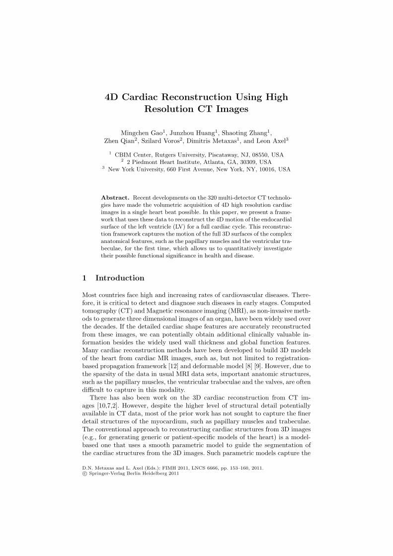
The model initialization framework is illustrated in Fig. 1.We use a 3D snake-based semi-automatic segmentation method to get the
initial model [11]. This segmentation process is very time consuming and couldnot be used to segment all frames. Usually it takes several hours to finish asemi-automatic segmentation on one time frame. However, once this model hasbeen generated, it is used to segment the rest of other frames automatically.
Segmentation results are represented as binary images. Isosurface detectionis then applied to generate the mesh. However, the resulting mesh is usuallybulky, noisy and irregular. To get a better initialization model, some geometricprocessing should be done on that mesh, such as decimating, detail-preservingsmoothing and isotropic remeshing. First, the initial model is too large to readilymodify, edge collapses are performed during decimation. After decimation, we geta mesh with much fewer vertices, but that still retains most of the shape details.The meshes have been decimated to about 20, 000 vertices. Detail-preservingsmoothing is then performed after decimation. The smoothing is restricted to
Decimation
IsosurfaceSemi-automaticSegmentationCT data
Isotropic
Remesher
Initialized
Mesh
Detail-preserving
Smoothing
Fig. 1. Initial model construction
156 M. Gao et al.
the tangential direction. Instead of moving each vertex towards the centroidof its neighbors, which would smooth out the shape details and sharp features,detail-preserving smoothing ensures higher quality meshes without losing details.Isotropic remeshing is important for the mesh quality. In irregular meshes, thevertices with high valences exert strong internal forces to drag other vertices,which can cause unrealistic results in deformable models [6]. An incrementalisotropic remeshing technique is used to remesh the given triangular mesh sothat all edges have approximately the same target edge length and the trianglesare as regular as possible. This process would generally be iterated several timesto get the final results.
After all these geometric processing steps, we finally get a high-quality tri-angular mesh with an appropriate number of vertices. This mesh is used as aninitialization for other frames.
2.2 Deformable Model Based Segmentation
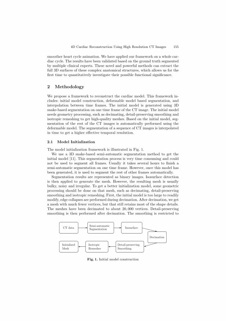
We want to deform our model normal to the boundaries during tracking. Todo so, we define an energy function, including a term, external energy, derivedfrom the image so that it is smaller at the boundaries. By minimizing the energyfunction, it drags the model towards the boundaries. We also want to keep theshape of the model unchanged during deformation. For that, we use anotherenergy term, model energy, which reflects the differences between the originalmodel and the deformed model.
Given a gray level image I(x, y) , viewed as a function of continuous positionvariables (x, y). The model Mt−1 derived from the previous frame is used to fitthe current frame Mt. The energy function we want to minimize is defined asfollows:
E(Mt, It, Mt−1) = Eext(Mt, It) + Emodel(Mt, Mt−1). (1)
The external energy Eext is designed to move the deformable model towardsobject boundaries.
Eext(Mt, It) = − |∇I|2 , (2)
where ∇ is the gradient operator.Model energy is defined as the differences of attribute vectors. An attribute
vector is attached to each vertex of the model [5], which reflects the geometricstructure of the model from a local to global level. In 3D, for a particular vertexVi, each attribute is defined as the volume of a tetrahedron on that vertex. Theother three vertices form the tetrahedron are randomly chosen from the lth levelneighborhood of Vi. Smaller tetrahedrons reflect the local structure near a vertexwhile larger tetrahedrons reflect a more global information around a vertex. Theattribute vector, if sufficient enough, uniquely characterizes different parts of asurface of a boundary. The normalized attribute vectors are affine-invariant [5].
The volume of a tetrahedron is defined as fl(Vi). The attribute vector on avertex is defined as:
F (Vi) = [f1(Vi), f2(Vi), ..., fR(Vi)(Vi)], (3)
where R(Vi) is the neighborhood layers we want to use around Vi.
4D Cardiac Reconstruction Using High Resolution CT Images 157
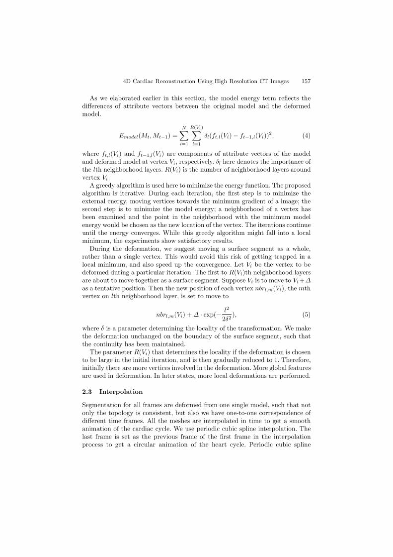
As we elaborated earlier in this section, the model energy term reflects thedifferences of attribute vectors between the original model and the deformedmodel.
Emodel(Mt, Mt−1) =N∑
i=1
R(Vi)∑
l=1
δl(ft,l(Vi) − ft−1,l(Vi))2, (4)
where ft,l(Vi) and ft−1,l(Vi) are components of attribute vectors of the modeland deformed model at vertex Vi, respectively. δl here denotes the importance ofthe lth neighborhood layers. R(Vi) is the number of neighborhood layers aroundvertex Vi.
A greedy algorithm is used here to minimize the energy function. The proposedalgorithm is iterative. During each iteration, the first step is to minimize theexternal energy, moving vertices towards the minimum gradient of a image; thesecond step is to minimize the model energy; a neighborhood of a vertex hasbeen examined and the point in the neighborhood with the minimum modelenergy would be chosen as the new location of the vertex. The iterations continueuntil the energy converges. While this greedy algorithm might fall into a localminimum, the experiments show satisfactory results.
During the deformation, we suggest moving a surface segment as a whole,rather than a single vertex. This would avoid this risk of getting trapped in alocal minimum, and also speed up the convergence. Let Vi be the vertex to bedeformed during a particular iteration. The first to R(Vi)th neighborhood layersare about to move together as a surface segment. Suppose Vi is to move to Vi+Δas a tentative position. Then the new position of each vertex nbrl,m(Vi), the mthvertex on lth neighborhood layer, is set to move to
nbrl,m(Vi) + Δ · exp(− l2
2δ2), (5)
where δ is a parameter determining the locality of the transformation. We makethe deformation unchanged on the boundary of the surface segment, such thatthe continuity has been maintained.
The parameter R(Vi) that determines the locality if the deformation is chosento be large in the initial iteration, and is then gradually reduced to 1. Therefore,initially there are more vertices involved in the deformation. More global featuresare used in deformation. In later states, more local deformations are performed.
2.3 Interpolation
Segmentation for all frames are deformed from one single model, such that notonly the topology is consistent, but also we have one-to-one correspondence ofdifferent time frames. All the meshes are interpolated in time to get a smoothanimation of the cardiac cycle. We use periodic cubic spline interpolation. Thelast frame is set as the previous frame of the first frame in the interpolationprocess to get a circular animation of the heart cycle. Periodic cubic spline
158 M. Gao et al.
interpolation makes heart meshes continuous on the second derivatives. Theinterpolation results are used in simulation of blood flow in the left ventricles.
3 Results and Validation
We applied our reconstruction framework to 10 cardiac CT volumes, which cap-tures a whole cycle of cardiac contraction. The CT data were acquired on a320-MDCT scanner using a conventional ECG-gated contrast-enhanced CT an-giography protocol. The imaging protocol parameters include: prospectively trig-gered, single-beat, volumetric acquisition; detector width 0.5 mm, voltage 120KV, current 200− 550 mA. Reconstructions were done at 10 equally distributedtime frames in a cardiac cycle. The resolution of each time frame is 512 by 512by 320.
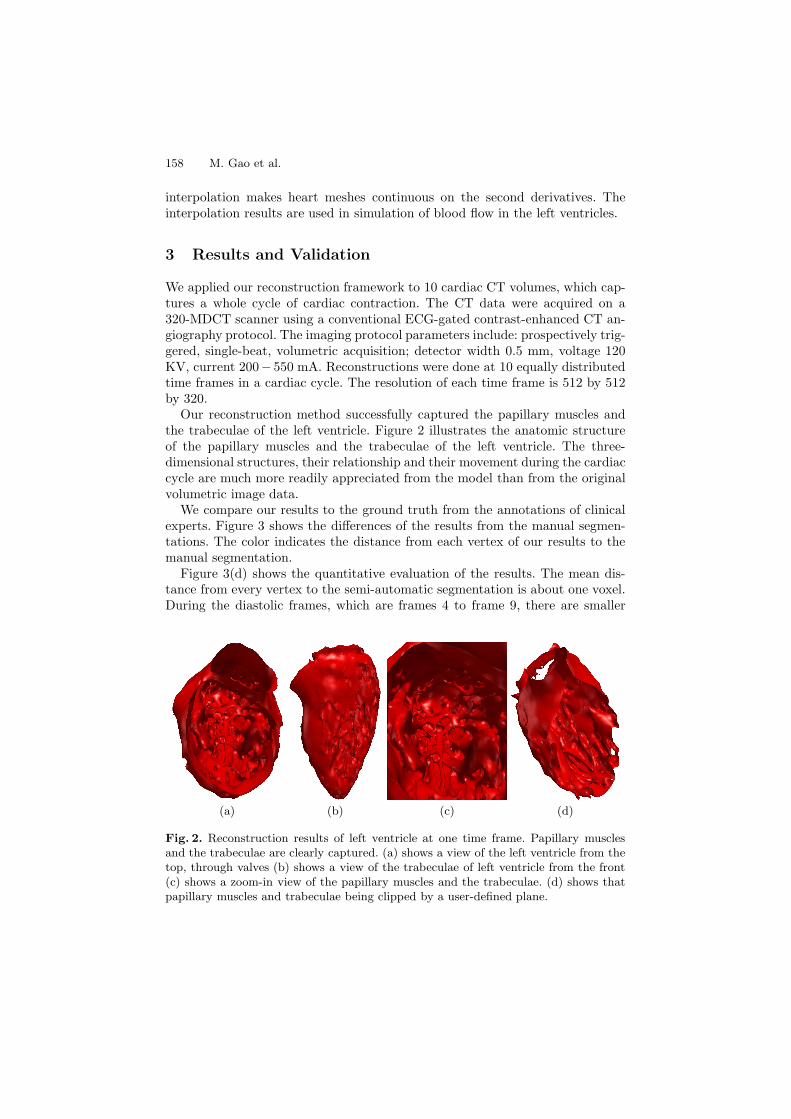
Our reconstruction method successfully captured the papillary muscles andthe trabeculae of the left ventricle. Figure 2 illustrates the anatomic structureof the papillary muscles and the trabeculae of the left ventricle. The three-dimensional structures, their relationship and their movement during the cardiaccycle are much more readily appreciated from the model than from the originalvolumetric image data.
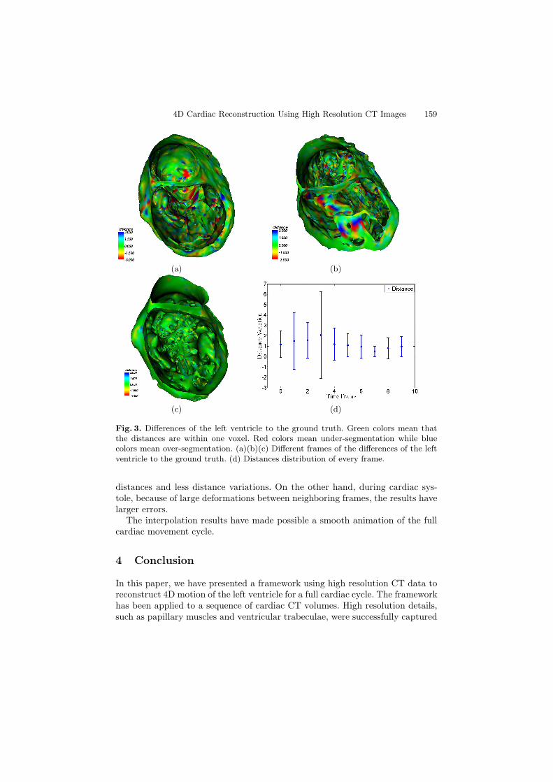
We compare our results to the ground truth from the annotations of clinicalexperts. Figure 3 shows the differences of the results from the manual segmen-tations. The color indicates the distance from each vertex of our results to themanual segmentation.
Figure 3(d) shows the quantitative evaluation of the results. The mean dis-tance from every vertex to the semi-automatic segmentation is about one voxel.During the diastolic frames, which are frames 4 to frame 9, there are smaller
(a) (b) (c) (d)
Fig. 2. Reconstruction results of left ventricle at one time frame. Papillary musclesand the trabeculae are clearly captured. (a) shows a view of the left ventricle from thetop, through valves (b) shows a view of the trabeculae of left ventricle from the front(c) shows a zoom-in view of the papillary muscles and the trabeculae. (d) shows thatpapillary muscles and trabeculae being clipped by a user-defined plane.
4D Cardiac Reconstruction Using High Resolution CT Images 159
(a) (b)
(c) (d)
Fig. 3. Differences of the left ventricle to the ground truth. Green colors mean thatthe distances are within one voxel. Red colors mean under-segmentation while bluecolors mean over-segmentation. (a)(b)(c) Different frames of the differences of the leftventricle to the ground truth. (d) Distances distribution of every frame.
distances and less distance variations. On the other hand, during cardiac sys-tole, because of large deformations between neighboring frames, the results havelarger errors.
The interpolation results have made possible a smooth animation of the fullcardiac movement cycle.
4 Conclusion
In this paper, we have presented a framework using high resolution CT data toreconstruct 4D motion of the left ventricle for a full cardiac cycle. The frameworkhas been applied to a sequence of cardiac CT volumes. High resolution details,such as papillary muscles and ventricular trabeculae, were successfully captured
160 M. Gao et al.
in this framework. In the future, we plan to use this framework to capture morefine structures of the heart, e.g., the valves and wall surfaces of all the fourchambers.
References
1. Chen, T., Metaxas, D., Axel, L.: 3D cardiac anatomy reconstruction using highresolution CT data. In: Barillot, C., Haynor, D.R., Hellier, P. (eds.) MICCAI 2004.LNCS, vol. 3216, pp. 411–418. Springer, Heidelberg (2004)
2. Lorenz, C., von Berg, J.: A comprehensive shape model of the heart. Medical ImageAnalysis 10(4), 657–670 (2006)
3. Mcinerney, T., Terzopoulos, D.: A dynamic finite element surface model for seg-mentation and tracking in multidimensional medical images with application tocardiac 4D image analysis. Computerized Medical Imaging and Graphics 19,69–83 (1995)
4. Montagnat, J., Delingette, H.: 4D deformable models with temporal constraints:application to 4D cardiac image segmentation. Medical Image Analysis 9(1),87–100 (2005)
5. Shen, D., Davatzikos, C.: Adaptive-focus statistical shape model for segmenta-tion of 3D MR structures. In: Delp, S.L., DiGoia, A.M., Jaramaz, B. (eds.)MICCAI 2000. LNCS, vol. 1935, pp. 206–215. Springer, Heidelberg (2000)
6. Shen, T., Li, H., Qian, Z., Huang, X.: Active volume models for 3d medical imagesegmentation. In: CVPR, pp. 707–714 (2009)
7. von Berg, J., Lorenz, C.: Multi-surface cardiac modelling, segmentation, and track-ing. In: Frangi, A.F., Radeva, P., Santos, A., Hernandez, M. (eds.) FIMH 2005.LNCS, vol. 3504, pp. 1–11. Springer, Heidelberg (2005)
8. Wang, X., Chen, T., Zhang, S., Metaxas, D., Axel, L.: LV motion and straincomputation from tMRI based on meshless deformable models. In: Metaxas, D.,Axel, L., Fichtinger, G., Szekely, G. (eds.) MICCAI 2008, Part I. LNCS, vol. 5241,pp. 636–644. Springer, Heidelberg (2008)
9. Zhang, S., Wang, X., Metaxas, D.N., Chen, T., Axel, L.: LV surface reconstructionfrom sparse TMRI using laplacian surface deformation and optimization. In: ISBI,pp. 698–701 (2009)
10. Zheng, Y., Barbu, A., Georgescu, B., Scheuering, M., Comaniciu, D.: Four-chamberheart modeling and automatic segmentation for 3D cardiac CT volumes usingmarginal space learning and steerable features. TMI 27(11), 1668–1681 (2008)
11. Zhu, S., Lee, T., Yuille, A.: Region competition: unifying snakes, region growing,energy/Bayes/MDL for multi-band image segmentation. In: ICCV, pp. 416–423(June 1995)
12. Zhuang, X., Rhode, K., Razavi, R., Hawkes, D., Ourselin, S.: A registration-basedpropagation framework for automatic whole heart segmentation of cardiac MRI.TMI 29(9), 1612–1625 (2010)