Embed Size (px)
Citation preview
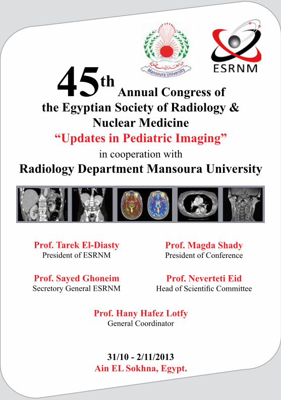
45th Annual Congress of
the Egyptian Society of Radiology &Nuclear Medicine
“Updates in Pediatric Imaging”in cooperation with
Radiology Department Mansoura University
31/10 - 2/11/2013Ain EL Sokhna, Egypt.
Prof. Magda ShadyPresident of Conference
Prof. Neverteti EidHead of Scientific Committee
Prof. Hany Hafez LotfyGeneral Coordinator
Prof. Tarek El-DiastyPresident of ESRNM
Prof. Sayed GhoneimSecretory General ESRNM
3
Scientific Committee
Prof. Nefertiti Kamal Eid (Head of scientific committee)Prof. Sabry El-MogyProf. Salwa EteibaProf. Magda El BakryProf. Amina SultanProf. Ahmed Abdel KhalekProf. Lamiaa ElSorougy
Organizing Committee
Dr. Germeen Albair AshamallahDr. Eman Mohamed Abd El-SalamDr. Merit El Maadawy
Advisory Board(Alphabetically arranged)
Prof. Abdelzaher HassanProf. Awad BesarProf. Emad NaguibProf. Fathy TantawyProf. Mohamad AbdelwahabProf. Mohamad ZahranProf. Omar HusseinProf. Samy ElbeblawyProf. Youssef Badran
4
ESRNM Board
Prof. Tarek EL-Diasty (President)
Prof. Sayed Ghoneim (Secretary General)
Prof. Hany Hafez Lotfy (Assistant Secretary General)
Prof. Ali Abdel Karim (Vice President)
Prof. Mohammed Abu El-Hoda (Treasurer)
Prof. Ashraf Selim
Prof. Hosny Abdelghany
Prof. Hossam Zaytoun
Prof. Khaled Shawky
Prof. Magdy El-Rakhawy
Prof. Mahmoud Dawood
Prof. Moharram El-Badawi
Prof. Saleh El-Esawy
Prof. Sameh Hanna
Prof. Wahid Tantawy
5
2:00pm-4:00pm Registration
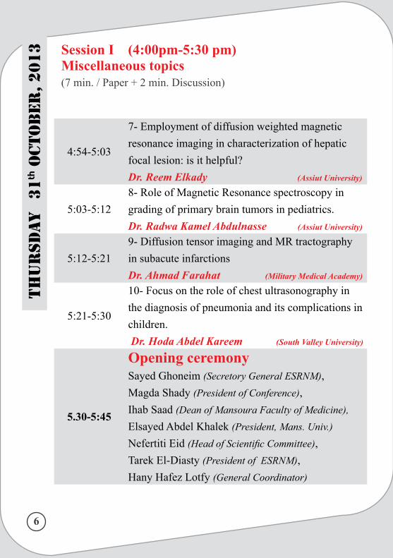
Session I (4:00pm-5:30 pm)Miscellaneous topics(7 min. / Paper + 2 min. Discussion)
Chairpersons:(Alphabetically)
Prof. Adel RezkProf. Lamiaa ElsorougyProf. Mahmoud DawoodProf. Mahmoud Elnahas Prof. Osama dawood
4:00-4:091- Aptness of CT wash out in pediatric adrenal masses: is it fact or fiction?Dr. Doaa Sharaf (Mansoura Urology&Nephrology Centre)
4:09-4:18
2- MRI CSF flowmetry; the golden modality in early detection of hydrocephalus in childrenProf. Hazem Moharram (Cairo University)
Dr. Mohamed Hafez (Cairo University)
4:18-4:27
3- The diagnostic performance of chest ultrasonography in the up-to-date work-up of the critical care settingDr. Rania Refaat (Ain Shams University)
4:27-4:36 4- Role of ulrasonography in pediatric female pelvisDr. Germeen Albeir (Mansoura University)
4:36-4:45
5-Does low dose MDCT have advantages in diagnosis of congenital megacolon in infants?Dr. Nagham Omar (Assuit University)
Dr. Gehan Saifeldin (Assuit University)
4:45-4:546- Estimation of how is 64 MDCT accurate inthe work-up of hepatoblastoma in pediatric patientsDr. Mohamed Gaber (El-Menia University)
Th
ur
sda
y 31
th OcT
Ob
er, 2013
6
4:54-5:03
7- Employment of diffusion weighted magnetic resonance imaging in characterization of hepatic focal lesion: is it helpful?Dr. Reem Elkady (Assiut University)
5:03-5:128- Role of Magnetic Resonance spectroscopy in grading of primary brain tumors in pediatrics.Dr. Radwa Kamel Abdulnasse (Assiut University)
5:12-5:219- Diffusion tensor imaging and MR tractography in subacute infarctionsDr. Ahmad Farahat (Military Medical Academy)
5:21-5:30
10- Focus on the role of chest ultrasonography in the diagnosis of pneumonia and its complications in children. Dr. Hoda Abdel Kareem (South Valley University)
5.30-5:45
Opening ceremonySayed Ghoneim (Secretory General ESRNM), Magda Shady (President of Conference), Ihab Saad (Dean of Mansoura Faculty of Medicine), Elsayed Abdel Khalek (President, Mans. Univ.)Nefertiti Eid (Head of Scientific Committee), Tarek El-Diasty (President of ESRNM), Hany Hafez Lotfy (General Coordinator)
Th
ur
sda
y
31th
OcT
Ob
er, 2
013 Session I (4:00pm-5:30 pm)
Miscellaneous topics(7 min. / Paper + 2 min. Discussion)
7
Chairpersons:(Alphabetically)
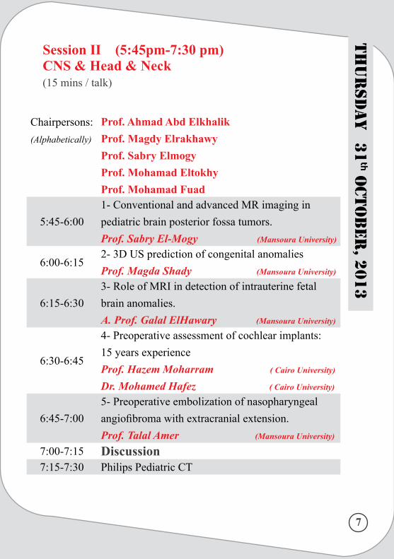
Prof. Ahmad Abd ElkhalikProf. Magdy ElrakhawyProf. Sabry ElmogyProf. Mohamad EltokhyProf. Mohamad Fuad
5:45-6:001- Conventional and advanced MR imaging in pediatric brain posterior fossa tumors.Prof. Sabry El-Mogy (Mansoura University)
6:00-6:152- 3D US prediction of congenital anomaliesProf. Magda Shady (Mansoura University)
6:15-6:303- Role of MRI in detection of intrauterine fetal brain anomalies.A. Prof. Galal ElHawary (Mansoura University)
6:30-6:45
4- Preoperative assessment of cochlear implants: 15 years experienceProf. Hazem Moharram ( Cairo University)
Dr. Mohamed Hafez ( Cairo University)
6:45-7:005- Preoperative embolization of nasopharyngeal angiofibroma with extracranial extension.Prof. Talal Amer (Mansoura University)
7:00-7:15 Discussion7:15-7:30 Philips Pediatric CT
Session II (5:45pm-7:30 pm)CNS & Head & Neck (15 mins / talk)
Th
ur
sda
y 31
th OcT
Ob
er, 2013
8
Th
ur
sda
y
31th
OcT
Ob
er, 2
013
Chairpersons:(Alphabetically)
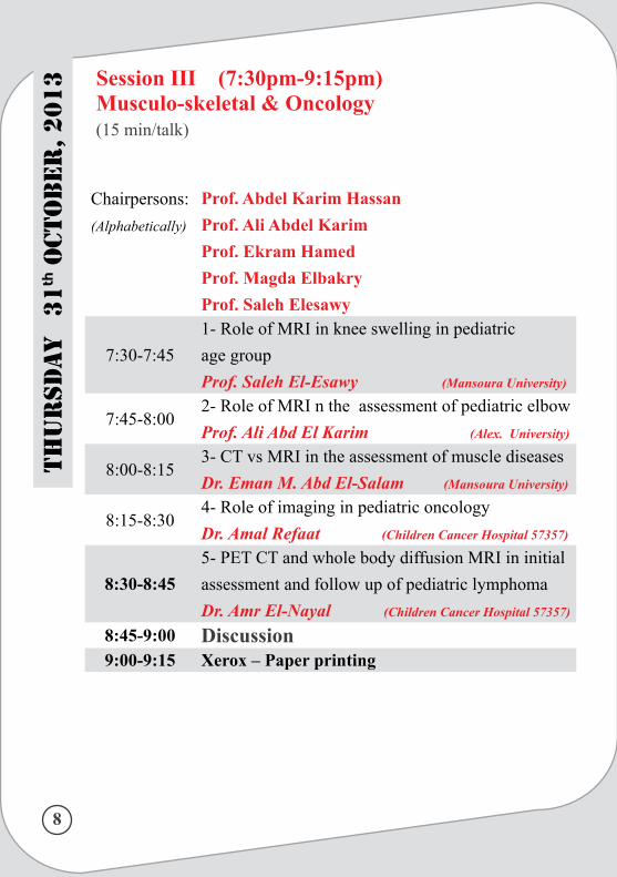
Prof. Abdel Karim HassanProf. Ali Abdel KarimProf. Ekram HamedProf. Magda ElbakryProf. Saleh Elesawy
7:30-7:451- Role of MRI in knee swelling in pediatric age groupProf. Saleh El-Esawy (Mansoura University)
7:45-8:002- Role of MRI n the assessment of pediatric elbowProf. Ali Abd El Karim (Alex. University)
8:00-8:153- CT vs MRI in the assessment of muscle diseasesDr. Eman M. Abd El-Salam (Mansoura University)
8:15-8:304- Role of imaging in pediatric oncologyDr. Amal Refaat (Children Cancer Hospital 57357)
8:30-8:455- PET CT and whole body diffusion MRI in initial assessment and follow up of pediatric lymphomaDr. Amr El-Nayal (Children Cancer Hospital 57357)
8:45-9:00 Discussion9:00-9:15 Xerox – Paper printing
Session III (7:30pm-9:15pm)Musculo-skeletal & Oncology(15 min/talk)
9
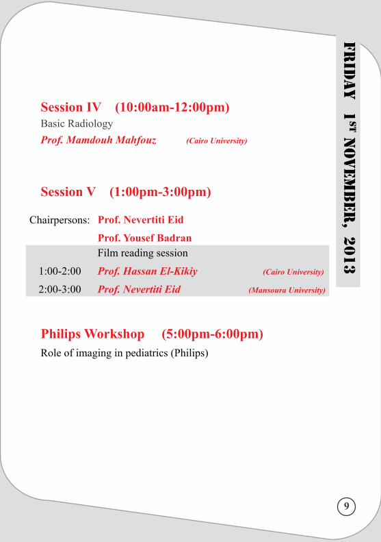
Chairpersons: Prof. Nevertiti Eid
Prof. Yousef Badran
1:00-2:00
2:00-3:00
Film reading session
Prof. Hassan El-Kikiy (Cairo University)
Prof. Nevertiti Eid (Mansoura University)
Session IV (10:00am-12:00pm)Basic RadiologyProf. Mamdouh Mahfouz (Cairo University)
Session V (1:00pm-3:00pm)
Philips Workshop (5:00pm-6:00pm)Role of imaging in pediatrics (Philips)
Frid
ay
1sT
NO
vem
ber
, 2013
10
Chairpersons:(Alphabetically)
Prof. Mahmoud Abd ElshahidProf. Mervat ShafeekDr. Amina SultanDr. Salwa Eteiba
6:00-6:151- Role of MDCT in imaging of congenital heart diseasesDr. Ahmad Ibrahim (Ain Shams University)
6:15-6:302- Role of MRI in imaging of congenital heart diseasesDr. Sara EL-Fawal ( Alex. University)
6:30-6:453- Pediatric mediastinal massesA.Prof. Hadeel Seif Eldeen (Qasr Eleiny Hospital)
6:45-7:004-Prenatal and postnatal imaging of lung lesionsProf. Jihan Mazroa (Mansoura University)
7:00-7:155- MDCT in congenital anomalies of the chestDr. Ashraf Abd El-Rahman (Mansoura Children Univ.)
7:15-7:30 Discussion
7:30-7:45SAMSUNG XGEO Clinical Review(World Wide)Dr. Miran Lee (Samsung HME Clinical Professor)
Session VI (6:00pm-7:45pm)Cardio-vascular & Chest Imaging(15 mins / talk)
Frid
ay
1sT
NO
vem
ber
, 20
13
11
Frid
ay
1sT
NO
vem
ber
, 2013
Chairpersons:
(Alphabetically)
Prof. Fathy Tantawy
Prof. Magdy Settin
Prof. Mohamad Borg
DR. Mohamad Aboelhoda
7:45-8:00
1- US findings in normal breast development and
breast masses in children
Prof. Nermin Soliman (Mansoura University)
8:00-8:15
2- Role of Diffusion-Weighted MR Imaging in
Pediatric Patients with Diffuse Liver Disease.
Prof. Adel El-Badrawy (Mansoura University)
8:15-8:303- Imaging in pediatric urinary tract
Prof. Mohamed Abu El-Ghar (Mansoura Urology&Nephrology centre)
8:30-8:45 Discussion8:45-9:00 Closing ceremony
Session VII (7:45pm-8:45 pm)Miscellaneous topics(15 mins / talk)
14
Abstracts
15
Aptness of CT Washout in Pediatric Adrenal Masses: Is It Fact or Fiction?
Authors: Doaa E. Sharaf. MSc, Tarek El-Diasty, MD
Purpose: To evaluate the accuracy of multi-detector computed tomogra-phy (MDCT) in diagnosis and differentiation of pediatric adrenal masses. Patients and methods: Between September 2011 and March 2013, charts of 50 pediatric patients were retrospectively reviewed. Mean age was 6 years (range 0.1-12 years). Twenty seven were girls. All had computed tomogra-phy scanning with a 64-MDCT scanner. They underwent unenhanced CT of the upper abdomen, then two enhanced acquisitions at 60 seconds (venous phase) and 15 minutes (delayed phase) after IV administration of iodinated contrast agent. Absolute percentage washout (APW) and relative percent-age washout (RPW) were calculated. The results were correlated with histo-pathological examination in children to who adrenalectomy was done. Results: Fifty patients with 53 adrenal masses were evaluated. The left side was involved in 33 children. Bilateral adrenal masses were noted in 3 cases. Adenomatous lesions demonstrated APW more than 60% and relative percentage washout RPW more than 40%, while APW less than 60% and RPW less than 40% in non-adenomatous lesions. The final diagnosis was hyperplasia in three cases, adrenal adenoma in 2 cases and non-adenoma-tous masses in 45 cases as follow: Neuroblastoma in 33 cases, Pheochromo-cytoma in 5 cases, Adreno-cortical carcinoma in one case, adrenal heamor-rhage in 4 cases, and adrenal cyst in 2 cases.
Conclusions: MDCT is the modality of choice in diagnosis of pediatric adrenal masses with high accuracy in differentiating benign from malignant masses with subsequent characterization of different types of non-adenom-atous masses by specific CT criteria with high sensitivity & specificity and with lesser need to use CT washout equation in pediatric adrenal masses.
16
MRI CSF Flowmetry; The golden modality in early detection of hydrocephalus in pediatrics.
Authors: Prof. Dr. Hazem Moharram MD, Dr. Mohamed Hafez MD, PhD
Abstract: Cine phase contrast magnetic resonance imaging (MRI) has been increas-ingly used during the last decade for evaluating cranial and spinal cerebro-spinal fluid (CSF) flow. Purpose: The aim of our study is to assess the value of cine-phase contrast MRI CSF flowmetry in early detection and diagnosis of hydrocephalus in children prior to the full established clinical features. Material and Methods: CSF flow study was performed with the use of an op-timized two dimensional cine phase-contrast MR technique. The protocols of examination include quantitative and qualitative analysis of CSF flow properties at the level of the aqueduct of Sylvius and applied on20 patients with relatively large head.Results and Conclusion: Correlative statistical analysis revealed that the sys-tolic peak and mean velocity as well as the systolic stroke volume values in normal pressure hydrocephalus (NPH) patients were statistically significant-ly higher than those of the normal controls denoting hyperdynamic aque-ductal CSF flow in non-obstructive hydro-cephalus. In cases of obstructive hydrocephalus either complete or partial, we found statistically significant markedly low systolic peak velocity, end-diastolic peak velocity and stroke volume values in comparison to healthy volunteers indicating a hypody-namic CSF circulation. Our results confirm the value of cine phase MRI as golden modality for characterizing CSF hydrodynamics in various types of hydrocephalus with early and accurate diagnosis prior to the established clinical calvarial de-formity features of hydrocephalus.
17
The diagnostic performance of chest ultrasonography in the up-to-date work-up of the critical care setting
Rania Refaat , Laila A. AbdurrahmanDepartment of Radiodiagnosis, Ain Shams University, Cairo,
Egypt
Objective: To compare the sensitivity, specificity and diagnostic perfor-mance of chest ultrasonography (US) for various pathologic pleuropulmo-nary abnormalities in intensive care unit (ICU) patients.Material and methods: Ninety consecutive patients admitted in chest ICU with respiratory distress were assessed clinically and by chest radiog-raphy (CXR).They were suspected to have a provisional diagnosis of any of the following pathologic entities: pneumonic consolidation, bronchogenic carcinoma, metastatic pulmonary nodules, pleural effusion, pneumothorax, hydropneumothorax and mesothelioma. These patients were scheduled for chest computed tomography (CT) and prospectively reviewed using chest US examination. The results of chest US were compared with the chest CT being the diagnostic standard of reference for each pathological entity to subsequently calculate its sensitivity, specificity and diagnostic performance. Results: The sensitivity, specificity and diagnostic accuracy of chest US were 100%, 96% & 97% for pneumonic consolidation, 71%, 100% & 98% for bronchogenic carcinoma and 92%, 100% & 99% for pneumothorax. The sensitivity, specificity and diagnostic accuracy of 100% for the rest of the included pathological entities were obtained.Conclusion: Chest ultrasonography has a considerable diagnostic perfor-mance for various pleuropulmonary pathologic conditions that may be en-countered in the ICU patients making it as an adjunct tool in the up-to-date work-up of the ICU setting.
18
Role of ultrasonography in pediatric female pelvis
Germeen A.Ashamallah, Ashraf Abd El-Rahman, Magda Elbakry
Department of Radiodiagnosis, Mansoura University,Egypt.
Abstract: Wide varieties of pathological processes affect the female pelvis in child-hood. Ultrasonography, a safe and available modality, provides real- time images of multiple planes and therefore is ideal for pediatric pelvic evalua-tion.Additional modalities such as CT and MRI should be reserved for cases in which ultrasonography is non-conclusive.Aim of the work: to evaluate the role of ultrasonography in diagnosis of pediatric female pelvic pathological conditions.Patients and methods: Thirty six female patients in pediatric age group, with variable complaints underwent pelvic ultrasonography exami-nation. They were divided into two groups. Group A underwent complemen-tary CT or MRI Group B were treated based on the diagnosis obtained by US. The results of US were compared with the result of CT & MRI in group A .While in group B they were compared the data obtained after clinical/surgical management. Results: This study included 36 cases divided in 2 groups. Group A in-cluded 21 cases (9 cases had non gynecological disorders and 12 cases had gynecological disorders). Group B included 15 cases; 4 of them had acute appendicitis and 11 had miscellaneous gynecological disorders. Pathologi-cal diagnosis was obtained in 17 cases.Conclusion: Ultrasound is the initial examination of choice for evalua-tion of pediatric gynecologic disorders; however, CT and MRI were more specific in characterization of disorder and determination of the extent of disease. The spectrum of gynecologic disorders in childhood mimics non gynecological disorders which necessitates accurate differentiation prior to treatment.
19
Does low dose MDCT has advantages in diagnosis of congenital megacolon in infants?
Dr.Nagham N Omar (rad.dept), Dr.gehan S Saifeldin(rad.dept), Dr. Nafisa I Alqusy(pediatric dept.) Assuit University
Objectives: evaluation of the applicability of low dose MDCT in detect-ing and diagnosis of congenital megacolon and assessing the postoperative status in infants.Patients and methods: Patients were recruited from the cohort of In-fants with chronic constipation from pediatric and surgical clinics in Assuit University. The recruited patients were sedated before examination. Exami-nations were done on 64-rows MDCT. Neither oral nor IV contrast media were administered for these examinations. Water soluble contrast enema was used. Scanning in the supine position cover the entire abdomen and pelvis.The images were sent to the workstation to be viewed in the axial, sagittal and coronal planes as well as for displaying volume rendering. Each MDCT examination was reviewed independently by two experienced radiologists for Interpretation of images by detecting and determining the location and length of the transition zone and detecting any associated anomalies. Evalu-ation of postoperative status.Results: Twenty- tow patients were recruited . A transition zone was found at rectosigmoid in 17(77.2%) cases, long segment (above sigmoid) in 2 (9%) cases, ultra short in 1 (4.5%) case, and total colone in 2 (9%) cases. We have found associated other gastrointestinal anomalies in 2 cases, spinal dysra-phism in 3 cases and urological anomalies in 2 cases. Postoperative evalua-tion is performed in 12 cases revealed good correction in 10 cases. Conclusion: low-dose MDCT has an essential role in diagnosis and as-sessing the postoperative status of congenital megacolon in infants.work-up of the ICU setting.
20
Estimation how 64 MDCT is accurate in the work up of hepatoblastoma in pediatric patients
Ahlam M. Ismail 1, Hoda Abdel Kareem 2, Mohamed Gaber3Departments of Pediatrics1 and Diagnostic Radiology2, South
Valley University & El-Minia University, EgyptBackground: Primary malignant liver tumors account for 1%–2% of all child-hood cancers. Hepatoblastoma is the most common primary hepatic malignancy of childhood. The probability of cure for hepatoblastoma is related strictly to the feasibility of achieving a complete mass definition and resection.Imaging plays a pivotal role in the diagnosis and management of children with hepatoblastoma. Objective:The aim of this work is to assess how MDCT is an accurate imaging modality for detecting hepatoblastoma surgical resectability and for the evaluation of the surgical bed in pediatric patients. Patients & methods: This study included 17 children,9 females and 8 males’ children with hepatoblastoma, (proved by biopsy), their ages ranged from 6 to 36 months. All patients were subjected to complete clinical examination, laboratory investigations including α-fetoprotein (AFP), bone scan, abdominal U/S and de-tailed MDCT for the chest and abdomen.Results: CT chest and abdomen was done using a 64 MDCT machine. The images were studied and reviewed on multi-planner & 3D reconstructed images for the tumor fo-cality, marginal definition, involvement of the vascular structures namely, inferior vena cava, hepatic veins and portal veins, lymph node metastases, and pulmonary metastatic nodules. The collected data were correlated with the surgical & pathologic findings. Post-operatively, MDCT liver was done to detect residual or recurrent tumor in the surgical bed. MDCT found to be accurate in detecting tumor resectability (16/17 cases), evaluation of the vascular structures (15/17 cases) and in detecting residual/recurrent tumor following surgery (3/4 cases).
Conclusion: MDCT of the abdomen and chest in children with hepatoblastoma is used to assess tumor resectability and to detect the presence of pulmonary metastasis, it can be performed quickly, easily, and usually without sedation. MDCT with its multi-planar image elegantly display the extent of hepatic involvement by tumor and its proximity to the vascular structures. Adding 3D CT imaging can provide high quality images of the tumors and its location relative to vital hepatic blood vessels that could help the surgeon to identify the tumor borders accurately and devise a comparative safe surgical strategy.
21
Employment of Diffusion weighted magnetic resonance imaging in characterization of hepatic focal lesions, is it
helpful?
Afaf A. Hassan, Reem M. Elkady, Moustafa M. Moustafa, Rafel Tappouni*
Assiut University, department of radiodiagnosis*Heshey Medical Center, Penn state University, USA
Objectives: The aim of this study is to investigate the role of diffusion weighted MRI (DWI) in improving hepatic focal lesion characterization and to determine the best ADC cutoff point to differentiate benign from malig-nant lesions.Materials and Methods: Eighty two patients with 122 hepatic focal lesions (31 metastases ,29 hemangiomas, 18 cysts ,16 hepatocellular carcinomas (HCC) , 5 abscesses, 5 focal nodular hyperplasias (FNH) , 5 adenomas,3 radiofrequency ablated lesions (RFA),2 regenerative nodules,2 lymphomas, 2 focal fatty lesions,2 undetermined benign lesions,1 cholangiocarcinoma and 1 confluent fibrosis) were included in our study. All patients were examined on 1.5 T MRI magnet and underwent standard liver MRI sequences in addition to respiratory triggered echo planner DWI sequence (b-values: 0, 100, 400,600 s/mm2) . Each lesion was evaluated qualitatively for its appearance in the 4 different b values images, and quantitatively by measuring the ADC values on the ADC maps. Results were correlated with histopathologic data and follow-up imaging .Receiver operating characteristic curve (ROC) was used to determine the best ADC cutoff point to differentiate benign from ma-lignant lesions.
Results: All malignant lesions and untreated abscesses showed restriction at high b value while cysts, necrotic tissue, RFA lesions and treated abscesses showed no restriction at high b value. Mean ADCs (×10−3mm2/s) were 3.16, 1.93 , 1.86 , 1.4 , 1.33 for cysts ,hemangiomas,RFA,FNH and adenomas respectively and 1.04 , 1.1, 0.83, 0.56 for HCC, cholangiocarcinoma,metastasis and lymphoma respectively . ADC measurements were ca-pable of differentiating benign from malignant liver lesions with a diagnostic accuracy of 94% using a cutoff ADC value of 1.29 x10⁻³ mm2/s.
Conclusion: DWI is a useful complementary tool that improves hepatic focal lesions characterization.
22
Role of Magnetic Resonance Spectroscopy inGrading of Primary Brain Tumors in Pediatric
By Nagham N Omar (Rad.dept.), Radwa K Abdelnasser
(Rad.dept.)and Nafeasa H Alqusy(Ped.dep) Assuit University
Objective: To assess the usefulness of MR spectroscopic imaging (MRS) in grading of primary brain tumors in Pediatric.Patients& Methods: MRS was performed in a 22 child with primary brain tumors. Metabolite ratios of Choline (Cho)/ N-acetyle –Laspartate (NAA), Cho/ Creatinine (Cr),Cho+Cr/NAA as well as lipids and lactate (LL)/Cr were calculated at short and intermediate echo time (TE). Addi-tionally, myo-inositol(mI)/Cr was calculated at short TE. Receiver operating characteristic analysis of metabolite ratios was performed to find a cut off values between high and low grade tumors. The resulting sensitivity, speci-ficity and accuracy were calculated. Results: At intermediate TE, Cho/NAA, Cho+Cr/NAA and Cho/Cr were significantly higher in high grade tumors than in low grade tumor. At short TE, Cho/Cr and LL/Cr ratios were significantly higher in high grade tumors than in low grade tumor. The diagnostic accuracy of the intermediate TE was 68% whereas at short TE, the diagnostic accuracy was 75%. Combination of both TEs revealed a diagnostic accuracy of 88%. Conclusion: Cho/NAA, Cho+Cr/NAA and Cho/Cr are reliable in deter-mining the tumor grade in children. LL/Cr is highly related to high grade tumors. Combination of both short and intermediate TEs provides better ac-curacy, in grading of brain neoplasm, compared to that when using each TE separately.
23
Diffusion tensor imaging and MR tractography in suba-cute infarctions
Ahmad Farahat ,Gehan Gouda, Mervat Elgohary.(Radiodiagnosis department, Ain shams University)
The DTI and the DT tractography provide feasible a non-invasive way to virtual re-construction of the white matter tracts in a reasonable time of acquisition, offering the FA which is a trustful method for the evaluation of the white matter integrity, and of useful contribution in the evaluation of ischemic cerebral strokes patients.Moreover, they open new era for a better knowledge for the brain anatomy and in the future they may give answers to a lot of non-explained neurological and psy-chiatric diseases.
24
Focus on the role of chest ultrasonography in the diagnosis of pneumonia and its complications in children
By Ahlam M. Ismail1, Hoda Abdel Kareem2
Departments of Pediatrics1 and Diagnostic Radiology2, South Valley University, Egypt
Background: The diagnosis of pneumonia in children is traditionally basedon physical examination, blood tests and chest X-rays. However, in-terpretation of chest X-ray is sometimes problematic, particularly in young infants in addition to the proved risk of repeated post-natal X-ray exposures especially acute lymphoblastic leukemia. Objective: This study aimed to highlight the role of chest ultrasonography (U/S) in the diagnosis and follow up of pediatric pneumonia and its compli-cations. Patients & methods: Our study included 50 children with pneumonia, 28 males and 22 females, their ages ranged between 3 months and 13 years. All patients subjected to complete clinical examination, laboratory investi-gations, plain chest radiography and chest U/S. Doppler U/S examination was added in selected cases. Results: The imaging findings on both chest X-ray and U/S were collected and analyzed guided by the clinical findings, laboratory results and response to treatment. The imaging findings were classified into parenchymal, pleural and pleuro-parenchymal. The chest U/S scored near equal result to chest radiography (98%) in diagnosing parenchymal lesions and was superior to it in pleural and pleuro-parenchymal lesions (100/78%). Conclusion: Chest ultrasonography in children proved a valuable role for diagnosis and follow up of pneumonia and its complications. It is rapid and can be easily repeated at the patient’s bedside with no risk of irradiation. Chest U/S can also be used to guide the thoracentesis in cases complicated with pleural fluid collections.
25
Imaging Approach of Pediatric Elbow
By: Prof. Dr. Ali Abd El-Karim FarahatProfessor and head of the MSK Unit Alexandria University Egypt
The radiological assessment of pediatric elbow especially in traumatic lesions is one of the diagnostic challenges due to the complexity and variability of the de-velopmental anatomy of the elbow in pediatric age group. The objectives of the presentation will cover the awareness of the developmental anatomy as an essential part in the imaging approach of pediatric elbow,proper imaging radiographs for identifications of pediatric elbow injuries and the important soft tissue signs as well as alignment considerations when dealing with traumatic elbow lesions in children.
IMAGING OF URINARY TRACT IN PEDIATRICS
Prof. Mohamed Abou El-Ghar Urology& Nephrology center – Mansoura University
• Imaging of the genitourinary system in children represents a cornerstone in the diagnosis of several congenital and acquired diseases.• The normal anatomy and many pathologies of the pediatric genitourinary system can be assessed with different imaging modalities. • At present, this work-up is performed by US , IVU, VCUG, CT and scintigraphy• These well-established methods still suffer from some restrictions.• Most of them are based on the use of ionizing radiation and invasive techniques. • The contribution of MR Imaging has opened new ways of approaching pathologi-cal conditions in this patient group.• The potential advantages of MRI for evaluating renal tract abnormalities in chil-dren are lack of ionizing radiation, the multiplanar capabilities and the excellent anatomic resolution and soft tissue contrast.• Our objective to review the imaging techniques used in imaging of the urinary tract in pediatrics including conventional techniques, Ultrasound, CT and scintig-raphy with special focus on the MRI and expressing its role in reducing the use of other techniques using ionizing radiation and iodinated contrast and also how can MRI serves as a problem solving specially in complex anomalies
LOGIC SERIES
Golden Sponsors
platinum Sponsor
Silver Sponsors
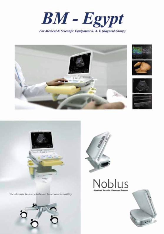
BM - Egypt
Organizing Office in Charge:12 Abo Bakr El-Sedek St., Ebad El Rahman City,Ring Road, El Mokatam, Cairo, Egypt.Email: [email protected]: 01006336368-01140229005
Aldamati Travel & Tourism
Travel Agent