Embed Size (px)
Citation preview
44 y/o female with dizziness
and dull headache
Edward Gillis, DO
Mark Kane, MD
Leo Wolansky, MD
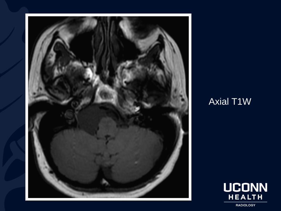
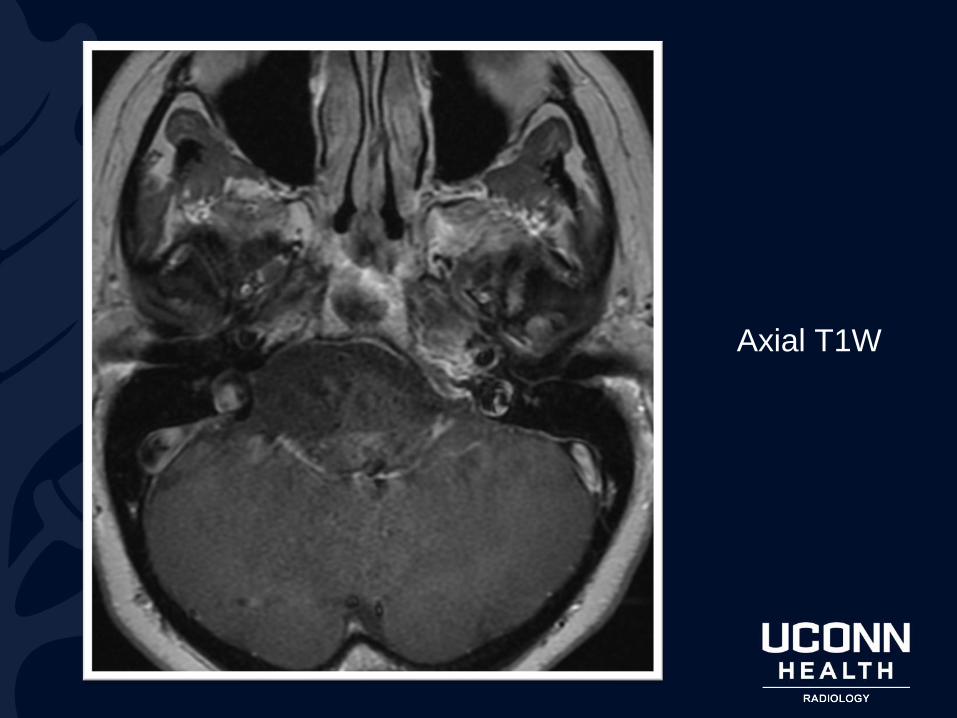
Axial T1W
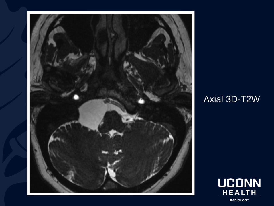
Axial 3D-T2W
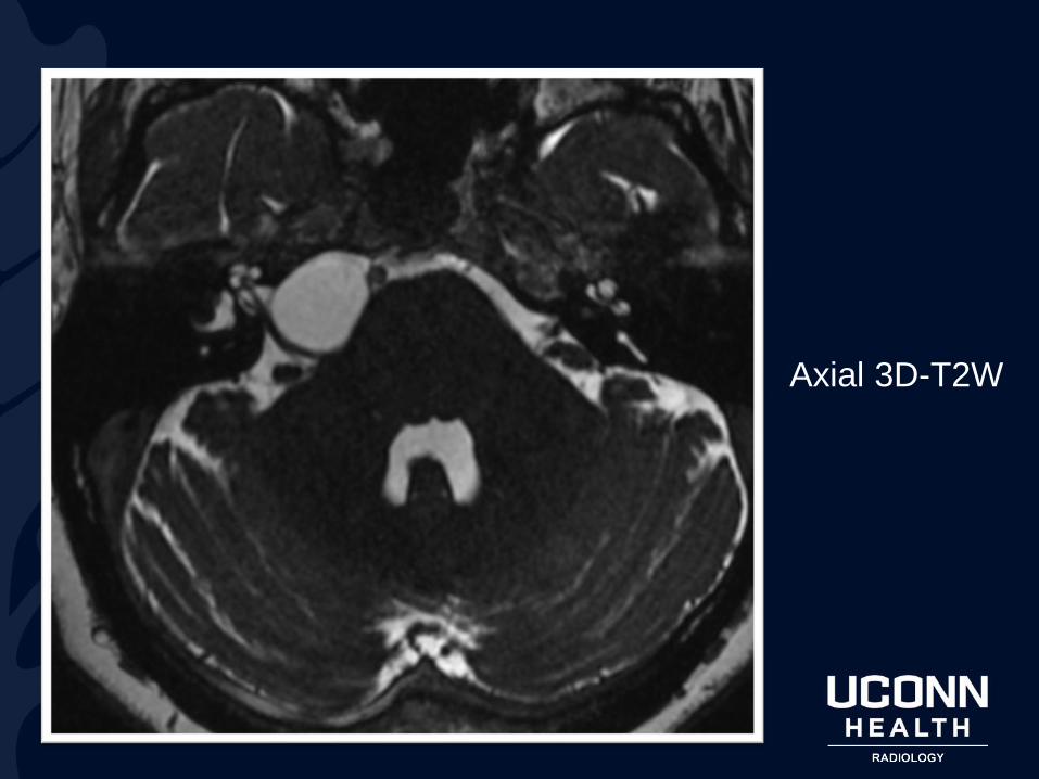
Axial 3D-T2W
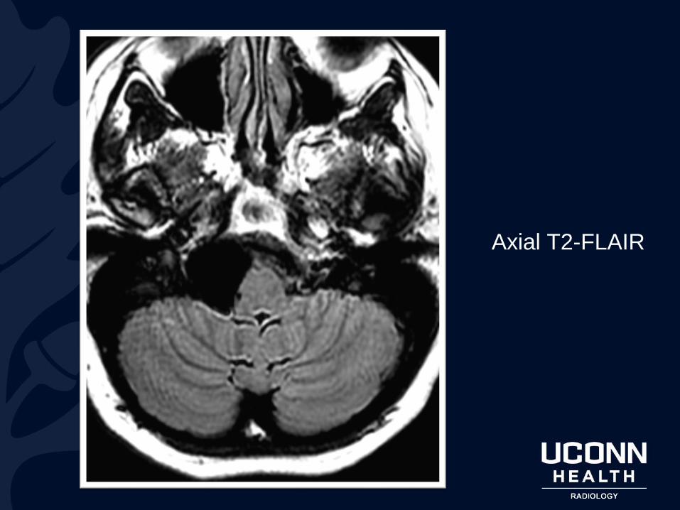
Axial T2-FLAIR
Axial T1W

?

Arachnoid Cyst
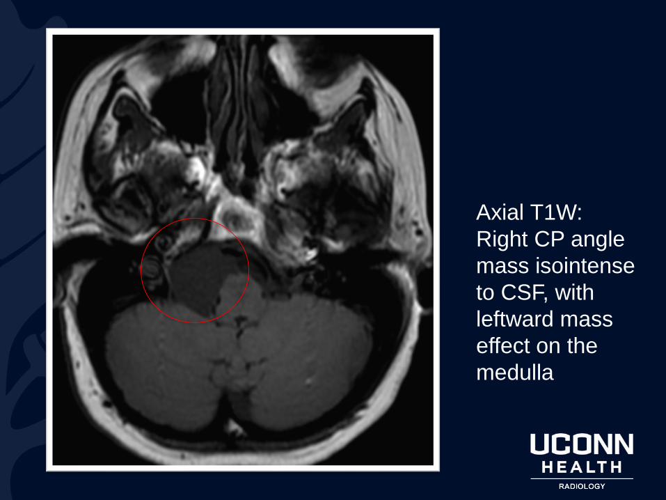
Axial T1W:
Right CP angle
mass isointense
to CSF, with
leftward mass
effect on the
medulla
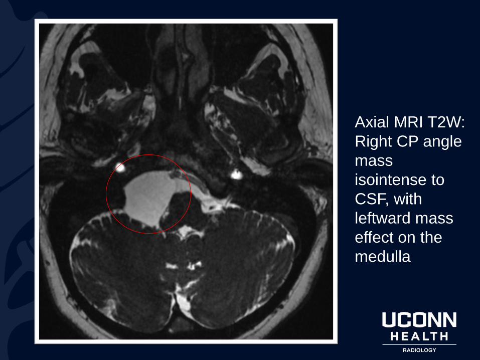
Axial MRI T2W:
Right CP angle
mass
isointense to
CSF, with
leftward mass
effect on the
medulla
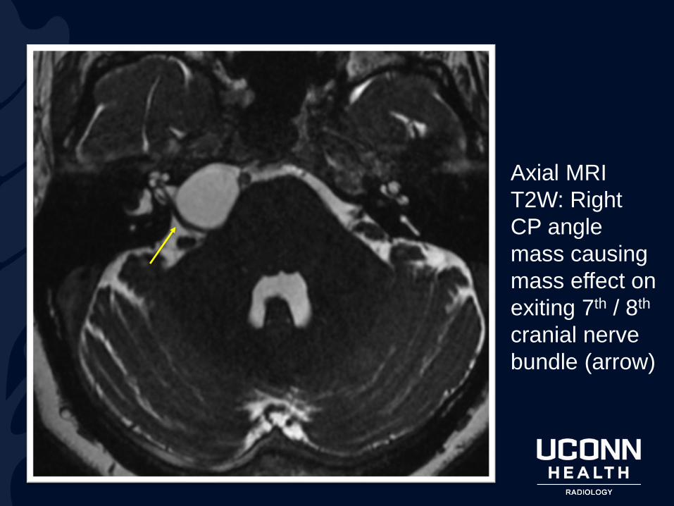
Axial MRI
T2W: Right
CP angle
mass causing
mass effect on
exiting 7th / 8th
cranial nerve
bundle (arrow)
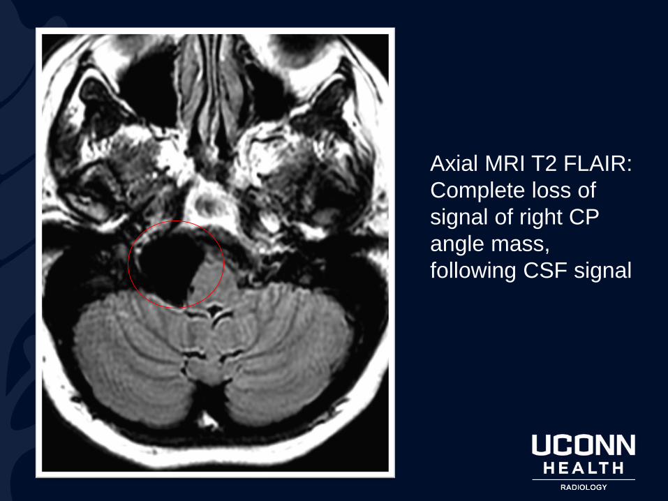
Axial MRI T2 FLAIR:
Complete loss of
signal of right CP
angle mass,
following CSF signal
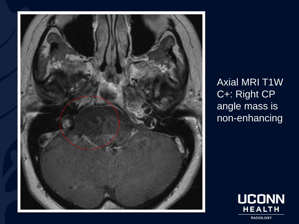
Axial MRI T1W
C+: Right CP
angle mass is
non-enhancing
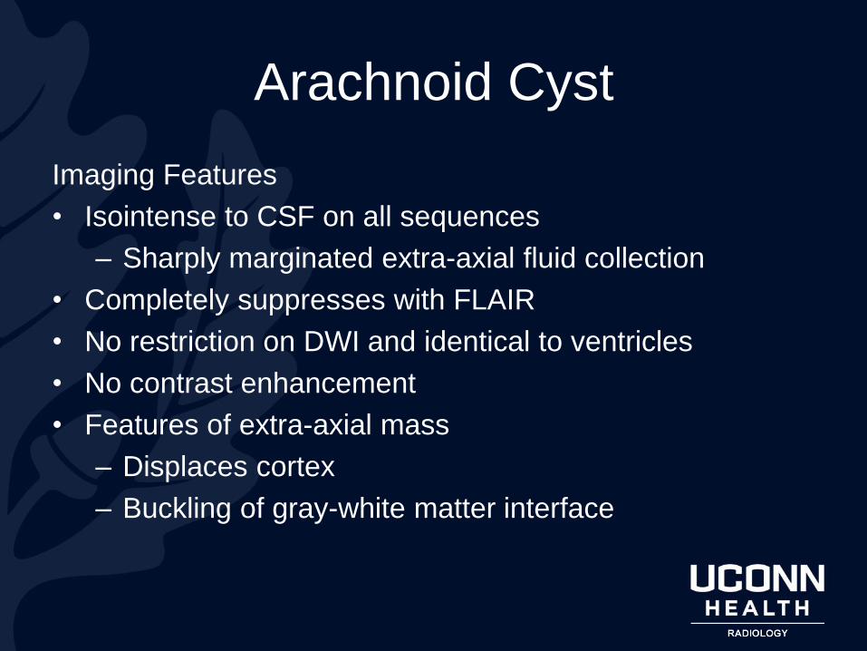
Arachnoid Cyst
Imaging Features
• Isointense to CSF on all sequences
– Sharply marginated extra-axial fluid collection
• Completely suppresses with FLAIR
• No restriction on DWI and identical to ventricles
• No contrast enhancement
• Features of extra-axial mass
– Displaces cortex
– Buckling of gray-white matter interface

Arachnoid CystGeneral Features
• Location
– Middle cranial fossa most commonly
– Cerebellopontine angle
– Suprasellar
• Giant periventricular arachnoid cysts can cause hydrocephalus
– Association with stenosis of Foramen of Monro & Aqueduct
• M:F is 3-5:1
• Usually asymptomatic, incidental finding, but depends on size & location
• DDx:
– Epidermoid cyst (Hyperintense on DWI & FLAIR)
– Chronic subdural hematoma (most SDH are not CSF signal)
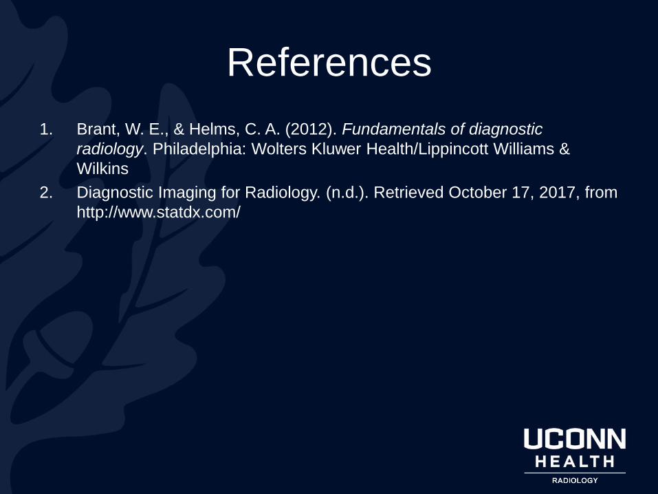
References
1. Brant, W. E., & Helms, C. A. (2012). Fundamentals of diagnostic
radiology. Philadelphia: Wolters Kluwer Health/Lippincott Williams &
Wilkins
2. Diagnostic Imaging for Radiology. (n.d.). Retrieved October 17, 2017, from
http://www.statdx.com/


![Concussion Education.ppt [Read-Only] Education...• Headache or “pressure” in head • Feeling sluggish, hazy, foggy, or • Nausea or vomiting • Balance problems or dizziness](https://img.pdfslide.us/doc/110x75/5f153a11f2d4a512a02f65a2/concussion-read-only-education-a-headache-or-aoepressurea-in-head-a-feeling.jpg)