Embed Size (px)
Citation preview

Chapter 4
-51-
4. WATER-MEMBRANE PARTITIONING AND DISTRIBUTION OF
BUPIVACAINE AND KETOPROFEN IN BILAYERS IN
DEPENDENCY ON pH AND ELECTROSTATICS
4.1. Introduction
The pharmacokinetic and pharmacodynamic characteristics of a new drug molecule, or a well-
established drug with a new indication or application form, are important and interesting in
therapeutic product development. The hydrophilicity
drug’s biological properties, as observed during Absorption, Distribution; Metabolism, and
Excretion. Beyond ADME, the drug’s interaction with specific and unspecific targets
(receptors, enzymes, proteins etc.) may also depend on relative hydrophilicity or lipophilicity
of the active agent.
The widely used determination of 1-octanol/water partition coefficient, Po/w
, can yield useful
information on the drug’s relative lipophilicity. However, 1-octanol is not a biological
molecule. It is therefore much better and more relevant to study the drug’s partition
coefficient between phospholipids and water, Pmem
, which can be measured using lipid bilayer
membrane vesicles. The membrane model simulates physiological drug surrounding more
precisely than an isotropic solvent, such as 1-octanol. In vitro pharmacokinetic and
pharmacodynamic properties are thus correlated better with an in vivo situation in which
numerous membrane during ADME, e.g the transcellular
passage of an orally applied drug through lipid bilayers in the intestinal epithelium.
Beyond pharmacological aspects, galenic problems can also be tackled by studying bilayer
membrane partition and distribution coefficient. Optimum loading of a drug on/into lipid
vesicles is among others a major key in the development of good pharmaceutical products
based on lipid bilayer vesicles (Langner et al. 1999; Cevc et al. 2001; 2004a; Allison 2007;
Rother et al. 2007). Knowledge of Pmem
will thus provide a good basis in galenic research
works.
In contrast to the common shake-flask method (Bouchard et al. 2002), which is used for Po/w
but inapplicable to Pmem
determination, the drug membrane partition coefficient can be
measured using different methods such as ultrafiltration (Austin et al. 1995), titration (Avdeef
et al. 1998), dialysis (Kramer et al. 1998), immobilized artificial membrane (IAM)-HPLC
(Ottiger et al. 1999), and the predictable quantitative structure activity relationship

Chapter 4
-52-
simulations (QSAR) (Patel et al. 2001). Each of these methods has its merits, but pH-metric
titration is the only one that allows determination of partition and distribution coefficients as a
function of the drug dissociation.
The current commercial drug substances can be divided into three groups (Balon et al. 1999):
bases (75 %), acids (20 %) and neutral molecules (5 %). The basic or acidic drugs often
coexist in a charged and neutral form, dependent on the formulation or ambient pH. Detailed
pH-metric titrations by Avdeef (2003) showed that for the neutral molecules the partition
coefficient in bilayer membranes is comparable with that in 1-octanol, PN
mem
N
o/w, while
for the ionized drug form the partition coefficient is significantly higher in a membrane than
in 1-octanol, P
I
mem >> P
I
o/w. Therefore, the resulting pH dependent drug distribution
coefficient, Dmem
, is higher in the membrane at pH values where the ionized species
predominates.
Figure 25: Structures of the acid ketoprofen (left) and the base bupivacaine (right).
(A) chemical structure; (B) 3-D structure; (C) surface lipophilicity; (D) surface charge
(Generated with HyperChem.)

Chapter 4
-53-
Drug partitioning into membranes is also sensitive to electrostatic and polarity effects, and to
interactions between the drug molecules and phospholipid bilayers (Cevc et al. 1987). This
explains why different salt concentrations (Bauerle et al. 1991; Alcorn et al. 1993; Thomas et
al. 1993; Austin et al. 1998) or different choice of phospholipids, such as
phosphatidylcholine, phosphatidylethanolamine, and phosphatidylserine (Langner et al. 1995;
Pedros et al. 1997; Takegami et al. 2005), influence PI
mem and D
mem.
I therefore studied partitioning and distribution of the basic local anaesthetic bupivacaine and
of the acidic non-steroidal anti-inflammatory drug (NSAID) ketoprofen (Figure 25) into
bilayers of soybean phosphatidylcholine. I measured the drug partitioning in this work in
dependency on the bulk pH but also explored the influence of salt concentration on equilibria.
To the best of my knowledge, no bilayer membrane partitioning data have been published for
these molecules to date, at least not considering electrostatic interactions between the drugs
and the membrane in parallel. Such interactions are important, however, in designing drug
containing ultradeformable bilayer membranes, with the intended use as vesicular carriers for
transdermal drug delivery (Cevc et al. 2003b; Cevc 2004).
The outcome of the present study will confirm observations of different authors which also
measured and considered electrostatic effect on drug membrane distribution. Unfortunately,
numerousness partition and distribution coefficient data published for various drugs and
excipients to date neglect such electrostatic interactions. With the results of the following
partition coefficient measurements, I tried to study drug partitioning as a function of bilayer’s
charge density, surface potential, and polarity, finding out that these interactions are
substantial when measuring and presenting PN
mem, P
I
mem, and D
mem.

Chapter 4
-54-
4.2. Theoretical background
The dissociation constant, pKa, of an ionizable acid or base in water is defined as the negative
common logarithm of the dissociation equilibrium, Ka,
aaKlogpK −=
[AH]
]][AO[H-
3
+
−= log for acids
][BH
][B]O[H3
+
+
−= log for bases
(18)
with a pH
110
10
+
=α−
−
apKpH
apKpH
(19)
Any drug’s dissociation is influenced by polar and electrostatic effects involving the
surrounding solvent as well as ions. Dissociation constant is therefore different in a lipid
bilayer membrane, pKmem
, and in water, pKa. pH-metric titration of an acid or base in lipid
bilayer vesicles suspension therefore yields an effective dissociation constant for the
molecules in contact with water and with lipid aggregates. The relationship is not a straight
one however, but rather a result of four coupled equations of equilibrium, as is illustrated in
Figure 26.
The resulting apparent dissociation constant, pKapp
, depends on partition coefficients of the
charged and the neutral drug forms, PI
memand P
N
mem, respectively, as well as on the pK
a and
pKmem
values, with the apparent value being between the two.
Figure 26: Schematic illustration of dissociation and partitioning of bases (i.e. cationic acids) and acids in
a system consisting of an aqueous bulk phase and lipid bilayer membranes.

Chapter 4
-55-
The charge-specific partition coefficients for the neutral and for the ionized molecules can be
calculated from the respective, directly measured, drug concentrations in the bilayer
membrane, cmem
, and in water, cw
(Miyazaki et al. 1992):
N
w
N
memN
mem
c
c
P =I
w
I
memI
mem
c
c
P = (20)
The associated pH dependent, phenomenological drug distribution coefficient, Dmem
,
considers both the neutral and ionized molecules (Miyazaki et al. 1992):
I
w
N
w
I
mem
N
mem
mem
cc
cc
D
+
+
= (21)
Alternatively, by measuring the apparent dissociation constants, pKapp
(j), for at least two
different lipid volume ratios vj = V
lipid / V
total ( j = 1 and 2), the neutral and ionized drug’s
partition coefficients in lipid bilayers can be calculated from the known dissociation constant
in water, pKa
(Avdeef 1992; Avdeef et al. 1998):
( )( ) ( )( )
( )( ) ( )( )
( )( ) ( )( )
−⋅
⋅−−⋅−⋅
=
−−
−+−−
apK
apppK
apK
apppK
apK
apppK
apppK
apK
apppK
apK
apppK
N
mem
vv
vvvv
P
21
21
221
12
1
1
2
2
1010
101010
(22)
( )( ) ( )( )
( )( ) ( )( )
−⋅
−+⋅−⋅
=
−−
−−
apK
apppK
apK
apppK
apK
apppK
apK
apppK
I
mem
vv
vvvv
P
21
21
12
1
2
2
1
1010
1010
(23)
Knowledge of both partition coefficients enables pH-dependent evaluation of the drug’s
distribution including both charged and neutral molecular forms:
( )
( )( )
( )pH
apKspH
apKsN
mem
I
memmemlogPPlogDlog
−−
+−⋅+= 10110 (24)
with the drug specific sign s = +1 for acidic and s = –1 for basic drug substances.
Equations (20) - (24) are valid so long as membrane properties are not affected significantly
by the membrane bound drug. Practically speaking, this is true for high lipid/drug ratios or
small partition coefficient PI
mem. Otherwise, when an appreciable drug quantity is bound to the
membrane, corrections must be made for the drug-induced changes in the membrane
properties, especially for the membrane’s electrostatic potential. The influence of
electrostatics on the drug partitioning must then be considered. This is typically done as a
function of the membrane surface charge density, , the bulk ions concentration, cw,i
, and the
polarity of the membrane/water interface, mem
.

Chapter 4
-56-
The surface charge density can be estimated from the known dissociation degree, , of an acid
or a base at the bilayer membrane surface (Austin et al. 1998):
lldrugdrug
drug
rArA
ezr
⋅+⋅
⋅⋅⋅α
=σ (25)
z is the drug’s charge number and e the elementary electronic charge. Adrug
and Al are the
surface area of the drug and the bilayer forming lipid in the membrane, respectively (Al >>
Adrug
within this work). rdrug
and rl are the mole ratios of the former and of the latter.
The Debye screening length, D, which gives the “thickness”
D of the diffuse electric double
layer near a charge, influences the surface potential as well, in dependency on ions, i, with
bulk concentrations cw,i
and the charge number zi(Cevc 1990):
∑ ⋅⋅⋅
⋅ε⋅ε
=λ
i
i,wiA
B
D
czeN
Tk
22
0
(26)
and 0 are the dielectric constant of the solution and the permittivity of free space,
respectively. The remaining parameters are the thermal energy, kBT, and the Avogadro
number, NA.
At 25 °C equation (26) simplifies for monovalent salts with total bulk salt concentration cw,t
(Israelachvili 1992):
50
3040.
t,wDc.
−
⋅= (27)
The surface potential,0,GC
, is usually calculated within the framework of the Gouy-Chapman
model (Gouy 1910; Chapman 1913):
GC,0ψ
⋅ε⋅ε⋅
λ⋅σ⋅⋅
⋅
⋅
⋅
=−
Tk
ez
ez
Tk
B
DB
0
1
2
sinh
2
0ε⋅ε
λ⋅σ
=D
for 0
< 25 mV
(28)
Within a membrane interface, e.g. near a phospholipid bilayer membrane, the polarity
changes. In terms of the dielectric constant, the difference is from mem
to mem
“bulk” (Marsh 2001).

Chapter 4
-57-
Therefore, the membrane surface potential 0 has to calculated based on equation (28)
0ψ
⋅ε⋅ε⋅
λ⋅σ⋅⋅
⋅
⋅
⋅
=−
Tk
ez
ez
Tk
Bmem
DB
0
1
2
sinh
2
0ε⋅ε
λ⋅σ
=
mem
D
for 0
< 25 mV
(29)
which only differs from the latter in the dielectric constant value.
In this work, I assumed the location of the charged bupivacaine and ketoprofen, i.e. the
position of the surface charge, near the headgroup and fixed the polarity to mem
= 30.
Knowledge of electrostatic surface potential then allows calculation of the drug’s
concentration at the membrane surface, based on equation (20), simply by:
ψ⋅⋅−
⋅⋅=
Tk
ez
Pcc
B
I
mem
I
w
I
mem
0
exp (30)
Coming back to the experimental determination and evaluation of partition coefficients via
pH-metric titrations, it is clear that, under electrostatic consideration and influence, the
apparent dissociation constant is a function of lipid volume ratio, vj. pK
app values determined
for at least two different lipid volume ratios thus allow derivation of the partition coefficients
PN
memand P
I
memfor the neutral and ionized drug forms, respectively:
( )
+⋅
ψ⋅⋅−
⋅
+⋅
⋅+=
1exp
1
log
0
j
B
I
mem
j
N
mem
aiapp
v
Tk
ez
P
vP
spKvpK (31)
Equation (31) must be solved together, and consistent, with equations (25),(29), and (30),
using least squares fitting procedure.
The pH dependent distribution of ionizable molecules in a lipid bilayer membrane is then
given, based on equation (24), by:
( ) ( )
[ ]pH
apKspH
apKsN
mem
B
I
memmemlogP
Tk
ez
expPlogDlog
−−
+−
⋅+
ψ⋅⋅−
⋅= 101100
(32)
Chapter 4
-58-
4.3. Material and Methods
4.3.1. Materials
Soybean phosphatidylcholine (SPC) was chosen as the bilayer forming lipid and was
purchased from Lipoid with the purity grade Lipoid S100 (Ludwigshafen, Germany).
Bupivacaine (hydrochloride monohydrate) and ketoprofen were supplied by Heumann PCS
(Feucht, Germany) and Bidachem (Fornovo S. Giovanni, Italy), respectively; both met the
quality specifications of the European Pharmacopoeia (EP). Potassium chloride, sodium
hydroxide, hydrochloric acid (all VWR, Darmstadt, Germany), and water for injection
(Deltaselect, Dreieich, Germany) had EP quality as well.
4.3.2. Preparation of drug containing large unilamellar lipid bilayer vesicles
and blank solutions
Large unilamellar vesicle suspensions with two different lipid concentrations
(160 mg/g and 50 mg/g, assuming = 1 g/ml) were used in the study. The corresponding SPC
quantity was interspersed into an aqueous solution of either bupivacaine or ketoprofen,
always keeping the final drug concentration constant (cd
= 20 mM). To determine the
influence of electrostatics on the drugs partitioning, different amounts of potassium chloride
were added to each suspension.
For better homogeneity, the suspensions were stirred for 2 h, and then extruded through a set
of different track-etched poly-carbonate membranes (PCTE, GE Osmonics) with decreasing
pore size (400 nm – 100 nm); a freeze-thaw cycle (180 min at -70 °C, 30 min at 40 °C) and
multi-extrusion through PCTE membranes with decreasing pore sizes (400 nm – 80 nm)
finished the vesicle preparation (Olson et al. 1979; Mayer et al. 1986; MacDonald et al.
1993). The resulting vesicle size and polydispersity were determined by the dynamic light
scattering (DLS), as is described in chapter 2.2.4. This yielded an average vesicle diameter of
dves
= 100 ± 15 nm and polydispersity index of PDI < 0.15.
The drug free blank solutions without any lipid vesicle were prepared by dissolving the
required potassium chloride quantities in water.
Directly before a pH-metric titration, the pH value of the vesicle suspension and its allocated
blank solution were adjusted to the starting pH < 3 and pH > 11 for bupivacaine and
ketoprofen titrations, respectively. This was done using adequate volumes of sodium
hydroxide (10 M) or hydrochloric acid (4 M). The resulting final ionic strength, comprising

Chapter 4
-59-
all ions (potassium chloride, sodium hydroxide, hydrochloric acid, and drug counter ions) was
calculated to be in the range of Ic = 35 mM to 265 mM.
4.3.3. pH-metric determination of the apparent dissociation constants,
membrane partition coefficients, and distribution coefficients
Determination of drug partition coefficients for the uncharged (neutral) molecule and for the
charged (ionized) entity, as well as analysis of the pH-dependent distribution coefficient, are
based on the pioneer work of Avdeef (1992; 1998).
According to the fundamental equations (22) and (23), the pKapp
of lipid vesicle suspensions
with two different total lipid volume ratios, vj, is needed to derive P
N
mem and P
I
mem. As pointed
out by Balon et al. (1999), the partition coefficients for the neutral and ionized drug forms are
independent on the tested lipid ratios; in contrast, the membrane partitioning of a drug is
typically temperature sensitive. Thus, all the measurements were done at 25 ± 0.3 °C. Blank
solutions provided controls, and supported the calculation of the mol equivalent of the titrated
drug substance.
The pH glass electrode DG111-SC was calibrated using the titrator DL67 and the LabX Pro
titration software (all from Mettler-Toledo, Giessen, Germany). This was done daily before
the measurements, which were done in triplicate for the drug vesicle suspension and its
reference blank solution. For the ketoprofen containing suspensions, a “down”-titration (from
high to low pH) was employed. For the oppositely charged bupivacaine, an “up”-titration in
opposite direction was chosen (lower panels in Figure 27). The analytes were determined with
two different titrants: sodium hydroxide (0.5 M) and hydrochloric acid (1 M) for bupivacaine
and ketoprofen samples, respectively.
In all experiments, the pH pH
done dynamically by the software. The pH equilibration between the lipid vesicle exterior and
interior was adjusted to be 1 - 2 min between each addition, as had been suggested in the
literature (Pauletti et al. 1994).
To evaluate the pKapp
values, the ratio of the ionized drug molecules
( )
totald
tstbt
ionized
Vc
VVsc
r
⋅
−⋅
= (33)
was first calculated. ct is the titrant concentration and V
tband V
tsare the titrant volumes used
for titrating blank solution and vesicle suspension in dependency on pH, respectively.
cd and V
totalare the total drug concentration and the measured volume, respectively.

Chapter 4
-60-
The ratio of the ionized drug molecules as a function of pH yields an apparent dissociation
constant, pKapp
, read-off at the inflexion point of the sigmoidal curve given in Figure 27. At
pH = pKapp
, the ratio of the charged and neutral molecules is 1:1.
0.0 0.5 1.0 1.5 2.0
0
7
14
4 6 8 10
0.0
0.2
0.4
0.6
0.8
1.0
0.0 0.5 1.0 1.5
0
7
14
0.0
0.2
0.4
0.6
0.8
1.0
pH
VNaOH
[mL]
pK
app
pH
r ionized
VHCl
[mL]
pK
app
4 6 8 10
pH
Figure 27: Illustration of pH-metric titrations for the determination of the apparent dissociation constant,
pKapp
, of bupivacaine (left panels) and ketoprofen (right panels) in bilayer vesicle suspensions.
The lower panels show the measurements of the drug containing lipid vesicle suspensions
(black dotted line) and the blank solutions (grey dotted line). The upper panels illustrate the
ratio of ionized drug (black line) and its derivation (grey line) in dependency on pH.
With the pKapp
derived from the experimental data measured with two different lipid ratios, vj,
and further considering the known aqueous dissociation constants of bupivacaine, pKa = 8.09
(Friberger et al. 1971)) or of ketoprofen, pKa = 4.36 (Rafols et al. 1997), the various partition
and distribution coefficients were derived in dependency on electrostatics with a least square
fitting procedure involving equations (31) and (32). The results are given in the following
section.

Chapter 4
-61-
4.4. Results and discussion
4.4.1. Apparent dissociation equilibrium of bupivacaine and ketoprofen
pH-metric titrations of bupivacaine or ketoprofen containing bilayer membranes yielded
different apparent dissociation equilibria in dependency on lipid/drug ratio and total ionic
strength, Ic (symbols in Figure 28).
7.2
7.4
7.6
7.8
8.0
8.2
Ic
= 35 mM
Ic
= 65 mM
Ic
= 265 mM
0.00 0.05 0.10 0.15 0.20
4.5
5.0
5.5
6.0
6.5
Ketoprofen
Bupivacaine
Ic
= 35 mM
Ic
= 85 mM
Ic
= 235 mM pKa
= 4.36
Lipid volume ratio, vj
0 5 10
pKa
= 8.09
Lipid/drug ratio [mol/mol]
pK
app
Figure 28: The apparent dissociation constant, pKapp
, of bupivacaine and ketoprofen in
bilayer membrane with different lipid/drug ratios.
With decreasing lipid/drug ratio, the pKapp
shift is reduced due to lower lipid
volume ratio, vj, and due to electrostatic repulsion between the charged bilayer
membrane and the drug molecules at the membrane/water interface.
( measured data ; fitted data ; reference data )
The pKa shift between the bilayer membrane and water is empirically in the range of –1 to –
1.5 units for bases (Miyazaki et al. 1992) and +2 to +3 units for acids (Austin et al. 1998). For
the base bupivacaine, which has an intrinsic dissociation constant of pKa
= 8.09 in water, the
apparent dissociation constant is shifted downwards to pKapp
= 7.15 – 7.33. For ketoprofen the
shift is upwards, from pKa
= 4.36 to pKapp
= 5.96 – 6.38. Figure 28 suggests, that the
magnitude of this effect depends, among others, on lipid/drug ratio, as is illustrated in Figure
26 for the four coupled equations of equilibrium. In addition, the data fitting curve based on
equation (31) shows an electrostatic influence on pKapp
shift, which will get attention in the
following section.

Chapter 4
-62-
4.4.2. Bilayer surface charge in dependency on pH
The fitted pKapp
curves given in Figure 28 highlight electrostatic effects on drug partitioning.
They show, for example, that at low lipid/drug ratio the pKapp
shift increases with salt
concentration. This is not surprising given that ions from the surrounding bulk solution not
only affect the Debye screening length, D; they moreover affect the surface charge density, ,
and the resulting surface potential, 0 (cf. equations (25),(26), and (29)).
Figure 29 for bupivacaine and Figure 30 for ketoprofen furthermore illustrate that for constant
drug concentration the drug dependent bilayer surface charge density decreases with
increasing lipid concentration. This is due to enlarged lipid area in case of high total lipid
concentration. High ionic strength, and the corresponding short screening length, reduce
repulsion of the equally charged drug molecules from the membrane/water interface, where
more such molecules reside, and increase pKapp
shift, as is shown in Figure 28.
0 3 6 9 12
0
5
10
15
20
25
30
35
Ic
[mM]
35
65
265
σ [e/nm
2
]
pH
σ [m
C/m
2
]
0 3 6 9 12
0.00
0.05
0.10
0.15
0.20
Ic
[mM]
35
65
265
lipid : drug = 3.125 [mol/mol] lipid : drug = 10 [mol/mol]
Figure 29: Surface charge density, , of bupivacaine containing bilayer membranes in dependency on
the bulk pH, ionic strength, Ic, or the drug/lipid ratio.
Bupivacaine, a tertiary amine base, is positively charged at low pH and neutral at high pH.
With increasing pH, the drug dependent surface charge density therefore changes.
Decreasing lipid/drug ratio and increasing Ic both results in higher surface charge
densities, due to weakened electrostatic repulsions.

Chapter 4
-63-
0 3 6 9 12
0
-10
-20
-30
-40
-50
Ic
[mM]
35
85
235
pH
σ [m
C/m
2
]
0 3 6 9 12
0.00
-0.05
-0.10
-0.15
-0.20
-0.25
-0.30
-0.35
Ic
[mM]
35
85
235
σ [e/nm
2
]
lipid : drug = 3.125 [mol/mol] lipid : drug = 10 [mol/mol]
Figure 30: Surface charge density, , of ketoprofen containing bilayer membranes in dependency on
the bulk pH, ionic strength, Ic, or the drug/lipid ratio.
Ketoprofen, a propionic acid derivative, is negatively charged at high pH and neutral at
low pH. With increasing pH, the drug dependent surface charge density therefore changes.
Decreasing lipid/drug ratio and increasing Ic both result in higher surface charge densities,
due to weakened electrostatic repulsions.
The deviation from sigmoidal curve progression, which is seen in Figure 29 and Figure 30 at
low ionic strengths, highlights a phenomenon. Theoretically, in absence of any repulsion, the
membrane bound drug molecules should change from neutral form into the charged one
according to dissociation equation, i.e. sigmoidally with decreasing or increasing pH for
bupivacaine or ketoprofen suspensions, respectively. In reality, the pH-induced changes of
bilayer surface charge density can actually force some of the charged drug molecules out of
the bilayer, thus reducing the final surface charge density. The propensity for this is higher at
low salt concentration, when electrostatic interactions are less screened.
Electrostatic surface potential is therefore one reason why charged and neutral drugs show
different membrane partitioning in dependency on the surrounding salt concentration, an
observation, which will be presented and discussed in the following section.

Chapter 4
-64-
4.4.3. Bilayer membrane partition and distribution in dependency on charge
state and electrostatics
The widely used 1-octanol/water partition coefficient (PN
o/w) of neutral molecules is similar to
the value measured with phospholipid bilayer membranes in water (PN
mem) (Avdeef et al.
1998). For the charged molecules, however, the partition coefficients PI
o/w and P
I
mem are
totally different for the most part (Miyazaki et al. 1992; Austin et al. 1995; Avdeef et al.
1998). The reason is that partitioning of amphiphilic acidic or basic drugs from water into a
membrane depends, among others, on electrostatic effects, on the drug’s charge state, and on
polarity. For example:
The propionic acid derivative ketoprofen in neutral form possesses an 1-octanol/water
partition coefficient log PN
o/w = 3.12 (La Rotonda et al. 1983). This is in good accord with
log PN
mem = 3.27 found in this study. For the ionized ketoprofen the partition values in octanol
and membrane differ significantly: log PI
o/w = -0.95 (Avdeef 2003) and log P
I
mem = 0.15 -
1.18, in dependency on ionic strength.
Bupivacaine, a tertiary amine base, has a similar log PN
o/w = 3.38 (Razak et al. 2001) but ten
times lower partition coefficient in the neutral form in a bilayer: log PN
mem = 2.43. However,
the partitioning of the positively charged bupivacaine is significantly higher compared to
ionized ketoprofen, particularly at low salt concentrations: log PI
mem = 1.23 - 1.58.
Unfortunately, to the best of my knowledge, there are no other comparable literature data for
the partitioning of charged bupivacaine in 1-octanol/water.
Ic [mM] log PN
o/w log PN
mem log PI
o/w log PI
mem
35 3.00 0.15
85 3.19 0.92Ketoprofen
235
3.12 a
3.27
- 0.95 b
1.18
35 2.08 1.23
65 2.17 1.41Bupivacaine
265
3.38 c
2.43
n/a
1.58
Table 8: Partition coefficients of neutral (N) and ionized (I) ketoprofen and bupivacaine
in a 1-octanol/water, Po/w
, or bilayer membrane/water, Pmem
, system in
dependency on the ionic strength, Ic.
a
(La Rotonda et al. 1983), b
(Avdeef 2003), c
(Razak et al. 2001)
Chapter 4
-65-
0 3 6 9 12
0
1
2
3
uncharged
charged
Ic
= 265 mM
Ic
= 65 mM
Ic
= 35 mM
Ketoprofen Bupivacaine
pH
log D
mem
el
0 3 6 9 12
0
1
2
3
uncharged
charged
Ic
= 235 mM
Ic
= 85 mM
Ic
= 35 mM
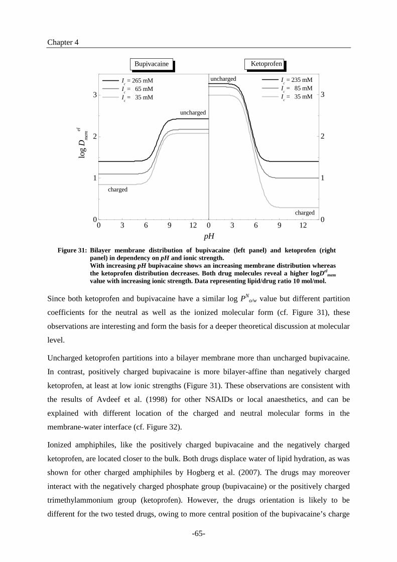
Figure 31: Bilayer membrane distribution of bupivacaine (left panel) and ketoprofen (right
panel) in dependency on pH and ionic strength.
With increasing pH bupivacaine shows an increasing membrane distribution whereas
the ketoprofen distribution decreases. Both drug molecules reveal a higher logDel
mem
value with increasing ionic strength. Data representing lipid/drug ratio 10 mol/mol.
Since both ketoprofen and bupivacaine have a similar log PN
o/w value but different partition
coefficients for the neutral as well as the ionized molecular form (cf. Figure 31), these
observations are interesting and form the basis for a deeper theoretical discussion at molecular
level.
Uncharged ketoprofen partitions into a bilayer membrane more than uncharged bupivacaine.
In contrast, positively charged bupivacaine is more bilayer-affine than negatively charged
ketoprofen, at least at low ionic strengths (Figure 31). These observations are consistent with
the results of Avdeef et al. (1998) for other NSAIDs or local anaesthetics, and can be
explained with different location of the charged and neutral molecular forms in the
membrane-water interface (cf. Figure 32).
Ionized amphiphiles, like the positively charged bupivacaine and the negatively charged
ketoprofen, are located closer to the bulk. Both drugs displace water of lipid hydration, as was
shown for other charged amphiphiles by Hogberg et al. (2007). The drugs may moreover
interact with the negatively charged phosphate group (bupivacaine) or the positively charged
trimethylammonium group (ketoprofen). However, the drugs orientation is likely to be
different for the two tested drugs, owing to more central position of the bupivacaine’s charge

Chapter 4
-66-
in comparison with ketoprofen. This makes the ionized ketoprofen more sensitive to
electrostatic effects, as is shown in Figure 31.
The neutral drug molecules are inserted deeper in the water-membrane interface, near the
fatty acid ester group. Ketoprofen can then bind via hydrogen bonds to the fatty acid carbonyl
group, finally resulting in an increased PN
mem(Avdeef et al. 1998; Hogberg et al. 2007). The
following section supports this conclusion and provides deeper insight into the electrostatic
and polarity induced contributions to the drug’s partitioning into lipid membranes.
Charged drug molecules within a monolayer
Uncharged drug molecules within a monolayer
Figure 32: Schematic illustration of ketoprofen and bupivacaine location within
a phosphatidylcholine membrane in dependency on their ionization.
(only a monolayer is presented). The upper graph shows charged
ketoprofen (left molecule) and charged bupivacaine (right molecule)
incorporated in the upper part of lipid headgroup region. The lower
graph illustrates the position of the neutral drugs in the deeper lipid
monolayer regions. (Generated with ACD/3D-viewer.)

Chapter 4
-67-
4.4.4. Influence of electrostatics and polarity on membrane dissociation
The dissociation equilibrium of acids and bases in bilayer vesicle suspensions is shifted by
polar and electrostatic effects compared to the bulk pKa
(Cevc et al. 1987):
pol
a
el
aaapppKpKpKpK ±+= (34)
pKa
el
pKa
pol
is the polarity dependent contribution to
the shift.
The electrostatic shift is given by:
Tk.
ez
pK
B
el
a
⋅
ψ⋅⋅
=
32
0
(35)
pKa
pol
depends on interfacial dielectric constant, mem
, at the drug binding site.
Its sign depends on the change in total number of charges on dissociation (Fernandez et al.
1977; Cevc et al. 1981). In other words, interaction of zwitterionic phosphatidylcholine
membranes with the molecular acid ketoprofen AH should cause a positive polarity induced
shift, due to increase of total charge from (+ -) to (+ - -). For dissociation of the cationic acid
bupivacaine BH+
the reverse should be true, owing to total charge decrease from (+ - +) to
(+ -).
The values for pKa
el
pKa
pol
for different salt concentrations at lipid/drug ratio of
10 mol/mol were calculated from the measured pKapp
values and the determined surface
potential 0 (cf. equations (29)) using equations (34) and (35).
Ic[mM]
0 [mV] pK
a
el
pKa
pol
35 61 1.03 - 1.95
65 55 0.93 - 1.79Bupivacaine
265 37 0.63 - 1.52
35 - 36 0.61 1.41
85 - 54 0.91 0.93Ketoprofen
235 - 38 0.64 1.18
Table 9: Surface potential 0 and the derived values of the electrostatic and polarity induced
contribution to the pKa shift for bupivacaine and ketoprofen in phosphatidylcholine
bilayer membranes at different ionic stengths with a lipid/drug ratio of 10 mol/mol.
Table 9 reveals that the polarity induced shift, which is an indicator of charged molecules
location in the membrane-water interface (Fernandez et al. 1977), is different for bupivacaine

Chapter 4
-68-
and ketoprofen. For ketoprofen pKa
pol
is in the range of 0.9 to 1.4, which is in good accord
with the values reported for other amphiphiles: ± 1.1 (Fernandez et al. 1977). The polarity
induced shift for bupivacaine is negative and somewhat larger, pKa
pol
= -1.5 – -2.0,
consistent with general expectations and the assumedly deeper insertion of this drug into the
interface.
Fernandez and Fromherz (1977) published polarity induced shifts as a function of different
dioxin-water mixtures representing various dielectric constants. Knowledge of the latter
information on dielectric profile near a phosphatidylcholine bilayer membrane (Marsh 2001)
enables semi-quantitative estimation of the drug’s location in the membrane-water interface.
The location of ketoprofen is then concluded to be between the trimethylammonium and
phosphate groups, having polarity of mem
– 35. Bupivacaine is located somewhat deeper
in the membrane, more precisely, between the phosphate and the fatty acid carbonyl groups
(mem
– 28). This observation is consistent with the discussion in section 4.4.3, where the
higher PI
memvalue for bupivacaine prompted me to assume its location more inside the
membrane compared to ketoprofen.
4.5. Conclusion and outlook
Knowledge of the dissociation equilibria of basic and acidic drug molecules, such as
bupivacaine and ketoprofen, and defined information on their partition and distribution
behaviour in bio-mimetic phosphatidylcholine membranes are important and useful for
solving pharmacological or galenic questions related to these drugs.
Primarily with regard to the development of modern pharmaceutical forms based on bilayer
membrane vesicles, the partition and distribution behaviour in dependency on pH,
electrostatics, and polarity will provide a good starting point for optimising drug loading,
improving formulation stability, and simulating in vivo behaviour. The pH and salt
concentration dependent effects studied herein are being used in the development of new
medicinal products based on Transfersome®
technology (Cevc et al. 2003b; Cevc 2004).
Especially the combination of bupivacaine or ketoprofen with such ultra adaptable, highly
fluctuating lipid vesicles, e.g. based on polyoxyethylene (20) oleyl ether saturated
phosphatidylcholine bilayer membranes, could generate new products for the local and carrier
mediated treatment of neuropathic pain or inflammation in near future.
Furthermore, membranes comprising phosphatidylcholine, which is the most common lipid in
living cells (Yorek 1993), are suitable models to study biologically relevant partitioning of

Chapter 4
-69-
new drugs or novel drug applications. Knowledge of the charged and uncharged drug
distribution into such bio-mimetic membrane can thus provide a good starting point for
optimisation of drug action, e.g. for potentiating local anaesthetics action or reducing adverse
side effects of NSAIDs.
Local anaesthetics of the bupivacaine type, both in neutral and ionized form, bind to the 6th
-IV subunit in the voltage-gated sodium ion channel
(Ragsdale et al. 1994). The resulting channel blocking hinders action potential building and
nerve pulse transmission (Chernoff et al. 1990). However, before interacting with the sodium
channel, a local anaesthetic must first diffuse, predominantly in its neutral form, through the
nerve cell membrane (Hogberg et al. 2007). This allows the drug to reach its binding site,
which is only accessible from the cell interior. An uncharged local anaesthetic unbinds much
faster from the channel, resulting in accelerated recovery from the nerve block (Schwarz et al.
1977). This relationship could be a prospective challenge in development of new or optimised
anaesthetics comprising both: a high membrane partitioning to reach the site of action and a
prolonged binding to the latter.
Partition coefficient measurements should therefore be used as a starting point for solving
such pharmacological questions. To be more reliable, such future studies should be performed
with a negatively charged membrane similar to that in nerve cell, e.g. composed of the
zwitterionic phosphatidylcholines and the negatively charged phospholipids (Inouye et al.
1988).
Ketoprofen belongs to the group of non-selective cyclooxygenase COX-I and -II inhibitors,
which are broadly used to treat any kind of mild to moderate pain and inflammation. Like the
other plentifully administered non-selective NSAIDs, ketoprofen can induce gastrointestinal
injuries due to COX-1 inhibition and to hindrance of the cytoprotective prostaglandin
expression in the gastrointestinal tract. The scientific evidence of this theory is inconsistent,
however, e.g. different drug potencies in COX-1 inhibition at comparable “COX-1 induced”
adverse side effects were reported (Lichtenberger 2001). The non-selective NSAID-caused
gastrointestinal injuries can therefore also be explained by chemical reaction with and
destabilisation of the protective phospholipid lining of the mucus gel layer (Lichtenberger et
al. 1995; Giraud et al. 1999). Therefore, knowledge of ketoprofen’s and any other NSAIDs
partitioning and distribution into lipid bilayers could support further development of “safe”
NSAIDs.

-70-


![The Protein Lipidation and its Analysis - Longdom · 2019-02-15 · Protein lipidation is not only essential for binding and partitioning in different membrane microdomains, [6,7]](https://img.pdfslide.us/doc/110x75/5e26944aaf965111d01e3446/the-protein-lipidation-and-its-analysis-longdom-2019-02-15-protein-lipidation.jpg)