Embed Size (px)
Citation preview

09/01/2013
1
UNcommon things occur UNcommonly
ERNEST LAM, DMD, MSc, PhD, FRCD(C)
Professor and the Dr. Lloyd & Mrs. Kay Chapman Chair in Clinical SciencesGraduate Program Director and Head,Discipline of Oral and Maxillofacial RadiologyFaculty of Dentistry, the University of Toronto
▫ 20 year old male.▫ chief complaint: “I have a cyst or abscess in my
jaw”.▫ history of the chief complaint: 1 month ago, an
oral and maxillofacial surgeon informed the patient he had a “cyst or abscess” in his jaw that should be investigated and managed prior to third molar extraction.
▫ intra-oral examination: there is a bony hard swelling in the right buccal vestibule.
09/01/2013
2
?

09/01/2013
3
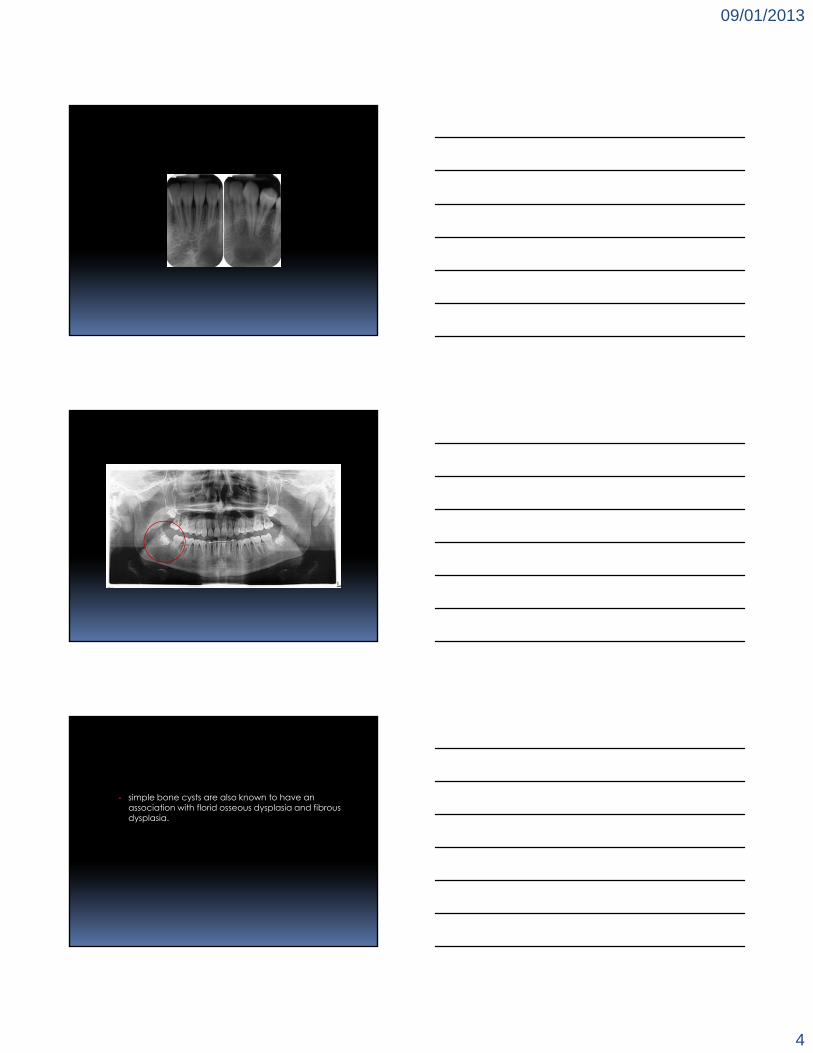
▫ simple (hemorrhagic/idiopathic/traumatic) bone cyst (cavity) an intra-bony cavity that lacks an epithelial lining.
▫ some believe that the formation of simple bone cysts represent a form of “aseptic necrosis” of the bone, the result of which is the reduced blood supply associated with these lesions.
radiographically, the cortex of the simple bone cyst is one of the most delicate.
classically, it shows an undulating path around the roots of the teeth, but this is not the case should the cavity develop away from the roots of teeth. lamina dura and tooth follicles are undisturbed.
09/01/2013
4
simple bone cysts are also known to have an association with florid osseous dysplasia and fibrous dysplasia.

09/01/2013
5
aug 2002
courtesy, DR. C. POON WOO
09/01/2013
6
mar 2006
courtesy, DR. C. POON WOO
dec 2008
courtesy, DR. C. POON WOO
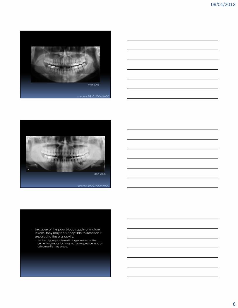
▫ because of the poor blood supply of mature lesions, they may be susceptible to infection if exposed to the oral cavity. this is a bigger problem with larger lesions, as the
cemento-osseous foci may act as sequestrae, and an osteomyelitis may ensue.
09/01/2013
7
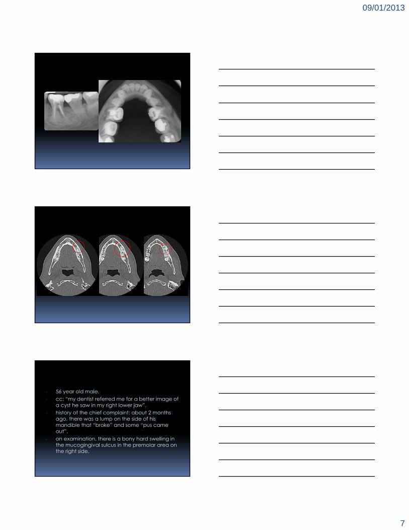
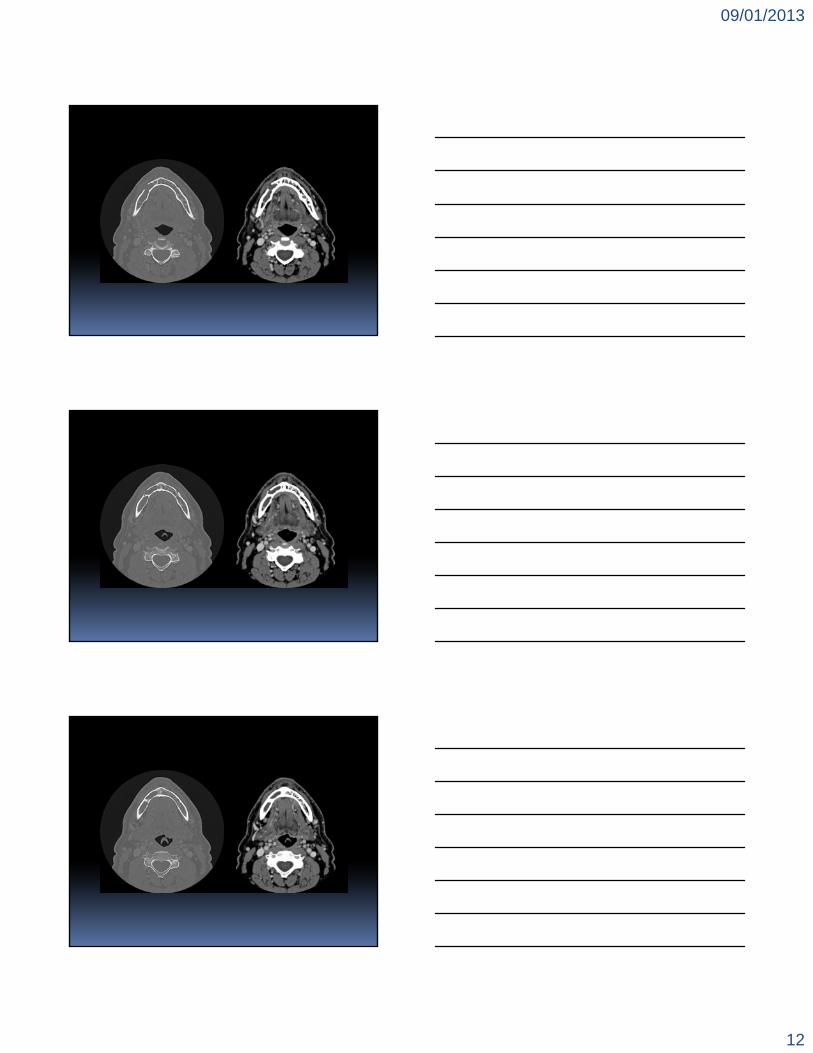
▫ 56 year old male.▫ cc: “my dentist referred me for a better image of
a cyst he saw in my right lower jaw”.▫ history of the chief complaint: about 2 months
ago, there was a lump on the side of his mandible that “broke” and some “pus came out”.
▫ on examination, there is a bony hard swelling in the mucogingival sulcus in the premolar area on the right side.

09/01/2013
8

09/01/2013
9
09/01/2013
10

09/01/2013
11
09/01/2013
12
09/01/2013
13

09/01/2013
14
?
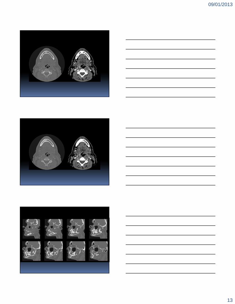
▫ keratocystic odontogenic tumour/odontogenic keratocyst) recently, the odontogenic keratocyst was reclassified
by the WHO as an odontogenic tumour because of its aggressive nature, its propensity to undergo “budding” of the basal epithelial cell layer and the identification of the putative tumour suppressor gene, PTCH.
the epithelial cells lining this entity are derived from remnants of the dental lamina.

09/01/2013
15
radiographically, keratocystic odontogenic tumours may occur in isolation, but they can also be associated with the crowns of unerupted or impacted teeth, mimicking a dentigerous cyst. some lesions may also spawn daughter cysts, so they
may take on a multilocular appearance.
the presence of an inflection in the contour of the follicle may be a clue that this is not a dentigerous cyst.
in the maxilla, keratocystic odontogenic tumours have a similar “hydraulic” appearance as a dentigerous cyst, but again, without the association with the cemento-enamel junction.
09/01/2013
16
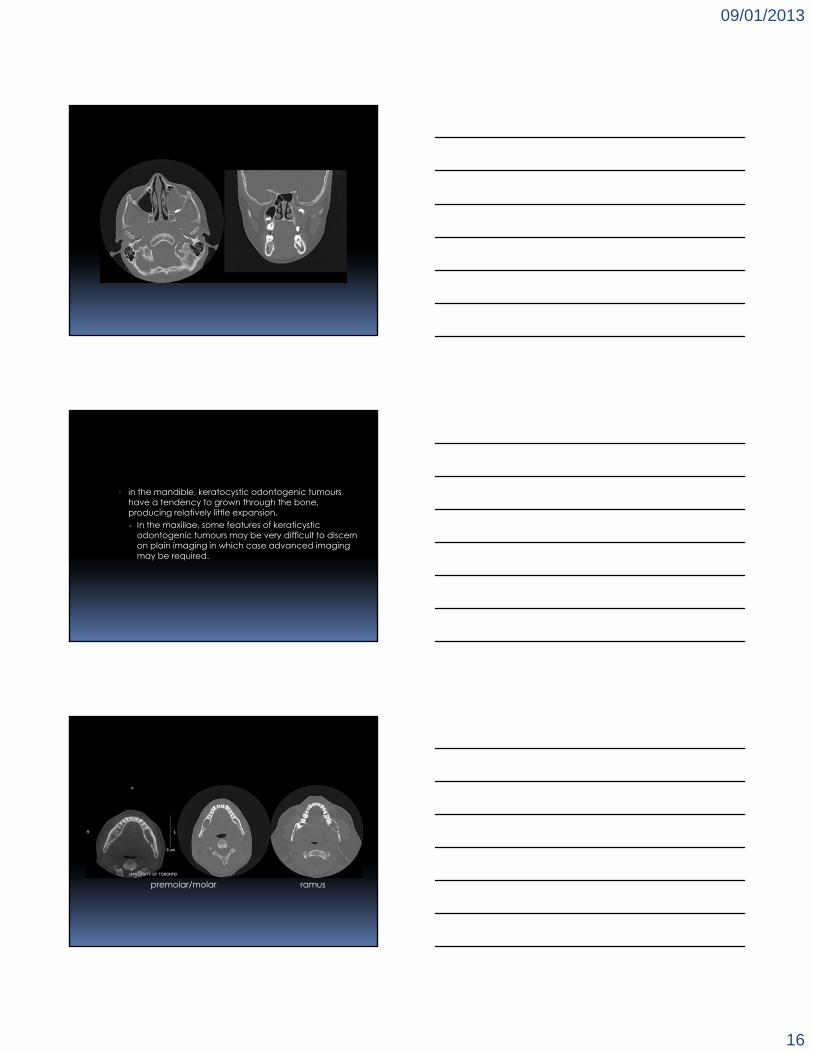
in the mandible, keratocystic odontogenic tumours have a tendency to grown through the bone, producing relatively little expansion.▫ In the maxillae, some features of keraticystic
odontogenic tumours may be very difficult to discern on plain imaging in which case advanced imaging may be required.
ramuspremolar/molar

09/01/2013
17
“trans”-mandibular
multiple keratocystic odontogenic tumours may occur in nevoid basal cell carcinoma syndrome (a.k.a. GORLIN GOLTZ basal cell nevus syndrome). some patients may develop as many as 28
keratocystic odontogenic tumours, although most develop 5 or 6.

09/01/2013
18
this is an autosomal dominant disorder that may become evident in childhood or early adolescence. affected individuals may present with multiple basal
cell tumours, bifid ribs, benign dermal cysts and ectopic calcification of the falx.
▫ cone beam CT

09/01/2013
19
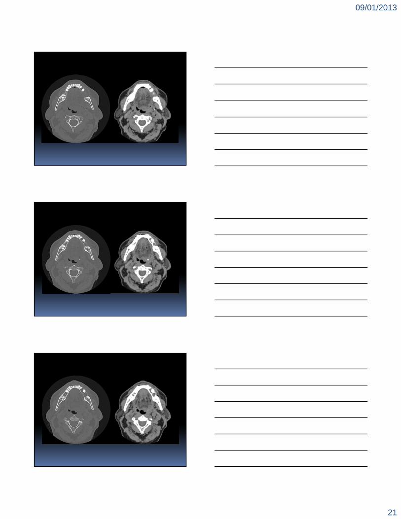
▫ 43 year old female with a mandibular radiolucency.

09/01/2013
20
09/01/2013
21

09/01/2013
22
09/01/2013
23

09/01/2013
24
?

09/01/2013
25
▫ ameloblastoma, the prototypical odontogenic tumour the mandible is the most favoured site (85%), especially
the molar/ramus area where incidence is approximately 60%.
most present as a painless swelling (75%), with pain (33%), loosening of teeth, malocclusion, ill-fitting dentures, or ulceration.
WORTH (1963) describes 4 radiographic patterns, one or all of which, may be seen. unilocular unilocular with coarse trabeculae multilocular honeycomb
WORTH also notes that identification of the following features greatly to the possibility of ameloblastoma: the presence of septation within a larger cavity. when a septum produces partial loculation within a
cavity. where there is loss of a bony margin, particularly the
retromolar alveolar crest or anterior border of the ramus.
09/01/2013
26
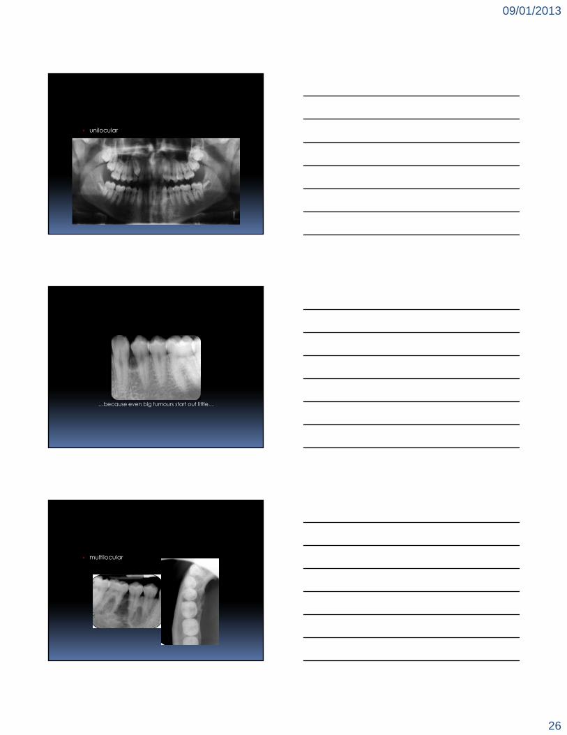
unilocular
…because even big tumours start out little…
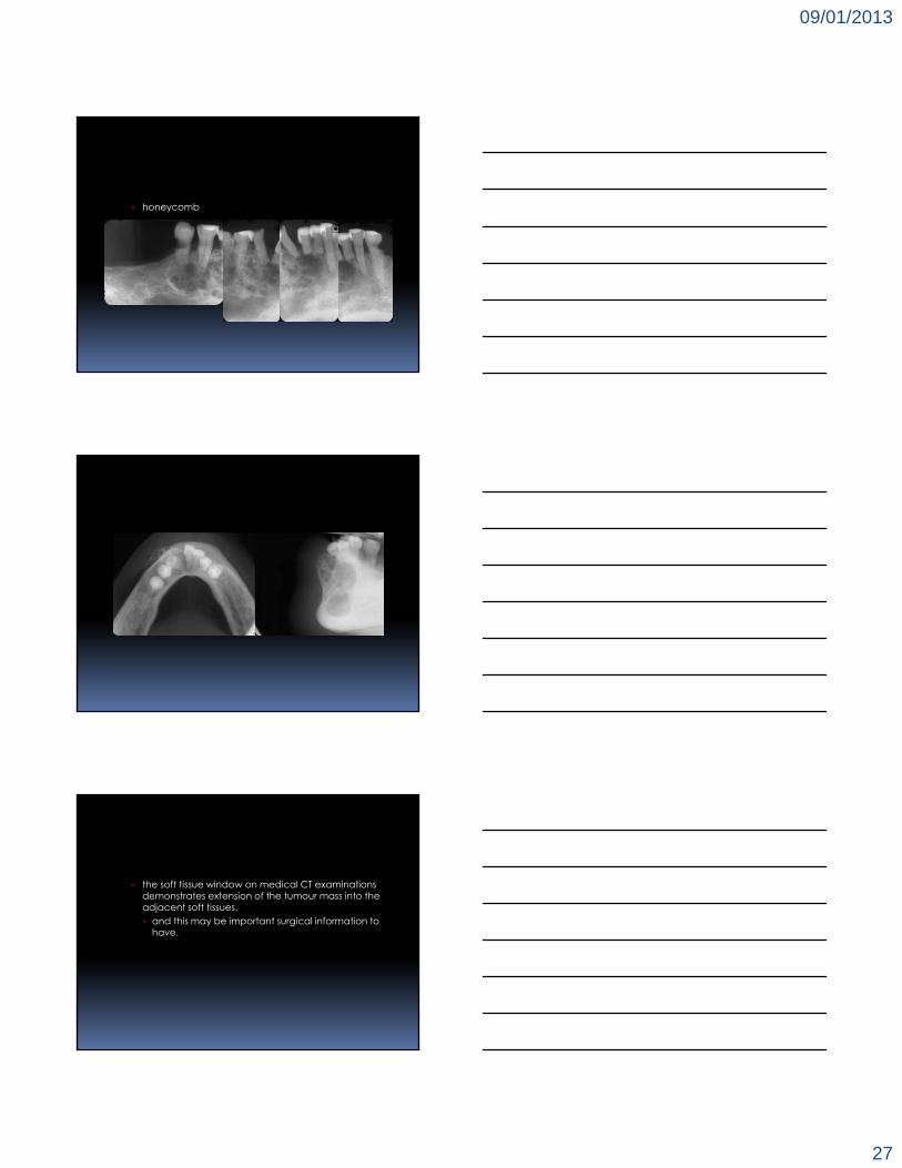
multilocular
09/01/2013
27
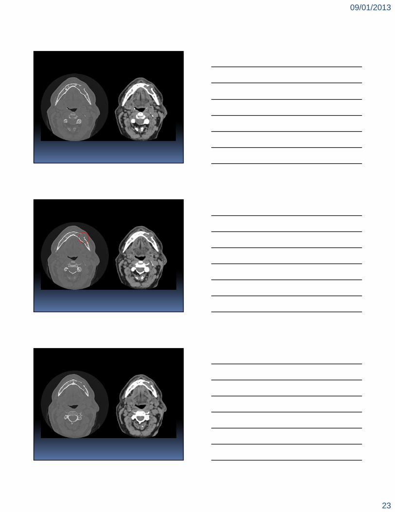
honeycomb
the soft tissue window on medical CT examinations demonstrates extension of the tumour mass into the adjacent soft tissues. and this may be important surgical information to
have.
09/01/2013
28
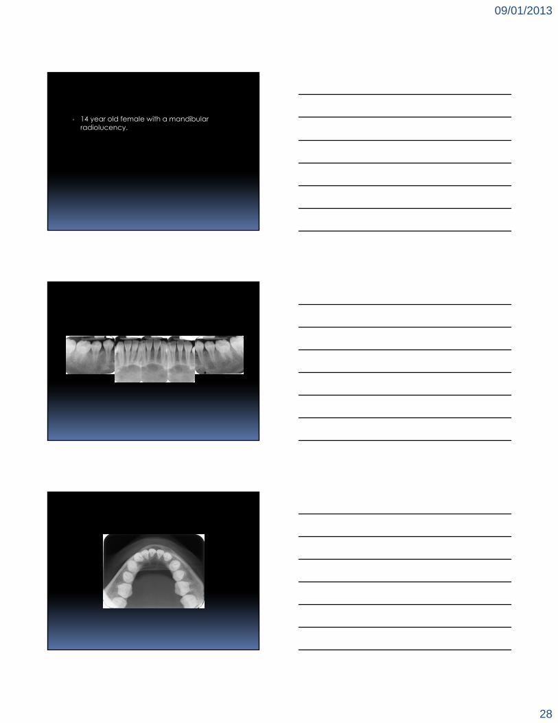
▫ 14 year old female with a mandibular radiolucency.

09/01/2013
29
?
central giant cell lesion these lesions have been thought to be reactive, yet
their stimulus is unknown. most commonly, individuals under the age of 20 are
affected, and these growths are characterized as being slow and painless.
central giant cell lesions occur with twice the frequency in the mandible as they do in the maxillae. maxillary lesions typically develop mesial to the
canines, while mandibular lesions develop mesial to first molars.

09/01/2013
30
radiographically, the lesions are radiolucent with a delicate network of straight septae that course through the radiolucency, but unlike myxomas, these lesions do produce considerable expansion of the bone. external resorption of teeth is a common feature with
the larger lesions.
giant cell lesions may occur with hyperparathyroidism, and occur bilaterally in the mandibular rami in cherubism and other syndromes.
▫ 11 year old female.▫ an orthodontist from the Hospital for Sick
Children would like you to localize the mandibular left third molar.

09/01/2013
31
▫ cone beam CT
▫ 41 year old female.▫ there is a history of pain and swelling in the left
posterior mandible following extraction of the mandibular right third molar.

09/01/2013
32
▫ cone beam CT
?
09/01/2013
33
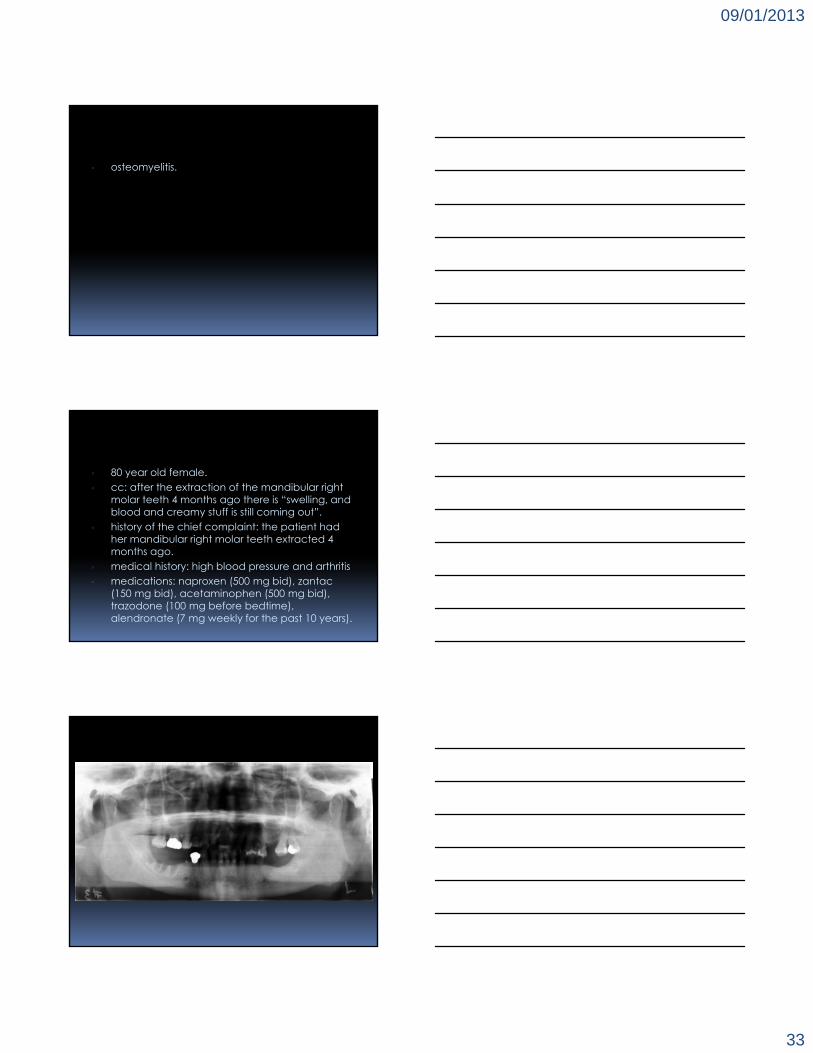
▫ osteomyelitis.
▫ 80 year old female.▫ cc: after the extraction of the mandibular right
molar teeth 4 months ago there is “swelling, and blood and creamy stuff is still coming out”.
▫ history of the chief complaint: the patient had her mandibular right molar teeth extracted 4 months ago.
▫ medical history: high blood pressure and arthritis▫ medications: naproxen (500 mg bid), zantac
(150 mg bid), acetaminophen (500 mg bid), trazodone (100 mg before bedtime), alendronate (7 mg weekly for the past 10 years).
09/01/2013
34
▫ cone beam CT
?

09/01/2013
35
▫ osteonecrosis (bisphosphonate-related osteonecrosis of the jaws).
▫ 34 year old male with orofacial pain.
▫ cone beam CT

09/01/2013
36
?
▫ left mandibular angle fracture.▫ left orbital floor blow-out fracture.
▫ 26 year old male with a mandibular swelling.
09/01/2013
37

09/01/2013
38

09/01/2013
39
?
▫ hard tissue sarcomas such as osteosarcoma and chondrosarcoma are very rare in the jaws. both these malignant tumours may undergo varying
degrees of mineralization of their soft tissue components tumour.
early bone changes in osteosarcoma and chondrosarcoma are best seen by intra-oral radiography.

09/01/2013
40
1sep 16jan 23jan
26jan 24feb 23mar
thank you
ERNEST LAM, DMD, MSc, PhD, FRCD(C)
Professor and the Dr. Lloyd & Mrs. Kay Chapman Chair in Clinical SciencesGraduate Program Director and Head,Discipline of Oral and Maxillofacial RadiologyFaculty of Dentistry, the University of Toronto


![Case Report - Hindawi Publishing Corporation1.Introduction. Foreign body aspiration is an uncommon problem in adults [1]. About 80 percent of reported cases occur in children under](https://img.pdfslide.us/doc/110x75/6107258152bc7237ff5c6bf2/case-report-hindawi-publishing-corporation-1introduction-foreign-body-aspiration.jpg)