Embed Size (px)
Citation preview
7/27/2019 4. Thrombocytopenia and Release of Activated von Willebrand Factor during Early Plasmodium falciparum Malaria…
http://slidepdf.com/reader/full/4-thrombocytopenia-and-release-of-activated-von-willebrand-factor-during-early 1/7
622 • JID 2007:196 (15 August) • de Mast et al.
M A J O R A R T I C L E
Thrombocytopenia and Release
of Activated von Willebrand Factorduring Early Plasmodium falciparum Malaria
Quirijn de Mast,1 Evelyn Groot,3 Peter J. Lenting,3 Philip G. de Groot,3 Matthew McCall,2 Robert W. Sauerwein,2
Rob Fijnheer,3,4 and Andre van der Ven1
Departments of 1General Internal Medicine and 2Medical Microbiology, Radboud University Nijmegen Medical Centre, Nijmegen, 3Laboratory
for Thrombosis and Haemostasis, Department of Clinical Chemistry and Haematology, University Medical Center Utrecht, Utrecht,
and 4Department of Internal Medicine, Jeroen Bosch Hospital, Location Groot Ziekengasthuis, ’s-Hertogenbosch, The Netherlands
Background. Thrombocytopenia occurs early during malarial infection, but its underlying mechanism is un-clear. Secretion of von Willebrand factor (vWF) occurs on endothelial cell activation, and it plays an important
role in platelet agglutination.Methods. In 14 healthy human volunteers who were experimentally infected with Plasmodium falciparum, we
studied vWF secretion and proteolysis as well as the relationship between changes in circulating platelet numbersand plasma levels of vWF and activated vWF.
Results. Platelet numbers started to decrease between days 7 and 9 after infection, which corresponded to theearliest phase of blood-stage infection. With the decrease in platelet numbers, levels of vWF, vWF propeptide(markers of chronic and acute endothelial cell activation, respectively), and activated vWF (exposing the glycoproteinIba platelet-binding domain) increased proportionally. A strong, reciprocal relationship was observed betweenplatelet numbers and levels of both vWF and activated vWF. Activity of the vWF-cleaving protease ADAMTS13(a disintegrin and metalloproteinase with a thrombospondin type 1 motif, member 13)—a regulator of vWFactivity—remained unchanged.
Conclusions. P. falciparum induces systemic acute endothelial cell activation and release of activated vWFimmediately after the onset of blood-stage infection. The resulting platelet agglutination may result in early thrombocytopenia and may play a role in the pathogenesis of malaria.
Clinical studies have shown the occurrence of throm-
bocytopenia in up to 80% of patients with malaria [1–
3], and platelet numbers have been negativelycorrelated
with both parasitemia [2] and disease severity [4]. We
have also observed a significant drop in platelet num-
bers in healthy volunteers who were experimentally
infected in our clinic with Plasmodium falciparum. Re-
markably, this decline started very early during blood-
stage infection—at the time when parasite densities
Received 27 November 2006; accepted 13 March 2007; electronically published
10 July 2007.
Potential conflicts of interest: none reported.
Financial support: Stichting Dioraphte (experimental human malaria infection);
Noaber and Hoge Dennen Foundations (laboratory work for von Willebrand factor).
Reprints or correspondence: Quirijn de Mast, Dept. of General Internal Medicine,
Radboud University Nijmegen Medical Center, PO Box 9101, 6500 HB, Nijmegen,
The Netherlands ([email protected]).
The Journal of Infectious Diseases 2007;196:622–8
2007 by the Infectious Diseases Society of America. All rights reserved.
0022-1899/2007/19604-0020$15.00
DOI: 10.1086/519844
were still at a submicroscopic detection level and before
symptoms of malaria were present. Platelets may play
a significant role in the pathogenesis of malaria. Platelet
accumulation in the brain microvasculature has been
described in children who died of cerebral malaria [5].
Intravascular platelet agglutination with increased plate-
let clearance from the circulation can be induced by
activated high-multimeric von Willebrand factor (vWF).
Elevated levels of vWF have been reported in field stud-
ies of patients with malaria [6–8], but its role in ma-
laria-induced thrombocytopenia and platelet clumpingis not known.
vWF is a large, multimeric glycoprotein that func-
tions as a carrier for factor VIII and as a bridging mol-
ecule for platelet adhesion and aggregation. It is pre-
dominantly produced in vascular endothelial cells, in
which it undergoes extensive processing after synthesis,
including dimerization, multimerization, and endopro-
teolytic cleavage of a propeptide. Large vWF multimers
7/27/2019 4. Thrombocytopenia and Release of Activated von Willebrand Factor during Early Plasmodium falciparum Malaria…
http://slidepdf.com/reader/full/4-thrombocytopenia-and-release-of-activated-von-willebrand-factor-during-early 2/7
von Willebrand Factor during Early Malaria • JID 2007:196 (15 August) • 623
and vWF propeptide are stored in specialized granules called
“Weibel-Palade bodies” [9], which are released during endo-
thelial cell activation. Therefore, both vWF and vWF propeptide
serve as markers of endothelial cell activation, although vWF
propeptide reflects acute endothelial cell perturbation because
of its shorter half-life. After release from Weibel-Palade bodies,
vWF multimers are rapidly proteolyzed to smaller and less-
active forms by the plasma metalloprotease ADAMTS13 (a dis-integrin and metalloproteinase with a thrombospondin type 1
motif, member 13) [10]. At least some released vWF is in an
activated conformation, which allows interaction with the
platelet receptor gpIba [11]. Determination of activated vWF
levels recently became possible with the introduction of a novel,
llama-derived nanobody (AU/VWFa-11) that specifically rec-
ognizes the vWF gpIba–binding conformation [12]. Increased
amounts of activated vWF were subsequently demonstrated in
the plasma of patients with thrombotic thrombocytopenic pur-
pura (TTP) [12]. This disease shares some similarities with
malaria: intravascular platelet agglutination with thrombocy-
topenia, nonimmune hemolysis, neurological symptoms, andfever.
Vascular endothelial cell activation is considered to be a com-
mon feature of malaria and plays an important role in the
pathogenesis of malaria by increasing sequestration of parasit-
ized red blood cells in the peripheral vasculature [6, 13, 14].
We hypothesized that endothelial cell activation with subse-
quent release of activated vWF is an early event in malaria and
is related to the development of early thrombocytopenia.There-
fore, we studied the role played by endothelial cell–derived
activated vWF in malaria-induced thrombocytopenia in healthy
volunteers who were experimentally infected with P. falciparum.
SUBJECTS, MATERIALS, AND METHODS
Experimental malarial infection, study subjects, and blood
sampling. Experimental human malarial infections are in-
valuable for phase 2a malaria-vaccine trials and are a powerful
tool for the study of the pathophysiology and immunology of
early malaria. By use of a stringent protocol, experimental hu-
man malarial infections have been proven to be reliable, safe,
and generally well tolerated [15, 16]. In addition to malaria-
induced thrombocytopenia, the current experimental infection
was used to study immunological responses during early P.
falciparum malaria. Fourteen healthy volunteers with no pre-
vious exposure to malaria were selected and infected with P.
falciparum parasites as described elsewhere by Verhage et al.
[16]. In short, Anopheles stephensi mosquitoes were maintained
in the insectary at the animal house of the Radboud University
Nijmegen Medical Centre and were infected with the NF54
strain of P. falciparum. Batches with 190% infected mosquitoes
were used for the experimental human infection. Volunteers
were infected by 5 P. falciparum–infected mosquitoes and were
followed-up via thick blood smears and assessment of malarial
symptoms twice a day from day 4 to 6 after infection. From
day 6 until 3 days after initiation of antimalarial treatment,
these assessments were done 3 times a day. Serial blood samples
were collected once daily in 4-mL Vacutainer glass tubes con-
taining 48 mL of EDTA-K3 (BD Diagnostics) at baseline, from
day 4 after infection until 3 days after the start of antimalarial
treatment, and on days 21 and 42 after infection. After cen-trifugation, plasma was stored at 80C. All assays were per-
formed on freshly thawed samples. Plasma from 40 healthy
donors was used as a control (normal pooled plasma [NPP]).
For the determination of ADAMTS13 activity, heparin plasma
from baseline and from day 8 after infection was used.
Treatment with a standard 6-dose curative regimen of ar-
temether-lumefantrine was immediately initiated on micro-
scopic detection of parasites in a thick blood smear. The In-
stitutional Review Board of the Radboud University Nijmegen
Medical Centre approved the protocol (CMO 2004/129), and
volunteers provided written informed consent.
Quantitative nucleic acid sequence–based amplification
(QT-NASBA) and vWF, vWF propeptide, activated vWF, and
ADAMTS13 activity. Three times daily, real-time QT-NASBA,
as described by Schneider et al. [17], was used for retrospective
determination of parasitemia. The sensitivity of this assay is 20
parasites/mL and is ∼1000 times more sensitive than standard
microscopy. vWF and vWF propeptide levels were determined
at baseline and from the last day before the onset of blood-
stage infection (as assessed by QT-NASBA) until 2 days after
the start of antimalarial treatment. vWF levels were determined
as described elsewhere [18]. vWF propeptide levels were mea-
sured as described by Borchiellini et al. [19]. Levels of activatedvWF were measured using the AU/VWFa-11 nanobody, as de-
scribed by Hulstein et al. [12]. The relative amount of activated
vWF was determined by calculating the ratio of the absorbance
slope of a plasma sample to the slope of the corresponding
baseline sample and then correcting for the amount of vWF.
ADAMTS13 activity was determined at baseline and on day 8
after infection by the FRETS-VWF73 assay (Peptides Interna-
tional) [20], in accordance with the manufacturer’s protocol.
NPP was used as a standard.
Statistical analysis. Platelet number and vWF, vWF pro-
peptide, activated vWF, and ADAMTS13 activity were expressed
as percentage of baseline values, and each participant servedas his or her own control. Means with corresponding 95%
confidence intervals (CIs) or SDs were calculated for normally
distributed data, whereas medians with ranges were determined
for data with a skewed distribution. Changes in serial levels of
vWF and vWF propeptide during the course of the infection
were tested by repeated-measures analysis of variance, and the
Wilcoxon matched-pairs test was used for activated vWF. Re-
lationships between the separate variables were examined by
7/27/2019 4. Thrombocytopenia and Release of Activated von Willebrand Factor during Early Plasmodium falciparum Malaria…
http://slidepdf.com/reader/full/4-thrombocytopenia-and-release-of-activated-von-willebrand-factor-during-early 3/7
624 • JID 2007:196 (15 August) • de Mast et al.
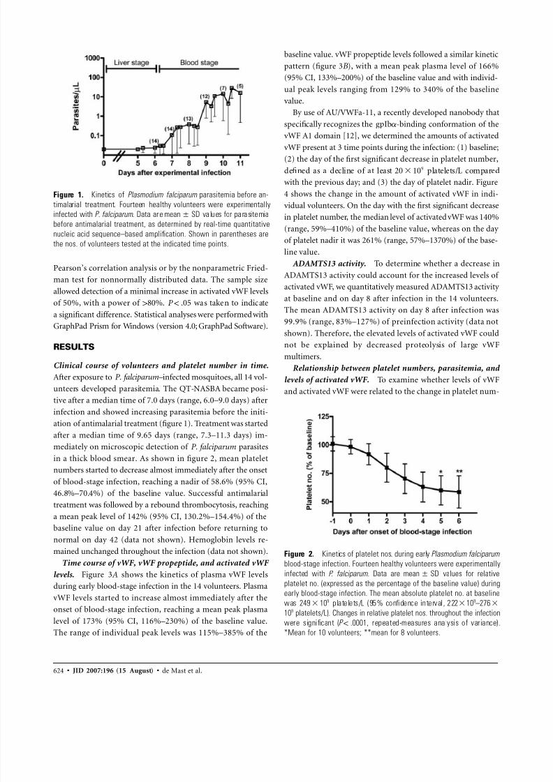
Figure 1. Kinetics of Plasmodium falciparum parasitemia before an-
timalarial treatment. Fourteen healthy volunteers were experimentally
infected with P. falciparum . Data are values for parasitemiamean SD
before antimalarial treatment, as determined by real-time quantitative
nucleic acid sequence–based amplification. Shown in parentheses are
the nos. of volunteers tested at the indicated time points.
Figure 2. Kinetics of platelet nos. during early Plasmodium falciparum
blood-stage infection. Fourteen healthy volunteers were experimentally
infected with P. falciparum. Data are values for relativemean SD
platelet no. (expressed as the percentage of the baseline value) during
early blood-stage infection. The mean absolute platelet no. at baseline
was platelets/L (95% confidence interval , 222 109–2769249 10
109 platelets/L). Changes in relative platelet nos. throughout the infection
were significant ( , repeated-measures analysis of variance).P ! .0001
*Mean for 10 volunteers; **mean for 8 volunteers.
Pearson’s correlation analysis or by the nonparametric Fried-
man test for nonnormally distributed data. The sample size
allowed detection of a minimal increase in activated vWF levels
of 50%, with a power of 180%. was taken to indicateP ! .05
a significant difference. Statistical analyses were performed with
GraphPad Prism for Windows (version 4.0; GraphPad Software).
RESULTS
Clinical course of volunteers and platelet number in time.
After exposure to P. falciparum– infected mosquitoes, all 14 vol-
unteers developed parasitemia. The QT-NASBA became posi-
tive after a median time of 7.0 days (range, 6.0–9.0 days) after
infection and showed increasing parasitemia before the initi-ation of antimalarial treatment (figure 1). Treatment was started
after a median time of 9.65 days (range, 7.3–11.3 days) im-
mediately on microscopic detection of P. falciparum parasites
in a thick blood smear. As shown in figure 2, mean platelet
numbers started to decrease almost immediately after the onset
of blood-stage infection, reaching a nadir of 58.6% (95% CI,
46.8%–70.4%) of the baseline value. Successful antimalarial
treatment was followed by a rebound thrombocytosis, reaching
a mean peak level of 142% (95% CI, 130.2%–154.4%) of the
baseline value on day 21 after infection before returning to
normal on day 42 (data not shown). Hemoglobin levels re-
mained unchanged throughout the infection (data not shown).
Time course of vWF, vWF propeptide, and activated vWF
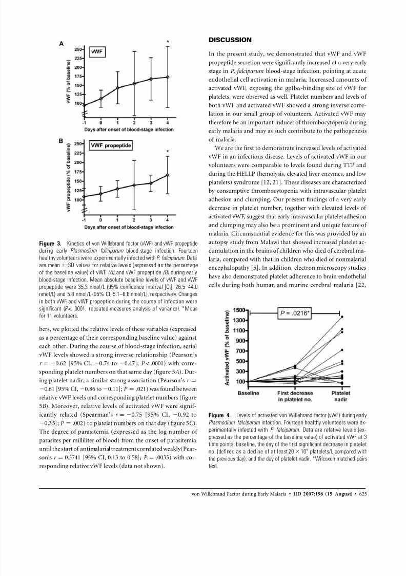
levels. Figure 3A shows the kinetics of plasma vWF levels
during early blood-stage infection in the 14 volunteers. Plasma
vWF levels started to increase almost immediately after the
onset of blood-stage infection, reaching a mean peak plasma
level of 173% (95% CI, 116%–230%) of the baseline value.
The range of individual peak levels was 115%–385% of the
baseline value. vWF propeptide levels followed a similar kinetic
pattern (figure 3B ), with a mean peak plasma level of 166%
(95% CI, 133%–200%) of the baseline value and with individ-
ual peak levels ranging from 129% to 340% of the baseline
value.
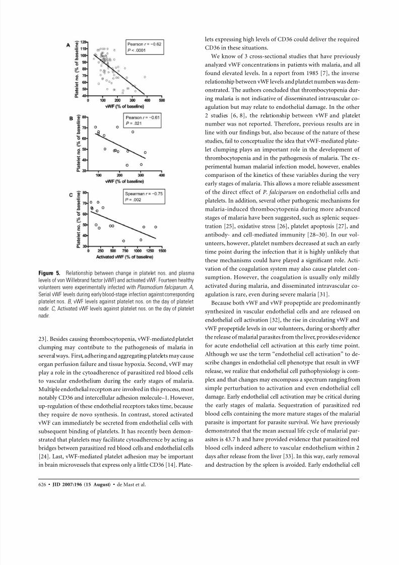
By use of AU/VWFa-11, a recently developed nanobody that
specifically recognizes the gpIba-binding conformation of the
vWF A1 domain [12], we determined the amounts of activatedvWF present at 3 time points during the infection: (1) baseline;
(2) the day of the first significant decrease in platelet number,
defined as a decline of at least platelets/L compared920 10
with the previous day; and (3) the day of platelet nadir. Figure
4 shows the change in the amount of activated vWF in indi-
vidual volunteers. On the day with the first significant decrease
in platelet number, the median level of activated vWF was 140%
(range, 59%–410%) of the baseline value, whereas on the day
of platelet nadir it was 261% (range, 57%–1370%) of the base-
line value.
ADAMTS13 activity. To determine whether a decrease in
ADAMTS13 activity could account for the increased levels of activated vWF, we quantitatively measured ADAMTS13 activity
at baseline and on day 8 after infection in the 14 volunteers.
The mean ADAMTS13 activity on day 8 after infection was
99.9% (range, 83%–127%) of preinfection activity (data not
shown). Therefore, the elevated levels of activated vWF could
not be explained by decreased proteolysis of large vWF
multimers.
Relationship between platelet numbers, parasitemia, and
levels of activated vWF. To examine whether levels of vWF
and activated vWF were related to the change in platelet num-
7/27/2019 4. Thrombocytopenia and Release of Activated von Willebrand Factor during Early Plasmodium falciparum Malaria…
http://slidepdf.com/reader/full/4-thrombocytopenia-and-release-of-activated-von-willebrand-factor-during-early 4/7
von Willebrand Factor during Early Malaria • JID 2007:196 (15 August) • 625
Figure 3. Kinetics of von Willebrand factor (vWF) and vWF propeptide
during early Plasmodium falciparum blood-stage infection. Fourteen
healthy volunteers were experimentally infected with P. falciparum. Data
are values for relative levels (expressed as the percentagemean SD
of the baseline value) of vWF (A) and vWF propeptide (B) during early
blood-stage infection. Mean absolute baseline levels of vWF and vWF
propeptide were 35.3 nmol/L (95% confidence interval [CI], 26.5–44.0
nmol/L) and 5.8 nmol/L (95% CI, 5.1–6.6 nmol/L), respectively. Changes
in both vWF and vWF propeptide during the course of infection weresignificant ( , repeated-measures analysis of variance). *MeanP ! .0001
for 11 volunteers.
Figure 4. Levels of activated von Willebrand factor (vWF) during early
Plasmodium falciparum infection. Fourteen healthy volunteers were ex-
perimentally infected with P. falciparum. Data are relative levels (ex-
pressed as the percentage of the baseline value) of activated vWF at 3
time points: baseline, the day of the first significant decrease in platelet
no. (defined as a decline of at least platelets/L compared with920 10
the previous day), and the day of platelet nadir. *Wilcoxon matched-pairs
test.
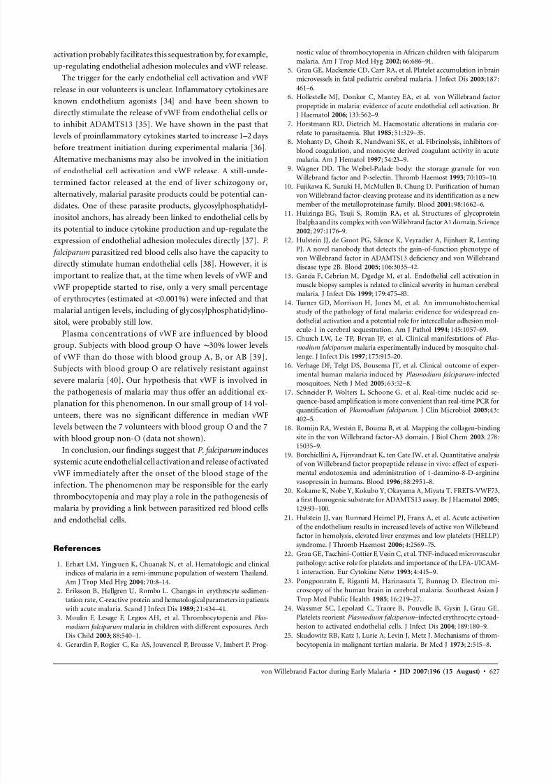
bers, we plotted the relative levels of these variables (expressed
as a percentage of their corresponding baseline value) against
each other. During the course of blood-stage infection, serial
vWF levels showed a strong inverse relationship (Pearson’s
[95% CI, 0.74 to 0.47]; ) with corre-r p0.62 P ! .0001
sponding platelet numbers on that same day (figure 5A ). Dur-
ing platelet nadir, a similar strong association (Pearson’s r p
[95% CI, 0.86 to 0.11]; ) was found between0.61 P p .021
relative vWF levels and corresponding platelet numbers (figure
5B ). Moreover, relative levels of activated vWF were signif-
icantly related (Spearman’s [95% CI, 0.92 tor p0.75
0.35]; ) to platelet numbers on that day (figure 5C ).P p .002
The degree of parasitemia (expressed as the log number of
parasites per milliliter of blood) from the onset of parasitemia
until the start of antimalarial treatment correlated weakly(Pear-
son’s [95% CI, 0.13 to 0.58]; ) with cor-r p 0.3741 P p .0035
responding relative vWF levels (data not shown).
DISCUSSION
In the present study, we demonstrated that vWF and vWF
propeptide secretion were significantly increased at a very early
stage in P. falciparum blood-stage infection, pointing at acute
endothelial cell activation in malaria. Increased amounts of
activated vWF, exposing the gpIba-binding site of vWF for
platelets, were observed as well. Platelet numbers and levels of
both vWF and activated vWF showed a strong inverse corre-
lation in our small group of volunteers. Activated vWF may
therefore be an important inducer of thrombocytopenia during
early malaria and may as such contribute to the pathogenesis
of malaria.
We are the first to demonstrate increased levels of activated
vWF in an infectious disease. Levels of activated vWF in our
volunteers were comparable to levels found during TTP and
during the HELLP (hemolysis, elevated liver enzymes, and low
platelets) syndrome [12, 21]. These diseases are characterized
by consumptive thrombocytopenia with intravascular platelet
adhesion and clumping. Our present findings of a very early decrease in platelet number, together with elevated levels of
activated vWF, suggest that early intravascular platelet adhesion
and clumping may also be a prominent and unique feature of
malaria. Circumstantial evidence for this was provided by an
autopsy study from Malawi that showed increased platelet ac-
cumulation in the brains of children who died of cerebral ma-
laria, compared with that in children who died of nonmalarial
encephalopathy [5]. In addition, electron microscopy studies
have also demonstrated platelet adherence to brain endothelial
cells during both human and murine cerebral malaria [22,
7/27/2019 4. Thrombocytopenia and Release of Activated von Willebrand Factor during Early Plasmodium falciparum Malaria…
http://slidepdf.com/reader/full/4-thrombocytopenia-and-release-of-activated-von-willebrand-factor-during-early 5/7
626 • JID 2007:196 (15 August) • de Mast et al.
Figure 5. Relationship between change in platelet nos. and plasma
levels of von Willebrand factor (vWF) and activated vWF. Fourteen healthy
volunteers were experimentally infected with Plasmodium falciparum. A,
Serial vWF levels during early blood-stage infection against correspondingplatelet nos. B, vWF levels against platelet nos. on the day of platelet
nadir. C, Activated vWF levels against platelet nos. on the day of platelet
nadir.
23]. Besides causing thrombocytopenia, vWF-mediated platelet
clumping may contribute to the pathogenesis of malaria in
several ways. First, adhering and aggregating platelets may cause
organ perfusion failure and tissue hypoxia. Second, vWF may
play a role in the cytoadherence of parasitized red blood cells
to vascular endothelium during the early stages of malaria.
Multiple endothelial receptors are involved in this process, most
notably CD36 and intercellular adhesion molecule–1. However,
up-regulation of these endothelial receptors takes time, because
they require de novo synthesis. In contrast, stored activated
vWF can immediately be secreted from endothelial cells with
subsequent binding of platelets. It has recently been demon-
strated that platelets may facilitate cytoadherence by acting as
bridges between parasitized red blood cells and endothelial cells
[24]. Last, vWF-mediated platelet adhesion may be important
in brain microvessels that express only a little CD36 [14]. Plate-
lets expressing high levels of CD36 could deliver the required
CD36 in these situations.
We know of 3 cross-sectional studies that have previously
analyzed vWF concentrations in patients with malaria, and all
found elevated levels. In a report from 1985 [7], the inverse
relationship between vWF levels and platelet numbers was dem-
onstrated. The authors concluded that thrombocytopenia dur-
ing malaria is not indicative of disseminated intravascular co-agulation but may relate to endothelial damage. In the other
2 studies [6, 8], the relationship between vWF and platelet
number was not reported. Therefore, previous results are in
line with our findings but, also because of the nature of these
studies, fail to conceptualize the idea that vWF-mediated plate-
let clumping plays an important role in the development of
thrombocytopenia and in the pathogenesis of malaria. The ex-
perimental human malarial infection model, however, enables
comparison of the kinetics of these variables during the very
early stages of malaria. This allows a more reliable assessment
of the direct effect of P. falciparum on endothelial cells and
platelets. In addition, several other pathogenic mechanisms for
malaria-induced thrombocytopenia during more advanced
stages of malaria have been suggested, such as splenic seques-
tration [25], oxidative stress [26], platelet apoptosis [27], and
antibody- and cell-mediated immunity [28–30]. In our vol-
unteers, however, platelet numbers decreased at such an early
time point during the infection that it is highly unlikely that
these mechanisms could have played a significant role. Acti-
vation of the coagulation system may also cause platelet con-
sumption. However, the coagulation is usually only mildly
activated during malaria, and disseminated intravascular co-
agulation is rare, even during severe malaria [31].Because both vWF and vWF propeptide are predominantly
synthesized in vascular endothelial cells and are released on
endothelial cell activation [32], the rise in circulating vWF and
vWF propeptide levels in our volunteers, during or shortly after
the release of malarial parasites from the liver, provides evidence
for acute endothelial cell activation at this early time point.
Although we use the term “endothelial cell activation” to de-
scribe changes in endothelial cell phenotype that result in vWF
release, we realize that endothelial cell pathophysiology is com-
plex and that changes may encompass a spectrum ranging from
simple perturbation to activation and even endothelial cell
damage. Early endothelial cell activation may be critical duringthe early stages of malaria. Sequestration of parasitized red
blood cells containing the more mature stages of the malarial
parasite is important for parasite survival. We have previously
demonstrated that the mean asexual life cycle of malarial par-
asites is 43.7 h and have provided evidence that parasitized red
blood cells indeed adhere to vascular endothelium within 2
days after release from the liver [33]. In this way, early removal
and destruction by the spleen is avoided. Early endothelial cell
7/27/2019 4. Thrombocytopenia and Release of Activated von Willebrand Factor during Early Plasmodium falciparum Malaria…
http://slidepdf.com/reader/full/4-thrombocytopenia-and-release-of-activated-von-willebrand-factor-during-early 6/7
von Willebrand Factor during Early Malaria • JID 2007:196 (15 August) • 627
activation probably facilitates this sequestration by, for example,
up-regulating endothelial adhesion molecules and vWF release.
The trigger for the early endothelial cell activation and vWF
release in our volunteers is unclear. Inflammatory cytokines are
known endothelium agonists [34] and have been shown to
directly stimulate the release of vWF from endothelial cells or
to inhibit ADAMTS13 [35]. We have shown in the past that
levels of proinflammatory cytokines started to increase 1–2 daysbefore treatment initiation during experimental malaria [36].
Alternative mechanisms may also be involved in the initiation
of endothelial cell activation and vWF release. A still-unde-
termined factor released at the end of liver schizogony or,
alternatively, malarial parasite products could be potential can-
didates. One of these parasite products, glycosylphosphatidyl-
inositol anchors, has already been linked to endothelial cells by
its potential to induce cytokine production and up-regulate the
expression of endothelial adhesion molecules directly [37]. P.
falciparum parasitized red blood cells also have the capacity to
directly stimulate human endothelial cells [38]. However, it is
important to realize that, at the time when levels of vWF andvWF propeptide started to rise, only a very small percentage
of erythrocytes (estimated at !0.001%) were infected and that
malarial antigen levels, including of glycosylphosphatidylino-
sitol, were probably still low.
Plasma concentrations of vWF are influenced by blood
group. Subjects with blood group O have ∼30% lower levels
of vWF than do those with blood group A, B, or AB [39].
Subjects with blood group O are relatively resistant against
severe malaria [40]. Our hypothesis that vWF is involved in
the pathogenesis of malaria may thus offer an additional ex-
planation for this phenomenon. In our small group of 14 vol-
unteers, there was no significant difference in median vWF
levels between the 7 volunteers with blood group O and the 7
with blood group non-O (data not shown).
In conclusion, our findings suggest that P. falciparum induces
systemic acute endothelial cell activation and release of activated
vWF immediately after the onset of the blood stage of the
infection. The phenomenon may be responsible for the early
thrombocytopenia and may play a role in the pathogenesis of
malaria by providing a link between parasitized red blood cells
and endothelial cells.
References
1. Erhart LM, Yingyuen K, Chuanak N, et al. Hematologic and clinical
indices of malaria in a semi-immune population of western Thailand.
Am J Trop Med Hyg 2004; 70:8–14.
2. Eriksson B, Hellgren U, Rombo L. Changes in erythrocyte sedimen-
tation rate, C-reactive protein and hematological parameters in patients
with acute malaria. Scand J Infect Dis 1989; 21:434–41.
3. Moulin F, Lesage F, Legros AH, et al. Thrombocytopenia and Plas-
modium falciparum malaria in children with different exposures. Arch
Dis Child 2003; 88:540–1.
4. Gerardin P, Rogier C, Ka AS, Jouvencel P, Brousse V, Imbert P. Prog-
nostic value of thrombocytopenia in African children with falciparum
malaria. Am J Trop Med Hyg 2002; 66:686–91.
5. Grau GE, Mackenzie CD, Carr RA, et al. Platelet accumulation in brain
microvessels in fatal pediatric cerebral malaria. J Infect Dis 2003;187:
461–6.
6. Hollestelle MJ, Donkor C, Mantey EA, et al. von Willebrand factor
propeptide in malaria: evidence of acute endothelial cell activation. Br
J Haematol 2006; 133:562–9.
7. Horstmann RD, Dietrich M. Haemostatic alterations in malaria cor-
relate to parasitaemia. Blut 1985; 51:329–35.
8. Mohanty D, Ghosh K, Nandwani SK, et al. Fibrinolysis, inhibitors of
blood coagulation, and monocyte derived coagulant activity in acute
malaria. Am J Hematol 1997; 54:23–9.
9. Wagner DD. The Weibel-Palade body: the storage granule for von
Willebrand factor and P-selectin. Thromb Haemost 1993; 70:105–10.
10. Fujikawa K, Suzuki H, McMullen B, Chung D. Purification of human
von Willebrand factor-cleaving protease and its identification as a new
member of the metalloproteinase family. Blood 2001; 98:1662–6.
11. Huizinga EG, Tsuji S, Romijn RA, et al. Structures of glycoprotein
Ibalpha and its complex with von Willebrand factor A1 domain. Science
2002; 297:1176–9.
12. Hulstein JJ, de Groot PG, Silence K, Veyradier A, Fijnheer R, Lenting
PJ. A novel nanobody that detects the gain-of-function phenotype of
von Willebrand factor in ADAMTS13 deficiency and von Willebrand
disease type 2B. Blood 2005; 106:3035–42.
13. Garcia F, Cebrian M, Dgedge M, et al. Endothelial cell activation inmuscle biopsy samples is related to clinical severity in human cerebral
malaria. J Infect Dis 1999; 179:475–83.
14. Turner GD, Morrison H, Jones M, et al. An immunohistochemical
study of the pathology of fatal malaria: evidence for widespread en-
dothelial activation and a potential role for intercellular adhesion mol-
ecule-1 in cerebral sequestration. Am J Pathol 1994; 145:1057–69.
15. Church LW, Le TP, Bryan JP, et al. Clinical manifestations of Plas-
modium falciparum malaria experimentally induced by mosquito chal-
lenge. J Infect Dis 1997; 175:915–20.
16. Verhage DF, Telgt DS, Bousema JT, et al. Clinical outcome of exper-
imental human malaria induced by Plasmodium falciparum-infected
mosquitoes. Neth J Med 2005; 63:52–8.
17. Schneider P, Wolters L, Schoone G, et al. Real-time nucleic acid se-
quence-based amplification is more convenient than real-time PCR for
quantification of Plasmodium falciparum. J Clin Microbiol 2005;43:402–5.
18. Romijn RA, Westein E, Bouma B, et al. Mapping the collagen-binding
site in the von Willebrand factor-A3 domain. J Biol Chem 2003; 278:
15035–9.
19. Borchiellini A, Fijnvandraat K, ten Cate JW, et al. Quantitative analysis
of von Willebrand factor propeptide release in vivo: effect of experi-
mental endotoxemia and administration of 1-deamino-8-D-arginine
vasopressin in humans. Blood 1996; 88:2951–8.
20. Kokame K, Nobe Y, Kokubo Y, Okayama A, Miyata T. FRETS-VWF73,
a first fluorogenic substrate for ADAMTS13 assay. Br J Haematol 2005;
129:93–100.
21. Hulstein JJ, van Runnard Heimel PJ, Franx A, et al. Acute activation
of the endothelium results in increased levels of active von Willebrand
factor in hemolysis, elevated liver enzymes and low platelets (HELLP)
syndrome. J Thromb Haemost 2006; 4:2569–75.
22. Grau GE, Tacchini-Cottier F, Vesin C, et al. TNF-induced microvascular
pathology: active role for platelets and importance of the LFA-1/ICAM-
1 interaction. Eur Cytokine Netw 1993; 4:415–9.
23. Pongponratn E, Riganti M, Harinasuta T, Bunnag D. Electron mi-
croscopy of the human brain in cerebral malaria. Southeast Asian J
Trop Med Public Health 1985; 16:219–27.
24. Wassmer SC, Lepolard C, Traore B, Pouvelle B, Gysin J, Grau GE.
Platelets reorient Plasmodium falciparum–infected erythrocyte cytoad-
hesion to activated endothelial cells. J Infect Dis 2004; 189:180–9.
25. Skudowitz RB, Katz J, Lurie A, Levin J, Metz J. Mechanisms of throm-
bocytopenia in malignant tertian malaria. Br Med J 1973; 2:515–8.
7/27/2019 4. Thrombocytopenia and Release of Activated von Willebrand Factor during Early Plasmodium falciparum Malaria…
http://slidepdf.com/reader/full/4-thrombocytopenia-and-release-of-activated-von-willebrand-factor-during-early 7/7
628 • JID 2007:196 (15 August) • de Mast et al.
26. Erel O, Vural H, Aksoy N, Aslan G, Ulukanligil M. Oxidative stress of
platelets and thrombocytopenia in patients with vivax malaria. Clin
Biochem 2001; 34:341–4.
27. Piguet PF, Kan CD, Vesin C. Thrombocytopenia in an animal model
of malaria is associated with an increased caspase-mediated death of
thrombocytes. Apoptosis 2002; 7:91–8.
28. Gramaglia I, Sahlin H, Nolan JP, Frangos JA, Intaglietta M, van der
Heyde HC. Cell- rather than antibody-mediated immunity leads to the
development of profound thrombocytopenia during experimentalPlas-
modium berghei malaria. J Immunol 2005; 175:7699–707.
29. Grau GE, Piguet PF, Gretener D, Vesin C, Lambert PH. Immuno-
pathology of thrombocytopenia in experimental malaria. Immunology
1988; 65:501–6.
30. Sorensen PG, Mickley H, Schmidt KG. Malaria-induced immune
thrombocytopenia. Vox Sang 1984; 47:68–72.
31. Vreeken J, Cremer-Goote TM. Haemostatic defect in non-immune
patients with falciparum malaria: no evidence of diffuse intravascular
coagulation. Br Med J 1978; 2:533–5.
32. van Mourik JA, Boertjes R, Huisveld IA, et al. von Willebrand factor
propeptide in vascular disorders: a tool to distinguish between acute
and chronic endothelial cell perturbation. Blood 1999; 94:179–85.
33. Hermsen CC, de Vlas SJ, van Gemert GJ, Telgt DS, Verhage DF, Sauer-
wein RW. Testing vaccines in human experimental malaria: statistical
analysis of parasitemia measured by a quantitative real-timepolymerase
chain reaction. Am J Trop Med Hyg 2004; 71:196–201.
34. Bevilacqua MP. Endothelial-leukocyte adhesion molecules. Annu Rev
Immunol 1993; 11:767–804.
35. Bernardo A, Ball C, Nolasco L, Moake JF, Dong JF. Effects of inflam-
matory cytokines on the release and cleavage of the endothelial cell-
derived ultralarge von Willebrand factor multimers under flow. Blood
2004; 104:100–6.
36. Hermsen CC, Konijnenberg Y, Mulder L, et al. Circulating concen-
trations of soluble granzyme A and B increase during natural and ex-
perimental Plasmodium falciparum infections. Clin Exp Immunol 2003;
132:467–72.37. Schofield L, Novakovic S, Gerold P, Schwarz RT, McConville MJ, Tach-
ado SD. Glycosylphosphatidylinositol toxin of Plasmodium up-regu-
lates intercellular adhesion molecule-1, vascular cell adhesion mole-
cule-1, and E-selectin expression in vascular endothelial cells and
increases leukocyte and parasite cytoadherence via tyrosine kinase-
dependent signal transduction. J Immunol 1996; 156:1886–96.
38. Viebig NK, Wulbrand U, Forster R, Andrews KT, Lanzer M, Knolle
PA. Direct activation of human endothelial cells by Plasmodium fal-
ciparum-infected erythrocytes. Infect Immun 2005; 73:3271–7.
39. Gill JC, Endres-Brooks J, Bauer PJ, Marks WJ Jr, Montgomery RR.
The effect of ABO blood group on the diagnosis of von Willebrand
disease. Blood 1987; 69:1691–5.
40. Fischer PR, Boone P. Short report: severe malaria associated withblood
group. Am J Trop Med Hyg 1998; 58:122–3.