Embed Size (px)
Citation preview
Binding of Levosimendan, a Calcium Sensitizer, to Cardiac Troponin C
Tia Sorsa1, Sami Heikkinen1, M. Bret Abbott2, Ekram Abusamhadneh2, Tero Laakso3, Carola
Tilgmann4, Ritva Serimaa3, Arto Annila5, Paul R. Rosevear2, Torbjörn Drakenberg5, 6, Piero
Pollesello4, and Ilkka Kilpeläinen1
1 NMR Laboratory, Institute of Biotechnology, University of Helsinki, P.O. Box 56, FIN-00014
Helsinki, Finland2 Department of Molecular Genetics, Biochemistry, and Microbiology, University of Cincinnati,
College of Medicine, Cincinnati, Ohio 45267, USA3 X-ray Laboratory, Department of Physics, University of Helsinki, P.O. Box 9, FIN-00014
Helsinki, Finland4 Orion Pharma, R & D, Cardiovascular Drug Discovery & Pharmacology, P.O. Box 65, FIN-
02101 Espoo, Finland5 VTT Biotechnology, P.O. Box 56, FIN-00014 Helsinki, Finland6 Department of Physical Chemistry 2, Chemical Centre, Lund University, P.O. Box 124, S-
22100 Lund, Sweden
RUNNING TITLE: Binding of levosimendan to cTnC
Corresponding Author: Ilkka Kilpeläinen
NMR Laboratory
Institute of Biotechnology, University of Helsinki
P.O. Box 56, FIN-00014 Helsinki, Finland
Phone: +358 9 191 595 40
Fax: +358 9 191 595 41
E-mail: [email protected]
Copyright 2000 by The American Society for Biochemistry and Molecular Biology, Inc.
JBC Papers in Press. Published on December 11, 2000 as Manuscript M007484200 by guest on July 6, 2018
http://ww
w.jbc.org/
Dow
nloaded from
2
SUMMARY
Levosimendan is an inodilatory drug that mediates its cardiac effect by the calcium
sensitization of contractile proteins. The target protein of levosimendan is cardiac troponin C
(cTnC). In the current work, we have studied the interaction of levosimendan with Ca2+-saturated
cTnC by heteronuclear NMR and small angle X-ray scattering. A specific interaction between
levosimendan and the Ca2+-loaded regulatory domain of human recombinant cTnCC35S was
observed. The changes in the NMR spectra of the N-domain of full-length cTnCC35S, due to the
binding of levosimendan to the primary site, were indicative of a slow conformational exchange.
In contrast, no binding of levosimendan to the regulatory domain of cTnCA-Cys, where all the
cysteine residues are mutated to serine, was detected. Moreover, it was shown that levosimendan
was in fast exchange, on the NMR time scale, with a secondary binding sites in the C-domain of
both cTnCC35S and cTnCA-Cys. The small angle X-ray scattering experiments confirm the binding
of levosimendan to Ca2+-saturated cTnC but shows that this binding does not introduce domain-
domain closure. The experiments were run in the absence of the reducing agent dithiothreitol
(DTT) and the preservative sodium azide (NaN3), since we found that levosimendan reacts with
these chemicals, commonly used for preparation of NMR protein samples.
Keywords: cardiac troponin C, cardiomyocyte, DTT, levosimendan, NaN3, nuclear magnetic
resonance, protein-drug binding, small angle X-ray scattering
by guest on July 6, 2018http://w
ww
.jbc.org/D
ownloaded from
3
INTRODUCTION
The number of patients suffering from heart failure is increasing along with the ageing of
population. Calcium sensitizers have been proposed as a treatment for congestive heart failure
since they exert a positive inotropic effect without increasing the intracellular calcium
concentration (1). Levosimendan, a potent calcium sensitizer which improves the force
development of the muscle contraction without increasing the cytosolic Ca2+-ion concentration
(2), was discovered using troponin C as target protein.
Troponin C (TnC) is responsible for the contraction trigger in the muscle. It belongs to
the family of calcium-binding EF-hand proteins and consists of two domains. The amino
terminal half (NTnC) is responsible for the calcium-dependent regulation of the contraction and
the carboxyl terminal half is a structural domain always loaded with divalent cations under
physiological conditions. Troponin C interacts with troponin I (TnI) and this interaction is
modulated by the binding of calcium. Studies of skeletal troponin C (sTnC), a homologous
protein, have shown that a hydrophobic patch is exposed in the open conformation of the
calcium-loaded regulatory domain, which is a binding site for TnI (3). This has also been
proposed to be a potential binding site for calcium sensitizers (4, 5). Contrary to sTnC, the
binding of Ca2+ to cTnC does not induce an opening of the conformation. Consequently there is,
in vitro, no exposure of a hydrophobic region (6-9). The simultaneous binding of cTnI and Ca2+
to cNTnC, however, opens the structure of the N-terminal domain (10, 11). This structural and
functional difference between TnC in skeletal and cardiac muscle is still to be clarified.
by guest on July 6, 2018http://w
ww
.jbc.org/D
ownloaded from
4
Levosimendan has been reported to bind to the regulatory domain of cardiac troponin C
in a calcium-dependent manner (5, 12). However, the interaction of levosimendan with cTnC has
been under debate for some time. Pollesello et al. (5) reported the binding of levosimendan to the
Ca2+-saturated form of cNTnC. A possible binding site for calcium sensitizers in the vicinity of
Asp88 was located by using point mutated and dansylated human recombinant cTnC, NMR and
molecular modelling (4, 5). However, very recently Kleerekoper and Putkey (13) reported that
levosimendan did not bind to cTnC. To clarify this controversial situation we studied the stability
of levosimendan and levosimendan-cTnC under various solution conditions and the interaction
of levosimendan with cTnC by heteronuclear NMR spectroscopy and small angle X-ray
scattering (SAXS). The results are also of general importance to studies of structure-activity
relationship (SAR) by NMR.
MATERIALS AND METHODS
Levosimendan Samples For every experiment with levosimendan, a fresh 30 mM stock
solution was prepared by dissolving dry levosimendan powder into 30 mM potassium carbonate.
The solution was gently shaken for approximately 30 s at room temperature until a clear solution
was obtained. The stock solution was analysed by HPLC and by mass spectrometry to ensure
that no degradation had occurred during the course of the sample preparation. Levosimendan
solutions were thereafter diluted in the same buffer solution used for protein samples (20 mM
Bis-Tris, 10 mM CaCl2, pH 6.8).
by guest on July 6, 2018http://w
ww
.jbc.org/D
ownloaded from
5
Protein Sample Preparation In this study, we used three different cTnC molecules.
Recombinant 15N-labelled N-terminal fragment of human cardiac troponin C (residues 1-91),
was cloned, expressed, and purified as previously described (8). The cDNA for cTnCA-Cys was
generated by site-directed mutagenesis of the cTnCC35S cDNA previously subcloned into the
pET23d+ expression vector (Novagen) . The PCR SOE strategy was used to incorporated base
changes encoding for Ser at codon 84 (14). The cTnCA-Cys cDNA was subsequently subcloned
into Nco I and Bam HI sites in the pET23d+ expression vector. Isotopically enriched cTnCC35S
and cTnCA-Cys were expressed and purified as previously described (15 and 16).
Protein samples were initially prepared in the presence of DTT to avoid disulfide
formation (17). Prior to the binding experiments protein solutions containing DTT were washed
with a large volume of DTT-free and NaN3-free buffer and concentrated by centrifuge
ultrafiltration (3K Centricon, 5 °C, Sorval SS-34 rotor, 7500 rpm). The washing buffer contained
20 mM Bis-Tris, 10 mM CaCl2 at pH 6.8. Protein concentrations, generally between 0.2 and 0.5
mM, were determined by the method of Bradford (18) using bovine serum albumin (BSA) as a
standard. The NMR samples were prepared to the volume of 300 µl, containing 5% D2O, and the
pH was adjusted to 6.8 at room temperature with a few microliters of dilute NaOH or HCl when
necessary (pH was not corrected for deuteron effects). An aliquot from the levosimendan stock
solution was instantly added after the preparation to the protein solution up to a 3 molar excess
compared to the protein concentration, and pH was readjusted to 6.8 with dilute HCl.
A small aliquot of the final Ca2+-saturated cTnC sample with levosimendan was kept at
40 °C and analysed with matrix-assisted laser desorption ionization time-of-flight mass
spectrometry (MALDI-TOF) at time points 0, 1, 4, 24 and 72 h.
by guest on July 6, 2018http://w
ww
.jbc.org/D
ownloaded from
6
NMR Spectroscopy All spectra were acquired by a Varian Unity Inova 600 or 800 MHz
spectrometers at 40 °C. One-dimensional proton spectra were collected to monitor the state of
levosimendan under various experimental conditions. Two dimensional 15N-heteronuclear
single-quantum correlation spectra (15N-HSQC) of cTnC and cNTnC were recorded at 800 MHz
using 256 time increments (ni) and 16 transients (nt) and spectral widths of 11000 Hz in proton
dimension and 2200 Hz in nitrogen dimension in the presence and absence of levosimendan. In
addition, constant time 13C-HSQC (13C-CT-HSQC) spectra of the double-labelled cTnCC35S were
recorded to measure chemical shifts of methionine methyl groups in the presence and absence of
levosimendan (nt=16, ni=135, spectral widths of 12000 Hz in proton dimension and 5000 Hz in
carbon dimension, 800 MHz). The 13C-edited NOESY spectra (ni=256, nt=48, spectral widths
for both dimension 10000 Hz, 800 MHz) of selectively labelled levosimendan were acquired for
the drug and protein-drug samples. Triple resonance spectra HNCACB and CBCA[CO]NH were
acquired from the sample of 15N/13C-labelled cTnCC35S complexed with levosimendan for the
sequence specific backbone assignment (ni=64 for 13C, ni=44 for 15N, nt=16, spectral widths of
10000 Hz in proton dimension, 12000 Hz in carbon dimension, and 2200 Hz in nitrogen
dimension, 600 MHz). The 1H chemical shifts were referenced to the water signal (4.62 ppm)
and the 13C and 15N chemical shifts were referenced indirectly relative to TSP (19). All spectra
were processed by Felix 97.0 software (Biosym Technologies, Inc.).
Small Angle X-ray Scattering For SAXS measurements, a fine focus Cu X-ray tube in line
focusing mode was utilized. CuKα radiation (1.54 Å) was monochromatized by using a Ni-filter
and a totally reflecting glass block (Huber small-angle chamber 701). The intensity curves were
measured using a linear one-dimensional position sensitive proportional counter (M. Braun
by guest on July 6, 2018http://w
ww
.jbc.org/D
ownloaded from
7
OED50M). The distance between the sample and the detector was 152 mm and the k-range was
from 0.03 to 0.7 1/Å. The magnitude of the scattering vector k is defined as k = 4πsinθ/λ, where
2θ is the scattering angle and λ is the wavelength. The instrumental function had a full width at
half maximum of 0.35 1/Å and 0.005 1/Å in vertical and horizontal directions, respectively. The
protein solution (80 µl of 0.3 mM cTnCC35S solution) was placed in a steel-framed cell with thin
polyimide windows. Measuring times of 2 h were used for cTnCC35S-levosimendan sample and 3
times 2 h for cTnCC35S sample. The background scattering due to solvent was measured
separately and subtracted from the intensity curves. The distance distribution function was
calculated by the indirect Fourier transform method using the program Gnom (20).
RESULTS
Stability of Levosimendan in the Samples The stability of levosimendan was monitored by
recording one-dimensional proton NMR spectra in the presence and absence of a reducing agent
dithiothreitol (DTT) and a bacterial inhibitor sodium azide (NaN3). The effects of these
substances on the stability of levosimendan were tested because they had been commonly used in
the preparation of protein samples for NMR analysis. A freshly made levosimendan sample (Fig.
1A) showed the characteristic AA’BB’ 1H-signal pattern of para-disubstituted phenyl ring in the
aromatic region of the proton NMR spectrum. After 20 h incubation, in the absence of NaN3 and
DTT, at 40 °C the signal pattern remained the same (Fig. 1B). Surprisingly, just a few minutes
after addition of NaN3 to a levosimendan solution, the 1H spectrum of the aromatic protons of
levosimendan showed a reduction in intensity combined with the appearance of new resonances
(Fig. 1C) indicative of a formation of a new compound. In an NMR tube this reaction proceeded
by guest on July 6, 2018http://w
ww
.jbc.org/D
ownloaded from
8
to completeness and a single product was obtained. We suspect that the cyano-groups reacted
with the azide-group to form a cyclic adduct (Fig. 1F) by a 1,3-dipolar addition mechanism (21).
In the presence of a large excess of DTT, sometimes a visible precipitate appeared in the
levosimendan sample, and the 1H signals of the aromatic protons of levosimendan almost
completely disappeared in a few minutes, as shown in Fig. 1D. The cyano-groups of
levosimendan might react by a reductive addition mechanism with the reducing agent DTT. A
possible reaction product with DTT is presented in Fig. 1G. Notably, both levosimendan and
DTT have two reactive groups, which can lead to formation of a polymer, causing the
precipitation of the sample and the broad lines in the 1H spectrum (Fig. 1D).
Binding of Levosimendan to (Ca2+)3-cTnC Two-dimensional 15N-HSQC spectra were
acquired to determine the interaction of levosimendan with the Ca2+-saturated form of cTnC.
The 15N-HSQC spectrum (Fig. 2) revealed changes in the chemical shifts of several cross
correlation peaks. Some cross peaks were split into two signals upon addition of levosimendan
whereas other cross peaks experienced frequency shifts. All resonance doublings took place in
the N-terminal half of the (Ca2+)3-cTnCC35S, whereas shift changes appeared also in the C-
terminal half.
The observed resonance doublings and chemical shift changes might have been due to at
least two different processes. One process has slow kinetics with a residence time of
levosimendan >0.1 s since it does not result in any observable broadening of the two resonances.
The other employs fast kinetics with a residence time of <0.001 s resulting in shift changes of up
to 20 Hz without any major line broadening. In the following we will assume that the binding
site with the slow kinetics is the primary binding site and that the fast kinetics are caused by
by guest on July 6, 2018http://w
ww
.jbc.org/D
ownloaded from
9
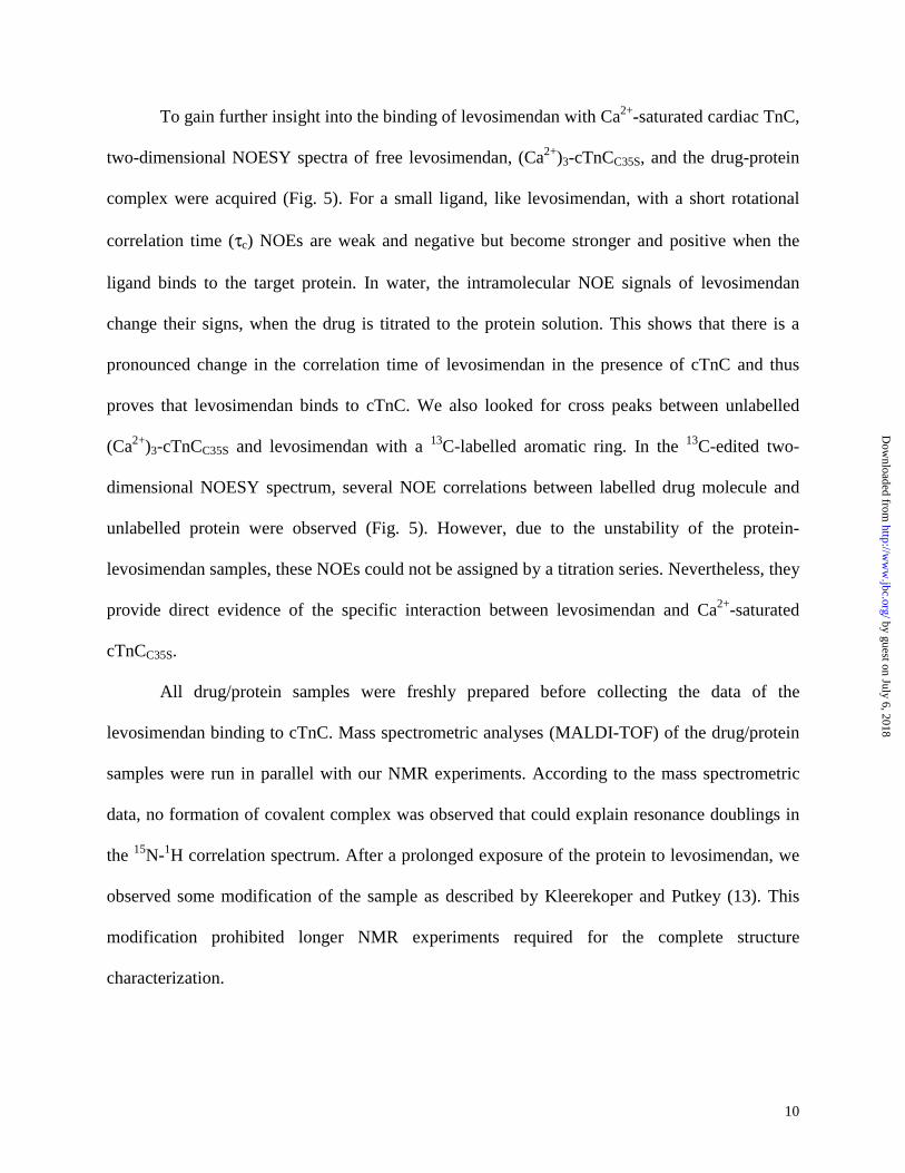
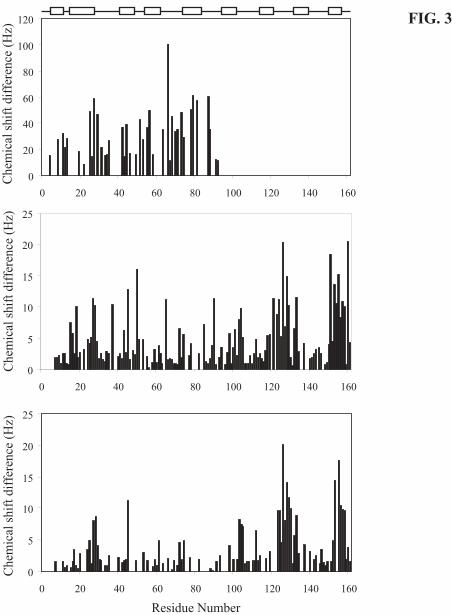
binding to one or more secondary binding sites. As can be seen from Fig. 3A, no resonance
doublings were observed beyond residue 92 and therefore we concluded that the primary binding
site is in the N-terminal domain. A comparison of the data in Figs. 2B and C shows that the
primary binding site did not exist in cTnCA-Cys. Evidently Cys84, but not Cys35, is important for
levosimendan binding to the primary site. It is also important to note that even though there are
no resonance doublings in the C-terminal half of cTnCC35S, this half of the molecule is essential
for the binding to the primary site since no resonance doubling is observed in the isolated N-
terminal half (1-91) of cTnC (Fig. 2D). These observations may explain the discrepancies in the
results obtained earlier when studying the binding of levosimendan to cardiac troponin C. In the
C-domain of cTnCC35S and cTnCA-Cys, there seems to be two secondary binding sites judged by
the relatively small chemical shift changes (Fig. 3B and C). By mapping these chemical shift
changes to the cTnC structure (1AJ4 from pdb), it appears that the two distinct interaction sites
in the C-domain of cTnC are not spatially related to each other.
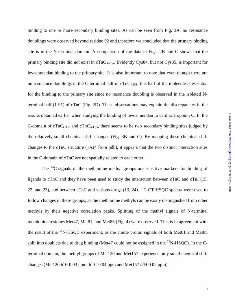
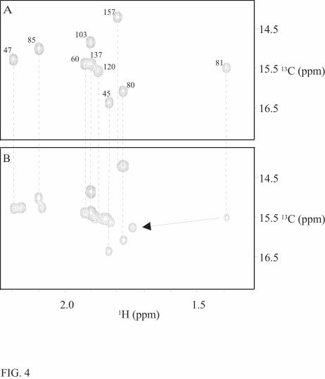
The 13C-signals of the methionine methyl groups are sensitive markers for binding of
ligands to cTnC and they have been used to study the interaction between cTnC and cTnI (15,
22, and 23), and between cTnC and various drugs (13, 24). 13C-CT-HSQC spectra were used to
follow changes in these groups, as the methionine methyls can be easily distinguished from other
methyls by their negative correlation peaks. Splitting of the methyl signals of N-terminal
methionine residues Met47, Met81, and Met85 (Fig. 4) were observed. This is in agreement with
the result of the 15N-HSQC experiment, as the amide proton signals of both Met81 and Met85
split into doublets due to drug binding (Met47 could not be assigned in the 15N-HSQC). In the C-
terminal domain, the methyl groups of Met120 and Met157 experience only small chemical shift
changes (Met120 δ1H 0.03 ppm, δ13C 0.04 ppm and Met157 δ1H 0.02 ppm).
by guest on July 6, 2018http://w
ww
.jbc.org/D
ownloaded from
10
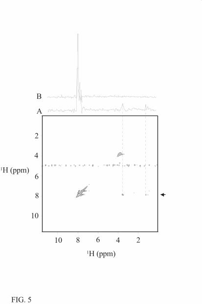
To gain further insight into the binding of levosimendan with Ca2+-saturated cardiac TnC,
two-dimensional NOESY spectra of free levosimendan, (Ca2+)3-cTnCC35S, and the drug-protein
complex were acquired (Fig. 5). For a small ligand, like levosimendan, with a short rotational
correlation time (τc) NOEs are weak and negative but become stronger and positive when the
ligand binds to the target protein. In water, the intramolecular NOE signals of levosimendan
change their signs, when the drug is titrated to the protein solution. This shows that there is a
pronounced change in the correlation time of levosimendan in the presence of cTnC and thus
proves that levosimendan binds to cTnC. We also looked for cross peaks between unlabelled
(Ca2+)3-cTnCC35S and levosimendan with a 13C-labelled aromatic ring. In the 13C-edited two-
dimensional NOESY spectrum, several NOE correlations between labelled drug molecule and
unlabelled protein were observed (Fig. 5). However, due to the unstability of the protein-
levosimendan samples, these NOEs could not be assigned by a titration series. Nevertheless, they
provide direct evidence of the specific interaction between levosimendan and Ca2+-saturated
cTnCC35S.
All drug/protein samples were freshly prepared before collecting the data of the
levosimendan binding to cTnC. Mass spectrometric analyses (MALDI-TOF) of the drug/protein
samples were run in parallel with our NMR experiments. According to the mass spectrometric
data, no formation of covalent complex was observed that could explain resonance doublings in
the 15N-1H correlation spectrum. After a prolonged exposure of the protein to levosimendan, we
observed some modification of the sample as described by Kleerekoper and Putkey (13). This
modification prohibited longer NMR experiments required for the complete structure
characterization.
by guest on July 6, 2018http://w
ww
.jbc.org/D
ownloaded from
11
All the NMR data were collected immediately after the addition of levosimendan to
troponin C. Interestingly, we noticed that the new signals and the shift changes induced by
levosimendan binding disappeared when the sample was stored for several days at 40 °C (data
not shown). Notably, the spectral changes reappeared upon addition of fresh levosimendan to the
sample.
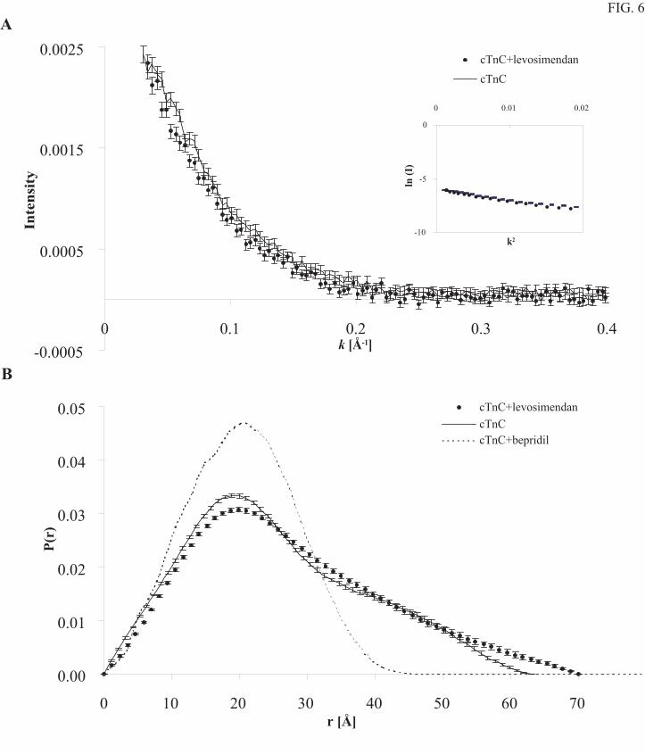
Small angle X-ray scattering (SAXS) of the Ca2+-saturated form of cTnCC35S and its
complex with levosimendan are rather similar. The scattered intensity obeys Guinier law at the
smallest k-values and there is no sign of protein aggregation (Fig. 6A) (26). There is only a small
change in the form of the distance distribution function, P(r), when levosimendan binds to
cTnCC35S (Fig. 6B). The maximum distance increases from 63 ± 5 Å to 70 ± 5 Å and the radius
of gyration from 20.2 ± 0.5 Å to 21.7 ± 0.6 Å. The determined radius of gyration, 20.2 ± 0.5 Å,
for troponin C without levosimendan, is in a good agreement with a previous study on troponin
C (27).
DISCUSSION
Our present finding that levosimendan reacts with common additives in protein solutions
used in NMR studies clearly shows the need for careful studies of the stability of molecules used
in binding experiments. In the drug discovery strategy SAR by NMR (28-30), for example, it
appears now of the utmost importance to know the stability of the compounds to be tested at the
experimental conditions which are used.
The binding of levosimendan to the cardiac troponin C has been under debate for some
time. Previous studies (5, 12, and 31) gave evidence for levosimendan binding. However,
by guest on July 6, 2018http://w
ww
.jbc.org/D
ownloaded from
12
contradictory results that show no binding have also been reported (13). In our hands, in the
course of the titration of (Ca2+)3-cTnCC35S with levosimendan, some odd behaviour were
observed. The pH of the protein-drug solution changed during the experiments, and sometimes
levosimendan precipitated out of the protein solution as a bright yellow precipitate, making it
difficult to reproduce the measurements (data not shown). We have now found that commonly
used additives in protein samples, DTT and NaN3, react with levosimendan (Fig. 1). Sodium
azide forms an adduct with levosimendan, and consequently it was no longer added to our
protein samples. To prepare cTnC samples without DTT was of concern because of the possible
formation of intra- and intermolecular disulfide bonds. However, we observed no intermolecular
disulfide bond formation in our DTT free cTnCC35S samples after a couple of days of incubation,
as analysed by MALDI-TOF. Moreover, intramolecular disulfide bonds are not possible in
cTnCC35S, with only one cysteine residue.
The controversial results of levosimendan binding to cTnC are difficult to explain only
by drug reactivity under different experimental conditions. We believe that the differences to
some extent also originate from the fact that various protein sequences have been used. In fact,
the recombinant N-terminal fragment of human cTnC contains two cysteine residues, Cys35 and
Cys84. In full-length chicken cTnCC35S, Cys35 is mutated to serine and in full-length chicken
cTnCA-Cys both cysteine residues are changed to serines. The residues Cys35 and Cys84 of cTnC
are conserved among various species, suggesting their importance for the function of the protein.
However, it has been previously reported that the conversion of cysteines to serines does not
alter calcium binding to cTnC, but might modify the structure of cTnC as indicated by changes
in its dye binding properties (17). The 15N-HSQC spectra show that binding of levosimendan to
Ca2+-saturated forms of cTnCC35S and cTnCA-Cys are different. The small chemical shift changes
by guest on July 6, 2018http://w
ww
.jbc.org/D
ownloaded from
13
attributed to the secondary binding sites are similar but the observed resonance doublings in the
N-domain of cTnCC35S are completely missing from the 15N-HSQC spectrum of the A-Cys form
of cTnC. This observation proves that the C84S mutant makes a difference in levosimendan
binding to the primary binding site. We therefore conclude that the primary binding site critically
depends on Cys84. The isolated N-terminal fragment of cTnC also shows interaction with
levosimendan (Fig. 2D). However, the binding seems to be different compared to the full-length
cTnCC35S. N-terminal fragment does not contain an intact primary binding site for levosimendan.
This is reasonable because Cys84 is only a few residues away from the chain end at Gly91.
It would naturally be very interesting to localise the primary binding site of levosimendan
in the cTnC. This is, however, presently not possible since there are effects all over the N-
terminal half of cTnC. The fact that most of the residues in the N-domain of cTnCC35S show
resonance doubling indicates that the binding of levosimendan to the primary site causes a
conformational change involving most of cNTnC. The exchange rate for this conformational
change is slow, k < 10 s-1, since we do not observe any line broadening effects. (Ca2+)3-cTnC
exists in two conformations, i.e. the open and closed states. The exchange between open and
closed conformations is intermediate on the NMR time scale and the equilibrium favours the
closed form (8 and 9). An obvious explanation for peak doublings would be that levosimendan
binds only to the open conformation, but there is a large difference between the Kon and Koff
values, the Koff being significantly slower as compared to Kon. Another possible explanation is
that levosimendan binds to both forms, but preferably to the open one, since in the presence of
levosimendan the equilibrium reaches about a 50:50 ratio for the two states. The exchange
between the two states of cTnC is significantly slower in the presence of levosimendan. At the
present stage of the work we were not able to completely rule out either possibility.
by guest on July 6, 2018http://w
ww
.jbc.org/D
ownloaded from
14
It is interesting to compare the levosimendan binding to troponin C with the binding of
other molecules (e.g. bepridil, EMD57033, and trifluoperazine, (24, 32)). Recently, it has been
shown by X-ray crystallography, that the structure of cNTnC opens in response to bepridil
binding (33). Three bepridil molecules bind to one cTnCA-Cys molecule. One bepridil molecule
binds to the N-terminal half and opens its structure and the other two bepridil molecules mimic
the TnI binding to the C-terminal half. The chemical shift changes induced by levosimendan
binding are smaller than those caused by bepridil binding (data not shown). In contrast to the
case of bepridil binding to cTnC our SAXS data show no sign of a domain-domain closure in the
presence of levosimendan (Fig. 6). If anything, it seems that levosimendan binding to the
primary binding site, located close to the end of N-domain, prevents the domains from moving
closer to each other and might actually increase the maximal distance (rmax). Alternatively, this
might be caused by a levosimendan induced structural change in the regulatory domain of cTnC.
In vivo cTnC is not expected to experience a large spatial reorganisation of domains within the
troponin complex. This is in accordance with Ca2+-sensitizers affecting only the regulatory
domain. However, the final evidence will be obtained once the structure of the levosimendan-
(Ca2+)3-cTnCC35S complex in solution is determined.
CONCLUSIONS
Our data unambiguously show several interaction sites for levosimendan on the Ca2+-
loaded form of cardiac troponin C (C35S). Levosimendan does bind to cTnCC35S but only in the
absence of NaN3 and DTT, which cause degradation of levosimendan. Thus, the current
observations explain the discrepancies between earlier studies of levosimendan binding to cTnC.
Our results suggest that the primary binding site is located in the regulatory domain (cNTnC) of
by guest on July 6, 2018http://w
ww
.jbc.org/D
ownloaded from
15
the cTnC and that there are two secondary binding sites at the C-terminal half of cTnC possibly
analogous to the case of three bepridil molecules binding to cTnCA-Cys (33). Likewise,
levosimendan may contribute to the opening of the regulatory domain. However, levosimendan
does not cause a domain-domain closure. At present, we are not able to determine the precise
locus of the primary binding site on the N-terminal domain due to the numerous changes in the
spectra upon levosimendan binding. However, results from experiments with cTnCA-Cys show
that the presence of Cys84 is of critical importance for levosimendan binding. The results
presented give us a reason to believe that the binding of levosimendan to the calcium saturated
regulatory domain of cTnC is the mechanism behind its known Ca2+-sensitizing effect.
by guest on July 6, 2018http://w
ww
.jbc.org/D
ownloaded from
16
REFERENCES
1 Endoh, M. (1995) Gen. Pharmacol. 26, 1-31
2 Hasenfuss, G., Pieske, B., Castell, M., Kretschmann, B., Maier, L.S., and Just, H. (1998)
Circulation 98:20, 2141-2147
3 Gagne, S.M., Li, M.X., McKay, R.T., and Sykes, B.D. (1998) Biochem. Cell Biol. 76, 302-
312
4 Ovaska, M. and Taskinen, J. (1991) Proteins 11, 79-94
5 Pollesello, P., Ovaska, M., Kaivola, J., Tilgmann, C., Lundstöm, K., Kalkkinen, N.,
Ulmanen, I., Nissinen, E., and Taskinen, J. (1994) J. Biol. Chem. 269, 28584-28590
6 Spyracopoulos, L., Li, M.X., Sia, S.K., Gagné, S.M., Chandra, M., Solaro, R.J., and Sykes,
B.D. (1997) Biochemistry 36, 12138-12146
7 Sia, S.K., Li, M.X., Spyracopoulos, L., Gagné, S.M., Liu, W., Putkey, J.A., and Sykes,
B.D. (1997) J. Biol. Chem. 272, 18216-18221
8 Pääkkönen, K., Annila, A., Sorsa, T., Pollesello, P., Tilgmann, C., Kilpeläinen, I., Karisola,
P., Ulmanen, I., and Drakenberg, T. (1998) J. Biol. Chem. 273, 15633-15638
by guest on July 6, 2018http://w
ww
.jbc.org/D
ownloaded from
17
9 Gaponenko, V., Abusamhadneh, E., Abbott, M.B., Finley, N., Gasmi-Seabrook, G., Solaro,
R.J., Rance, M., and Rosevear, P.R. (1999) J. Biol. Chem. 274, 16681-16684
10 Li, M.X., Spyracopoulos, L., and Sykes, B.D. (1999) Biochemistry 38, 8289-8298
11 Dong, W.J., Xing, J., Villain, M., Hellinger, M., Robinson, J.M., Chandra, M., Solaro, R.J.,
Umeda, P.K., and Cheung, H.C. (1999) J. Biol. Chem. 274, 31382-31390
12 Haikala, H., Kaivola, J., Nissinen, E., Wall, P., Levijoki, J., and Linden, I.-B. (1995) J.
Mol. Cell. Cardiol. 27, 1859-1866
13 Kleerekoper, Q. and Putkey, J.A. (1999) J. Biol. Chem. 274, 23932-23939
14 Horton, R.M. (1995) Mol. Biotechnol. 3(2), 93-99
15 Krudy, G.A., Kleerekoper, Q., Guo, X., Howarth, J.W., Solaro, R.J., and Rosevear, P.R.
(1994) J. Biol. Chem. 269, 23731-23735
16 Finley, N., Abbott, M.B., Abusamhadneh, E., Gaponenko, V., Dong, W., Gasmi-Seabrook,
G., Howarth, J.W., Rance, M., Solaro, R.J., Cheung, H.C., and Rosevear, P.R. (1999)
FEBS Lett. 453, 107-112
by guest on July 6, 2018http://w
ww
.jbc.org/D
ownloaded from
18
17 Putkey, J.A., Dotson, D.G., and Mouawad, P. (1993) J. Biol. Chem. 268, 6827-6830
18 Bradford, M.M. (1976) Anal. Biochem. 72, 248-254
19 Wishart, D.S., Bigam, C.G., Yao, J., Abildgaard, F., Dyson, H.J., Oldfield, E., Markley,
J.L., and Sykes, B.D. (1995) J. Biomol. NMR 6, 135-140
20 Svergun, D.L., Semenyuk, A.V., and Feigin, L.A. (1988) Acta Crystallogr. A44, 244-250
21 March, J. (1985) Advanced Organic Chemistry: Reactions, Mechanisms, and Structure, 3rd
edition, John Wiley & Sons, Inc., New York, 743-744
22 Howarth, J., Krudy, G.A., Lin, X., Putkey, J.A., and Rosevear, P.R. (1995) Protein Sci. 4,
671-680
23 Kleerekoper, Q., Howarth, J.W., Guo, X., Solaro, R.J., and Rosevear, P.R. (1995)
Biochemistry 34, 13343-13352
24 Kleerekoper, Q., Liu, W., Choi, D., and Putkey, J.A. (1998) J. Biol. Chem. 273, 8153-8160
25 Lin, X., Krudy, G.A., Howarth, J., Brito, R.M.M., Rosevear, P.R., and Putkey, J.A. (1994)
Biochemistry 33, 14434-14442
by guest on July 6, 2018http://w
ww
.jbc.org/D
ownloaded from
19
26 Guinier, A. and Fournet, G. (1955) Small-Angle Scattering of X-rays, John Wiley & Sons,
Inc., New York
27 Fujisawa, T., Ueki, T., and Inoko, Y. (1987) J. Appl. Cryst. 20, 349-355
28 Shuker, S.B., Hajduk, P.J., Meadows, R.P., and Fesik, S.W. (1996) Science 274, 1531-
1534
29 Hajduk, P.J., Meadows, R.P., and Fesik, S.W. (1997) Science 278, 497-499
30 Hajduk, P.J., Gerfin, T., Boehlen, J.-M., Häberli, M., Marek, D., and Fesik, S.W. (1999)
J. Med. Chem. 42, 2315-2317
31 Levijoki, J., Pollesello, P., Kaivola, J., Tilgmann, C., Sorsa, T., Annila, A., Kilpeläinen, I.,
and Haikala, H., (2000) J. Mol. Cell. Cardiol. 32, 479-491
32 Pan, B.-S. and Johnson, R.G., Jr. (1996) J. Biol. Chem. 271, 817-823
33 Li, Y., Love, M.L., Putkey, J.A., and Cohen, C. (2000) Proc. Natl. Acad. Sci. 97, 5140-
5145
by guest on July 6, 2018http://w
ww
.jbc.org/D
ownloaded from
20
FIGURE LEGENDS
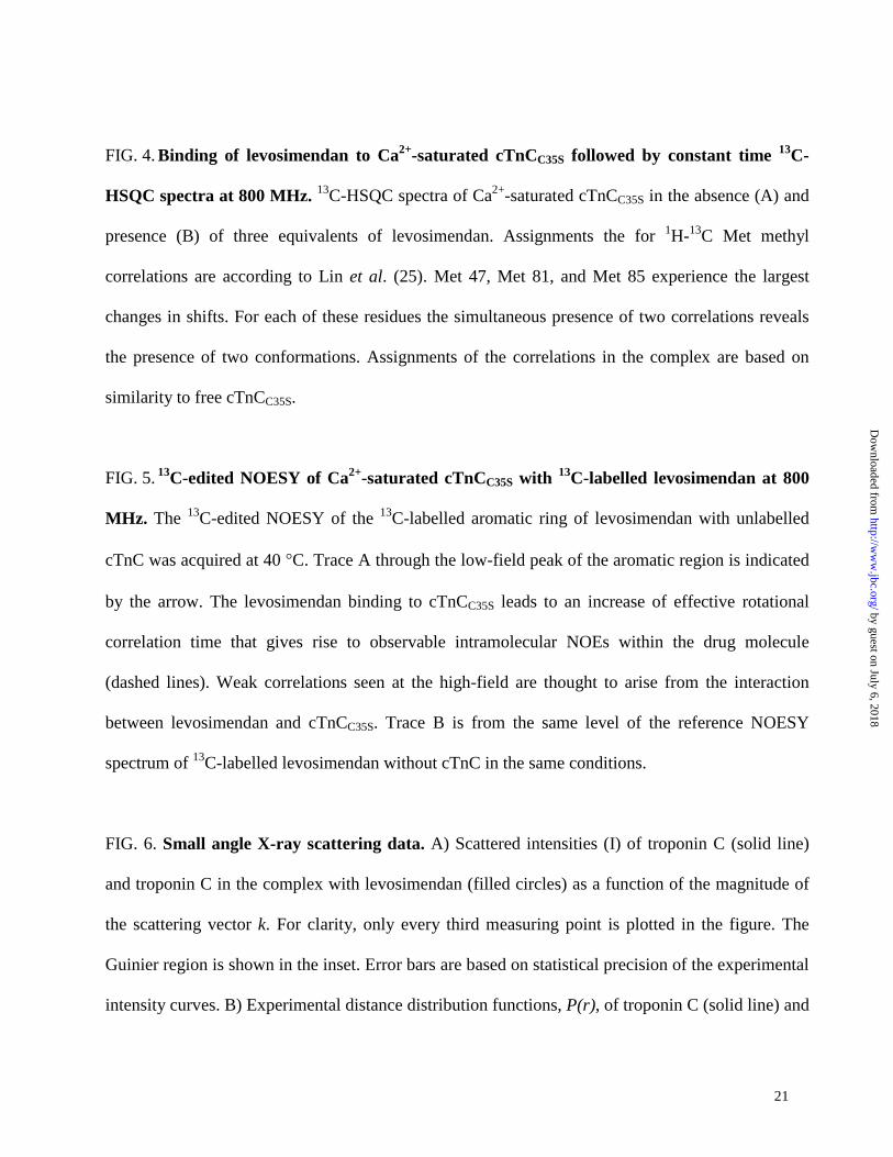
FIG. 1. Stability of levosimendan. A freshly prepared levosimendan sample in water without
additives (A) and the same sample after an incubation of 20 hrs at 40 °C (B) show the characteristic
AA’BB’ spectrum of levosimendan in an aromatic region of the 1D-proton spectra acquired at 600
MHz. Stability of the drug was tested in the presence of 0.05% NaN3 (1 mM levosimendan) (C) and
8 mM DTT (1 mM levosimendan) (D). The spectrum of levosimendan in the presence of NaN3 is
referenced according to the left doublet. Other spectra were referenced to the water signal.
Levosimendan molecule (E) and its adduct with sodium azide (F) as well as a degradation product
of levosimendan in the presence of DTT (G) are shown on the right.
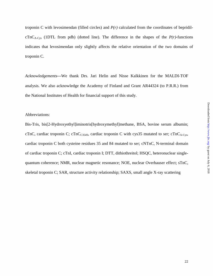
FIG. 2. Binding of levosimendan to Ca2+-saturated cTnC observed by 15N-HSQC spectra at
800 MHz. The spectrum of cTnCC35S shows chemical shift changes and resonance doublings upon
levosimendan binding (A). Expansions of the well-resolved region of Ca2+-saturated cTnCC35S (B),
cTnCA-Cys (C) and cNTnC (D) in the presence (blue) and absence (red) of three equivalents of
levosimendan. Chemical shift changes are larger upon levosimendan binding for Ca2+-saturated
cTnCC35S (B) than for cTnCA-Cys (C), and also the isolated N-terminal fragment (1-91) of cTnC (D).
FIG. 3. Chemical shift changes due to levosimendan binding to Ca2+-saturated cTnC.
Levosimendan binding to Ca2+-saturated cTnCC35S gave rise to resonance doublings with large
splittings (A) and to small shift changes (B) presented as a function of amino acid sequence. For
cTnCA-Cys the chemical shift changes are small (C) and no resonance doublings are observed. The
splittings are presented as a distance in Hz between the cross correlation peaks of the cTnCC35S.
by guest on July 6, 2018http://w
ww
.jbc.org/D
ownloaded from
21
FIG. 4. Binding of levosimendan to Ca2+-saturated cTnCC35S followed by constant time 13C-
HSQC spectra at 800 MHz. 13C-HSQC spectra of Ca2+-saturated cTnCC35S in the absence (A) and
presence (B) of three equivalents of levosimendan. Assignments the for 1H-13C Met methyl
correlations are according to Lin et al. (25). Met 47, Met 81, and Met 85 experience the largest
changes in shifts. For each of these residues the simultaneous presence of two correlations reveals
the presence of two conformations. Assignments of the correlations in the complex are based on
similarity to free cTnCC35S.
FIG. 5. 13C-edited NOESY of Ca2+-saturated cTnCC35S with 13C-labelled levosimendan at 800
MHz. The 13C-edited NOESY of the 13C-labelled aromatic ring of levosimendan with unlabelled
cTnC was acquired at 40 °C. Trace A through the low-field peak of the aromatic region is indicated
by the arrow. The levosimendan binding to cTnCC35S leads to an increase of effective rotational
correlation time that gives rise to observable intramolecular NOEs within the drug molecule
(dashed lines). Weak correlations seen at the high-field are thought to arise from the interaction
between levosimendan and cTnCC35S. Trace B is from the same level of the reference NOESY
spectrum of 13C-labelled levosimendan without cTnC in the same conditions.
FIG. 6. Small angle X-ray scattering data. A) Scattered intensities (I) of troponin C (solid line)
and troponin C in the complex with levosimendan (filled circles) as a function of the magnitude of
the scattering vector k. For clarity, only every third measuring point is plotted in the figure. The
Guinier region is shown in the inset. Error bars are based on statistical precision of the experimental
intensity curves. B) Experimental distance distribution functions, P(r), of troponin C (solid line) and
by guest on July 6, 2018http://w
ww
.jbc.org/D
ownloaded from
22
troponin C with levosimendan (filled circles) and P(r) calculated from the coordinates of bepridil-
cTnCA-Cys (1DTL from pdb) (dotted line). The difference in the shapes of the P(r)-functions
indicates that levosimendan only slightly affects the relative orientation of the two domains of
troponin C.
AcknowledgementsWe thank Drs. Jari Helin and Nisse Kalkkinen for the MALDI-TOF
analysis. We also acknowledge the Academy of Finland and Grant AR44324 (to P.R.R.) from
the National Institutes of Health for financial support of this study.
Abbreviations:
Bis-Tris, bis[2-Hydroxyethyl]iminotris[hydroxymethyl]methane, BSA, bovine serum albumin;
cTnC, cardiac troponin C; cTnCC354S, cardiac troponin C with cys35 mutated to ser; cTnCA-Cys,
cardiac troponin C both cysteine residues 35 and 84 mutated to ser; cNTnC, N-terminal domain
of cardiac troponin C; cTnI, cardiac troponin I; DTT, dithiothreitol; HSQC, heteronuclear single-
quantum coherence; NMR, nuclear magnetic resonance; NOE, nuclear Overhauser effect; sTnC,
skeletal troponin C; SAR, structure activity relationship; SAXS, small angle X-ray scattering
by guest on July 6, 2018http://w
ww
.jbc.org/D
ownloaded from
p p m8 . 0 7 . 5
B
C
D
EA
F
G
F I G . 1
H N
N N H
O
H 3 C
N
N
N H
N
N
N
H N
N N H
O
H 3 C
N
N
N
H N
N N H
O
H 3 C
N
N
N H
SHO
HO
H S
H O
H O
by guest on July 6, 2018http://w
ww
.jbc.org/D
ownloaded from
1 0 . 4 9 . 2 8 . 0
1 1 1 . 0
1 2 9 . 0
1 5 N ( p p m )
9 . 8 9 . 0 8 . 2
1 3 0 . 0
1 2 5 . 0
1 5 N ( p p m )
E 3 2
F 1 5 3
I 1 4 8I 1 1 2 V 7 2
D 1 4 9 D 1 1 3
D 7 3
E 6 6
B
C
1 3 0 . 0
1 2 5 . 0
E 3 2F 1 5 3
I 1 4 8I 1 1 2V 7 2
D 1 4 9 D 1 1 3
D 7 3
E 6 6
A
1 5 N ( p p m )
D 1 2 6 . 0
1 2 7 . 0
E 6 6
D 7 3
V 7 2
8 . 49 . 4
1 5 N ( p p m )
1 H ( p p m )
F I G . 2
by guest on July 6, 2018http://w
ww
.jbc.org/D
ownloaded from
0
5
1 0
1 5
2 0
2 5
0 2 0 4 0 6 0 8 0 1 0 0 1 2 0 1 4 0 1 6 0
0
5
1 0
1 5
2 0
2 5
0 2 0 4 0 6 0 8 0 1 0 0 1 2 0 1 4 0 1 6 0
0
2 0
4 0
6 0
8 0
1 0 0
1 2 0
0 2 0 4 0 6 0 8 0 1 0 0 1 2 0 1 4 0 1 6 0
Chemical shift difference (Hz)
Chemical shift difference (Hz)
Chemical shift difference (Hz)
R e s i d u e N u m b e r
F I G . 3
by guest on July 6, 2018http://w
ww
.jbc.org/D
ownloaded from
2 . 0 1 . 51 H ( p p m )
1 6 . 5
1 3 C ( p p m )
4 78 5
1 5 7
1 0 3
6 0 1 3 7
1 2 0
4 58 0
8 1
A
B
1 4 . 5
1 5 . 5
1 6 . 5
1 3 C ( p p m )
1 4 . 5
1 5 . 5
F I G . 4
by guest on July 6, 2018http://w
ww
.jbc.org/D
ownloaded from
1 H ( p p m )
1 H ( p p m )
A
B
2
4
6
8
1 0
1 0 8 6 4 2
F I G . 5
by guest on July 6, 2018http://w
ww
.jbc.org/D
ownloaded from
F I G . 6
B
A
- 0 . 0 0 0 5
0 . 0 0 0 5
0 . 0 0 1 5
0 . 0 0 2 5
0 0 . 1 0 . 2 0 . 3 0 . 4
c T n C + l e v o s i m e n d a n
c T n C
0 . 0 0
0 . 0 1
0 . 0 2
0 . 0 3
0 . 0 4
0 . 0 5
0 1 0 2 0 3 0 4 0 5 0 6 0 7 0
c T n C + l e v o s i m e n d a n
c T n Cc T n C + b e p r i d i l
- 1 0
- 5
0
0 0 . 0 1 0 . 0 2
I nt ens it y
P( r)
r [ Å ]
k [ Å - 1 ]
k 2
l n (I )
by guest on July 6, 2018http://w
ww
.jbc.org/D
ownloaded from
Pollesello and Ilkka KilpeläinenTilgmann, Ritva Serimaa, Arto Annila, Paul R. Rosevear, Torbjörn Drakenberg, Piero
Tia Sorsa, Sami Heikkinen, M. Bret Abbott, Ekram Abusamhadneh, Tero Laakso, CarolaBinding of levosimendan, a calcium sensitizer, to cardiac troponin C
published online December 11, 2000J. Biol. Chem.
10.1074/jbc.M007484200Access the most updated version of this article at doi:
Alerts:
When a correction for this article is posted•
When this article is cited•
to choose from all of JBC's e-mail alertsClick here
by guest on July 6, 2018http://w
ww
.jbc.org/D
ownloaded from