Embed Size (px)
Citation preview
APPROVAL SHEET
Complete report of general biology with title “Anatomy of Vertebrate Animals”,
created by :
Name : Ummi Qalsum
Reg. Number : 091204174
Class : B (ICP)
Departement : Physics
Group : V (Five)
After checked by Assistant and Assistant Coordinator, so this is report accepted.
Makassar, November 2009
Assistant Coordinator Assistant
(Djumarirmanto, S.Pd) (Zaidah . R)
Nim.061404026
Known By
Lecturer of Responsibility
(Ir. Muh. Wiharto, M.Si)
NIP.132 006 81

CHAPTER IINTRODUCTION
A. Background
Amphibious name comes from the Greek word (Amphi = double +
bios = life). Most of this class shows that a phase of life in the water and then
have a phase of life on land. In the second phase shows the nature of fish and
reptiles and shows that Amphibia are a group of Chordata was the first out of
life in the water.
One example of amphibious animals are most representative of
toads and toads. One will be confused to distinguish between these toads and
toads. Both kinds of these animals looks like. Difference toads short, stout or
thin, slightly bent-backed, four-legged and no tail (Anura: a no, ura tail).
Toads generally smooth-skinned, moist, with long hind legs. Instead toad or
rough-skinned bangkong berbintil-nodule until berbingkul-bingkul, often dry,
and the hind legs are often short, so many less intelligent leap. But the two
terms are often interchangeable use.
Frogs and toads generally have organs that are specialized to
support life. Among the pulmo for life on land, slimy skin and webbed feet to
facilitate swimming in water, 2 nostrils directly related to the cavum oris used
for breathing when these frogs and toads in the water.
In terms of the anatomy of frogs also differ with other living creatures. For
example heart consists of three different room from the living land consisting
of 4 rooms and living creatures like fish water teridiri only of 2 rooms.
According to scientists this is what causes that frogs are considered as
animals transition from water to land.
B. Experiment’s Purpose
The students can recognice shapes, colours, and locatioan of organ
and its relationship to other organs in an organ system.
C. Experiment’s Benefit
Students will be able to explain recognice shapes, colours, and
locatioan of organ and its relationship to other organs in an organ system
CHAPTER IIPREVIEW OF LITERATURE
Morphology of the Toad
Clasification
Based on morphological characteristics of toads above classification is as follows:
Kingdom Animalia
Class Amphibia
Order Annura
Family Bufodae
Genus Bufo
Species Bufo melanostictu (Merrem, 1982).
Width of the head and body are united, there are two pairs of legs or
members, no neck and tail. The inside is covered by soft smooth moist skin. Head
has a wide mouth pliers to retrieve the food, 2 nostrils / nares small externa near the
nose that functions in respiration, 2 big eyes spherik, 2-hole behind closed by a flat
tympani membrane that serves as the ear to receive sound waves. Each eye has a lid
top and bottom, and inside it has a clear cornea nictitans membrane to cover the eyes
if they are in the water. At the end of the back of the body found anus, a small hole to
remove the remnants of food which not digested, urine and sex cells / egg or sperm
from the reproductive (Kastowo, 1982: 32)
toad legs consists of a pair of front feet and rear legs. Front legs of the upper
arm (brancium), forearm (antebrancium), hand (manus), and the fingers (digiti). At
the rear leg of the thigh (femur), lower leg (crus), feet (plague) and fingers (digiti)
(Radiopoetro, 1996: 474).
In general, the number of fingers toad front legs are usually four fingers and
five fingers back legs. At the rear leg lengthening with the potential to jump.
Sometimes found as prehaluk extra finger on the ventral side of the foot. Prehaluk on
Spadefoot (toad diggers land) in the form of hard bones that are used to dig the
ground as a place to hide (Radiopoetro, 1996: 474).
Amphibious skin is very important in respiration and protection. Flexible thin
skin that divides the outer body to protect the organism against disease, in respiratory
function, the absorption of water, because the toads do not ever drink. On equipped
with mucous glands that causes the skin humidity maintained, for the species that
live in water, mucus provides oil for the body. Most have granular glands and
mucous glands. Both are similar, but different products. Granular glands produce a
substance or toxin abnoxious to protect themselves from enemies. Both classified as
alveolar glands (glands that have no spending channel, but products on out through
the cell wall itself naturally). Racundapat menimbukan glands in the skin irritation
(Sukiya, 2005: 47).
Toad is bilaterally symmetrical, with the left and right sides equal. The
middle part is called the medial, side / lateral, front face of the body is the end of the
anterior, posterior disebutujung the rear, the back or dorsal, are part of the ventral
face. Body part consists of the head / caput, esophagus / cervik, chest / thorax or
pectoral, stomach or abdomen, pelvis and buttocks kaudal the short (Kastowo, 1982:
32).
Toads and frogs are amphibious animals the best known people in Indonesia.
Both kinds of these animals looks like. Frog was short, stout or thin, slightly bent-
backed, four-legged and no tail. Toads generally smooth-skinned, moist, with long
hind legs. Instead frog or rough-skinned bangkong berbintil-nodule until berbingkul-
bingkul, often dry and hind legs are often short, so many less intelligent leap. But the
two terms are often interchangeable use. Frogs and toads start life as eggs laid in the
water mother, the nest of foam or in other damp places. Frog eggs and frogs hatch
into tadpoles or tadpoles, who was like a fat fish, with gills and breathe for a while
lived in the water. Will slowly grow back legs, which was followed by the growth of
the front legs, tail, and passing the disappearance of the gills with lungs. After a time,
these tadpoles will jump to the ground as a toad or a small frog (Inger and Iskandar,
2005).
The toad is an animal intermediate between aquatic and terrestrial animals.
Therefore, the beginning of his life began in the waters and then moved to the
mainland. Frog habitat varies from the swamp to the mountains. Most live in forested
areas because the toads need a moist place to protect themselves from drought. There
are species of toads in his life always in the water and also living on the mainland
and the high trees. toads that live out of water is usually at a certain period will be
visiting the waters for breeding. Taxonomic levels in the toad can show dikertahiui
with morphology character as a reference for the identification and determination
(Kurniati, 2003).
Adult toad, if observed carefully, will see clearly the diversity of species
Atara variation from one to another has kataki wide body equipped with two pairs of
limbs. The front limbs are shorter and smaller, and has 4 fingers, while the back of a
much larger and longer. This is in accordance with its function is to jump. Members
of this movement are also equipped with a membrane pool to facilitate the toads
swim (Mahardono, 1980).
All the organs of motion associated with the movement of the toad jump.
Long rear legs provide strength to move forward. Form the back legs are adapted to
the process of landing, and long hind legs adapted to leap toad. Regular toad can
jump 2-10 times its body length and jump length can reach 30-40 times as much
length. Long jump depends on the physiology and morphology of a species and the
interests of the movement. Some rare species like the jumping but walking in other
vertebrates, and species that lives in water, adjusted to belakngnya foot swimming
(Zug, 1997).
Metamorphosis in Amphibious is one variation on the toad species.
Metamorphosis of his own understanding that change is the development of overall
shape, physiological and biochemical individual, whereas in some insects,
metamorphosis is complete only with the larval form of equipment for the adult
form. Metamorphic changes actually change the entire tissues and organs. Examples
of other animals such as frogs have such variation in the butterfly (Lepidoptera) are
also experiencing metamorphosis (Mahardono, 1980).
CHAPTER IIIOBSERVATION METHOD
A. Place and Date
This experiment’s is done at:
Day and Date : Wednesday, November 18th 2009
Time : at 13.30-15.00
Place : Laboratory of Biology
Faculty Mathematis and Science
Makassar State University
(at the 2nd east floor part)
B. Tools and Materials
1. Tools
1) Bottle killer
2) Surgical Tray
3) Surgical Instrument
a) Scissors
b) Straws Limon
c) Tweezers
d) Needles
e) Scalpels
2. Materials
1) Toads
2) Cotton
3) Kloroform/eter
C. Work Procedure
1) We took a wad of cotton soaked with chloroform or eter and entered
into a killer bottle, closed tightly.
2) Removing the toads had not moved and put on a surgical tray.
3) Observing the outside of toads
4) Drawing the toads from the outside
5) Gap opened up with a scalpel and tweezers, so that the mouth is open,
we observe and draw the parts.
6) For surgery, placing the toads on his back on the tray of surgical,
nailing all four feet, so not easy to shake.
7) With tweezers, pinching the skin it the abdomen, lengthwise near the
thighs, lift slightly and cut across. The skin under a pair of tweezers
thus forming a gap in the skin of the abdomen.
8) Through the skin opening, insert the blunt tip scissors and cut the skin
to the head until the scissors fell. Flipped in to this gap, a pair of
scissors in to the base of both thighs.
9) Cutting the skin side toward the right and left, so skin can be exposed
belly.
10) For Observations digestive system
a. Opened the cavity his mouth with tweezers so skapel and open
the oral cavity. Observing ther form of teeth and felt with the
fingers of teeth and jaws and teeth fomer.
b. Tongue pulled out with tweezers and then observe the shape and
pelekatannya.
c. Continue observing the abdominal cavity contains the bowels,
observing the shape and the color of :
1) Kana tone heart how sebelah lobe, search and view kantun-
gan bile its color.
2) Stomach on the left, lifted hearts and will appear dendenum
and pangkreas .
3) Runut intestine small intestine until thick.
4) The turn anus to rectum.
11) Observations of blood circulation system
a. The head of the heart was the heart of the membranes
b. Membrane covering the heart pierced with a needle or the tip
skapel to break, and then watched part - parts, namely :
1) Booth (ventrikal)
2) Porch (atrium)
3) The main arteri (truncis abterious) that came out of ventrikal
then branched into two aortic (left and right)
c. Drawing the heart and the member name of the above
12) For Observations of breathing system
a. Nothing the left side of the stomach and the right heart, it will be
hidden part of the lung - the lung.
b. Releasing the heart with scissors so that it will appear windpipe
(trachea).
c. Drawing the respiratory system of frogs.
13) For Observations excretory and reproductive system (uregentalia)
a. Removing the organs - digestive organs begin at the stomach to
the rectum, and menstrum (connective tissue) that holds.
b. Would seem a pair of oval kidney attached to the back of the ab-
domen further observed :
1. Kidneys and adrenal glands
2. Body fat (corpus adiposum) frilly yellow)
3. Renal tract (ureters) from the kidney to the bladder to the
pocket.
c. In female frogs, there is a pair of avaries on the left and right, up
slightly ovarian ovinduct it would seem that lies heart part.
d. Creating a picture of a frog uregenitalia system and member
name or the part.
CHAPTER IVOBSERVATION RESULT
A. Observation Result
From the experiment, we can get the result such as :
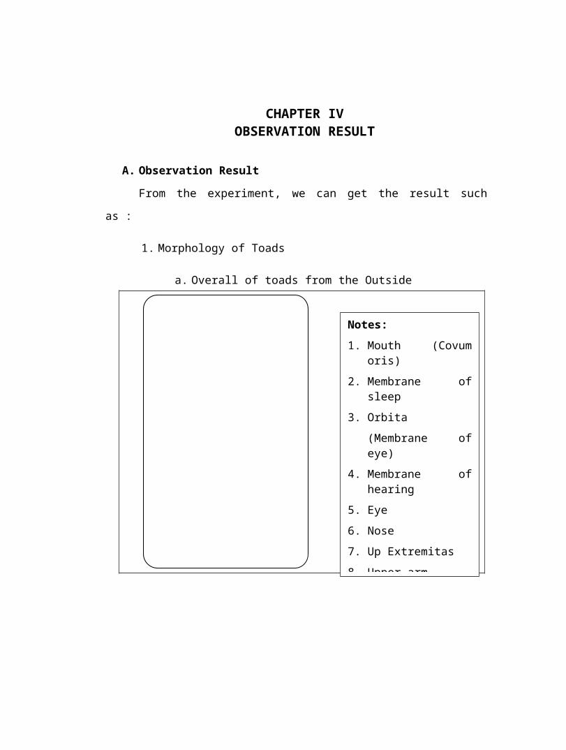
1. Morphology of Toads
a. Overall of toads from the Outside
Notes:
1. Mouth (Covum oris)
2. Membrane of sleep
3. Orbita
(Membrane of eye)
4. Membrane of hearing
5. Eye
6. Nose
7. Up Extremitas
8. Upper arm
9. Fore arm
10. Palm
11. Digiti
12. Down Extremitas
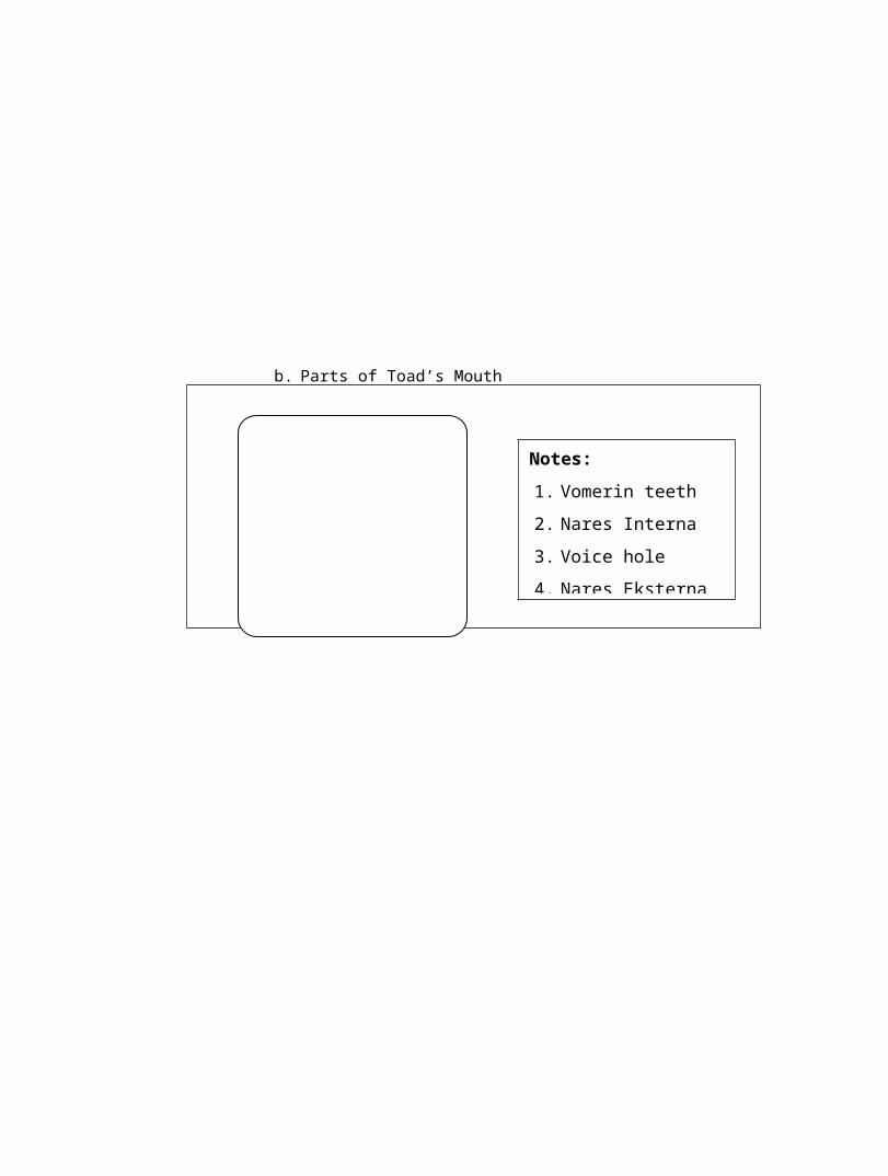
b. Parts of Toad’s Mouth
Notes:
1. Vomerin teeth
2. Nares Interna
3. Voice hole
4. Nares Eksterna
5. Tongue
2. Anatomy of Toads for overall
3. Circulatory System
Notes:
1. Throat
2. Heart
3. Liver
4. Lung
5. Bile
6. Kidneys
7. Pancreas
8. Stomach
9. Intestine
10. Ovary
Notes:
1. Left Aorta
2. Right Aorta
3. Atrium Sinixter (left)
4. Atrium Dexter (Right)
5. Venrikel
4. Digestive System
5. Respiratory System
Notes :
1. Throat
2. liver
3. Bile
4. Pancreas
5. Stomach
6. Small Intestine
7. Large intestine
Notes :
1. Tracea
2. Branch of Broncus
3. Left Pulmo
4. Right Pulmo
5. Alveolus
6. Blood capiler
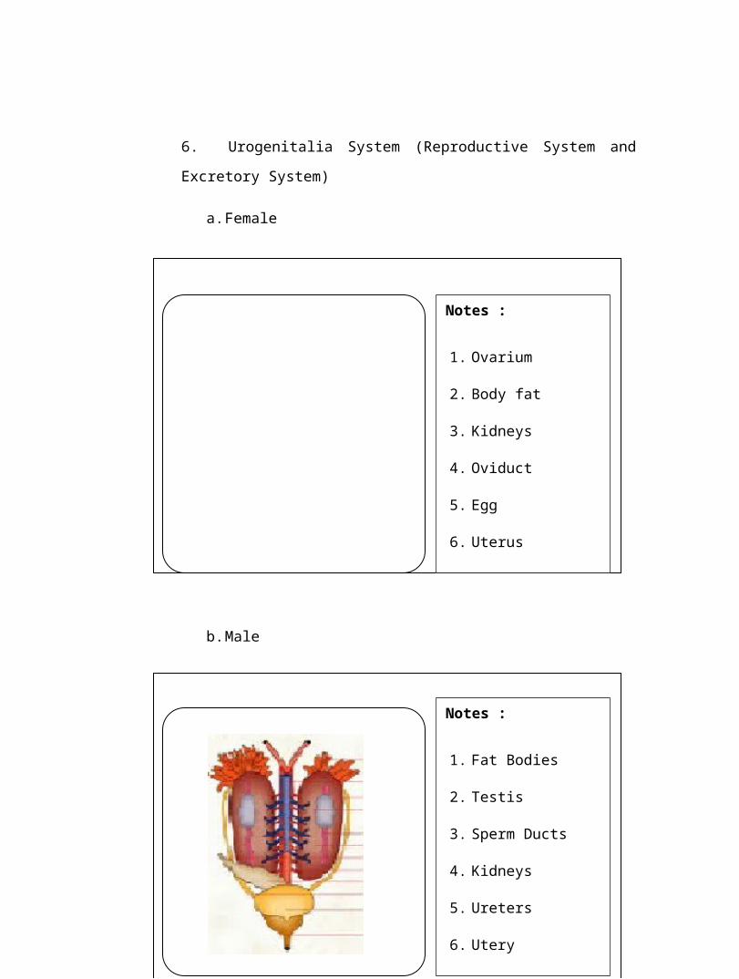
6. Urogenitalia System (Reproductive System and Excretory System)
a. Female
b. Male
Notes :
1. Fat Bodies
2. Testis
3. Sperm Ducts
4. Kidneys
5. Ureters
6. Utery
7. Bladder
Notes :
1. Ovarium
2. Body fat
3. Kidneys
4. Oviduct
5. Egg
6. Uterus
7. Cloaca
B. Discussion
1) The Mouth
Vomarine and Maxillary Teeth: Used for holding prey
Internal Nares (nostrils) breathing
Eustachian Tubes: equalize pressure in inner ear
Glottis : Tube leading to the lungs
Esophagus: Tube leading to the stomach
Tongue: Front attached, aids in grabbing prey
Tympanic Membrane: eardrum, located behind eyes
Nictitating Membrane: clear eyelid, protects the eye
2) Circulation System
toad heart contains:
- 3 rooms: 2 atria and 1 ventricle
- Sinus venosus: blood accommodate the large vessels that will go into the right
atrium.
The direction of blood flow:
O2-rich blood from the lungs and the skin into the left atrium. O2 poor blood into
the right atrium by means of sinus venosus. Of atrial blood into the ventricles,
causing a rich mixture of blood and blood O2 poor O2. Of ventricular O2-rich
blood is pumped into the body tissue and at O2 poor blood flow to the lungs to
the skin to get the O2.
toad circulatory including double circulation (in a one-time circulation, the blood
through the heart 2 times)
3) Digestive System
Food in the digestive system of amphibians, nearly equal to the fish, including the
digestive tract and digestive glands. one animal
amphibians are toads. Toad food of small animals (insects). Respectively on the
toad digestive system include:
1. the oral cavity: there is a cone-shaped teeth to hold prey and the tongue
to catch prey,
2. esophagus; a short channel,
3. ventrikulus (stomach), the shape of the bag when filled with food
becomes wide. toad stomach can be categorized into 2, namely the entry
of the esophagus and holes out to the intestine,
4. intestinum (bowel): can be distinguished on the small intestine and
colon thick. Intestine includes: the duodenum. jejenum, and ileum, but not
clearly demarcated.
5. Thick intestine ends in the rectum and into the kloata, and
6. cloaca: an estuary along the digestive tract of food, reproductive tract,
and urine.
The toad's mouth is where digestion begins. It is equipped with feeble, practically
useless teeth. These are present only in the upper jaw. The toad's tongue is highly
specialized. Normally, the tip of its tongue is folded backward toward the throat.
From this position the toad can flick it out rapidly to grasp any passing prey. To
better hold this prey, the tongue is sticky. (See also Tongue.)
Food passes from the toad's mouth into the stomach by way of the esophagus.
From the stomach, the food moves into the small intestine, where most of the
digestion occurs. Large digestive glands, the liver and the pancreas, are attached
to the digestive system by ducts. A gall bladder is also present (see Digestive
System).
4) Respiratory System
Respirator cavity lining of the mouth, skin and lungs. This respirator has
a thin, wet layer adjacent to the blood vessels so oxygen can diffuse.
Lining of the oral cavity: when the pharyngeal cavity of the mouth moves, nostrils
open and glottis closed so the air in the oral cavity through the mouth lining thin.
Skin: skin of oxygen into the skin through a vein (venous kutanea) and then to the
next jantun gdan circulated throughout the body. CO2 from tissue taken to heart
and then into the skin and lungs through the skin arteries lungs (pulmo artery
kutenea).
The lungs: a pair of lungs which leads to a bubble-shaped blood capillaries. The
toad has no ribs and diaphragm, thus breathing mechanism is governed by the
lower jaw muscles and abdominal muscles. toad inspiration when expiration took
place at the mouth closed.
Oxygen diffuses through the skin, and lungs. Except in phase tadpoles
breathe with gills that lives in water. Lining of the oral cavity can serve as a
respirator because of the thin and there are many capillaries which empties into
the place. In the event of movement of the oral cavity and pharynx, nose Iubang
open and glottis closed so the air in the oral cavity and diffuse through the lining
of the mouth cavity is thin. In addition to breathing with the mouth membrane, the
toad skin to breathe well, it is possible karna skin always wet and contains many
capillaries so that respiratory gases diffuse easily .
5) Reproductive System and Excretory System
Reproduction in toads is by way of external fertilization, the male frog clamping
female frogs when the marriage (ie when the egg is released and the sperm
sprayed)
The function of male genitalia:
1. Testis -> produce spermatozoa
2. Vasdefferns -> channel of spermatozoa from the testes to the penis
3. Penis -> spermatozoa as a tool to enter the female genitalia.
4. Cloaca->issued spermatoza
The function of female genitalia:
1. Ovary -> producing ova
2. Oviduct -> channel ova
3. Fallopian tube -> where fertilization
4. The uterus / womb -> breeding of embryos
5. Placenta -> channel nutrients, waste, gases between the embryo parent dg
6. Amnion -> keep the embryo from shock
7. Vaginas -> where copulation and expenditure channels babies
8. Cloaca -> yg channel as a function other than excretion jg as breeding
places
CHAPTER VCONCLUSSION AND SUGGESTION
A. Conclussion
Toads have a complete metabolism because toads including higher animals in this
case is the vertebrate animals, which have a complete system in the body, among
others: the digestive system, respiratory system or the respiratory, circulatory system,
or Urogenitalia system Reproductive system and the system eksresi Sekretori Tissue.
B. Suggestion
For praktikan, before starting the practicum should understand the metabolic
systems of the body of an animal that will be used.
For the assistant to the praktikan attention in observing the shape and
characteristics of the observed organ.
For the laboratory to prepare equipment and materials to be used properly, such
as a complete surgical instruments and materials ready to use.
BIBLIOGHRAPY
Anonim. 2009. Katak. http://gurungeblog.files.wordpress.com/2008/11/jantung-katak.jpg
Anonim. 2009. Vertebrata. http://2.bp.blogspot.com
Anonim. 2009. Kodok dan Katak. http://www.e-smartschool.com
Djuhanda, T. 1982. Anatomi dari Empat Species Hewan Vertebrata. Armico, Bandung
Inger, R.F. and Iskandar, J. T. 2005. A Collection of Amphibians From West Sumatra With Description of A New Species of Megrophys (Amphibia:Anura). The Raffles Bulletin Zoology. 53(1)133-142.
Kurniati, H. 2003. Amphibians and Reptiles of Gunung Halimun Nation Park West Java Indonesia (Frogs, Lizards and Snakes). An Illustrated Guide Bokk. Researc Center For Biology-LIPI, Bogor.
Mahardono, A. 1980. Anatomi Katak. PT Intermasa, Jakarta
Radiopoetro. 1986. Zoologi. Erlangga, Jakarta.
Zug, G. R. 1997. Herpetology : An Introduction Biology of Amphibian and Reptiles. Academic press, Inc., New York.