Embed Size (px)
Citation preview
4. Isolation of Wolbachia from Trichogramma pretiosum – culturing
and preparation for transmission studies
Rijesh, K. and Swaminathan, K.
Department of Microbial Biotechnology, Bharathiar University, Coimbatore
Abstract
Maternally inherited Wolbachia induces sex manipulations in the host insect.
Parthenogenesis induced by Wolbachia is exploited in biological control programs to
increase the number of females in the population of biocontrol agents such as
Trichogramma. Trichogramma is very species specific and was not detected across
the species. In the present study, Wolbachia detected in T. pretiosum of Andhra
Pradesh population was isolated and cultured in Sf21 ovarian cell line, since the
bacterium is an obligatory intracellular endosymbiont. The survival of bacterium was
estimated by semi quantitative RT PCR was found to be 5-7 days after which the
bacterium was expelled from the cells. The metabolic activity of the infected Sf21
was also analyzed by MTT assay to see the effect of infectin in the cell line. For long
trerm use of Wolbachia in transmission studies, cryopreserved Wolbachia was viable
after the storage for six months and could establish infection in Sf21 cell lines.
Key words: Semi quantitative RT PCR, MTT, Sf21 cell lines, Shell vial technique,
cryopreservation
4.1. Introduction
Wolbachia are a group of endosymbiotic bacteria first reported in 1924 by
Marshall Hertig an entomologist and Samuel Wolbach, a pathologist, through a
collaborative study on the presence and identification of microorganisms in
arthropods (Hertig and Wolbach, 1924). Wolbachia is one of the most common
parasitic microbes of many arthropods, nematodes and insect species. A survey
conducted by a team of scientists detected Wolbachia in 16.9% of insect species
which corresponds to a total number of 1.69 – 5.07 million insect species present
globally (Werren et al., 1995).
The maternally inherited Wolbachia, for its obligatory intracellular nature
cannot be cultured in any artificial medium and requires established cell line for
culture and maintenance (Munderloh and Kurtti, 1995). The inability to culture these
81
Please purchase PDF Split-Merge on www.verypdf.com to remove this watermark.
bacteria outside of the invertebrate host has also led to the existing bias of Wolbachia
research towards infections that occur in host insects that are easily reared. A
continuous cell line was established from the eggs of Aedes albopictus tiger mosquito
naturally infected with Wolbachia (O‘Neill et al., 1997). In vitro cultivation of
various strains of Wolbachia was achieved by (Dobson et al., 2002) using several
insect cell lines. Shell vial technique, a diagnostic protocol to detect rickettsial
infection was used to introduce Wolbachia in cell lines. In this technique the
bacterium is centrifuged onto a monolayer of uninfected cells, for the initial
establishment of infection. This method requires rather large quantities of infected
host materials for in vitro infection. Wolbachia infect small-sized arthropods;
therefore it is often difficult to obtain sufficient amount of host materials and to keep
whole sample materials from other bacterial contaminants. Later, a simplified
protocol of infecting Wolbachia was introduced by Noda et al., (2002), overlaying the
homogenate of plant hopper ovary on two insect and one mammalian cell lines to
establish infection. The infection was confirmed visually by electron microscopy and
ftsZ diagnostic PCR. Wolbachia apparently have a broader in vitro host range, as
suggested by Dobson et al., (2002). Wolbachia was also cultured in human embryonic
lung fibroblast cell lines (Fenollar et al., 2003) confirming that the host cell range is
broader than initially thought.
The need for horizontal transfer of Wolbachia bacterium between invertebrate
host hinges on the ability to adapt to new intracellular environments (McMeniman et
al., 2008). The experimental transfer of Wolbachia between distantly related host
species often results in the loss of infection, presumably due to an inability of
Wolbachia to adapt quickly to the new host. McMeniman et al., (2008) cultured a life
shortening Wolbachia from Drosophila in cell lines derived from A. albopictus
mosquito. After several serial passages in the cell line, the bacterium was reintroduced
to its native host and observed that the ability to cause life-shortening infection and
cytoplasmic incompatibility have reduced compared to the original strain. This study
suggests that for horizontal transfer Wolbachia should not be maintained in the cell
lines for several passages.
Fallon (2008) studied the cytological properties of mosquito cell line infected
with Wolbachia and observed that the cells grew as patchy monolayers comprising of
non-contiguous clusters of cells that give rise to solid clumps of tightly adherent cells.
82
Please purchase PDF Split-Merge on www.verypdf.com to remove this watermark.
He also observed free floating multicellular clumps in the medium. The presence of
Wolbachia was monitored by PCR amplification of specific genes and the cytological
properties did not alter even after the removal of bacterium by antibiotic treatment.
Since few of the Wolbachia infected cells detach from the surface and float in
the medium, quantification is impossible. Fallon and Hellestad (2008) performed an
assay based on conversion of methylthiazole tetrazolium [MTT; 3-(4, 5-
dimethylthiazole-2-yl)-2,5-dipheny tetrazolium bromide] to a colored formazan
product in the mitochondria of cells. This MTT assay also reflects the mitochondrial
function (Berridge et al., 2005) and hence tetrazolium dyes are widely used as
indicators of cell metabolism.
The objective of the present study is to isolate the Wolbachia detected in
Trichogramma pretiosum, purify and culture in Sf21 (Spodoptera frugiperda) insect
ovarian cell line. The viability of the bacterium in the cell line was checked and
cryopreserved for long term use.
4.2. Material and methods
4.2.1. Wolbachia isolation from Trichogramma pretiosum
Wolbachia was isolated from T. pretiosum infested Corcyra eggs. Infested
Corcyra eggs were collected after six days of infestion, aseptically surface sterilized
with 1% sodium hypochlorite and then washed twice with 70% ethanol. To remove
the ethanol content, the eggs were rinsed twice with serum free Grace‘s insect
medium. The eggs were then individually crushed in 200µl of fresh serum free
Grace‘s insect medium, transferred and homogenized for 10 strokes in a 0.1ml
homogenizer (Wheaton, USA). The suspension was centrifuged at 2500 rpm for 3
minutes to pellet the egg debris and filtered through a 5µm filter (Merck Millipore,
Germany). The filtrate is then centrifuged at 11000 rpm for 5 minutes through a
250mM sucrose gradient. The Wolbachia pellet was suspended in 100 µl medium and
was used for infecting the Sf21 insect ovarian cell lines
4.2.2. Sf21 insect ovarian cell line maintenance and infection of Wolbachia
Spodoptera frugiperda (Sf21) ovarian cell line procured from National Centre
for Cell Science (NCCS), Pune, India was maintained in Grace‘s Insect medium
83
Please purchase PDF Split-Merge on www.verypdf.com to remove this watermark.
(HiMedia Laboratories, India) supplemented with 10% Fetal Bovine Serum (FBS)
and without any antibiotics. The cells on attaining confluence were sub-cultured by
sloughing with a micropipette and were incubated at 28oC in a CO2 independent
incubator.
Wolbachia infection was done by a modified Shell vial technique. The isolated
Wolbachia suspension (100 µl) was transferred and mixed in 2ml of medium
containing 60% confluent Sf21 cells maintained in a 25cm2 culture flask. The flask
was centrifuged in bucket rotor (Hettich) with proper balance at 2500rpm for 15
minutes at 15oC. After centrifugation, 5ml of fresh culture medium was added to the
flask and kept for incubation for 24 hours. As a control, Sf21 cells were added with
100 µl of sterile water and followed the same infection process.
4.2.3. Detection of Wolbachia by Microscopy and PCR
Wolbachia infected cell lines were observed for surface granulation in an
inverted microscope (Olympus IX70) at 4x and 10x magnification.
The Wolbachia infected Sf21cell line was slowly flushed out to a microfuge
tube and centrifuged at 2000rpm for 2 minutes at room temperature. The pellet was
washed twice with 1X Phosphate buffer saline (8g of NaCl, 0.2g of KCl, 1.44g of
Na2HPO4, 0.24g of KH2PO4 in 800 ml of distilled water and autoclaved. pH 7.4). The
pellet was then suspended in 100µl of STE buffer (100mM-NaCl, 10mM-TrisCl,
10mM-EDTA) and quickly freeze thawed twice in liquid Nitrogen. The suspension
was then homogenized in a 0.1ml homogenizer for 10 strokes and centrifuged at
3000rpm for 2 minutes. The supernatant was transferred to a fresh vial and added with
double the volume of ethanol and incubated at -20oC for 2h. After incubation, it was
centrifuged at 12000rpm for 10min at 4oC. The pellet was suspended in 20µl of TE
Buffer. Fifty nano grams of T. pretiosum DNA isolated by above method was used for
detecting the presence of Wolbachia in all the populations. T. embryophagum which
naturally harbors Wolbachia is considered as a positive control for the PCR based
detection. Specific Wolbachia Surface Protein (wsp) primers (Forward- 81F
5‘TGGTCCAATAAGTGATGAAGAAAC-3‘, Reverse- 691R
5‘AAAAATTAAACGCTACTCCA-3‘) were used for the detection and confirmation.
The PCR reaction was carried out in a 25µl reaction volume with 1x PCR buffer,
2.5mM dNTPs, 10pM of forward and reverse primer and 1U of Taq polymerase
84
Please purchase PDF Split-Merge on www.verypdf.com to remove this watermark.
(NEB, England). The amplification conditions for wsp was 3 min initial denaturation
at 94oC followed by 35 cycles of 1min denaturation at 94
oC, 1min annealing at 53
oC
and 1min elongation at 72oC. The PCR amplicon was excised from the gel using
Qiaex II Gel Extraction Kit (Qiagen, India) following manufacturers instruction. The
extracted product was then sequenced at Chromous Biotech, India by Sanger‘s
method.
4.2.4. Growth curve of Wolbachia in Sf21 cell line
Sf21 cell lines were infected with Wolbachia as above in multiple flasks and
were incubated for 1, 3, 5, 7, 9, and 10 days. After the respective day of incubation,
infected cells were collected, pelleted and suspended in 1ml of TRI Reagent (Sigma
Aldrich, India) and RNA was isolated as per manufacturer‘s protocol. The cells in
TRI were freeze thawed multiple times in liquid nitrogen to make sure proper rupture
of cells.
To synthesize first strand cDNA, 5µg of RNA was added with 2µl of oligo dT
and incubated at 65ºC for 15min and immediately transferred to ice. Two microlitres
of 10mM dNTP mix, 5µl of M-MLV buffer and 5 units of M-MLV reverse
transcriptase were added and the reaction volume made up to 25µl. The reaction
mixture was incubated at 42ºC for 90 min. The enzyme was inactivated by adding
0.5mM of EDTA and incubating at 75ºC for 15 min. A control reaction without
reverse transcriptase was performed to examine the DNA contamination. Twenty five
ng of cDNA was taken for the semi-quantitative RT PCR with wsp primers as per the
above PCR conditions. A set of primers were developed for Spodoptera actin
(Forward 5’ ACG ATA TGG AGA AGA TCT GGC 3’ and Reverse 5’ GGC GTA GCC
CTC GTA GAT 3’) to use as an internal control with the same parameters except the
annealing temperature of 48ºC. The amplicons were analyzed in an agarose gel
electrophoresis. The intensity of bands at different days was quantified using UVI
BANDMAP software (Uvipro Platinum, Cambridge, UK). A growth curve was
plotted for the values obtained.
4.2.5. Metabolic analysis of Wolbachia infected Sf21 cell lines by MTT assay
The Sf21 cells were plated in 35 mm culture plates with 2ml of Grace‘s
Insect medium with FBS. Wolbachia was isolated as previous and infected the cell
85
Please purchase PDF Split-Merge on www.verypdf.com to remove this watermark.
lines as described above and incubated for 11 days. MTT (3-(4,5-dimethylthiazol-2-
yl)-2,5-diphenyltetrazolium bromide, Sigma) was dissolved in Phosphate buffered
saline (PBS) at a concentration of 5mg/ml. MTT (100 µl) was added to infected cells
at 1, 3, 5, 7, 9 and 10 days of incubation and were incubated for 1h. The cells were
directly suspended in the medium by sloughing with a micropipette and then collected
by centrifuging at 10000rpm for 5 min in a 2 ml microfuge tube. The supernatant was
discarded and the pellets were stored at -20oC until processed in acid isopropanol.
One ml of acid isopropanol (0.04N HCl in isopropanol) was added to the
pellets and incubated at 50oC for 15min to 30 min with intermittent vortexing, to
leach the formazan product. The tubes were centrifuged at 10000 rpm for 5 min to
clarify the supernatant by pelleting the white cellular debris. The supernatant was
measured at 570nm in a spectrophotometer against an acid isopropanol blank.
4.2.6. Cryopreservation of Wolbachia
Wolbachia infected by above mentioned method, after incubating for 24h was
collected by gently sloughing the cells in a 2ml of spent medium and transferred to a
sterile centrifuge tube. The cells were collected by spinning at 500rpm for 2mins to
the pellet 1ml of freezing medium (60% Grace‘s Insect medium, 30% of FBS, 10% of
DMSO) was added and mixed gently. It was then transferred to a sterile Cryovial
(Nunc) and transferred to a Cryocooler (Thermo Fischer) kept at -80oC for four hours.
The cryo-cooler decreases the temperature gradually at 1oC/min. After 4h incubation,
the cryo-vial was immediately transferred to cryo-can containing liquid Nitrogen.
4.2.7. Viability confirmation of cryopreserved Wolbachia
The Wolbachia infected Sf21 cell preserved in liquid Nitrogen was taken after
three months and immediately thawed at 37oC and transferred to a centrifuge tube
containing 9ml of fresh Grace‘s Insect medium. After mixing gently, it was then spun
at 500rpm for 2 min. The pellet was suspended in 200µl of fresh media and
homogenized in a 0.1ml homogenizer for 10 strokes. The suspension was spun at
1000 rpm for 5 min and supernatant transferred to a fresh tube. This supernatant was
used to infect previously seeded Sf21 cell lines as per Shell vial technique.
86
Please purchase PDF Split-Merge on www.verypdf.com to remove this watermark.
After 24h of incubation, the cells were observed for granulation and viability
of Wolbachia was analyzed by isolating RNA using TRI reagent and performing wsp
PCR.
4.3. Results
4.3.1. Sf21 insect ovarian cell line maintenance and infection of Wolbachia
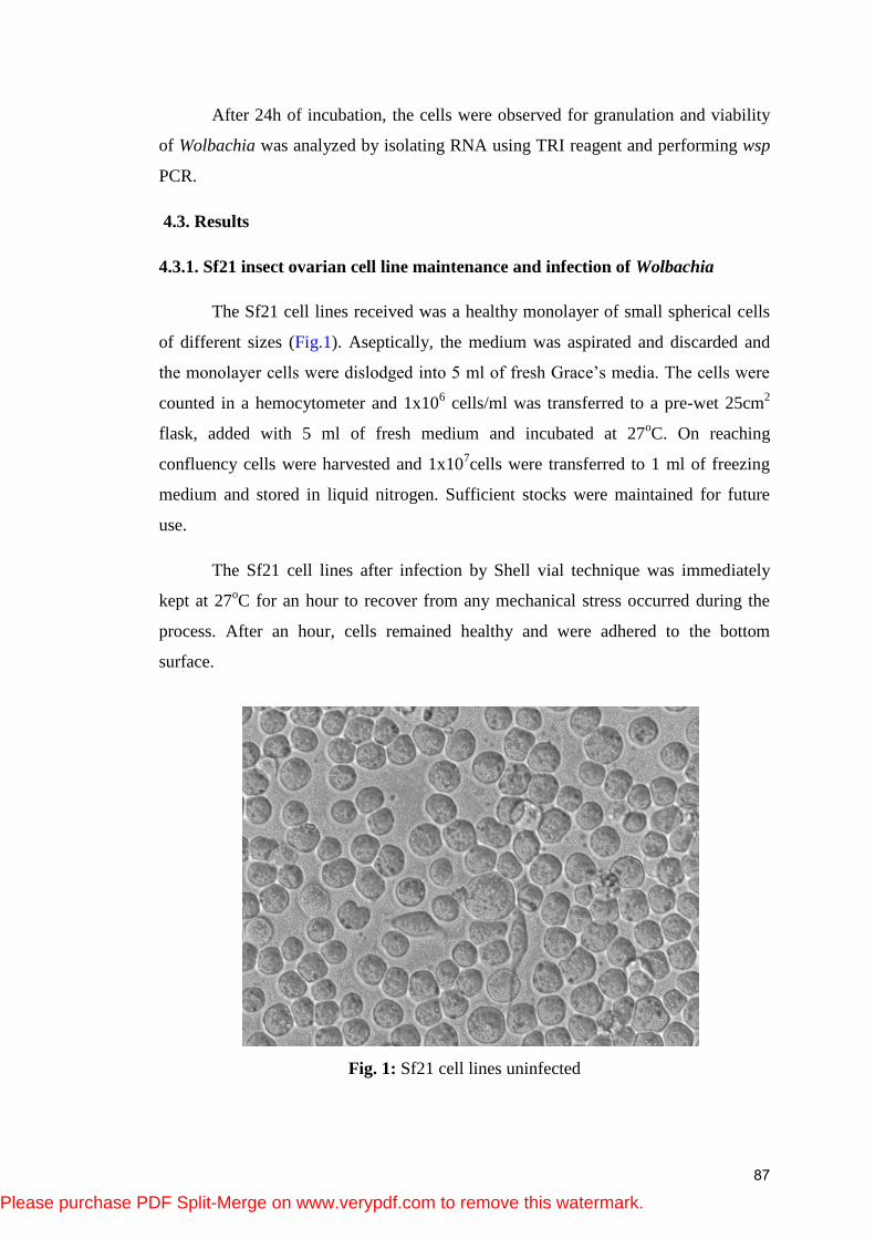
The Sf21 cell lines received was a healthy monolayer of small spherical cells
of different sizes (Fig.1). Aseptically, the medium was aspirated and discarded and
the monolayer cells were dislodged into 5 ml of fresh Grace‘s media. The cells were
counted in a hemocytometer and 1x10⁶ cells/ml was transferred to a pre-wet 25cm
2
flask, added with 5 ml of fresh medium and incubated at 27oC. On reaching
confluency cells were harvested and 1x10⁷cells were transferred to 1 ml of freezing
medium and stored in liquid nitrogen. Sufficient stocks were maintained for future
use.
The Sf21 cell lines after infection by Shell vial technique was immediately
kept at 27oC for an hour to recover from any mechanical stress occurred during the
process. After an hour, cells remained healthy and were adhered to the bottom
surface.
Fig. 1: Sf21 cell lines uninfected
87
Please purchase PDF Split-Merge on www.verypdf.com to remove this watermark.
4.3.2. Detection of Wolbachia by Microscopy and PCR
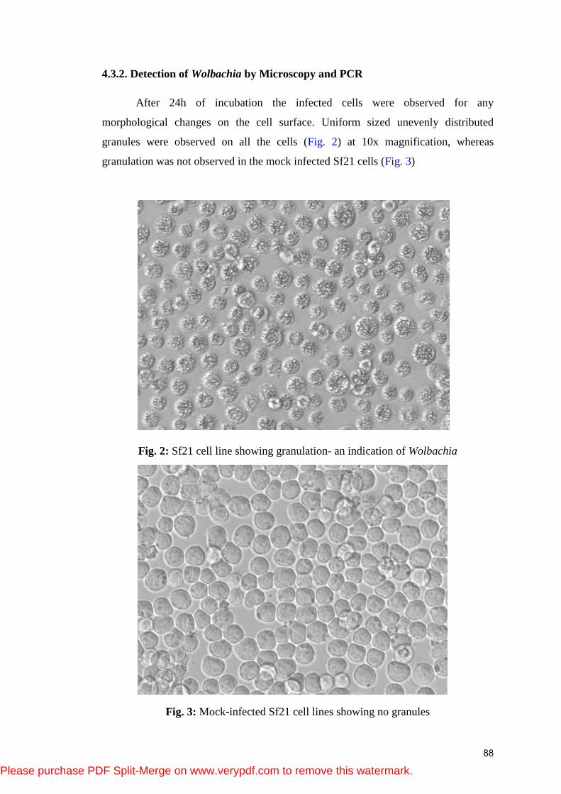
After 24h of incubation the infected cells were observed for any
morphological changes on the cell surface. Uniform sized unevenly distributed
granules were observed on all the cells (Fig. 2) at 10x magnification, whereas
granulation was not observed in the mock infected Sf21 cells (Fig. 3)
Fig. 2: Sf21 cell line showing granulation- an indication of Wolbachia
Fig. 3: Mock-infected Sf21 cell lines showing no granules
88
Please purchase PDF Split-Merge on www.verypdf.com to remove this watermark.
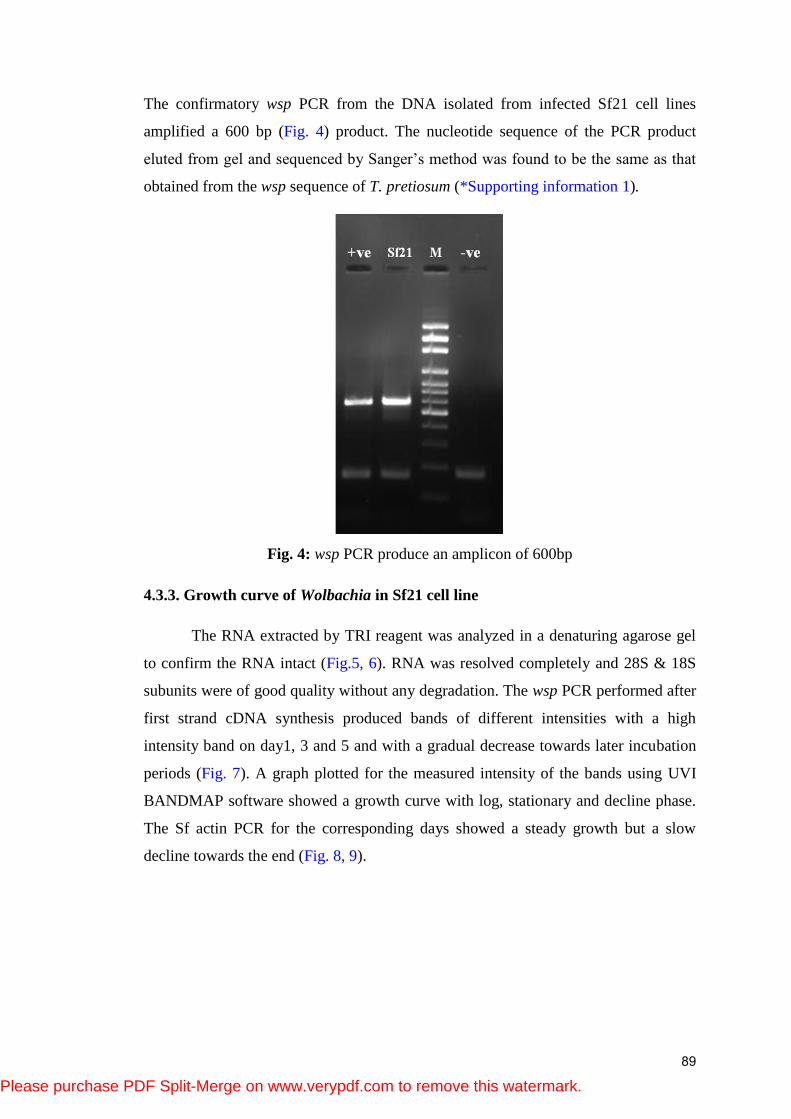
The confirmatory wsp PCR from the DNA isolated from infected Sf21 cell lines
amplified a 600 bp (Fig. 4) product. The nucleotide sequence of the PCR product
eluted from gel and sequenced by Sanger‘s method was found to be the same as that
obtained from the wsp sequence of T. pretiosum (*Supporting information 1).
Fig. 4: wsp PCR produce an amplicon of 600bp
4.3.3. Growth curve of Wolbachia in Sf21 cell line
The RNA extracted by TRI reagent was analyzed in a denaturing agarose gel
to confirm the RNA intact (Fig.5, 6). RNA was resolved completely and 28S & 18S
subunits were of good quality without any degradation. The wsp PCR performed after
first strand cDNA synthesis produced bands of different intensities with a high
intensity band on day1, 3 and 5 and with a gradual decrease towards later incubation
periods (Fig. 7). A graph plotted for the measured intensity of the bands using UVI
BANDMAP software showed a growth curve with log, stationary and decline phase.
The Sf actin PCR for the corresponding days showed a steady growth but a slow
decline towards the end (Fig. 8, 9).
89
Please purchase PDF Split-Merge on www.verypdf.com to remove this watermark.
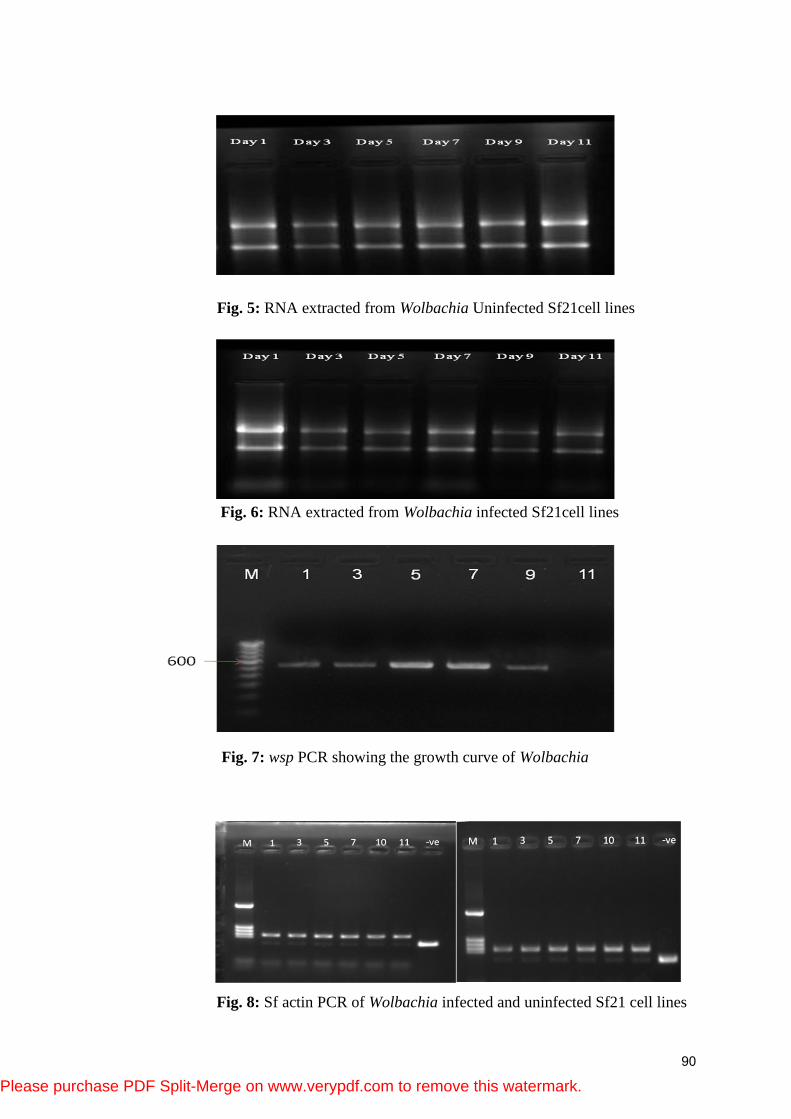
Fig. 5: RNA extracted from Wolbachia Uninfected Sf21cell lines
Fig. 6: RNA extracted from Wolbachia infected Sf21cell lines
Fig. 7: wsp PCR showing the growth curve of Wolbachia
Fig. 8: Sf actin PCR of Wolbachia infected and uninfected Sf21 cell lines
90
Please purchase PDF Split-Merge on www.verypdf.com to remove this watermark.
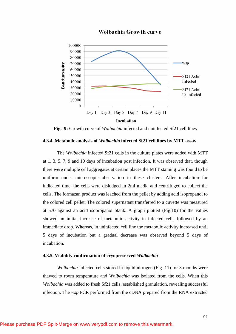
Fig. 9: Growth curve of Wolbachia infected and uninfected Sf21 cell lines
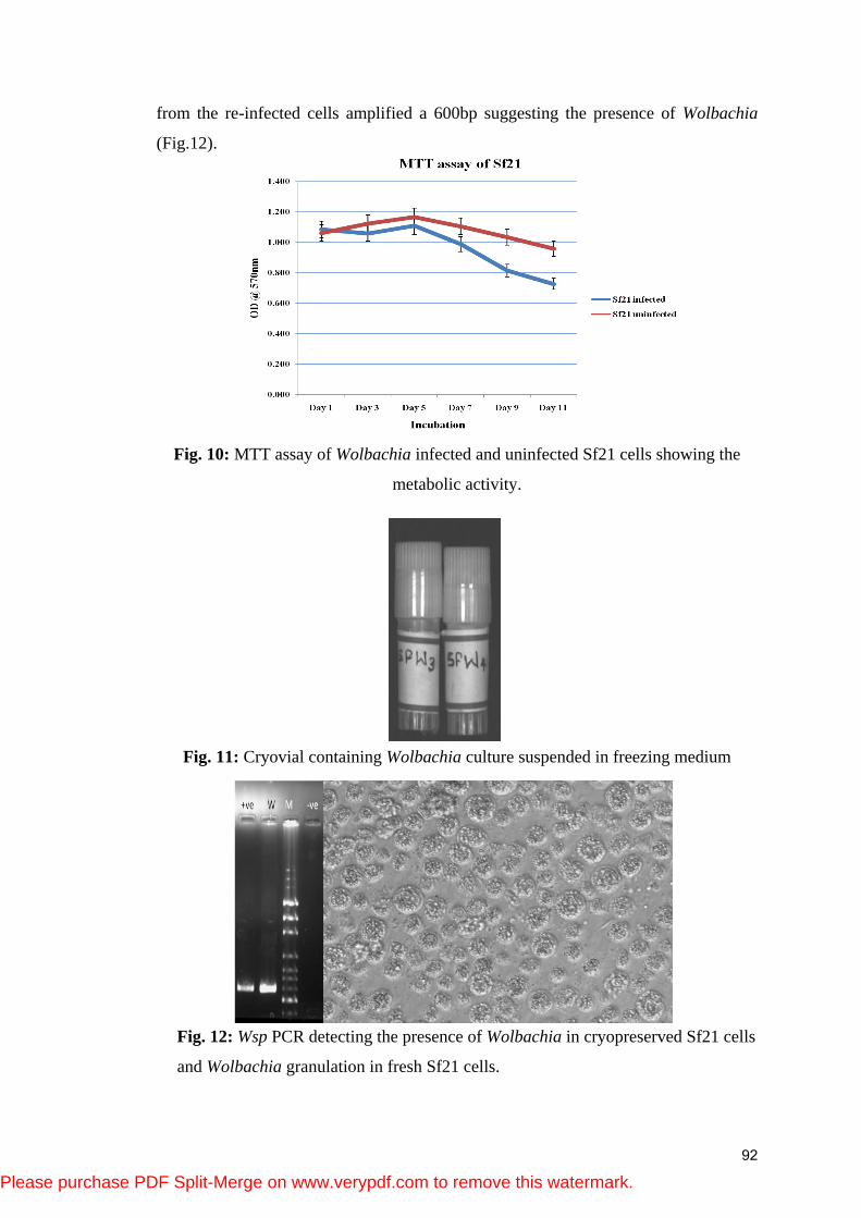
4.3.4. Metabolic analysis of Wolbachia infected Sf21 cell lines by MTT assay
The Wolbachia infected Sf21 cells in the culture plates were added with MTT
at 1, 3, 5, 7, 9 and 10 days of incubation post infection. It was observed that, though
there were multiple cell aggregates at certain places the MTT staining was found to be
uniform under microscopic observation in these clusters. After incubation for
indicated time, the cells were dislodged in 2ml media and centrifuged to collect the
cells. The formazan product was leached from the pellet by adding acid isopropanol to
the colored cell pellet. The colored supernatant transferred to a cuvette was measured
at 570 against an acid isopropanol blank. A graph plotted (Fig.10) for the values
showed an initial increase of metabolic activity in infected cells followed by an
immediate drop. Whereas, in uninfected cell line the metabolic activity increased until
5 days of incubation but a gradual decrease was observed beyond 5 days of
incubation.
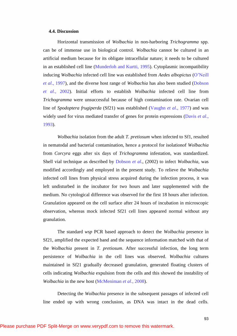
4.3.5. Viability confirmation of cryopreserved Wolbachia
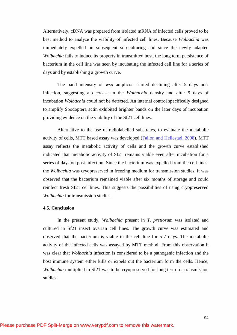
Wolbachia infected cells stored in liquid nitrogen (Fig. 11) for 3 months were
thawed to room temperature and Wolbachia was isolated from the cells. When this
Wolbachia was added to fresh Sf21 cells, established granulation, revealing successful
infection. The wsp PCR performed from the cDNA prepared from the RNA extracted
91
Please purchase PDF Split-Merge on www.verypdf.com to remove this watermark.
from the re-infected cells amplified a 600bp suggesting the presence of Wolbachia
(Fig.12).
Fig. 10: MTT assay of Wolbachia infected and uninfected Sf21 cells showing the
metabolic activity.
Fig. 11: Cryovial containing Wolbachia culture suspended in freezing medium
Fig. 12: Wsp PCR detecting the presence of Wolbachia in cryopreserved Sf21 cells
and Wolbachia granulation in fresh Sf21 cells.
92
Please purchase PDF Split-Merge on www.verypdf.com to remove this watermark.
4.4. Discussion
Horizontal transmission of Wolbachia in non-harboring Trichogramma spp.
can be of immense use in biological control. Wolbachia cannot be cultured in an
artificial medium because for its obligate intracellular nature; it needs to be cultured
in an established cell line (Munderloh and Kurtti, 1995). Cytoplasmic incompatibility
inducing Wolbachia infected cell line was established from Aedes albopictus (O‘Neill
et al., 1997), and the diverse host range of Wolbachia has also been studied (Dobson
et al., 2002). Initial efforts to establish Wolbachia infected cell line from
Trichogramma were unsuccessful because of high contamination rate. Ovarian cell
line of Spodoptera frugiperda (Sf21) was established (Vaughn et al., 1977) and was
widely used for virus mediated transfer of genes for protein expressions (Davis et al.,
1993).
Wolbachia isolation from the adult T. pretiosum when infected to Sf1, resulted
in nematodal and bacterial contamination, hence a protocol for isolationof Wolbachia
from Corcyra eggs after six days of Trichogramma infestation, was standardized.
Shell vial technique as described by Dobson et al., (2002) to infect Wolbachia, was
modified accordingly and employed in the present study. To relieve the Wolbachia
infected cell lines from physical stress acquired during the infection process, it was
left undisturbed in the incubator for two hours and later supplemented with the
medium. No cytological difference was observed for the first 18 hours after infection.
Granulation appeared on the cell surface after 24 hours of incubation in microscopic
observation, whereas mock infected Sf21 cell lines appeared normal without any
granulation.
The standard wsp PCR based approach to detect the Wolbachia presence in
Sf21, amplified the expected band and the sequence information matched with that of
the Wolbachia present in T. pretiosum. After successful infection, the long term
persistence of Wolbachia in the cell lines was observed. Wolbachia cultures
maintained in Sf21 gradually decreased granulation, generated floating clusters of
cells indicating Wolbachia expulsion from the cells and this showed the instability of
Wolbachia in the new host (McMeniman et al., 2008).
Detecting the Wolbachia presence in the subsequent passages of infected cell
line ended up with wrong conclusion, as DNA was intact in the dead cells.
93
Please purchase PDF Split-Merge on www.verypdf.com to remove this watermark.
Alternatively, cDNA was prepared from isolated mRNA of infected cells proved to be
best method to analyze the viability of infected cell lines. Because Wolbachia was
immediately expelled on subsequent sub-culturing and since the newly adapted
Wolbachia fails to induce its property in transmitted host, the long term persistence of
bacterium in the cell line was seen by incubating the infected cell line for a series of
days and by establishing a growth curve.
The band intensity of wsp amplicon started declining after 5 days post
infection, suggesting a decrease in the Wolbachia density and after 9 days of
incubation Wolbachia could not be detected. An internal control specifically designed
to amplify Spodoptera actin exhibited brighter bands on the later days of incubation
providing evidence on the viability of the Sf21 cell lines.
Alternative to the use of radiolabelled substrates, to evaluate the metabolic
activity of cells, MTT based assay was developed (Fallon and Hellestad, 2008). MTT
assay reflects the metabolic activity of cells and the growth curve established
indicated that metabolic activity of Sf21 remains viable even after incubation for a
series of days on post infection. Since the bacterium was expelled from the cell lines,
the Wolbachia was cryopreserved in freezing medium for transmission studies. It was
observed that the bacterium remained viable after six months of storage and could
reinfect fresh Sf21 cel lines. This suggests the possibilities of using cryopreserved
Wolbachia for transmission studies.
4.5. Conclusion
In the present study, Wolbachia present in T. pretiosum was isolated and
cultured in Sf21 insect ovarian cell lines. The growth curve was estimated and
observed that the bacterium is viable in the cell line for 5-7 days. The metabolic
activity of the infected cells was assayed by MTT method. From this observation it
was clear that Wolbachia infection is considered to be a pathogenic infection and the
host immune system either kills or expels out the bacterium form the cells. Hence,
Wolbachia multiplied in Sf21 was to be cryopreserved for long term for transmission
studies.
94
Please purchase PDF Split-Merge on www.verypdf.com to remove this watermark.
4.6. References
Berridge, M. V., Herst, P. M., & Tan, A. S. (2005). Tetrazolium dyes as tools in cell
biology: new insights into their cellular reduction. Biotechnology Annual Review, 11,
127–152. doi:10.1016/S1387-2656(05)11004-7
Davis, T. R., Wickham, T. J., McKenna, K. A., Granados, R. R., Shuler, M. L., &
Wood, H. A. (1993). Comparative recombinant protein production of eight insect cell
lines. In Vitro Cellular & Developmental Biology - Animal, 29(5), 388–390.
doi:10.1007/BF02633986
Dobson, S. L., Marsland, E. J., Veneti, Z., Bourtzis, K., & O‘Neill, S. L. (2002).
Characterization of Wolbachia host cell range via the in vitro establishment of
infections. Applied and Environmental Microbiology, 68(2), 656–660.
doi:10.1128/AEM.68.2.656-660.2002
Fallon, A. M. (2008). Cytological properties of an Aedes albopictus mosquito cell line
infected with Wolbachia strain wAlbB. In Vitro Cellular & Developmental Biology.
Animal, 44(5-6), 154–161. doi:10.1007/s11626-008-9090-4
Fallon, A. M., & Hellestad, V. J. (2008). Standardization of a colorimetric method to
quantify growth and metabolic activity of Wolbachia-infected mosquito cells. In Vitro
Cellular & Developmental Biology. Animal, 44(8-9), 351–356. doi:10.1007/s11626-
008-9129-6
Fenollar, F., La Scola, B., Inokuma, H., Dumler, J. S., Taylor, M. J., & Raoult, D.
(2003). Culture and phenotypic characterization of a Wolbachia pipientis isolate.
Journal of Clinical Microbiology, 41(12), 5434–5441. doi:10.1128/JCM.41.12.5434-
5441.2003
Hertig, M., & Wolbach, S. B. (1924). Studies on Rickettsia-like micro-organisms in
insects. The Journal of Medical Research, 44(3), 329–374.7.
McMeniman, C. J., Lane, A. M., Fong, A. W. C., Voronin, D. A., Iturbe-Ormaetxe, I.,
Yamada, R., O‘Neill, S. L. (2008). Host adaptation of a Wolbachia strain after long-
term serial passage in mosquito cell lines. Applied and Environmental Microbiology,
74(22), 6963–6969. doi:10.1128/AEM.01038-08
Munderloh, U. G., & Kurtti, T. J. (1995). Cellular and molecular interrelationships
between ticks and prokaryotic tick-borne pathogens. Annual Review of Entomology,
40(1), 221–243. doi:10.1146/annurev.en.40.010195.001253
95
Please purchase PDF Split-Merge on www.verypdf.com to remove this watermark.
Noda, H., Miyoshi, T., & Koizumi, Y. (2002). In vitro Cultivation of Wolbachia in
Insect and Mammalian Cell Lines. In Vitro Cellular & Developmental Biology.
Animal, 38(7), 423–427.
O‘Neill, S. L., Pettigrew, M. M., Sinkins, S. P., Braig, H. R., Andreadis, T. G., &
Tesh, R. B. (1997). In vitro cultivation of Wolbachia pipientis in an Aedes albopictus
cell line. Insect Molecular Biology, 6(1), 33–39.
Vaughn, J. L., Goodwin, R. H., Tompkins, G. J., & McCawley, P. (1977). The
establishment of two cell lines from the insect Spodoptera frugiperda (lepidoptera;
noctuidae). In Vitro, 13(4), 213–217. doi:10.1007/BF02615077
Werren, J. H., Windsor, D., & Guo, L. (1995). Distribution of Wolbachia among
Neotropical Arthropods. Proceedings of the Royal Society of London. Series B:
Biological Sciences, 262(1364), 197–204. doi:10.1098/rspb.1995.0196
96
Please purchase PDF Split-Merge on www.verypdf.com to remove this watermark.

*Supporting information (wsp sequence of Wolbachia present in T. pretiosum)
TGGTCCAATAAGTGATGAAGAAACTAGCTACTACGTTCGTTTACAATACAACGGTGAAAT
TTTACCTTTTTATACAAAAGTTGATGGTATTAAAAATGCAACAAGTAAAGAGAAGGATAG
TCCTTTAAAAAGATCTTTTATAGCTGGTGGTGTTGCGTTTGGTTATAAAATGGATGACAT
CAGAGTTGATGTTGAAGGGCTTTACTCACGATTGGCTAAAAATAAAGCTGTAATAGATGC
TTCTGAAGCAAATGTTGCAGACAGTTTAACAGCATTTTCAGGATTGGTTAACGTTTATTA
TGATATAGTGATTGAAGATATGCCTATCACTCCATACGTTGGTGTTGGTGTTGGTGCAGC
ATATATCAGCAATCCTTCAAACGCTGCTGACGTTAAAGATCAAAGGAGATTTGGTTTTGC
TTATCAAGCAAAAGCTGGTGTTAGTTATGATGTAGCCCCAGAAAACAAACTCTTTGCTGG
AGCTCGTTACTTCGGTTCTTATGGTGCTAGTTTTGATAAGGCAGCTAAGGGTGATGATGG
TATCAAAAATATTCTTACAACACTTGTGCAGAGCCT
97
Please purchase PDF Split-Merge on www.verypdf.com to remove this watermark.


![Wolbachia Seminar Master [Compatibility Mode]](https://img.pdfslide.us/doc/110x75/54679b73b4af9f623f8b588c/wolbachia-seminar-master-compatibility-mode.jpg)