Embed Size (px)
Citation preview

12/15/13 edugen.wileyplus.com/edugen/shared/resource/view_resource.uni?id=rsd4897326
edugen.wileyplus.com/edugen/shared/resource/view_resource.uni?id=rsd4897326 1/14
E X E R C I S E
4 Cell Structure and CellCycle
Materials
model or diagram of a cell compound microscopes and lens paper prepared slides of human skeletal muscle cells, pseudostratified ciliatedcolumnar epithelium (trachea), nonciliated simple columnar epithelium withmicrovilli (small intestine), motor neurons, sperm, and blood
3dimensional models of mitosis whitefish blastula slides
Objectives
After completing this exercise, you should be able to:
1. Identify cellular components on a model or diagram
2. Describe the function of the plasma membrane and cellular organelles
3. Identify cells and observable cellular structures on prepared microscope slides
4. Identify the stages of mitosis
5. Describe the events of each stage of mitosis
The human body contains over a trillion cells. These cells form the organs of the human bodyand are responsible for organ function. Cells take in nutrients delivered to them by the blood anduse these nutrients to make carbohydrates, proteins, lipids, and nucleic acids. Cells use these

12/15/13 edugen.wileyplus.com/edugen/shared/resource/view_resource.uni?id=rsd4897326
edugen.wileyplus.com/edugen/shared/resource/view_resource.uni?id=rsd4897326 2/14
macromolecules to make cellular and extracellular structures, repair themselves, and to performthe tasks required for organ function.
A CELL STRUCTURECells are the smallest structural and functional units of living organisms. They are enclosed by a plasmamembrane which controls the movement of substances into and out of the cell. The interior of the cell isfilled with cytoplasm that contains cytosol (a viscous fluid) and organelles (little organs). Like anautomobile, a cell has different parts or organelles that perform different functions. A “generalized” animalcell is shown in Figure 41, and functions of cellular organelles are described in Table 41.
Try the Interactive Version.
Figure 41 Sectional drawing of a cell.
1. mitochondrion
2. peroxisome
3. smooth endoplasmic reticulum
4. lysosome
5. plasma membrane
6. centrioles
7. microvillus
8. flagellum
9. cilium
10. secretory vesicle
11. chromatin
12. nuclear membrane
13. nucleolus
14. nucleus

12/15/13 edugen.wileyplus.com/edugen/shared/resource/view_resource.uni?id=rsd4897326
edugen.wileyplus.com/edugen/shared/resource/view_resource.uni?id=rsd4897326 3/14
15. cytoplasm
16. rough endoplasmic reticulum
17. ribosome
18. Golgi complex
Table 41 Function of Cell StructuresSTRUCTURE FUNCTION
Plasma Membrane Controls movement of substances into and out of the cell
Microvilli Folds of the plasma membrane that increase the surface area of the cellto increase absorption or secretion
Nucleus Contains DNA molecules and nucleolus
Nucleolus Assembly site for ribosomes
Chromatin Long thin strands within nucleus. Each strand composed of one DNAmolecule and associated proteins.
Cytoplasm Area of the cell that includes the cytosol and organelles
Cytosol Fluid portion of cytoplasm that surrounds organelles
Organelles

12/15/13 edugen.wileyplus.com/edugen/shared/resource/view_resource.uni?id=rsd4897326
edugen.wileyplus.com/edugen/shared/resource/view_resource.uni?id=rsd4897326 4/14
• Mitochondria Makes ATP via aerobic cellular respiration
• Ribosomes Site of protein synthesis
• Rough endoplasmicreticulum (RER)
Processes and transports proteins made at attached ribosomes;synthesizes phospholipids
• Smoothendoplasmicreticulum (SER)
Fatty acid and steroid synthesis; detoxifies toxic substances
• Golgi complex Receives and modifies proteins from RER; sorts and transports them
• Secretory vesicles Secrete substances outside the cell by exocytosis
• Lysosomes Enzymes digest and recycle worn out organelles and substancesentering the cell; can digest the cell
• Peroxisomes Produce hydrogen peroxide; detoxify harmful substances
• Cytoskeleton Three kinds of protein filaments; maintain cell shape and involved incell movement and movement of organelles
• Centrosomes(centrioles)
Form mitotic spindle; needed to form cilia and flagella
• Cilia Abundant, hairlike cell projections that move fluids and particlesalong the cell surface
• Flagella Long cell projection; whiplike motion moves sperm
Activity 1 Cell Structure
1. Point to each cell structure shown in Figure 41 on a cell model or chart.2. Describe the function of each organelle as you point to it.
Discussion Questions: Cell Structures
Which cell structures from Table 41 are not found in most human cells?
B CELL SPECIALIZATIONThe human body contains over 200 different types of cells with different functions. These differences infunction are reflected in cell structure. Cells of the human body differ from the generalized animal cell inshape, size, or number and type of organelles present. In the next activity you will observe cells of skeletalmuscle, pseudostratified ciliated columnar epithelium, nonciliated simple columnar epithelium withmicrovilli, motor neurons, sperm, and blood.
12/15/13 edugen.wileyplus.com/edugen/shared/resource/view_resource.uni?id=rsd4897326
edugen.wileyplus.com/edugen/shared/resource/view_resource.uni?id=rsd4897326 5/14
Skeletal muscle cells are long, cylindrical cells that contain specialized proteins (contractile proteins)that enable them to contract (shorten in length) to move bones. The contractile proteins are organizedinto repeating units that can be observed in the light microscope as striations.
Pseudostratified ciliated columnar epithelial cells have cilia that move substances like mucus alongthe surface of the cells. Mucus is produced by specialized cells called goblet cells.
Nonciliated simple columnar epithelium with microvilli. Microvilli increase the surface area of theplasma membrane which provides a larger area for absorption of nutrients along the gastrointestinaltract or secretion of product from glands.
Motor neurons are nervous tissue cells with many processes (cell extensions) that receiveinformation from other neurons and send electrical signals to muscle cells causing them to contract.
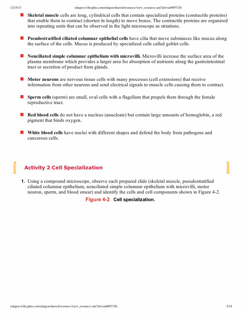
Sperm cells (sperm) are small, oval cells with a flagellum that propels them through the femalereproductive tract.
Red blood cells do not have a nucleus (anucleate) but contain large amounts of hemoglobin, a redpigment that binds oxygen.
White blood cells have nuclei with different shapes and defend the body from pathogens andcancerous cells.
Activity 2 Cell Specialization
1. Using a compound microscope, observe each prepared slide (skeletal muscle, pseudostratifiedciliated columnar epithelium, nonciliated simple columnar epithelium with microvilli, motorneuron, sperm, and blood smear) and identify the cells and cell components shown in Figure 42.
Figure 42 Cell specialization.

12/15/13 edugen.wileyplus.com/edugen/shared/resource/view_resource.uni?id=rsd4897326
edugen.wileyplus.com/edugen/shared/resource/view_resource.uni?id=rsd4897326 6/14
12/15/13 edugen.wileyplus.com/edugen/shared/resource/view_resource.uni?id=rsd4897326
edugen.wileyplus.com/edugen/shared/resource/view_resource.uni?id=rsd4897326 7/14
2. Describe each cell's shape and list the cell structures that can be seen with the light microscope ineach cell type.

12/15/13 edugen.wileyplus.com/edugen/shared/resource/view_resource.uni?id=rsd4897326
edugen.wileyplus.com/edugen/shared/resource/view_resource.uni?id=rsd4897326 8/14
(a) skeletal muscle cell: _________
(b) pseudostratified ciliated columnar epithelial cell: _________
(c) motor neuron: _________
(d) sperm cell: _________
(e) red blood cell _________
(f) white blood cell _________
(g) nonciliated simple columnar epithelium: _________
C SOMATIC CELL DIVISION: MITOSIS AND CYTOKINESISSomatic (soma = body) cell division occurs when one cell divides to produce two genetically identical cells.Cell division is needed for growth of the individual and cell replacement.
The cell cycle, a period during which a cell grows and divides into two genetically identical cells (daughtercells), begins when a cell is produced by cell division and ends when the cell divides. The length of the cellcycle differs according to the type of cell, with some cells dividing more frequently than others. The cellcycle can be divided into two basic periods: interphase, a long period during which the cell conducts itsnormal activity, grows, and prepares for cell division; and the mitotic phase, when the cell is dividing. Themitotic phase consists of mitosis, or nuclear division, and cytokinesis, or cytoplasmic division. The fourstages of mitosis are: prophase, metaphase, anaphase, and telophase (Table 42).
Table 42 Phases of Somatic Cell CyclePHASE DESCRIPTION OF ACTIVITY
Interphase (inter =between)
Normal cell work; cell metabolically active and growing; DNA replicates
Mitotic Phase Cell division
Mitosis (mitos =thread)
Nuclear division
• Prophase (pro =first)
Nucleolus and nuclear membrane disappear; chromatin condenses intochromosomes; centrioles move to opposite poles; spindle fibers form
• Metaphase(meta = next)
Chromosomes line up at metaphasal plate; spindle fibers attach tocentromeres of chromatids
• Anaphase (ana= apart)
Chromatids of chromosomes separate; move to opposite poles

12/15/13 edugen.wileyplus.com/edugen/shared/resource/view_resource.uni?id=rsd4897326
edugen.wileyplus.com/edugen/shared/resource/view_resource.uni?id=rsd4897326 9/14
• Telophase (telo= end)
Cell reverses prophase activities
Cytokinesis (cyto =cell; kinesio =movement)
Cytoplasmic division into two genetically identical daughter cells
To observe interphase and the stages of mitosis, you will examine a prepared microscope slide containingseveral sections of a whitefish blastula. The blastula is an early embryonic stage in which cells are dividingrapidly, providing many cells in different stages of mitosis.
Activity 3 Mitotic Phases
1. Observe the 3dimensional models of the mitotic phases, noting the changes in each phase.2. Using Table 42, identify interphase, each phase of mitosis, and cytokinesis in Figure 43(a)–(e).
Try the Interactive Version.
Figure 43 Mitotic phases.
anaphase (ANafaze)
interphase (INterfaze)
metaphase (MEHtafaze)
prophase (PROfaze)
telephase and cytokinesis (TELLofaze andcytokihNEEsis)
(a) _________
(b) _________
(c) _________
(d) _________
(e) _________

12/15/13 edugen.wileyplus.com/edugen/shared/resource/view_resource.uni?id=rsd4897326
edugen.wileyplus.com/edugen/shared/resource/view_resource.uni?id=rsd4897326 10/14

12/15/13 edugen.wileyplus.com/edugen/shared/resource/view_resource.uni?id=rsd4897326
edugen.wileyplus.com/edugen/shared/resource/view_resource.uni?id=rsd4897326 11/14

12/15/13 edugen.wileyplus.com/edugen/shared/resource/view_resource.uni?id=rsd4897326
edugen.wileyplus.com/edugen/shared/resource/view_resource.uni?id=rsd4897326 12/14
3. Obtain a prepared whitefish blastula slide and hold it up to the light. Notice that there are manyblastula sections on each slide. It will be necessary to view several of these sections to find all ofthe phases.
4. Using a compound microscope, begin looking at your slide with the lowpower objective lens.Use the highpower objective lens to identify interphase, the four stages of mitosis, andcytokinesis.
Reviewing Your Knowledge
Cellular StructureFill in the blank with the name of the cell structure that fits the description.
1. short, hairlike projections for movement of substances along cell surface2. intracellular fluid3. site of energy production by cellular respiration4. site of protein synthesis5. site of steroid and fatty acid synthesis6. small vesicle with digestive enzymes7. organelles needed to form cilia and flagella8. threadlike strand of DNA with associated proteins9. site of secretory and membrane protein synthesis10. site where protein products are stored, packaged, and exported11. contains DNA that control cellular activities12. site of ribosome synthesis

12/15/13 edugen.wileyplus.com/edugen/shared/resource/view_resource.uni?id=rsd4897326
edugen.wileyplus.com/edugen/shared/resource/view_resource.uni?id=rsd4897326 13/14
13. gives the cell shape, support, movement, and holds organelles in position14. controls movement of substances into or out of the cell15. folds of the plasma membrane that increase the cell's surface area16. detoxifies harmful substances, produces hydrogen peroxide, and oxidizes amino acids17. double membrane that separates the nucleus from the cytoplasm18. a small membranous sac that delivers proteins to the plasma membrane to exit the cell
Phases of the Cell CycleWrite the phase of the cell cycle that the phrase describes.
1. cytoplasmic division2. cell performing normal functions; longest phase3. nuclear division4. chromatid pairs line up at equatorial plate5. chromatin condenses into chromosomes6. spindle fibers break up; nucleus reappears7. centromeres divide; chromosomes move to opposite poles8. nuclear membrane disassembles and disappears9. chromosomes unravel to form chromatin10. mitotic spindle forms11. DNA replicates
Using Your Knowledge
Cellular Organelles and Their FunctionWrite the letter for the correct answer in the blank.
1. _________ A cell makes and secretes a proteinbased hormone. This particular cell would have a greatamount of Golgi complex and:
(a) SER(b) RER(c) mitochondria(d) lysosomes
2. _________ Cells that make steroids (lipids) would have a larger amount of:
(a) SER(b) RER(c) mitochondria(d) lysosomes
3. _________ Cells that need large amounts of ATP would have many:

12/15/13 edugen.wileyplus.com/edugen/shared/resource/view_resource.uni?id=rsd4897326
edugen.wileyplus.com/edugen/shared/resource/view_resource.uni?id=rsd4897326 14/14
(a) Golgi complexes(b) ribosomes(c) SER(d) mitochondria
4. _________ Cells that line the small intestine are specialized for absorption and secretion. The plasmamembrane structure they have to accomplish this is:
(a) centrioles(b) cilia(c) flagella(d) microvilli
5. _________ Immune cells that destroy bacteria with chemicals need an abundance of:
(a) SER(b) ribosomes(c) lysosomes(d) centrioles
The Cell Cycle and MitosisAnswer each question with a short answer.
6. Explain the role of somatic cell division as a person ages from infancy to adulthood.7. Explain the role of cell division in wound healing.8. List the cell structures involved in mitosis.9. Can red blood cells undergo mitosis? Explain.10. Sperm and eggs have onehalf the number of chromosomes of the somatic cells that divided to form
them. Are sperm and eggs formed by mitosis? Explain.