Embed Size (px)
Citation preview
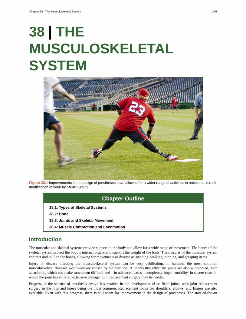
38 | THEMUSCULOSKELETALSYSTEM
Figure 38.1 Improvements in the design of prostheses have allowed for a wider range of activities in recipients. (credit:modification of work by Stuart Grout)
Chapter Outline
38.1: Types of Skeletal Systems
38.2: Bone
38.3: Joints and Skeletal Movement
38.4: Muscle Contraction and Locomotion
Introduction
The muscular and skeletal systems provide support to the body and allow for a wide range of movement. The bones of theskeletal system protect the body’s internal organs and support the weight of the body. The muscles of the muscular systemcontract and pull on the bones, allowing for movements as diverse as standing, walking, running, and grasping items.
Injury or disease affecting the musculoskeletal system can be very debilitating. In humans, the most commonmusculoskeletal diseases worldwide are caused by malnutrition. Ailments that affect the joints are also widespread, suchas arthritis, which can make movement difficult and—in advanced cases—completely impair mobility. In severe cases inwhich the joint has suffered extensive damage, joint replacement surgery may be needed.
Progress in the science of prosthesis design has resulted in the development of artificial joints, with joint replacementsurgery in the hips and knees being the most common. Replacement joints for shoulders, elbows, and fingers are alsoavailable. Even with this progress, there is still room for improvement in the design of prostheses. The state-of-the-art
Chapter 38 | The Musculoskeletal System 1091
prostheses have limited durability and therefore wear out quickly, particularly in young or active individuals. Currentresearch is focused on the use of new materials, such as carbon fiber, that may make prostheses more durable.
38.1 | Types of Skeletal Systems
By the end of this section, you will be able to:
• Discuss the different types of skeletal systems
• Explain the role of the human skeletal system
• Compare and contrast different skeletal systems
A skeletal system is necessary to support the body, protect internal organs, and allow for the movement of an organism.There are three different skeleton designs that fulfill these functions: hydrostatic skeleton, exoskeleton, and endoskeleton.
Hydrostatic Skeleton
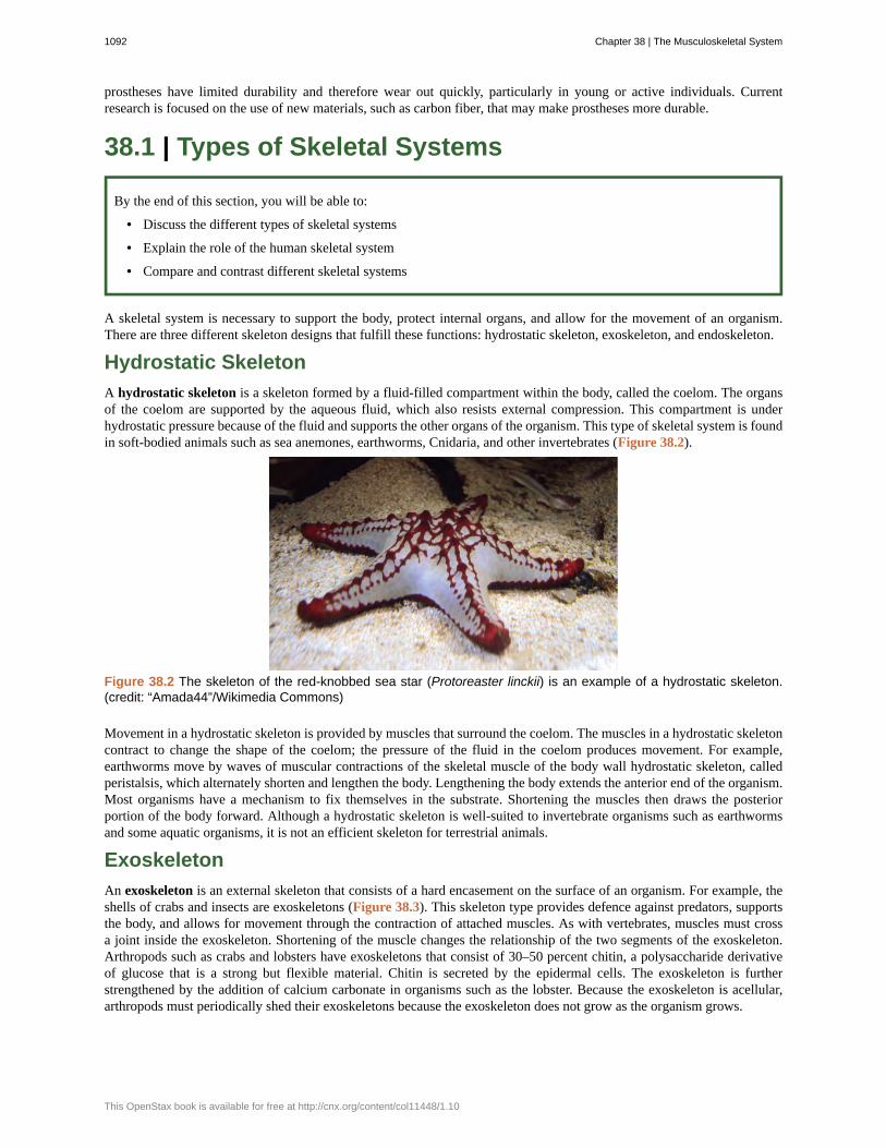
A hydrostatic skeleton is a skeleton formed by a fluid-filled compartment within the body, called the coelom. The organsof the coelom are supported by the aqueous fluid, which also resists external compression. This compartment is underhydrostatic pressure because of the fluid and supports the other organs of the organism. This type of skeletal system is foundin soft-bodied animals such as sea anemones, earthworms, Cnidaria, and other invertebrates (Figure 38.2).
Figure 38.2 The skeleton of the red-knobbed sea star (Protoreaster linckii) is an example of a hydrostatic skeleton.(credit: “Amada44”/Wikimedia Commons)
Movement in a hydrostatic skeleton is provided by muscles that surround the coelom. The muscles in a hydrostatic skeletoncontract to change the shape of the coelom; the pressure of the fluid in the coelom produces movement. For example,earthworms move by waves of muscular contractions of the skeletal muscle of the body wall hydrostatic skeleton, calledperistalsis, which alternately shorten and lengthen the body. Lengthening the body extends the anterior end of the organism.Most organisms have a mechanism to fix themselves in the substrate. Shortening the muscles then draws the posteriorportion of the body forward. Although a hydrostatic skeleton is well-suited to invertebrate organisms such as earthwormsand some aquatic organisms, it is not an efficient skeleton for terrestrial animals.
Exoskeleton
An exoskeleton is an external skeleton that consists of a hard encasement on the surface of an organism. For example, theshells of crabs and insects are exoskeletons (Figure 38.3). This skeleton type provides defence against predators, supportsthe body, and allows for movement through the contraction of attached muscles. As with vertebrates, muscles must crossa joint inside the exoskeleton. Shortening of the muscle changes the relationship of the two segments of the exoskeleton.Arthropods such as crabs and lobsters have exoskeletons that consist of 30–50 percent chitin, a polysaccharide derivativeof glucose that is a strong but flexible material. Chitin is secreted by the epidermal cells. The exoskeleton is furtherstrengthened by the addition of calcium carbonate in organisms such as the lobster. Because the exoskeleton is acellular,arthropods must periodically shed their exoskeletons because the exoskeleton does not grow as the organism grows.
1092 Chapter 38 | The Musculoskeletal System
This OpenStax book is available for free at http://cnx.org/content/col11448/1.10
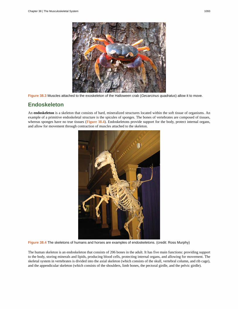
Figure 38.3 Muscles attached to the exoskeleton of the Halloween crab (Gecarcinus quadratus) allow it to move.
Endoskeleton
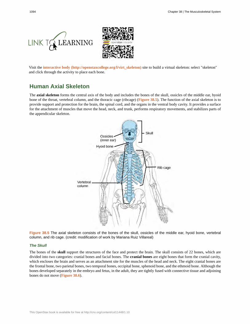
An endoskeleton is a skeleton that consists of hard, mineralized structures located within the soft tissue of organisms. Anexample of a primitive endoskeletal structure is the spicules of sponges. The bones of vertebrates are composed of tissues,whereas sponges have no true tissues (Figure 38.4). Endoskeletons provide support for the body, protect internal organs,and allow for movement through contraction of muscles attached to the skeleton.
Figure 38.4 The skeletons of humans and horses are examples of endoskeletons. (credit: Ross Murphy)
The human skeleton is an endoskeleton that consists of 206 bones in the adult. It has five main functions: providing supportto the body, storing minerals and lipids, producing blood cells, protecting internal organs, and allowing for movement. Theskeletal system in vertebrates is divided into the axial skeleton (which consists of the skull, vertebral column, and rib cage),and the appendicular skeleton (which consists of the shoulders, limb bones, the pectoral girdle, and the pelvic girdle).
Chapter 38 | The Musculoskeletal System 1093
Visit the interactive body (http://openstaxcollege.org/l/virt_skeleton) site to build a virtual skeleton: select "skeleton"and click through the activity to place each bone.
Human Axial Skeleton
The axial skeleton forms the central axis of the body and includes the bones of the skull, ossicles of the middle ear, hyoidbone of the throat, vertebral column, and the thoracic cage (ribcage) (Figure 38.5). The function of the axial skeleton is toprovide support and protection for the brain, the spinal cord, and the organs in the ventral body cavity. It provides a surfacefor the attachment of muscles that move the head, neck, and trunk, performs respiratory movements, and stabilizes parts ofthe appendicular skeleton.
Figure 38.5 The axial skeleton consists of the bones of the skull, ossicles of the middle ear, hyoid bone, vertebralcolumn, and rib cage. (credit: modification of work by Mariana Ruiz Villareal)
The Skull
The bones of the skull support the structures of the face and protect the brain. The skull consists of 22 bones, which aredivided into two categories: cranial bones and facial bones. The cranial bones are eight bones that form the cranial cavity,which encloses the brain and serves as an attachment site for the muscles of the head and neck. The eight cranial bones arethe frontal bone, two parietal bones, two temporal bones, occipital bone, sphenoid bone, and the ethmoid bone. Although thebones developed separately in the embryo and fetus, in the adult, they are tightly fused with connective tissue and adjoiningbones do not move (Figure 38.6).
1094 Chapter 38 | The Musculoskeletal System
This OpenStax book is available for free at http://cnx.org/content/col11448/1.10
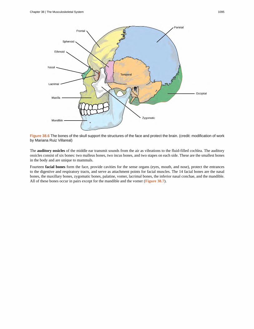
Figure 38.6 The bones of the skull support the structures of the face and protect the brain. (credit: modification of workby Mariana Ruiz Villareal)
The auditory ossicles of the middle ear transmit sounds from the air as vibrations to the fluid-filled cochlea. The auditoryossicles consist of six bones: two malleus bones, two incus bones, and two stapes on each side. These are the smallest bonesin the body and are unique to mammals.
Fourteen facial bones form the face, provide cavities for the sense organs (eyes, mouth, and nose), protect the entrancesto the digestive and respiratory tracts, and serve as attachment points for facial muscles. The 14 facial bones are the nasalbones, the maxillary bones, zygomatic bones, palatine, vomer, lacrimal bones, the inferior nasal conchae, and the mandible.All of these bones occur in pairs except for the mandible and the vomer (Figure 38.7).
Chapter 38 | The Musculoskeletal System 1095
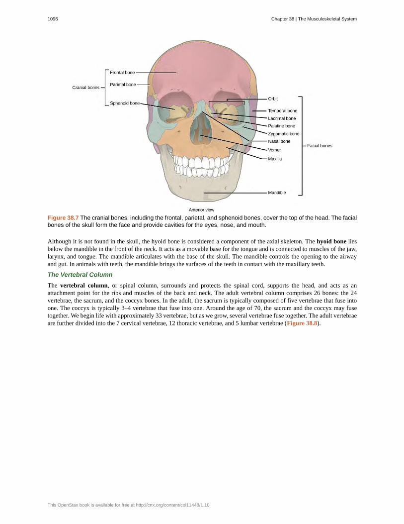
Figure 38.7 The cranial bones, including the frontal, parietal, and sphenoid bones, cover the top of the head. The facialbones of the skull form the face and provide cavities for the eyes, nose, and mouth.
Although it is not found in the skull, the hyoid bone is considered a component of the axial skeleton. The hyoid bone liesbelow the mandible in the front of the neck. It acts as a movable base for the tongue and is connected to muscles of the jaw,larynx, and tongue. The mandible articulates with the base of the skull. The mandible controls the opening to the airwayand gut. In animals with teeth, the mandible brings the surfaces of the teeth in contact with the maxillary teeth.
The Vertebral Column
The vertebral column, or spinal column, surrounds and protects the spinal cord, supports the head, and acts as anattachment point for the ribs and muscles of the back and neck. The adult vertebral column comprises 26 bones: the 24vertebrae, the sacrum, and the coccyx bones. In the adult, the sacrum is typically composed of five vertebrae that fuse intoone. The coccyx is typically 3–4 vertebrae that fuse into one. Around the age of 70, the sacrum and the coccyx may fusetogether. We begin life with approximately 33 vertebrae, but as we grow, several vertebrae fuse together. The adult vertebraeare further divided into the 7 cervical vertebrae, 12 thoracic vertebrae, and 5 lumbar vertebrae (Figure 38.8).
1096 Chapter 38 | The Musculoskeletal System
This OpenStax book is available for free at http://cnx.org/content/col11448/1.10
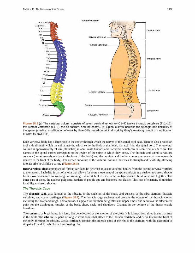
Figure 38.8 (a) The vertebral column consists of seven cervical vertebrae (C1–7) twelve thoracic vertebrae (Th1–12),five lumbar vertebrae (L1–5), the os sacrum, and the coccyx. (b) Spinal curves increase the strength and flexibility ofthe spine. (credit a: modification of work by Uwe Gille based on original work by Gray's Anatomy; credit b: modificationof work by NCI, NIH)
Each vertebral body has a large hole in the center through which the nerves of the spinal cord pass. There is also a notch oneach side through which the spinal nerves, which serve the body at that level, can exit from the spinal cord. The vertebralcolumn is approximately 71 cm (28 inches) in adult male humans and is curved, which can be seen from a side view. Thenames of the spinal curves correspond to the region of the spine in which they occur. The thoracic and sacral curves areconcave (curve inwards relative to the front of the body) and the cervical and lumbar curves are convex (curve outwardsrelative to the front of the body). The arched curvature of the vertebral column increases its strength and flexibility, allowingit to absorb shocks like a spring (Figure 38.8).
Intervertebral discs composed of fibrous cartilage lie between adjacent vertebral bodies from the second cervical vertebrato the sacrum. Each disc is part of a joint that allows for some movement of the spine and acts as a cushion to absorb shocksfrom movements such as walking and running. Intervertebral discs also act as ligaments to bind vertebrae together. Theinner part of discs, the nucleus pulposus, hardens as people age and becomes less elastic. This loss of elasticity diminishesits ability to absorb shocks.
The Thoracic Cage
The thoracic cage, also known as the ribcage, is the skeleton of the chest, and consists of the ribs, sternum, thoracicvertebrae, and costal cartilages (Figure 38.9). The thoracic cage encloses and protects the organs of the thoracic cavity,including the heart and lungs. It also provides support for the shoulder girdles and upper limbs, and serves as the attachmentpoint for the diaphragm, muscles of the back, chest, neck, and shoulders. Changes in the volume of the thorax enablebreathing.
The sternum, or breastbone, is a long, flat bone located at the anterior of the chest. It is formed from three bones that fusein the adult. The ribs are 12 pairs of long, curved bones that attach to the thoracic vertebrae and curve toward the front ofthe body, forming the ribcage. Costal cartilages connect the anterior ends of the ribs to the sternum, with the exception ofrib pairs 11 and 12, which are free-floating ribs.
Chapter 38 | The Musculoskeletal System 1097
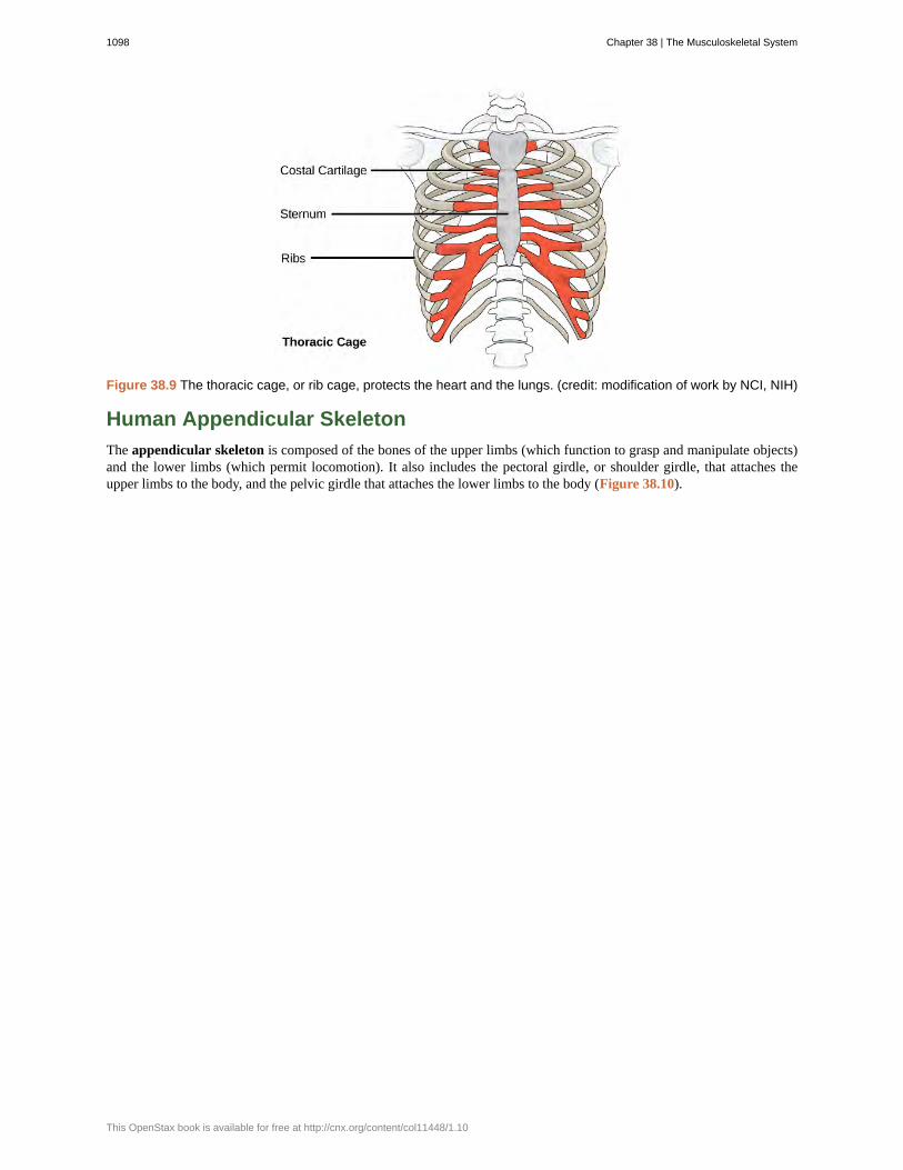
Figure 38.9 The thoracic cage, or rib cage, protects the heart and the lungs. (credit: modification of work by NCI, NIH)
Human Appendicular Skeleton
The appendicular skeleton is composed of the bones of the upper limbs (which function to grasp and manipulate objects)and the lower limbs (which permit locomotion). It also includes the pectoral girdle, or shoulder girdle, that attaches theupper limbs to the body, and the pelvic girdle that attaches the lower limbs to the body (Figure 38.10).
1098 Chapter 38 | The Musculoskeletal System
This OpenStax book is available for free at http://cnx.org/content/col11448/1.10
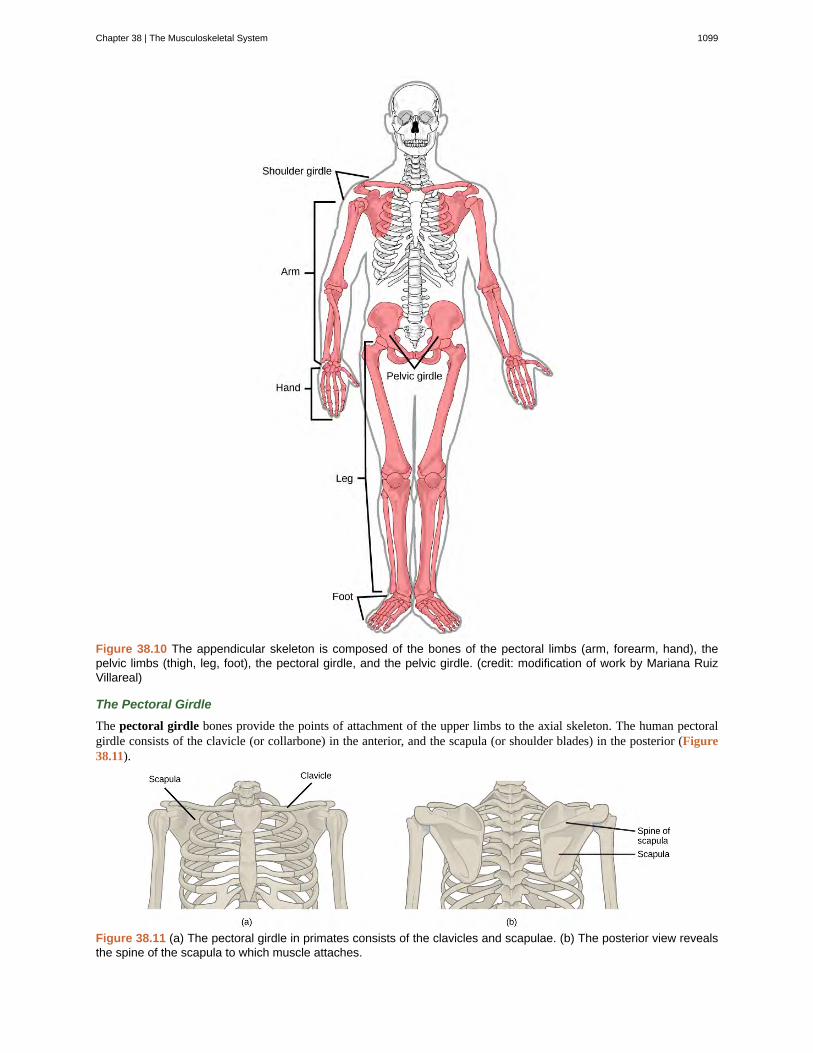
Figure 38.10 The appendicular skeleton is composed of the bones of the pectoral limbs (arm, forearm, hand), thepelvic limbs (thigh, leg, foot), the pectoral girdle, and the pelvic girdle. (credit: modification of work by Mariana RuizVillareal)
The Pectoral Girdle
The pectoral girdle bones provide the points of attachment of the upper limbs to the axial skeleton. The human pectoralgirdle consists of the clavicle (or collarbone) in the anterior, and the scapula (or shoulder blades) in the posterior (Figure38.11).
Figure 38.11 (a) The pectoral girdle in primates consists of the clavicles and scapulae. (b) The posterior view revealsthe spine of the scapula to which muscle attaches.
Chapter 38 | The Musculoskeletal System 1099
The clavicles are S-shaped bones that position the arms on the body. The clavicles lie horizontally across the front of thethorax (chest) just above the first rib. These bones are fairly fragile and are susceptible to fractures. For example, a fallwith the arms outstretched causes the force to be transmitted to the clavicles, which can break if the force is excessive. Theclavicle articulates with the sternum and the scapula.
The scapulae are flat, triangular bones that are located at the back of the pectoral girdle. They support the muscles crossingthe shoulder joint. A ridge, called the spine, runs across the back of the scapula and can easily be felt through the skin(Figure 38.11). The spine of the scapula is a good example of a bony protrusion that facilitates a broad area of attachmentfor muscles to bone.
The Upper Limb
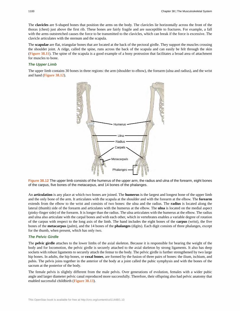
The upper limb contains 30 bones in three regions: the arm (shoulder to elbow), the forearm (ulna and radius), and the wristand hand (Figure 38.12).
Figure 38.12 The upper limb consists of the humerus of the upper arm, the radius and ulna of the forearm, eight bonesof the carpus, five bones of the metacarpus, and 14 bones of the phalanges.
An articulation is any place at which two bones are joined. The humerus is the largest and longest bone of the upper limband the only bone of the arm. It articulates with the scapula at the shoulder and with the forearm at the elbow. The forearmextends from the elbow to the wrist and consists of two bones: the ulna and the radius. The radius is located along thelateral (thumb) side of the forearm and articulates with the humerus at the elbow. The ulna is located on the medial aspect(pinky-finger side) of the forearm. It is longer than the radius. The ulna articulates with the humerus at the elbow. The radiusand ulna also articulate with the carpal bones and with each other, which in vertebrates enables a variable degree of rotationof the carpus with respect to the long axis of the limb. The hand includes the eight bones of the carpus (wrist), the fivebones of the metacarpus (palm), and the 14 bones of the phalanges (digits). Each digit consists of three phalanges, exceptfor the thumb, when present, which has only two.
The Pelvic Girdle
The pelvic girdle attaches to the lower limbs of the axial skeleton. Because it is responsible for bearing the weight of thebody and for locomotion, the pelvic girdle is securely attached to the axial skeleton by strong ligaments. It also has deepsockets with robust ligaments to securely attach the femur to the body. The pelvic girdle is further strengthened by two largehip bones. In adults, the hip bones, or coxal bones, are formed by the fusion of three pairs of bones: the ilium, ischium, andpubis. The pelvis joins together in the anterior of the body at a joint called the pubic symphysis and with the bones of thesacrum at the posterior of the body.
The female pelvis is slightly different from the male pelvis. Over generations of evolution, females with a wider pubicangle and larger diameter pelvic canal reproduced more successfully. Therefore, their offspring also had pelvic anatomy thatenabled successful childbirth (Figure 38.13).
1100 Chapter 38 | The Musculoskeletal System
This OpenStax book is available for free at http://cnx.org/content/col11448/1.10
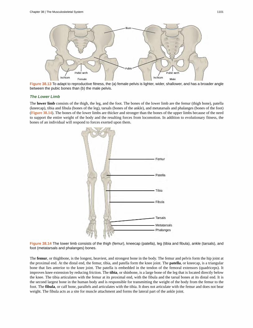
Figure 38.13 To adapt to reproductive fitness, the (a) female pelvis is lighter, wider, shallower, and has a broader anglebetween the pubic bones than (b) the male pelvis.
The Lower Limb
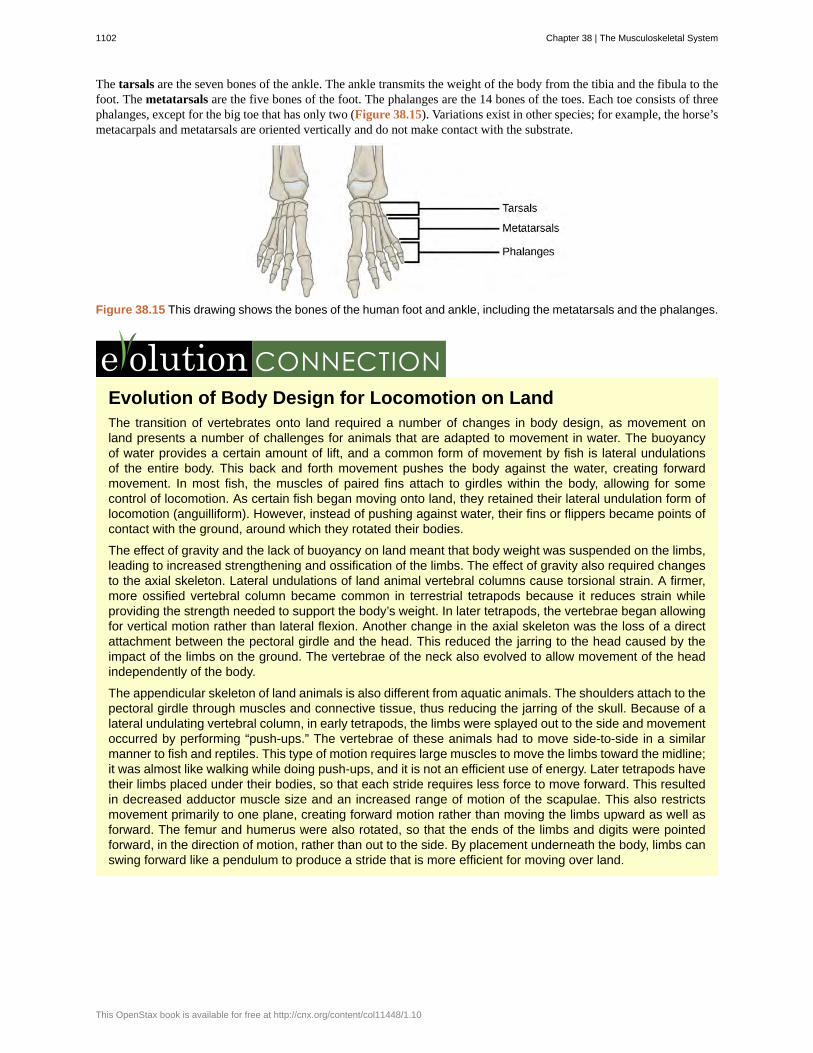
The lower limb consists of the thigh, the leg, and the foot. The bones of the lower limb are the femur (thigh bone), patella(kneecap), tibia and fibula (bones of the leg), tarsals (bones of the ankle), and metatarsals and phalanges (bones of the foot)(Figure 38.14). The bones of the lower limbs are thicker and stronger than the bones of the upper limbs because of the needto support the entire weight of the body and the resulting forces from locomotion. In addition to evolutionary fitness, thebones of an individual will respond to forces exerted upon them.
Figure 38.14 The lower limb consists of the thigh (femur), kneecap (patella), leg (tibia and fibula), ankle (tarsals), andfoot (metatarsals and phalanges) bones.
The femur, or thighbone, is the longest, heaviest, and strongest bone in the body. The femur and pelvis form the hip joint atthe proximal end. At the distal end, the femur, tibia, and patella form the knee joint. The patella, or kneecap, is a triangularbone that lies anterior to the knee joint. The patella is embedded in the tendon of the femoral extensors (quadriceps). Itimproves knee extension by reducing friction. The tibia, or shinbone, is a large bone of the leg that is located directly belowthe knee. The tibia articulates with the femur at its proximal end, with the fibula and the tarsal bones at its distal end. It isthe second largest bone in the human body and is responsible for transmitting the weight of the body from the femur to thefoot. The fibula, or calf bone, parallels and articulates with the tibia. It does not articulate with the femur and does not bearweight. The fibula acts as a site for muscle attachment and forms the lateral part of the ankle joint.
Chapter 38 | The Musculoskeletal System 1101
The tarsals are the seven bones of the ankle. The ankle transmits the weight of the body from the tibia and the fibula to thefoot. The metatarsals are the five bones of the foot. The phalanges are the 14 bones of the toes. Each toe consists of threephalanges, except for the big toe that has only two (Figure 38.15). Variations exist in other species; for example, the horse’smetacarpals and metatarsals are oriented vertically and do not make contact with the substrate.
Figure 38.15 This drawing shows the bones of the human foot and ankle, including the metatarsals and the phalanges.
Evolution of Body Design for Locomotion on LandThe transition of vertebrates onto land required a number of changes in body design, as movement onland presents a number of challenges for animals that are adapted to movement in water. The buoyancyof water provides a certain amount of lift, and a common form of movement by fish is lateral undulationsof the entire body. This back and forth movement pushes the body against the water, creating forwardmovement. In most fish, the muscles of paired fins attach to girdles within the body, allowing for somecontrol of locomotion. As certain fish began moving onto land, they retained their lateral undulation form oflocomotion (anguilliform). However, instead of pushing against water, their fins or flippers became points ofcontact with the ground, around which they rotated their bodies.
The effect of gravity and the lack of buoyancy on land meant that body weight was suspended on the limbs,leading to increased strengthening and ossification of the limbs. The effect of gravity also required changesto the axial skeleton. Lateral undulations of land animal vertebral columns cause torsional strain. A firmer,more ossified vertebral column became common in terrestrial tetrapods because it reduces strain whileproviding the strength needed to support the body’s weight. In later tetrapods, the vertebrae began allowingfor vertical motion rather than lateral flexion. Another change in the axial skeleton was the loss of a directattachment between the pectoral girdle and the head. This reduced the jarring to the head caused by theimpact of the limbs on the ground. The vertebrae of the neck also evolved to allow movement of the headindependently of the body.
The appendicular skeleton of land animals is also different from aquatic animals. The shoulders attach to thepectoral girdle through muscles and connective tissue, thus reducing the jarring of the skull. Because of alateral undulating vertebral column, in early tetrapods, the limbs were splayed out to the side and movementoccurred by performing “push-ups.” The vertebrae of these animals had to move side-to-side in a similarmanner to fish and reptiles. This type of motion requires large muscles to move the limbs toward the midline;it was almost like walking while doing push-ups, and it is not an efficient use of energy. Later tetrapods havetheir limbs placed under their bodies, so that each stride requires less force to move forward. This resultedin decreased adductor muscle size and an increased range of motion of the scapulae. This also restrictsmovement primarily to one plane, creating forward motion rather than moving the limbs upward as well asforward. The femur and humerus were also rotated, so that the ends of the limbs and digits were pointedforward, in the direction of motion, rather than out to the side. By placement underneath the body, limbs canswing forward like a pendulum to produce a stride that is more efficient for moving over land.
1102 Chapter 38 | The Musculoskeletal System
This OpenStax book is available for free at http://cnx.org/content/col11448/1.10
38.2 | Bone
By the end of this section, you will be able to:
• Classify the different types of bones in the skeleton
• Explain the role of the different cell types in bone
• Explain how bone forms during development
Bone, or osseous tissue, is a connective tissue that constitutes the endoskeleton. It contains specialized cells and a matrixof mineral salts and collagen fibers.
The mineral salts primarily include hydroxyapatite, a mineral formed from calcium phosphate. Calcification is theprocess of deposition of mineral salts on the collagen fiber matrix that crystallizes and hardens the tissue. The process ofcalcification only occurs in the presence of collagen fibers.
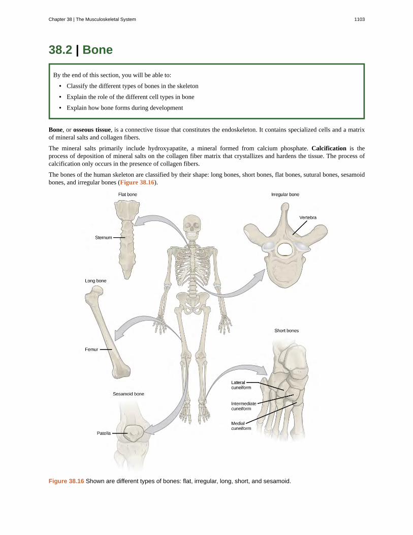
The bones of the human skeleton are classified by their shape: long bones, short bones, flat bones, sutural bones, sesamoidbones, and irregular bones (Figure 38.16).
Figure 38.16 Shown are different types of bones: flat, irregular, long, short, and sesamoid.
Chapter 38 | The Musculoskeletal System 1103
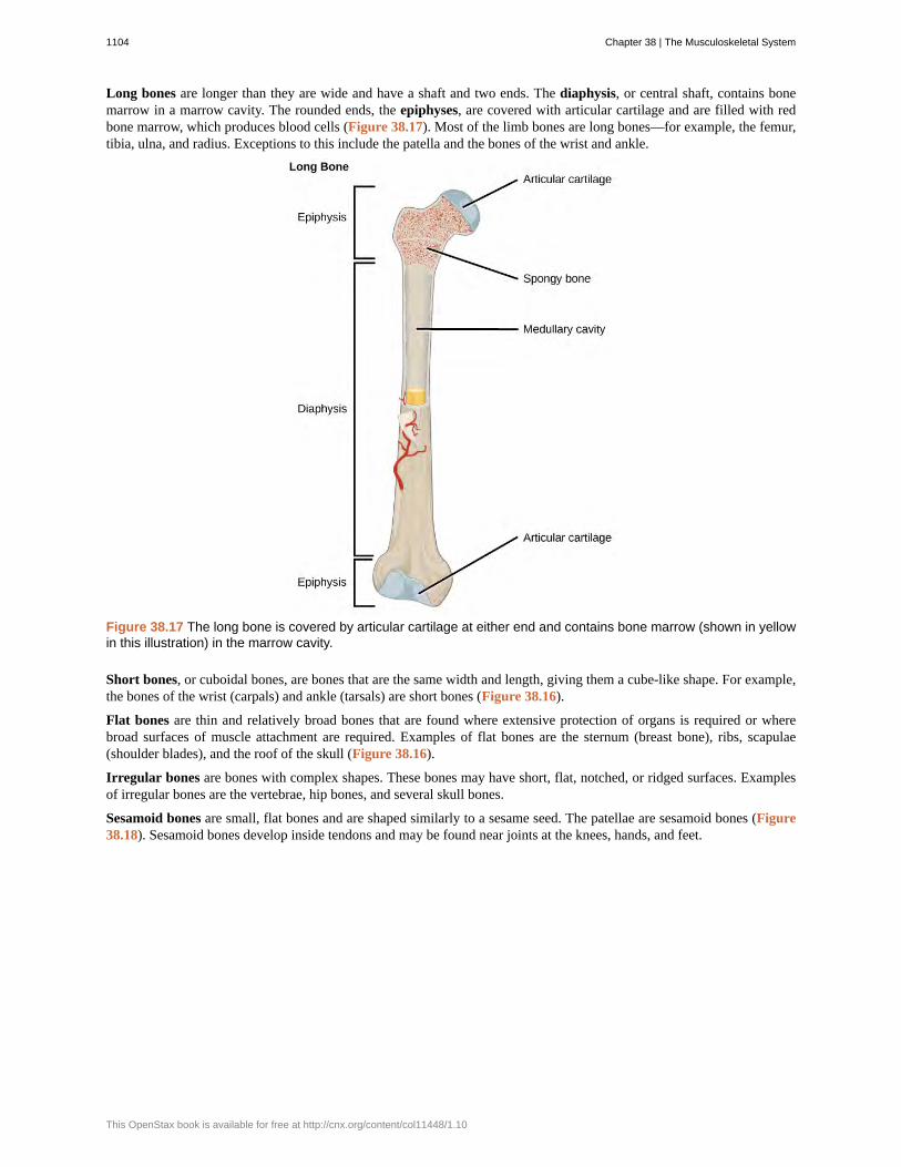
Long bones are longer than they are wide and have a shaft and two ends. The diaphysis, or central shaft, contains bonemarrow in a marrow cavity. The rounded ends, the epiphyses, are covered with articular cartilage and are filled with redbone marrow, which produces blood cells (Figure 38.17). Most of the limb bones are long bones—for example, the femur,tibia, ulna, and radius. Exceptions to this include the patella and the bones of the wrist and ankle.
Figure 38.17 The long bone is covered by articular cartilage at either end and contains bone marrow (shown in yellowin this illustration) in the marrow cavity.
Short bones, or cuboidal bones, are bones that are the same width and length, giving them a cube-like shape. For example,the bones of the wrist (carpals) and ankle (tarsals) are short bones (Figure 38.16).
Flat bones are thin and relatively broad bones that are found where extensive protection of organs is required or wherebroad surfaces of muscle attachment are required. Examples of flat bones are the sternum (breast bone), ribs, scapulae(shoulder blades), and the roof of the skull (Figure 38.16).
Irregular bones are bones with complex shapes. These bones may have short, flat, notched, or ridged surfaces. Examplesof irregular bones are the vertebrae, hip bones, and several skull bones.
Sesamoid bones are small, flat bones and are shaped similarly to a sesame seed. The patellae are sesamoid bones (Figure38.18). Sesamoid bones develop inside tendons and may be found near joints at the knees, hands, and feet.
1104 Chapter 38 | The Musculoskeletal System
This OpenStax book is available for free at http://cnx.org/content/col11448/1.10
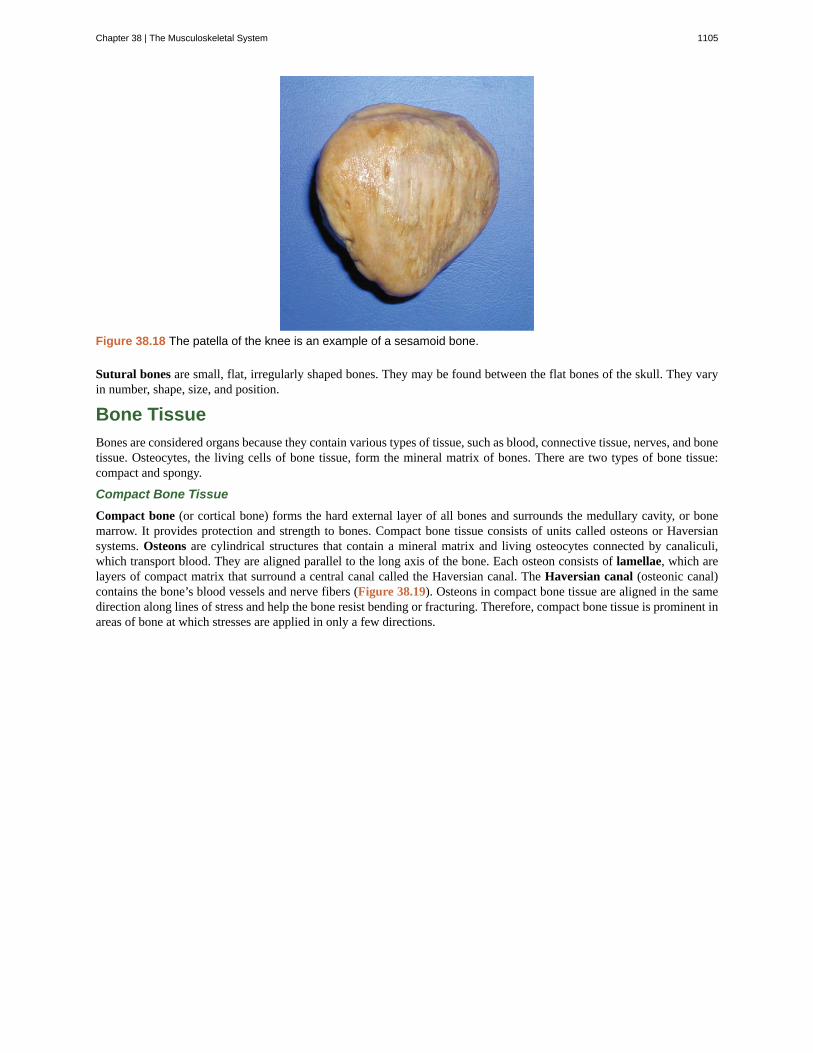
Figure 38.18 The patella of the knee is an example of a sesamoid bone.
Sutural bones are small, flat, irregularly shaped bones. They may be found between the flat bones of the skull. They varyin number, shape, size, and position.
Bone Tissue
Bones are considered organs because they contain various types of tissue, such as blood, connective tissue, nerves, and bonetissue. Osteocytes, the living cells of bone tissue, form the mineral matrix of bones. There are two types of bone tissue:compact and spongy.
Compact Bone Tissue
Compact bone (or cortical bone) forms the hard external layer of all bones and surrounds the medullary cavity, or bonemarrow. It provides protection and strength to bones. Compact bone tissue consists of units called osteons or Haversiansystems. Osteons are cylindrical structures that contain a mineral matrix and living osteocytes connected by canaliculi,which transport blood. They are aligned parallel to the long axis of the bone. Each osteon consists of lamellae, which arelayers of compact matrix that surround a central canal called the Haversian canal. The Haversian canal (osteonic canal)contains the bone’s blood vessels and nerve fibers (Figure 38.19). Osteons in compact bone tissue are aligned in the samedirection along lines of stress and help the bone resist bending or fracturing. Therefore, compact bone tissue is prominent inareas of bone at which stresses are applied in only a few directions.
Chapter 38 | The Musculoskeletal System 1105
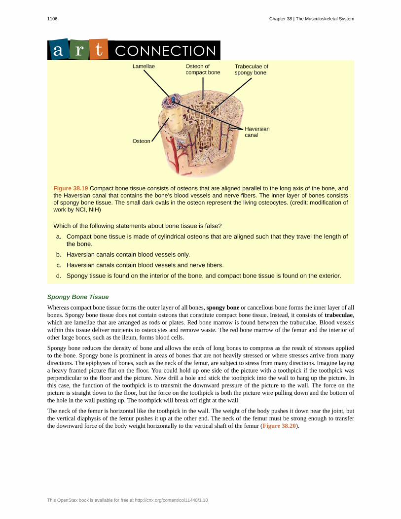
Figure 38.19 Compact bone tissue consists of osteons that are aligned parallel to the long axis of the bone, andthe Haversian canal that contains the bone’s blood vessels and nerve fibers. The inner layer of bones consistsof spongy bone tissue. The small dark ovals in the osteon represent the living osteocytes. (credit: modification ofwork by NCI, NIH)
Which of the following statements about bone tissue is false?
a. Compact bone tissue is made of cylindrical osteons that are aligned such that they travel the length ofthe bone.
b. Haversian canals contain blood vessels only.
c. Haversian canals contain blood vessels and nerve fibers.
d. Spongy tissue is found on the interior of the bone, and compact bone tissue is found on the exterior.
Spongy Bone Tissue
Whereas compact bone tissue forms the outer layer of all bones, spongy bone or cancellous bone forms the inner layer of allbones. Spongy bone tissue does not contain osteons that constitute compact bone tissue. Instead, it consists of trabeculae,which are lamellae that are arranged as rods or plates. Red bone marrow is found between the trabuculae. Blood vesselswithin this tissue deliver nutrients to osteocytes and remove waste. The red bone marrow of the femur and the interior ofother large bones, such as the ileum, forms blood cells.
Spongy bone reduces the density of bone and allows the ends of long bones to compress as the result of stresses appliedto the bone. Spongy bone is prominent in areas of bones that are not heavily stressed or where stresses arrive from manydirections. The epiphyses of bones, such as the neck of the femur, are subject to stress from many directions. Imagine layinga heavy framed picture flat on the floor. You could hold up one side of the picture with a toothpick if the toothpick wasperpendicular to the floor and the picture. Now drill a hole and stick the toothpick into the wall to hang up the picture. Inthis case, the function of the toothpick is to transmit the downward pressure of the picture to the wall. The force on thepicture is straight down to the floor, but the force on the toothpick is both the picture wire pulling down and the bottom ofthe hole in the wall pushing up. The toothpick will break off right at the wall.
The neck of the femur is horizontal like the toothpick in the wall. The weight of the body pushes it down near the joint, butthe vertical diaphysis of the femur pushes it up at the other end. The neck of the femur must be strong enough to transferthe downward force of the body weight horizontally to the vertical shaft of the femur (Figure 38.20).
1106 Chapter 38 | The Musculoskeletal System
This OpenStax book is available for free at http://cnx.org/content/col11448/1.10
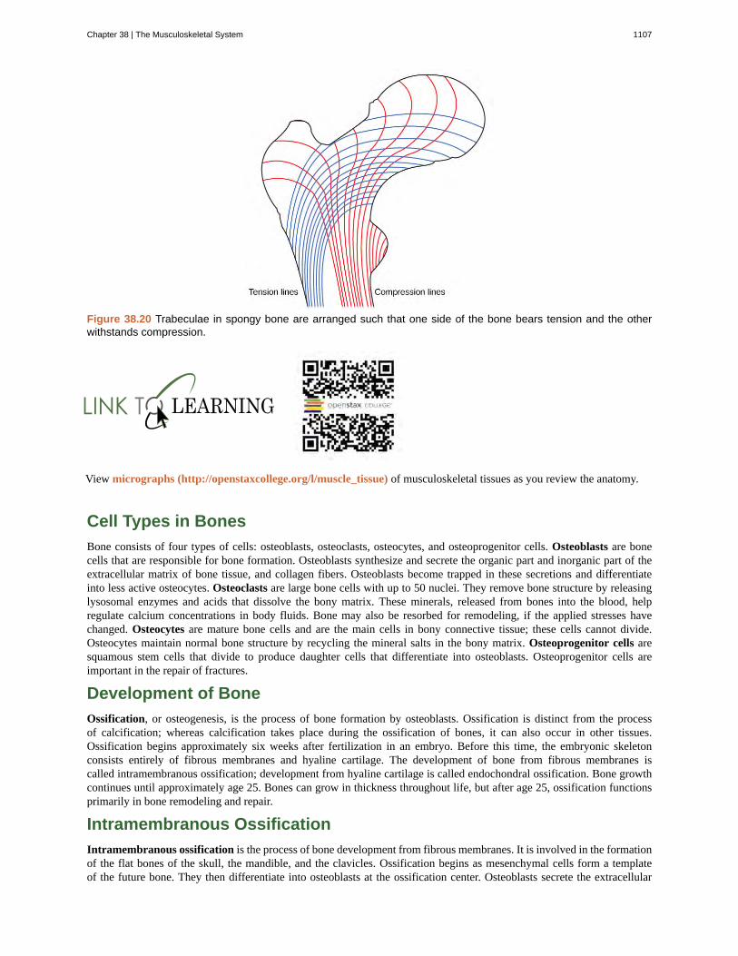
Figure 38.20 Trabeculae in spongy bone are arranged such that one side of the bone bears tension and the otherwithstands compression.
View micrographs (http://openstaxcollege.org/l/muscle_tissue) of musculoskeletal tissues as you review the anatomy.
Cell Types in Bones
Bone consists of four types of cells: osteoblasts, osteoclasts, osteocytes, and osteoprogenitor cells. Osteoblasts are bonecells that are responsible for bone formation. Osteoblasts synthesize and secrete the organic part and inorganic part of theextracellular matrix of bone tissue, and collagen fibers. Osteoblasts become trapped in these secretions and differentiateinto less active osteocytes. Osteoclasts are large bone cells with up to 50 nuclei. They remove bone structure by releasinglysosomal enzymes and acids that dissolve the bony matrix. These minerals, released from bones into the blood, helpregulate calcium concentrations in body fluids. Bone may also be resorbed for remodeling, if the applied stresses havechanged. Osteocytes are mature bone cells and are the main cells in bony connective tissue; these cells cannot divide.Osteocytes maintain normal bone structure by recycling the mineral salts in the bony matrix. Osteoprogenitor cells aresquamous stem cells that divide to produce daughter cells that differentiate into osteoblasts. Osteoprogenitor cells areimportant in the repair of fractures.
Development of Bone
Ossification, or osteogenesis, is the process of bone formation by osteoblasts. Ossification is distinct from the processof calcification; whereas calcification takes place during the ossification of bones, it can also occur in other tissues.Ossification begins approximately six weeks after fertilization in an embryo. Before this time, the embryonic skeletonconsists entirely of fibrous membranes and hyaline cartilage. The development of bone from fibrous membranes iscalled intramembranous ossification; development from hyaline cartilage is called endochondral ossification. Bone growthcontinues until approximately age 25. Bones can grow in thickness throughout life, but after age 25, ossification functionsprimarily in bone remodeling and repair.
Intramembranous Ossification
Intramembranous ossification is the process of bone development from fibrous membranes. It is involved in the formationof the flat bones of the skull, the mandible, and the clavicles. Ossification begins as mesenchymal cells form a templateof the future bone. They then differentiate into osteoblasts at the ossification center. Osteoblasts secrete the extracellular
Chapter 38 | The Musculoskeletal System 1107
matrix and deposit calcium, which hardens the matrix. The non-mineralized portion of the bone or osteoid continues to formaround blood vessels, forming spongy bone. Connective tissue in the matrix differentiates into red bone marrow in the fetus.The spongy bone is remodeled into a thin layer of compact bone on the surface of the spongy bone.
Endochondral Ossification
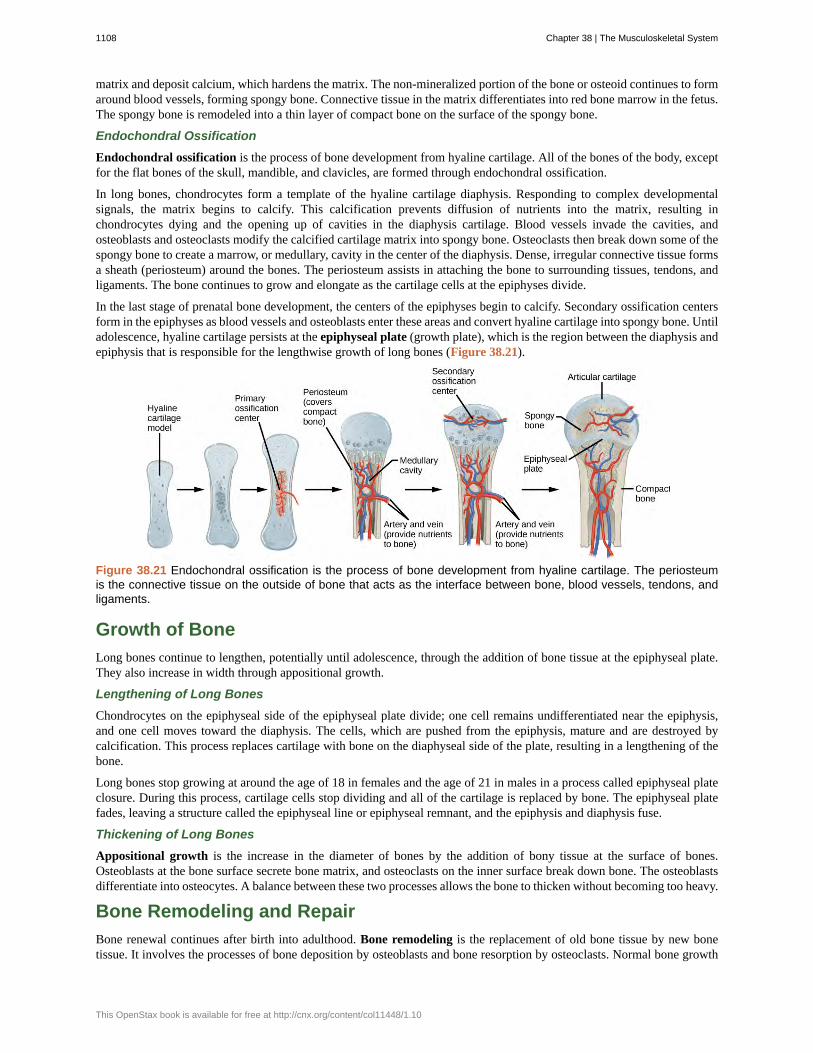
Endochondral ossification is the process of bone development from hyaline cartilage. All of the bones of the body, exceptfor the flat bones of the skull, mandible, and clavicles, are formed through endochondral ossification.
In long bones, chondrocytes form a template of the hyaline cartilage diaphysis. Responding to complex developmentalsignals, the matrix begins to calcify. This calcification prevents diffusion of nutrients into the matrix, resulting inchondrocytes dying and the opening up of cavities in the diaphysis cartilage. Blood vessels invade the cavities, andosteoblasts and osteoclasts modify the calcified cartilage matrix into spongy bone. Osteoclasts then break down some of thespongy bone to create a marrow, or medullary, cavity in the center of the diaphysis. Dense, irregular connective tissue formsa sheath (periosteum) around the bones. The periosteum assists in attaching the bone to surrounding tissues, tendons, andligaments. The bone continues to grow and elongate as the cartilage cells at the epiphyses divide.
In the last stage of prenatal bone development, the centers of the epiphyses begin to calcify. Secondary ossification centersform in the epiphyses as blood vessels and osteoblasts enter these areas and convert hyaline cartilage into spongy bone. Untiladolescence, hyaline cartilage persists at the epiphyseal plate (growth plate), which is the region between the diaphysis andepiphysis that is responsible for the lengthwise growth of long bones (Figure 38.21).
Figure 38.21 Endochondral ossification is the process of bone development from hyaline cartilage. The periosteumis the connective tissue on the outside of bone that acts as the interface between bone, blood vessels, tendons, andligaments.
Growth of Bone
Long bones continue to lengthen, potentially until adolescence, through the addition of bone tissue at the epiphyseal plate.They also increase in width through appositional growth.
Lengthening of Long Bones
Chondrocytes on the epiphyseal side of the epiphyseal plate divide; one cell remains undifferentiated near the epiphysis,and one cell moves toward the diaphysis. The cells, which are pushed from the epiphysis, mature and are destroyed bycalcification. This process replaces cartilage with bone on the diaphyseal side of the plate, resulting in a lengthening of thebone.
Long bones stop growing at around the age of 18 in females and the age of 21 in males in a process called epiphyseal plateclosure. During this process, cartilage cells stop dividing and all of the cartilage is replaced by bone. The epiphyseal platefades, leaving a structure called the epiphyseal line or epiphyseal remnant, and the epiphysis and diaphysis fuse.
Thickening of Long Bones
Appositional growth is the increase in the diameter of bones by the addition of bony tissue at the surface of bones.Osteoblasts at the bone surface secrete bone matrix, and osteoclasts on the inner surface break down bone. The osteoblastsdifferentiate into osteocytes. A balance between these two processes allows the bone to thicken without becoming too heavy.
Bone Remodeling and Repair
Bone renewal continues after birth into adulthood. Bone remodeling is the replacement of old bone tissue by new bonetissue. It involves the processes of bone deposition by osteoblasts and bone resorption by osteoclasts. Normal bone growth
1108 Chapter 38 | The Musculoskeletal System
This OpenStax book is available for free at http://cnx.org/content/col11448/1.10
requires vitamins D, C, and A, plus minerals such as calcium, phosphorous, and magnesium. Hormones such as parathyroidhormone, growth hormone, and calcitonin are also required for proper bone growth and maintenance.
Bone turnover rates are quite high, with five to seven percent of bone mass being recycled every week. Differences inturnover rate exist in different areas of the skeleton and in different areas of a bone. For example, the bone in the head ofthe femur may be fully replaced every six months, whereas the bone along the shaft is altered much more slowly.
Bone remodeling allows bones to adapt to stresses by becoming thicker and stronger when subjected to stress. Bones thatare not subject to normal stress, for example when a limb is in a cast, will begin to lose mass. A fractured or broken boneundergoes repair through four stages:
1. Blood vessels in the broken bone tear and hemorrhage, resulting in the formation of clotted blood, or a hematoma, atthe site of the break. The severed blood vessels at the broken ends of the bone are sealed by the clotting process, andbone cells that are deprived of nutrients begin to die.
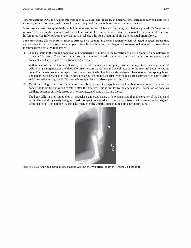
2. Within days of the fracture, capillaries grow into the hematoma, and phagocytic cells begin to clear away the deadcells. Though fragments of the blood clot may remain, fibroblasts and osteoblasts enter the area and begin to reformbone. Fibroblasts produce collagen fibers that connect the broken bone ends, and osteoblasts start to form spongy bone.The repair tissue between the broken bone ends is called the fibrocartilaginous callus, as it is composed of both hyalineand fibrocartilage (Figure 38.22). Some bone spicules may also appear at this point.
3. The fibrocartilaginous callus is converted into a bony callus of spongy bone. It takes about two months for the brokenbone ends to be firmly joined together after the fracture. This is similar to the endochondral formation of bone, ascartilage becomes ossified; osteoblasts, osteoclasts, and bone matrix are present.
4. The bony callus is then remodelled by osteoclasts and osteoblasts, with excess material on the exterior of the bone andwithin the medullary cavity being removed. Compact bone is added to create bone tissue that is similar to the original,unbroken bone. This remodeling can take many months, and the bone may remain uneven for years.
Figure 38.22 After this bone is set, a callus will knit the two ends together. (credit: Bill Rhodes)
Chapter 38 | The Musculoskeletal System 1109
Decalcification of BonesQuestion: What effect does the removal of calcium and collagen have on bone structure?
Background: Conduct a literature search on the role of calcium and collagen in maintaining bone structure.Conduct a literature search on diseases in which bone structure is compromised.
Hypothesis: Develop a hypothesis that states predictions of the flexibility, strength, and mass of bones thathave had the calcium and collagen components removed. Develop a hypothesis regarding the attempt toadd calcium back to decalcified bones.
Test the hypothesis: Test the prediction by removing calcium from chicken bones by placing them in a jarof vinegar for seven days. Test the hypothesis regarding adding calcium back to decalcified bone by placingthe decalcified chicken bones into a jar of water with calcium supplements added. Test the prediction bydenaturing the collagen from the bones by baking them at 250°C for three hours.
Analyze the data: Create a table showing the changes in bone flexibility, strength, and mass in the threedifferent environments.
Report the results: Under which conditions was the bone most flexible? Under which conditions was thebone the strongest?
Draw a conclusion: Did the results support or refute the hypothesis? How do the results observed in thisexperiment correspond to diseases that destroy bone tissue?
38.3 | Joints and Skeletal Movement
By the end of this section, you will be able to:
• Classify the different types of joints on the basis of structure
• Explain the role of joints in skeletal movement
The point at which two or more bones meet is called a joint, or articulation. Joints are responsible for movement, such asthe movement of limbs, and stability, such as the stability found in the bones of the skull.
Classification of Joints on the Basis of Structure
There are two ways to classify joints: on the basis of their structure or on the basis of their function. The structuralclassification divides joints into bony, fibrous, cartilaginous, and synovial joints depending on the material composing thejoint and the presence or absence of a cavity in the joint.
Fibrous Joints
The bones of fibrous joints are held together by fibrous connective tissue. There is no cavity, or space, present betweenthe bones and so most fibrous joints do not move at all, or are only capable of minor movements. There are three typesof fibrous joints: sutures, syndesmoses, and gomphoses. Sutures are found only in the skull and possess short fibers ofconnective tissue that hold the skull bones tightly in place (Figure 38.23).
1110 Chapter 38 | The Musculoskeletal System
This OpenStax book is available for free at http://cnx.org/content/col11448/1.10
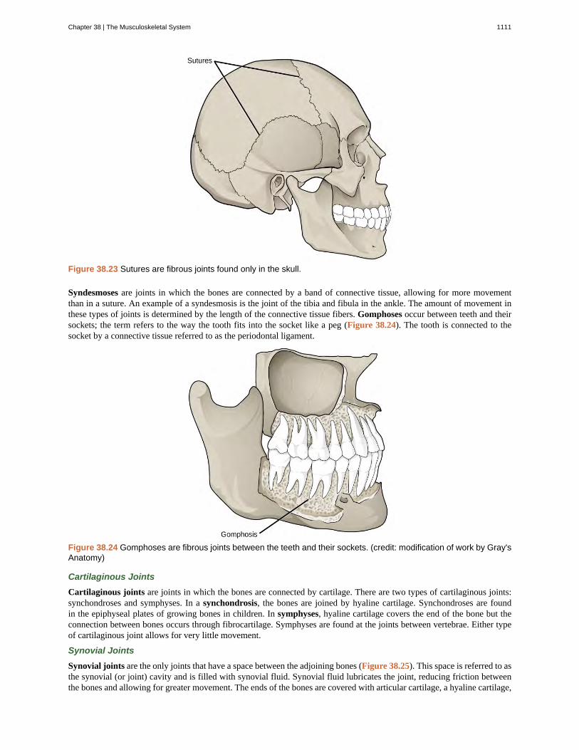
Figure 38.23 Sutures are fibrous joints found only in the skull.
Syndesmoses are joints in which the bones are connected by a band of connective tissue, allowing for more movementthan in a suture. An example of a syndesmosis is the joint of the tibia and fibula in the ankle. The amount of movement inthese types of joints is determined by the length of the connective tissue fibers. Gomphoses occur between teeth and theirsockets; the term refers to the way the tooth fits into the socket like a peg (Figure 38.24). The tooth is connected to thesocket by a connective tissue referred to as the periodontal ligament.
Figure 38.24 Gomphoses are fibrous joints between the teeth and their sockets. (credit: modification of work by Gray'sAnatomy)
Cartilaginous Joints
Cartilaginous joints are joints in which the bones are connected by cartilage. There are two types of cartilaginous joints:synchondroses and symphyses. In a synchondrosis, the bones are joined by hyaline cartilage. Synchondroses are foundin the epiphyseal plates of growing bones in children. In symphyses, hyaline cartilage covers the end of the bone but theconnection between bones occurs through fibrocartilage. Symphyses are found at the joints between vertebrae. Either typeof cartilaginous joint allows for very little movement.
Synovial Joints
Synovial joints are the only joints that have a space between the adjoining bones (Figure 38.25). This space is referred to asthe synovial (or joint) cavity and is filled with synovial fluid. Synovial fluid lubricates the joint, reducing friction betweenthe bones and allowing for greater movement. The ends of the bones are covered with articular cartilage, a hyaline cartilage,
Chapter 38 | The Musculoskeletal System 1111
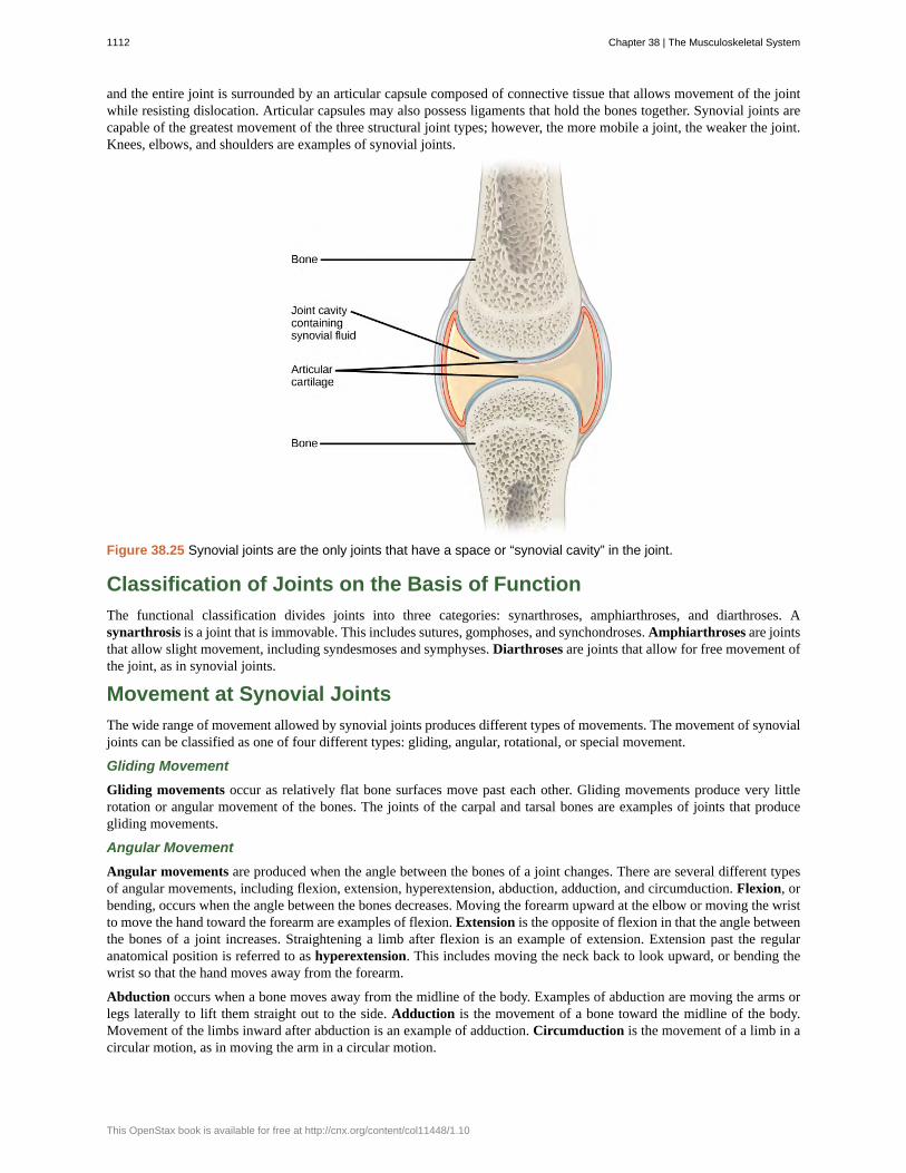
and the entire joint is surrounded by an articular capsule composed of connective tissue that allows movement of the jointwhile resisting dislocation. Articular capsules may also possess ligaments that hold the bones together. Synovial joints arecapable of the greatest movement of the three structural joint types; however, the more mobile a joint, the weaker the joint.Knees, elbows, and shoulders are examples of synovial joints.
Figure 38.25 Synovial joints are the only joints that have a space or “synovial cavity” in the joint.
Classification of Joints on the Basis of Function
The functional classification divides joints into three categories: synarthroses, amphiarthroses, and diarthroses. Asynarthrosis is a joint that is immovable. This includes sutures, gomphoses, and synchondroses. Amphiarthroses are jointsthat allow slight movement, including syndesmoses and symphyses. Diarthroses are joints that allow for free movement ofthe joint, as in synovial joints.
Movement at Synovial Joints
The wide range of movement allowed by synovial joints produces different types of movements. The movement of synovialjoints can be classified as one of four different types: gliding, angular, rotational, or special movement.
Gliding Movement
Gliding movements occur as relatively flat bone surfaces move past each other. Gliding movements produce very littlerotation or angular movement of the bones. The joints of the carpal and tarsal bones are examples of joints that producegliding movements.
Angular Movement
Angular movements are produced when the angle between the bones of a joint changes. There are several different typesof angular movements, including flexion, extension, hyperextension, abduction, adduction, and circumduction. Flexion, orbending, occurs when the angle between the bones decreases. Moving the forearm upward at the elbow or moving the wristto move the hand toward the forearm are examples of flexion. Extension is the opposite of flexion in that the angle betweenthe bones of a joint increases. Straightening a limb after flexion is an example of extension. Extension past the regularanatomical position is referred to as hyperextension. This includes moving the neck back to look upward, or bending thewrist so that the hand moves away from the forearm.
Abduction occurs when a bone moves away from the midline of the body. Examples of abduction are moving the arms orlegs laterally to lift them straight out to the side. Adduction is the movement of a bone toward the midline of the body.Movement of the limbs inward after abduction is an example of adduction. Circumduction is the movement of a limb in acircular motion, as in moving the arm in a circular motion.
1112 Chapter 38 | The Musculoskeletal System
This OpenStax book is available for free at http://cnx.org/content/col11448/1.10
Rotational Movement
Rotational movement is the movement of a bone as it rotates around its longitudinal axis. Rotation can be toward themidline of the body, which is referred to as medial rotation, or away from the midline of the body, which is referred to aslateral rotation. Movement of the head from side to side is an example of rotation.
Special Movements
Some movements that cannot be classified as gliding, angular, or rotational are called special movements. Inversioninvolves the soles of the feet moving inward, toward the midline of the body. Eversion is the opposite of inversion,movement of the sole of the foot outward, away from the midline of the body. Protraction is the anterior movement ofa bone in the horizontal plane. Retraction occurs as a joint moves back into position after protraction. Protraction andretraction can be seen in the movement of the mandible as the jaw is thrust outwards and then back inwards. Elevation isthe movement of a bone upward, such as when the shoulders are shrugged, lifting the scapulae. Depression is the oppositeof elevation—movement downward of a bone, such as after the shoulders are shrugged and the scapulae return to theirnormal position from an elevated position. Dorsiflexion is a bending at the ankle such that the toes are lifted toward theknee. Plantar flexion is a bending at the ankle when the heel is lifted, such as when standing on the toes. Supination is themovement of the radius and ulna bones of the forearm so that the palm faces forward. Pronation is the opposite movement,in which the palm faces backward. Opposition is the movement of the thumb toward the fingers of the same hand, makingit possible to grasp and hold objects.
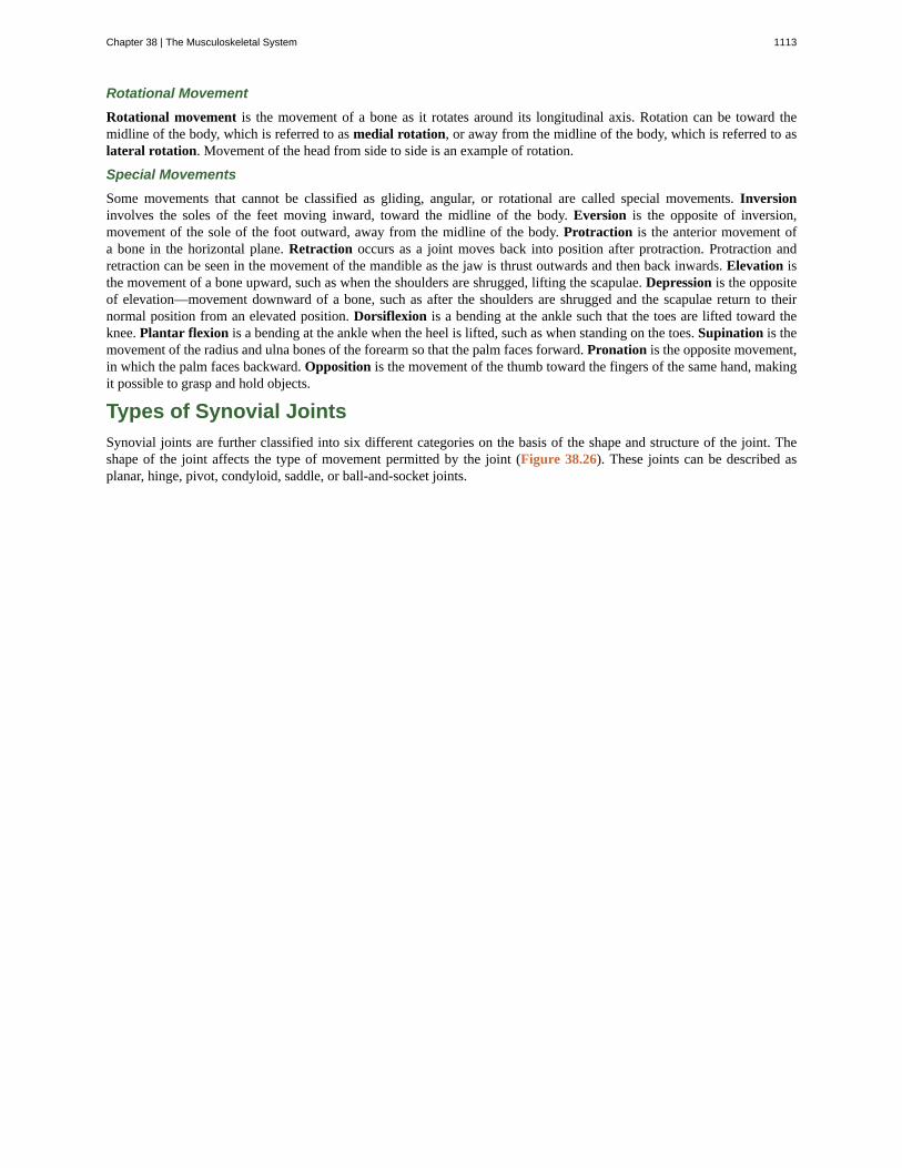
Types of Synovial Joints
Synovial joints are further classified into six different categories on the basis of the shape and structure of the joint. Theshape of the joint affects the type of movement permitted by the joint (Figure 38.26). These joints can be described asplanar, hinge, pivot, condyloid, saddle, or ball-and-socket joints.
Chapter 38 | The Musculoskeletal System 1113
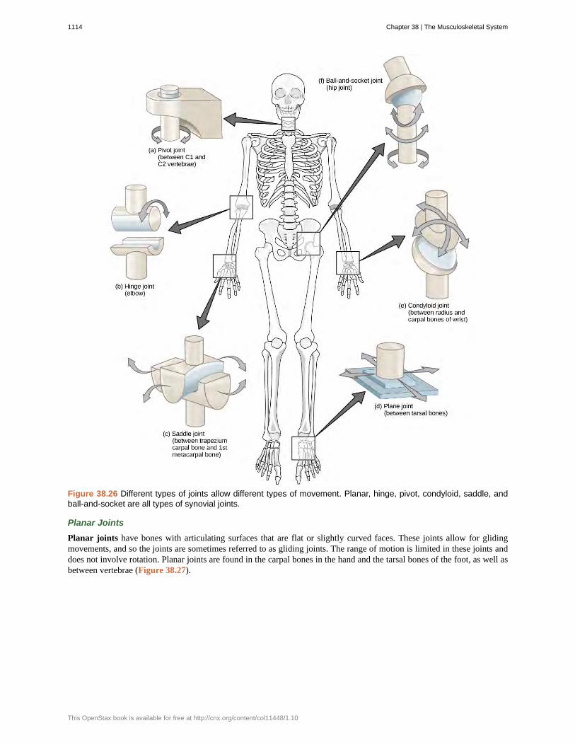
Figure 38.26 Different types of joints allow different types of movement. Planar, hinge, pivot, condyloid, saddle, andball-and-socket are all types of synovial joints.
Planar Joints
Planar joints have bones with articulating surfaces that are flat or slightly curved faces. These joints allow for glidingmovements, and so the joints are sometimes referred to as gliding joints. The range of motion is limited in these joints anddoes not involve rotation. Planar joints are found in the carpal bones in the hand and the tarsal bones of the foot, as well asbetween vertebrae (Figure 38.27).
1114 Chapter 38 | The Musculoskeletal System
This OpenStax book is available for free at http://cnx.org/content/col11448/1.10
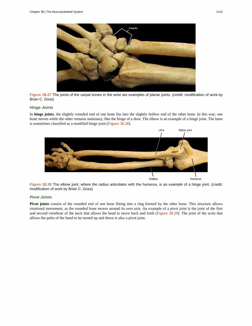
Figure 38.27 The joints of the carpal bones in the wrist are examples of planar joints. (credit: modification of work byBrian C. Goss)
Hinge Joints
In hinge joints, the slightly rounded end of one bone fits into the slightly hollow end of the other bone. In this way, onebone moves while the other remains stationary, like the hinge of a door. The elbow is an example of a hinge joint. The kneeis sometimes classified as a modified hinge joint (Figure 38.28).
Figure 38.28 The elbow joint, where the radius articulates with the humerus, is an example of a hinge joint. (credit:modification of work by Brian C. Goss)
Pivot Joints
Pivot joints consist of the rounded end of one bone fitting into a ring formed by the other bone. This structure allowsrotational movement, as the rounded bone moves around its own axis. An example of a pivot joint is the joint of the firstand second vertebrae of the neck that allows the head to move back and forth (Figure 38.29). The joint of the wrist thatallows the palm of the hand to be turned up and down is also a pivot joint.
Chapter 38 | The Musculoskeletal System 1115
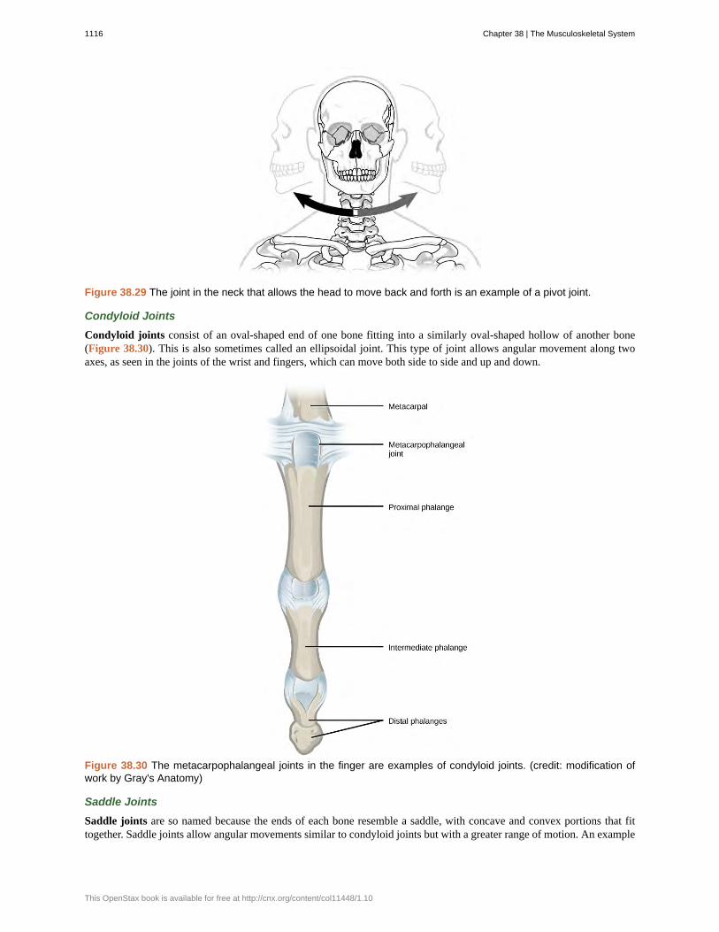
Figure 38.29 The joint in the neck that allows the head to move back and forth is an example of a pivot joint.
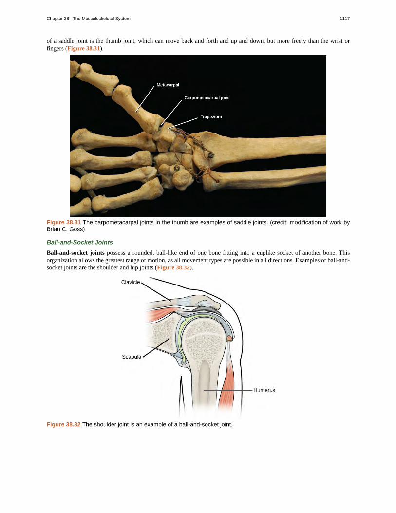
Condyloid Joints
Condyloid joints consist of an oval-shaped end of one bone fitting into a similarly oval-shaped hollow of another bone(Figure 38.30). This is also sometimes called an ellipsoidal joint. This type of joint allows angular movement along twoaxes, as seen in the joints of the wrist and fingers, which can move both side to side and up and down.
Figure 38.30 The metacarpophalangeal joints in the finger are examples of condyloid joints. (credit: modification ofwork by Gray's Anatomy)
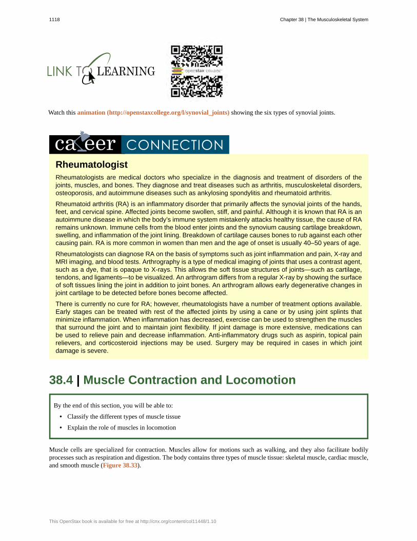
Saddle Joints
Saddle joints are so named because the ends of each bone resemble a saddle, with concave and convex portions that fittogether. Saddle joints allow angular movements similar to condyloid joints but with a greater range of motion. An example
1116 Chapter 38 | The Musculoskeletal System
This OpenStax book is available for free at http://cnx.org/content/col11448/1.10
of a saddle joint is the thumb joint, which can move back and forth and up and down, but more freely than the wrist orfingers (Figure 38.31).
Figure 38.31 The carpometacarpal joints in the thumb are examples of saddle joints. (credit: modification of work byBrian C. Goss)
Ball-and-Socket Joints
Ball-and-socket joints possess a rounded, ball-like end of one bone fitting into a cuplike socket of another bone. Thisorganization allows the greatest range of motion, as all movement types are possible in all directions. Examples of ball-and-socket joints are the shoulder and hip joints (Figure 38.32).
Figure 38.32 The shoulder joint is an example of a ball-and-socket joint.
Chapter 38 | The Musculoskeletal System 1117
Watch this animation (http://openstaxcollege.org/l/synovial_joints) showing the six types of synovial joints.
RheumatologistRheumatologists are medical doctors who specialize in the diagnosis and treatment of disorders of thejoints, muscles, and bones. They diagnose and treat diseases such as arthritis, musculoskeletal disorders,osteoporosis, and autoimmune diseases such as ankylosing spondylitis and rheumatoid arthritis.
Rheumatoid arthritis (RA) is an inflammatory disorder that primarily affects the synovial joints of the hands,feet, and cervical spine. Affected joints become swollen, stiff, and painful. Although it is known that RA is anautoimmune disease in which the body’s immune system mistakenly attacks healthy tissue, the cause of RAremains unknown. Immune cells from the blood enter joints and the synovium causing cartilage breakdown,swelling, and inflammation of the joint lining. Breakdown of cartilage causes bones to rub against each othercausing pain. RA is more common in women than men and the age of onset is usually 40–50 years of age.
Rheumatologists can diagnose RA on the basis of symptoms such as joint inflammation and pain, X-ray andMRI imaging, and blood tests. Arthrography is a type of medical imaging of joints that uses a contrast agent,such as a dye, that is opaque to X-rays. This allows the soft tissue structures of joints—such as cartilage,tendons, and ligaments—to be visualized. An arthrogram differs from a regular X-ray by showing the surfaceof soft tissues lining the joint in addition to joint bones. An arthrogram allows early degenerative changes injoint cartilage to be detected before bones become affected.
There is currently no cure for RA; however, rheumatologists have a number of treatment options available.Early stages can be treated with rest of the affected joints by using a cane or by using joint splints thatminimize inflammation. When inflammation has decreased, exercise can be used to strengthen the musclesthat surround the joint and to maintain joint flexibility. If joint damage is more extensive, medications canbe used to relieve pain and decrease inflammation. Anti-inflammatory drugs such as aspirin, topical painrelievers, and corticosteroid injections may be used. Surgery may be required in cases in which jointdamage is severe.
38.4 | Muscle Contraction and Locomotion
By the end of this section, you will be able to:
• Classify the different types of muscle tissue
• Explain the role of muscles in locomotion
Muscle cells are specialized for contraction. Muscles allow for motions such as walking, and they also facilitate bodilyprocesses such as respiration and digestion. The body contains three types of muscle tissue: skeletal muscle, cardiac muscle,and smooth muscle (Figure 38.33).
1118 Chapter 38 | The Musculoskeletal System
This OpenStax book is available for free at http://cnx.org/content/col11448/1.10
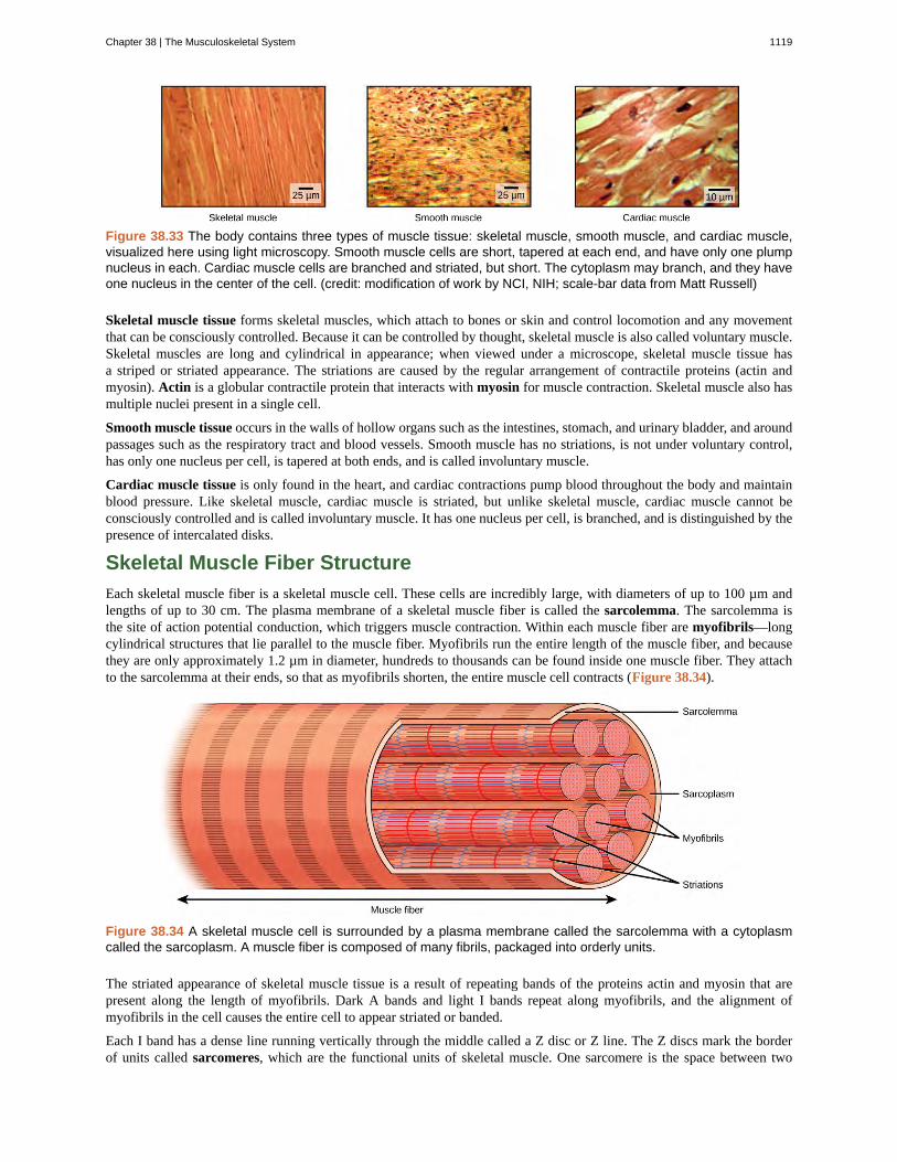
Figure 38.33 The body contains three types of muscle tissue: skeletal muscle, smooth muscle, and cardiac muscle,visualized here using light microscopy. Smooth muscle cells are short, tapered at each end, and have only one plumpnucleus in each. Cardiac muscle cells are branched and striated, but short. The cytoplasm may branch, and they haveone nucleus in the center of the cell. (credit: modification of work by NCI, NIH; scale-bar data from Matt Russell)
Skeletal muscle tissue forms skeletal muscles, which attach to bones or skin and control locomotion and any movementthat can be consciously controlled. Because it can be controlled by thought, skeletal muscle is also called voluntary muscle.Skeletal muscles are long and cylindrical in appearance; when viewed under a microscope, skeletal muscle tissue hasa striped or striated appearance. The striations are caused by the regular arrangement of contractile proteins (actin andmyosin). Actin is a globular contractile protein that interacts with myosin for muscle contraction. Skeletal muscle also hasmultiple nuclei present in a single cell.
Smooth muscle tissue occurs in the walls of hollow organs such as the intestines, stomach, and urinary bladder, and aroundpassages such as the respiratory tract and blood vessels. Smooth muscle has no striations, is not under voluntary control,has only one nucleus per cell, is tapered at both ends, and is called involuntary muscle.
Cardiac muscle tissue is only found in the heart, and cardiac contractions pump blood throughout the body and maintainblood pressure. Like skeletal muscle, cardiac muscle is striated, but unlike skeletal muscle, cardiac muscle cannot beconsciously controlled and is called involuntary muscle. It has one nucleus per cell, is branched, and is distinguished by thepresence of intercalated disks.
Skeletal Muscle Fiber Structure
Each skeletal muscle fiber is a skeletal muscle cell. These cells are incredibly large, with diameters of up to 100 µm andlengths of up to 30 cm. The plasma membrane of a skeletal muscle fiber is called the sarcolemma. The sarcolemma isthe site of action potential conduction, which triggers muscle contraction. Within each muscle fiber are myofibrils—longcylindrical structures that lie parallel to the muscle fiber. Myofibrils run the entire length of the muscle fiber, and becausethey are only approximately 1.2 µm in diameter, hundreds to thousands can be found inside one muscle fiber. They attachto the sarcolemma at their ends, so that as myofibrils shorten, the entire muscle cell contracts (Figure 38.34).
Figure 38.34 A skeletal muscle cell is surrounded by a plasma membrane called the sarcolemma with a cytoplasmcalled the sarcoplasm. A muscle fiber is composed of many fibrils, packaged into orderly units.
The striated appearance of skeletal muscle tissue is a result of repeating bands of the proteins actin and myosin that arepresent along the length of myofibrils. Dark A bands and light I bands repeat along myofibrils, and the alignment ofmyofibrils in the cell causes the entire cell to appear striated or banded.
Each I band has a dense line running vertically through the middle called a Z disc or Z line. The Z discs mark the borderof units called sarcomeres, which are the functional units of skeletal muscle. One sarcomere is the space between two
Chapter 38 | The Musculoskeletal System 1119
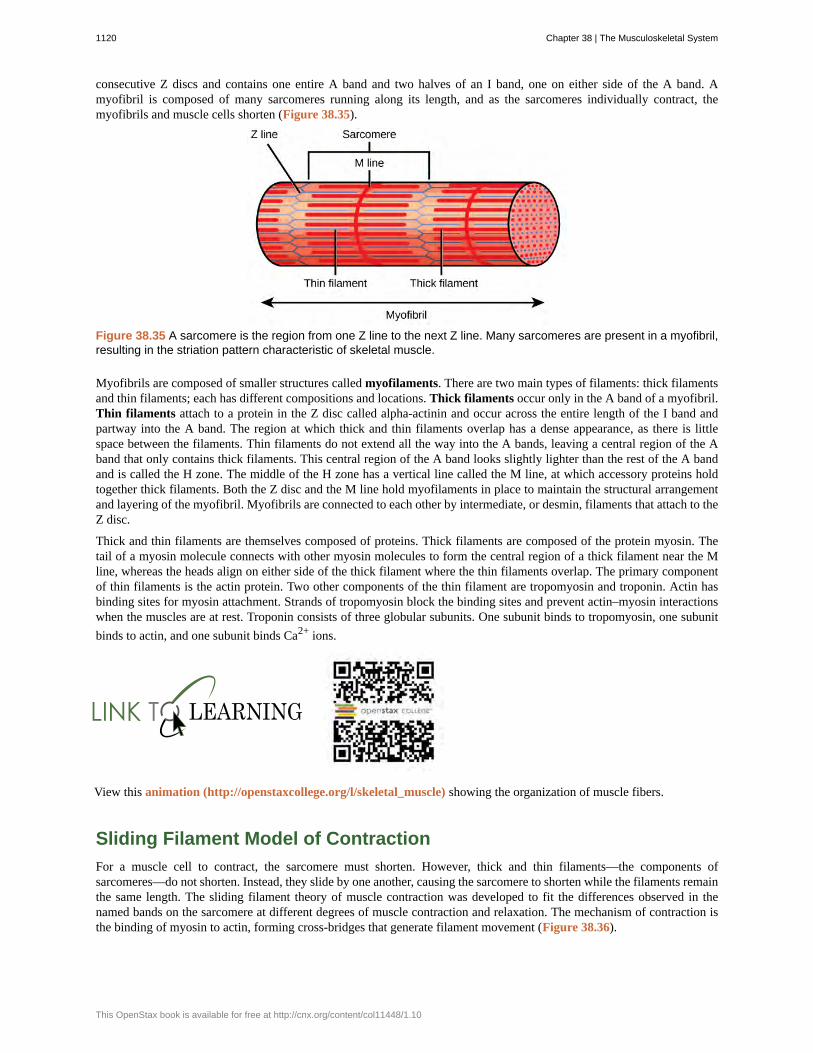
consecutive Z discs and contains one entire A band and two halves of an I band, one on either side of the A band. Amyofibril is composed of many sarcomeres running along its length, and as the sarcomeres individually contract, themyofibrils and muscle cells shorten (Figure 38.35).
Figure 38.35 A sarcomere is the region from one Z line to the next Z line. Many sarcomeres are present in a myofibril,resulting in the striation pattern characteristic of skeletal muscle.
Myofibrils are composed of smaller structures called myofilaments. There are two main types of filaments: thick filamentsand thin filaments; each has different compositions and locations. Thick filaments occur only in the A band of a myofibril.Thin filaments attach to a protein in the Z disc called alpha-actinin and occur across the entire length of the I band andpartway into the A band. The region at which thick and thin filaments overlap has a dense appearance, as there is littlespace between the filaments. Thin filaments do not extend all the way into the A bands, leaving a central region of the Aband that only contains thick filaments. This central region of the A band looks slightly lighter than the rest of the A bandand is called the H zone. The middle of the H zone has a vertical line called the M line, at which accessory proteins holdtogether thick filaments. Both the Z disc and the M line hold myofilaments in place to maintain the structural arrangementand layering of the myofibril. Myofibrils are connected to each other by intermediate, or desmin, filaments that attach to theZ disc.
Thick and thin filaments are themselves composed of proteins. Thick filaments are composed of the protein myosin. Thetail of a myosin molecule connects with other myosin molecules to form the central region of a thick filament near the Mline, whereas the heads align on either side of the thick filament where the thin filaments overlap. The primary componentof thin filaments is the actin protein. Two other components of the thin filament are tropomyosin and troponin. Actin hasbinding sites for myosin attachment. Strands of tropomyosin block the binding sites and prevent actin–myosin interactionswhen the muscles are at rest. Troponin consists of three globular subunits. One subunit binds to tropomyosin, one subunitbinds to actin, and one subunit binds Ca2+ ions.
View this animation (http://openstaxcollege.org/l/skeletal_muscle) showing the organization of muscle fibers.
Sliding Filament Model of Contraction
For a muscle cell to contract, the sarcomere must shorten. However, thick and thin filaments—the components ofsarcomeres—do not shorten. Instead, they slide by one another, causing the sarcomere to shorten while the filaments remainthe same length. The sliding filament theory of muscle contraction was developed to fit the differences observed in thenamed bands on the sarcomere at different degrees of muscle contraction and relaxation. The mechanism of contraction isthe binding of myosin to actin, forming cross-bridges that generate filament movement (Figure 38.36).
1120 Chapter 38 | The Musculoskeletal System
This OpenStax book is available for free at http://cnx.org/content/col11448/1.10
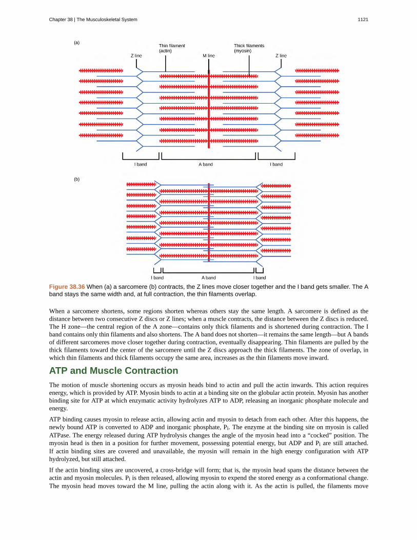
Figure 38.36 When (a) a sarcomere (b) contracts, the Z lines move closer together and the I band gets smaller. The Aband stays the same width and, at full contraction, the thin filaments overlap.
When a sarcomere shortens, some regions shorten whereas others stay the same length. A sarcomere is defined as thedistance between two consecutive Z discs or Z lines; when a muscle contracts, the distance between the Z discs is reduced.The H zone—the central region of the A zone—contains only thick filaments and is shortened during contraction. The Iband contains only thin filaments and also shortens. The A band does not shorten—it remains the same length—but A bandsof different sarcomeres move closer together during contraction, eventually disappearing. Thin filaments are pulled by thethick filaments toward the center of the sarcomere until the Z discs approach the thick filaments. The zone of overlap, inwhich thin filaments and thick filaments occupy the same area, increases as the thin filaments move inward.
ATP and Muscle Contraction
The motion of muscle shortening occurs as myosin heads bind to actin and pull the actin inwards. This action requiresenergy, which is provided by ATP. Myosin binds to actin at a binding site on the globular actin protein. Myosin has anotherbinding site for ATP at which enzymatic activity hydrolyzes ATP to ADP, releasing an inorganic phosphate molecule andenergy.
ATP binding causes myosin to release actin, allowing actin and myosin to detach from each other. After this happens, thenewly bound ATP is converted to ADP and inorganic phosphate, Pi. The enzyme at the binding site on myosin is calledATPase. The energy released during ATP hydrolysis changes the angle of the myosin head into a “cocked” position. Themyosin head is then in a position for further movement, possessing potential energy, but ADP and Pi are still attached.If actin binding sites are covered and unavailable, the myosin will remain in the high energy configuration with ATPhydrolyzed, but still attached.
If the actin binding sites are uncovered, a cross-bridge will form; that is, the myosin head spans the distance between theactin and myosin molecules. Pi is then released, allowing myosin to expend the stored energy as a conformational change.The myosin head moves toward the M line, pulling the actin along with it. As the actin is pulled, the filaments move
Chapter 38 | The Musculoskeletal System 1121
approximately 10 nm toward the M line. This movement is called the power stroke, as it is the step at which force isproduced. As the actin is pulled toward the M line, the sarcomere shortens and the muscle contracts.
When the myosin head is “cocked,” it contains energy and is in a high-energy configuration. This energy is expended as themyosin head moves through the power stroke; at the end of the power stroke, the myosin head is in a low-energy position.After the power stroke, ADP is released; however, the cross-bridge formed is still in place, and actin and myosin are boundtogether. ATP can then attach to myosin, which allows the cross-bridge cycle to start again and further muscle contractioncan occur (Figure 38.37).
Watch this video (http://openstaxcollege.org/l/contract_muscle) explaining how a muscle contraction is signaled.
1122 Chapter 38 | The Musculoskeletal System
This OpenStax book is available for free at http://cnx.org/content/col11448/1.10
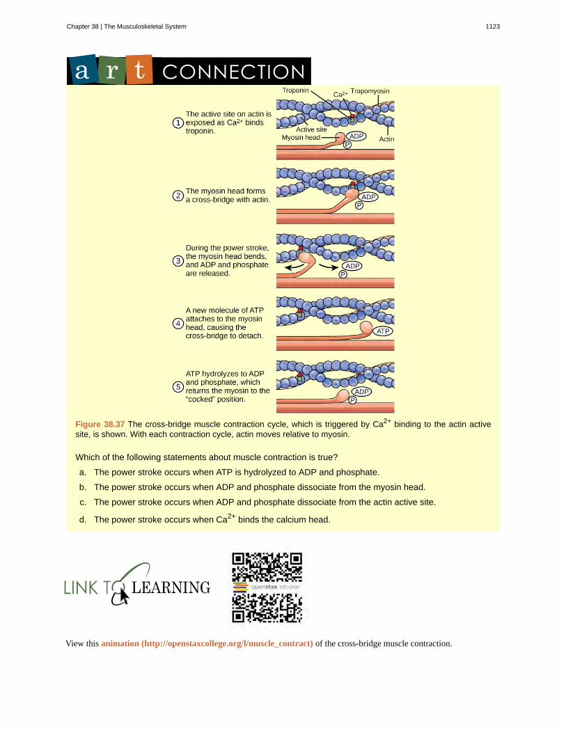
Figure 38.37 The cross-bridge muscle contraction cycle, which is triggered by Ca2+ binding to the actin activesite, is shown. With each contraction cycle, actin moves relative to myosin.
Which of the following statements about muscle contraction is true?
a. The power stroke occurs when ATP is hydrolyzed to ADP and phosphate.
b. The power stroke occurs when ADP and phosphate dissociate from the myosin head.
c. The power stroke occurs when ADP and phosphate dissociate from the actin active site.
d. The power stroke occurs when Ca2+ binds the calcium head.
View this animation (http://openstaxcollege.org/l/muscle_contract) of the cross-bridge muscle contraction.
Chapter 38 | The Musculoskeletal System 1123
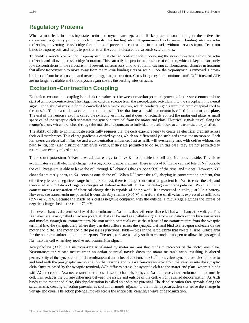
Regulatory Proteins
When a muscle is in a resting state, actin and myosin are separated. To keep actin from binding to the active siteon myosin, regulatory proteins block the molecular binding sites. Tropomyosin blocks myosin binding sites on actinmolecules, preventing cross-bridge formation and preventing contraction in a muscle without nervous input. Troponinbinds to tropomyosin and helps to position it on the actin molecule; it also binds calcium ions.
To enable a muscle contraction, tropomyosin must change conformation, uncovering the myosin-binding site on an actinmolecule and allowing cross-bridge formation. This can only happen in the presence of calcium, which is kept at extremelylow concentrations in the sarcoplasm. If present, calcium ions bind to troponin, causing conformational changes in troponinthat allow tropomyosin to move away from the myosin binding sites on actin. Once the tropomyosin is removed, a cross-bridge can form between actin and myosin, triggering contraction. Cross-bridge cycling continues until Ca2+ ions and ATPare no longer available and tropomyosin again covers the binding sites on actin.
Excitation–Contraction Coupling
Excitation–contraction coupling is the link (transduction) between the action potential generated in the sarcolemma and thestart of a muscle contraction. The trigger for calcium release from the sarcoplasmic reticulum into the sarcoplasm is a neuralsignal. Each skeletal muscle fiber is controlled by a motor neuron, which conducts signals from the brain or spinal cord tothe muscle. The area of the sarcolemma on the muscle fiber that interacts with the neuron is called the motor end plate.The end of the neuron’s axon is called the synaptic terminal, and it does not actually contact the motor end plate. A smallspace called the synaptic cleft separates the synaptic terminal from the motor end plate. Electrical signals travel along theneuron’s axon, which branches through the muscle and connects to individual muscle fibers at a neuromuscular junction.
The ability of cells to communicate electrically requires that the cells expend energy to create an electrical gradient acrosstheir cell membranes. This charge gradient is carried by ions, which are differentially distributed across the membrane. Eachion exerts an electrical influence and a concentration influence. Just as milk will eventually mix with coffee without theneed to stir, ions also distribute themselves evenly, if they are permitted to do so. In this case, they are not permitted toreturn to an evenly mixed state.
The sodium–potassium ATPase uses cellular energy to move K+ ions inside the cell and Na+ ions outside. This aloneaccumulates a small electrical charge, but a big concentration gradient. There is lots of K+ in the cell and lots of Na+ outsidethe cell. Potassium is able to leave the cell through K+ channels that are open 90% of the time, and it does. However, Na+
channels are rarely open, so Na+ remains outside the cell. When K+ leaves the cell, obeying its concentration gradient, thateffectively leaves a negative charge behind. So at rest, there is a large concentration gradient for Na+ to enter the cell, andthere is an accumulation of negative charges left behind in the cell. This is the resting membrane potential. Potential in thiscontext means a separation of electrical charge that is capable of doing work. It is measured in volts, just like a battery.However, the transmembrane potential is considerably smaller (0.07 V); therefore, the small value is expressed as millivolts(mV) or 70 mV. Because the inside of a cell is negative compared with the outside, a minus sign signifies the excess ofnegative charges inside the cell, −70 mV.
If an event changes the permeability of the membrane to Na+ ions, they will enter the cell. That will change the voltage. Thisis an electrical event, called an action potential, that can be used as a cellular signal. Communication occurs between nervesand muscles through neurotransmitters. Neuron action potentials cause the release of neurotransmitters from the synapticterminal into the synaptic cleft, where they can then diffuse across the synaptic cleft and bind to a receptor molecule on themotor end plate. The motor end plate possesses junctional folds—folds in the sarcolemma that create a large surface areafor the neurotransmitter to bind to receptors. The receptors are actually sodium channels that open to allow the passage ofNa+ into the cell when they receive neurotransmitter signal.
Acetylcholine (ACh) is a neurotransmitter released by motor neurons that binds to receptors in the motor end plate.Neurotransmitter release occurs when an action potential travels down the motor neuron’s axon, resulting in alteredpermeability of the synaptic terminal membrane and an influx of calcium. The Ca2+ ions allow synaptic vesicles to move toand bind with the presynaptic membrane (on the neuron), and release neurotransmitter from the vesicles into the synapticcleft. Once released by the synaptic terminal, ACh diffuses across the synaptic cleft to the motor end plate, where it bindswith ACh receptors. As a neurotransmitter binds, these ion channels open, and Na+ ions cross the membrane into the musclecell. This reduces the voltage difference between the inside and outside of the cell, which is called depolarization. As AChbinds at the motor end plate, this depolarization is called an end-plate potential. The depolarization then spreads along thesarcolemma, creating an action potential as sodium channels adjacent to the initial depolarization site sense the change involtage and open. The action potential moves across the entire cell, creating a wave of depolarization.
1124 Chapter 38 | The Musculoskeletal System
This OpenStax book is available for free at http://cnx.org/content/col11448/1.10
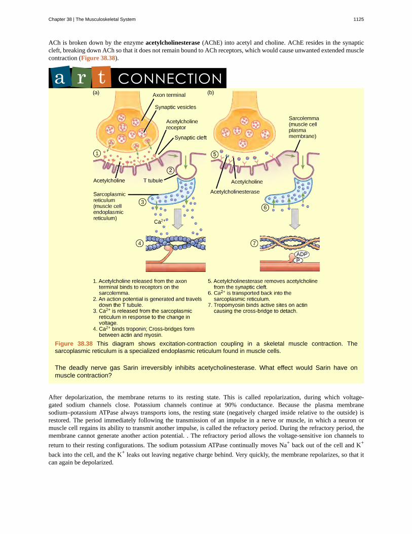
ACh is broken down by the enzyme acetylcholinesterase (AChE) into acetyl and choline. AChE resides in the synapticcleft, breaking down ACh so that it does not remain bound to ACh receptors, which would cause unwanted extended musclecontraction (Figure 38.38).
Figure 38.38 This diagram shows excitation-contraction coupling in a skeletal muscle contraction. Thesarcoplasmic reticulum is a specialized endoplasmic reticulum found in muscle cells.
The deadly nerve gas Sarin irreversibly inhibits acetycholinesterase. What effect would Sarin have onmuscle contraction?
After depolarization, the membrane returns to its resting state. This is called repolarization, during which voltage-gated sodium channels close. Potassium channels continue at 90% conductance. Because the plasma membranesodium–potassium ATPase always transports ions, the resting state (negatively charged inside relative to the outside) isrestored. The period immediately following the transmission of an impulse in a nerve or muscle, in which a neuron ormuscle cell regains its ability to transmit another impulse, is called the refractory period. During the refractory period, themembrane cannot generate another action potential. . The refractory period allows the voltage-sensitive ion channels toreturn to their resting configurations. The sodium potassium ATPase continually moves Na+ back out of the cell and K+
back into the cell, and the K+ leaks out leaving negative charge behind. Very quickly, the membrane repolarizes, so that itcan again be depolarized.
Chapter 38 | The Musculoskeletal System 1125
Control of Muscle Tension
Neural control initiates the formation of actin–myosin cross-bridges, leading to the sarcomere shortening involved in musclecontraction. These contractions extend from the muscle fiber through connective tissue to pull on bones, causing skeletalmovement. The pull exerted by a muscle is called tension, and the amount of force created by this tension can vary. Thisenables the same muscles to move very light objects and very heavy objects. In individual muscle fibers, the amount oftension produced depends on the cross-sectional area of the muscle fiber and the frequency of neural stimulation.
The number of cross-bridges formed between actin and myosin determine the amount of tension that a muscle fiber canproduce. Cross-bridges can only form where thick and thin filaments overlap, allowing myosin to bind to actin. If morecross-bridges are formed, more myosin will pull on actin, and more tension will be produced.
The ideal length of a sarcomere during production of maximal tension occurs when thick and thin filaments overlap tothe greatest degree. If a sarcomere at rest is stretched past an ideal resting length, thick and thin filaments do not overlapto the greatest degree, and fewer cross-bridges can form. This results in fewer myosin heads pulling on actin, and lesstension is produced. As a sarcomere is shortened, the zone of overlap is reduced as the thin filaments reach the H zone,which is composed of myosin tails. Because it is myosin heads that form cross-bridges, actin will not bind to myosin inthis zone, reducing the tension produced by this myofiber. If the sarcomere is shortened even more, thin filaments beginto overlap with each other—reducing cross-bridge formation even further, and producing even less tension. Conversely, ifthe sarcomere is stretched to the point at which thick and thin filaments do not overlap at all, no cross-bridges are formedand no tension is produced. This amount of stretching does not usually occur because accessory proteins, internal sensorynerves, and connective tissue oppose extreme stretching.
The primary variable determining force production is the number of myofibers within the muscle that receive an actionpotential from the neuron that controls that fiber. When using the biceps to pick up a pencil, the motor cortex of the brainonly signals a few neurons of the biceps, and only a few myofibers respond. In vertebrates, each myofiber responds fullyif stimulated. When picking up a piano, the motor cortex signals all of the neurons in the biceps and every myofiberparticipates. This is close to the maximum force the muscle can produce. As mentioned above, increasing the frequency ofaction potentials (the number of signals per second) can increase the force a bit more, because the tropomyosin is floodedwith calcium.
1126 Chapter 38 | The Musculoskeletal System
This OpenStax book is available for free at http://cnx.org/content/col11448/1.10
abduction
acetylcholinesterase
actin
adduction
amphiarthrosis
angular movement
appendicular skeleton
appositional growth
articulation
auditory ossicle
axial skeleton
ball-and-socket joint
bone
bone remodeling
calcification
cardiac muscle
carpus
cartilaginous joint
circumduction
clavicle
compact bone
condyloid joint
coxal bone
cranial bone
depression
diaphysis
diarthrosis
dorsiflexion
KEY TERMS
when a bone moves away from the midline of the body
(AChE) enzyme that breaks down ACh into acetyl and choline
globular contractile protein that interacts with myosin for muscle contraction
movement of the limbs inward after abduction
joint that allows slight movement; includes syndesmoses and symphyses
produced when the angle between the bones of a joint changes
composed of the bones of the upper limbs, which function to grasp and manipulate objects, andthe lower limbs, which permit locomotion
increase in the diameter of bones by the addition of bone tissue at the surface of bones
any place where two bones are joined
(also, middle ear) transduces sounds from the air into vibrations in the fluid-filled cochlea
forms the central axis of the body and includes the bones of the skull, the ossicles of the middle ear, thehyoid bone of the throat, the vertebral column, and the thoracic cage (ribcage)
joint with a rounded, ball-like end of one bone fitting into a cuplike socket of another bone
(also, osseous tissue) connective tissue that constitutes the endoskeleton
replacement of old bone tissue by new bone tissue
process of deposition of mineral salts in the collagen fiber matrix that crystallizes and hardens the tissue
tissue muscle tissue found only in the heart; cardiac contractions pump blood throughout the body andmaintain blood pressure
eight bones that comprise the wrist
joint in which the bones are connected by cartilage
movement of a limb in a circular motion.
S-shaped bone that positions the arms laterally
forms the hard external layer of all bones
oval-shaped end of one bone fitting into a similarly oval-shaped hollow of another bone
hip bone
one of eight bones that form the cranial cavity that encloses the brain and serves as an attachment site for themuscles of the head and neck
movement downward of a bone, such as after the shoulders are shrugged and the scapulae return to theirnormal position from an elevated position; opposite of elevation
central shaft of bone, contains bone marrow in a marrow cavity
joint that allows for free movement of the joint; found in synovial joints
bending at the ankle such that the toes are lifted toward the knee
Chapter 38 | The Musculoskeletal System 1127
elevation
endochondral ossification
endoskeleton
epiphyseal plate
epiphysis
eversion
exoskeleton
extension
facial bone
femur
fibrous joint
fibula
flat bone
flexion
forearm
gliding movement
gomphosis
Haversian canal
hinge joint
humerus
hydrostatic skeleton
hyoid bone
hyperextension
intervertebral disc
intramembranous ossification
inversion
irregular bone
joint
lamella
lateral rotation
movement of a bone upward, such as when the shoulders are shrugged, lifting the scapulae
process of bone development from hyaline cartilage
skeleton of living cells that produce a hard, mineralized tissue located within the soft tissue of organisms
region between the diaphysis and epiphysis that is responsible for the lengthwise growth of long bones
rounded end of bone, covered with articular cartilage and filled with red bone marrow, which produces bloodcells
movement of the sole of the foot outward, away from the midline of the body; opposite of inversion
a secreted cellular product external skeleton that consists of a hard encasement on the surface of an organism
movement in which the angle between the bones of a joint increases; opposite of flexion
one of the 14 bones that form the face; provides cavities for the sense organs (eyes, mouth, and nose) andattachment points for facial muscles
(also, thighbone) longest, heaviest, and strongest bone in the body
joint held together by fibrous connective tissue
(also, calf bone) parallels and articulates with the tibia
thin and relatively broad bone found where extensive protection of organs is required or where broad surfaces ofmuscle attachment are required
movement in which the angle between the bones decreases; opposite of extension
extends from the elbow to the wrist and consists of two bones: the ulna and the radius
when relatively flat bone surfaces move past each other
the joint in which the tooth fits into the socket like a peg
contains the bone’s blood vessels and nerve fibers
slightly rounded end of one bone fits into the slightly hollow end of the other bone
only bone of the arm
skeleton that consists of aqueous fluid held under pressure in a closed body compartment
lies below the mandible in the front of the neck
extension past the regular anatomical position
composed of fibrous cartilage; lies between adjacent vertebrae from the second cervical vertebra tothe sacrum
process of bone development from fibrous membranes
soles of the feet moving inward, toward the midline of the body
bone with complex shapes; examples include vertebrae and hip bones
point at which two or more bones meet
layer of compact tissue that surrounds a central canal called the Haversian canal
rotation away from the midline of the body
1128 Chapter 38 | The Musculoskeletal System
This OpenStax book is available for free at http://cnx.org/content/col11448/1.10
long bone
lower limb
medial rotation
metacarpus
metatarsal
motor end plate
myofibril
myofilament
myosin
opposition
osseous tissue
ossification
osteoblast
osteoclast
osteocyte
osteon
patella
pectoral girdle
pelvic girdle
phalange
pivot joint
planar joint
plantar flexion
pronation
protraction
radius
resorption
retraction
rib
rotational movement
saddle joint
bone that is longer than wide, and has a shaft and two ends
consists of the thigh, the leg, and the foot
rotation toward the midline of the body
five bones that comprise the palm
one of the five bones of the foot
sarcolemma of the muscle fiber that interacts with the neuron
long cylindrical structures that lie parallel to the muscle fiber
small structures that make up myofibrils
contractile protein that interacts with actin for muscle contraction
movement of the thumb toward the fingers of the same hand, making it possible to grasp and hold objects
connective tissue that constitutes the endoskeleton
(also, osteogenesis) process of bone formation by osteoblasts
bone cell responsible for bone formation
large bone cells with up to 50 nuclei, responsible for bone remodeling
mature bone cells and the main cell in bone tissue
cylindrical structure aligned parallel to the long axis of the bone
(also, kneecap) triangular bone that lies anterior to the knee joint
bones that transmit the force generated by the upper limbs to the axial skeleton
bones that transmit the force generated by the lower limbs to the axial skeleton
one of the bones of the fingers or toes
joint with the rounded end of one bone fitting into a ring formed by the other bone
joint with bones whose articulating surfaces are flat
bending at the ankle such that the heel is lifted, such as when standing on the toes
movement in which the palm faces backward
anterior movement of a bone in the horizontal plane
bone located along the lateral (thumb) side of the forearm; articulates with the humerus at the elbow
process by which osteoclasts release minerals stored in bones
movement in which a joint moves back into position after protraction
one of 12 pairs of long, curved bones that attach to the thoracic vertebrae and curve toward the front of the body toform the ribcage
movement of a bone as it rotates around its own longitudinal axis
joint with concave and convex portions that fit together; named because the ends of each bone resemble asaddle
Chapter 38 | The Musculoskeletal System 1129
sarcolemma
sarcomere
scapula
sesamoid bone
short bone
skeletal muscle tissue
skull
smooth muscle
spongy bone tissue
sternum
supination
suture
suture bone
symphysis
synarthrosis
synchondrosis
syndesmosis
synovial joint
tarsal
thick filament
thin filament
thoracic cage
tibia
trabeculae
tropomyosin
troponin
ulna
vertebral column
plasma membrane of a skeletal muscle fiber
functional unit of skeletal muscle
flat, triangular bone located at the posterior pectoral girdle
small, flat bone shaped like a sesame seed; develops inside tendons
bone that has the same width and length, giving it a cube-like shape
forms skeletal muscles, which attach to bones and control locomotion and any movement thatcan be consciously controlled
bone that supports the structures of the face and protects the brain
tissue occurs in the walls of hollow organs such as the intestines, stomach, and urinary bladder, andaround passages such as the respiratory tract and blood vessels
forms the inner layer of all bones
(also, breastbone) long, flat bone located at the front of the chest
movement of the radius and ulna bones of the forearm so that the palm faces forward
short fiber of connective tissue that holds the skull bones tightly in place; found only in the skull
small, flat, irregularly shaped bone that forms between the flat bones of the cranium
hyaline cartilage covers the end of the bone, but the connection between bones occurs through fibrocartilage;symphyses are found at the joints between vertebrae
joint that is immovable
bones joined by hyaline cartilage; synchondroses are found in the epiphyseal plates of growing bones inchildren
joint in which the bones are connected by a band of connective tissue, allowing for more movement than ina suture
only joint that has a space between the adjoining bones
one of the seven bones of the ankle
a group of myosin molecules
two polymers of actin wound together along with tropomyosin and troponin
(also, ribcage) skeleton of the chest, which consists of the ribs, thoracic vertebrae, sternum, and costalcartilages
(also, shinbone) large bone of the leg that is located directly below the knee
lamellae that are arranged as rods or plates
acts to block myosin binding sites on actin molecules, preventing cross-bridge formation and preventingcontraction until a muscle receives a neuron signal
binds to tropomyosin and helps to position it on the actin molecule, and also binds calcium ions
bone located on the medial aspect (pinky-finger side) of the forearm
(also, spine) surrounds and protects the spinal cord, supports the head, and acts as an attachment pointfor ribs and muscles of the back and neck
1130 Chapter 38 | The Musculoskeletal System
This OpenStax book is available for free at http://cnx.org/content/col11448/1.10
CHAPTER SUMMARY
38.1 Types of Skeletal Systems
The three types of skeleton designs are hydrostatic skeletons, exoskeletons, and endoskeletons. A hydrostatic skeleton isformed by a fluid-filled compartment held under hydrostatic pressure; movement is created by the muscles producingpressure on the fluid. An exoskeleton is a hard external skeleton that protects the outer surface of an organism and enablesmovement through muscles attached on the inside. An endoskeleton is an internal skeleton composed of hard, mineralizedtissue that also enables movement by attachment to muscles. The human skeleton is an endoskeleton that is composed ofthe axial and appendicular skeleton. The axial skeleton is composed of the bones of the skull, ossicles of the ear, hyoidbone, vertebral column, and ribcage. The skull consists of eight cranial bones and 14 facial bones. Six bones make up theossicles of the middle ear, while the hyoid bone is located in the neck under the mandible. The vertebral column contains26 bones, and it surrounds and protects the spinal cord. The thoracic cage consists of the sternum, ribs, thoracic vertebrae,and costal cartilages. The appendicular skeleton is made up of the limbs of the upper and lower limbs. The pectoral girdleis composed of the clavicles and the scapulae. The upper limb contains 30 bones in the arm, the forearm, and the hand.The pelvic girdle attaches the lower limbs to the axial skeleton. The lower limb includes the bones of the thigh, the leg,and the foot.
38.2 Bone
Bone, or osseous tissue, is connective tissue that includes specialized cells, mineral salts, and collagen fibers. The humanskeleton can be divided into long bones, short bones, flat bones, and irregular bones. Compact bone tissue is composed ofosteons and forms the external layer of all bones. Spongy bone tissue is composed of trabeculae and forms the inner partof all bones. Four types of cells compose bony tissue: osteocytes, osteoclasts, osteoprogenitor cells, and osteoblasts.Ossification is the process of bone formation by osteoblasts. Intramembranous ossification is the process of bonedevelopment from fibrous membranes. Endochondral ossification is the process of bone development from hyalinecartilage. Long bones lengthen as chondrocytes divide and secrete hyaline cartilage. Osteoblasts replace cartilage withbone. Appositional growth is the increase in the diameter of bones by the addition of bone tissue at the surface of bones.Bone remodeling involves the processes of bone deposition by osteoblasts and bone resorption by osteoclasts. Bone repairoccurs in four stages and can take several months.
38.3 Joints and Skeletal Movement
The structural classification of joints divides them into bony, fibrous, cartilaginous, and synovial joints. The bones offibrous joints are held together by fibrous connective tissue; the three types of fibrous joints are sutures, syndesomes, andgomphoses. Cartilaginous joints are joints in which the bones are connected by cartilage; the two types of cartilaginousjoints are synchondroses and symphyses. Synovial joints are joints that have a space between the adjoining bones. Thefunctional classification divides joints into three categories: synarthroses, amphiarthroses, and diarthroses. The movementof synovial joints can be classified as one of four different types: gliding, angular, rotational, or special movement. Glidingmovements occur as relatively flat bone surfaces move past each other. Angular movements are produced when the anglebetween the bones of a joint changes. Rotational movement is the movement of a bone as it rotates around its ownlongitudinal axis. Special movements include inversion, eversion, protraction, retraction, elevation, depression,dorsiflexion, plantar flexion, supination, pronation, and opposition. Synovial joints are also classified into six differentcategories on the basis of the shape and structure of the joint: planar, hinge, pivot, condyloid, saddle, and ball-and-socket.
38.4 Muscle Contraction and Locomotion
The body contains three types of muscle tissue: skeletal muscle, cardiac muscle, and smooth muscle. Skeleton muscletissue is composed of sarcomeres, the functional units of muscle tissue. Muscle contraction occurs when sarcomeresshorten, as thick and thin filaments slide past each other, which is called the sliding filament model of muscle contraction.ATP provides the energy for cross-bridge formation and filament sliding. Regulatory proteins, such as troponin andtropomyosin, control cross-bridge formation. Excitation–contraction coupling transduces the electrical signal of theneuron, via acetylcholine, to an electrical signal on the muscle membrane, which initiates force production. The number ofmuscle fibers contracting determines how much force the whole muscle produces.
ART CONNECTION QUESTIONS
1. Figure 38.19Which of the following statements aboutbone tissue is false?
Chapter 38 | The Musculoskeletal System 1131
a. Compact bone tissue is made of cylindricalosteons that are aligned such that they travel thelength of the bone.
b. Haversian canals contain blood vessels only.c. Haversian canals contain blood vessels and
nerve fibers.d. Spongy tissue is found on the interior of the
bone, and compact bone tissue is found on theexterior.
2. Figure 38.37 Which of the following statements aboutmuscle contraction is true?
a. The power stroke occurs when ATP ishydrolyzed to ADP and phosphate.
b. The power stroke occurs when ADP andphosphate dissociate from the myosin head.
c. The power stroke occurs when ADP andphosphate dissociate from the actin active site.
d. The power stroke occurs when Ca2+ binds thecalcium head.
3. Figure 38.38 The deadly nerve gas Sarin irreversiblyinhibits acetycholinesterase. What effect would Sarin haveon muscle contraction?
REVIEW QUESTIONS
4. The forearm consists of the:a. radius and ulnab. radius and humerusc. ulna and humerusd. humerus and carpus
5. The pectoral girdle consists of the:a. clavicle and sternumb. sternum and scapulac. clavicle and scapulad. clavicle and coccyx
6. All of the following are groups of vertebrae except________, which is a curvature.
a. thoracicb. cervicalc. lumbard. pelvic
7. Which of these is a facial bone?a. frontalb. occipitalc. lacrimald. temporal
8. The Haversian canal:a. is arranged as rods or platesb. contains the bone’s blood vessels and nerve
fibersc. is responsible for the lengthwise growth of long
bonesd. synthesizes and secretes matrix
9. The epiphyseal plate:a. is arranged as rods or platesb. contains the bone’s blood vessels and nerve
fibersc. is responsible for the lengthwise growth of long
bonesd. synthesizes and secretes bone matrix
10. The cells responsible for bone resorption are________.
a. osteoclastsb. osteoblastsc. fibroblastsd. osteocytes
11. Compact bone is composed of ________.a. trabeculaeb. compacted collagenc. osteonsd. calcium phosphate only
12. Synchondroses and symphyses are:a. synovial jointsb. cartilaginous jointsc. fibrous jointsd. condyloid joints
13. The movement of bone away from the midline of thebody is called ________.
a. circumductionb. extensionc. adductiond. abduction
14. Which of the following is not a characteristic of thesynovial fluid?
a. lubricationb. shock absorptionc. regulation of water balance in the jointd. protection of articular cartilage
15. The elbow is an example of which type of joint?
a. hingeb. pivotc. saddled. gliding
16. In relaxed muscle, the myosin-binding site on actin isblocked by ________.
a. titinb. troponinc. myoglobind. tropomyosin
17. The cell membrane of a muscle fiber is called a________.
a. myofibrilb. sarcolemmac. sarcoplasmd. myofilament
1132 Chapter 38 | The Musculoskeletal System
This OpenStax book is available for free at http://cnx.org/content/col11448/1.10
18. The muscle relaxes if no new nerve signal arrives.However the neurotransmitter from the previousstimulation is still present in the synapse. The activity of________ helps to remove this neurotransmitter.
a. myosinb. action potentialc. tropomyosind. acetylcholinesterase
19. The ability of a muscle to generate tensionimmediately after stimulation is dependent on:
a. myosin interaction with the M lineb. overlap of myosin and actinc. actin attachments to the Z lined. none of the above
CRITICAL THINKING QUESTIONS
20. What are the major differences between the malepelvis and female pelvis that permit childbirth in females?
21. What are the major differences between the pelvicgirdle and the pectoral girdle that allow the pelvic girdle tobear the weight of the body?
22. What are the major differences between spongy boneand compact bone?
23. What are the roles of osteoblasts, osteocytes, andosteoclasts?
24. What movements occur at the hip joint and knees asyou bend down to touch your toes?
25. What movement(s) occur(s) at the scapulae when youshrug your shoulders?
26. How would muscle contractions be affected if ATPwas completely depleted in a muscle fiber?
27. What factors contribute to the amount of tensionproduced in an individual muscle fiber?
28. What effect will low blood calcium have on neurons?What effect will low blood calcium have on skeletalmuscles?
Chapter 38 | The Musculoskeletal System 1133