Embed Size (px)
Citation preview

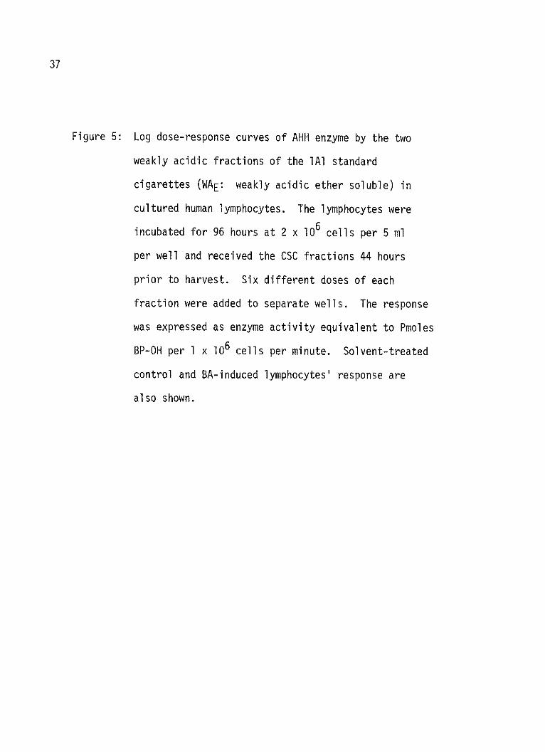
37I
EFFECTS OF CIGARETTE SMOKE CONDENSATES ON
CULTURED HUMAN LYMPHOCYTES AND SEPARATION
OF BENZO-a-PYRENE METABOLITES BY
HIGH PRESSURE LIQUID CHROMATOGRAPHY
THESIS
Presented to the Graduate Council of the
North Texas State University in Partial
Fulfillment of the Requirements
For the Degree of
MASTER OF SCIENCE
By
Burhan I. Ghanayem, B.S.
Denton, Texas
August, 1979

Ghanayem, Burhan, Effects of Cigarette Smoke Condensates on
Cultured Human Lymphocytes and Separation of Benzo-a-pyrene
Metabolites by High Pressure Liquid Chromatography. Master of
Science (Basic Health Sciences), August 1979, 82 pp., 16 figures,
1 table, bibliography, 91 titles.
Cigarette smoke condensates from all cigarettes tested were
found to be potent inducers of AHH enzyme in cultured human
lymphocytes and, with the exception of Kent Lights and Carlton
CSC's, all were found to be toxic under the experiment conditions.
Most of the AHH inducing activity was found in basic and neutral
fractions of the lAl standard cigarettes.
A radiometric assay of BP metabolites in cultured human
lymphocytes was developed in which we were able to separate the
primary metabolites and the secondary metabolites from the parent
compound (BP) by neutral alumnia HPLC. The primary metabolites
were further separated by a selective enzyme hydrolysis and/or
reverse phase HPLC.

1979
BURHAN IBRAHIEM GHANAYEM
ALL RIGHTS RESERVED
Univer siyMicrofilms
International300 N. ZEEB ROAD, ANN ARBOR, MI 48106
TABLE OF CONTENTS
PageLIST OF TABLES................................................iv
LIST OF ILLUSTRATIONS...........................................v
Chapter
I. INTRODUCTION............................................1
II. MATERIALS AND METHODS...........................-- -......... 15
III. RESULTS ..........................---..---------..... 24
IV. DISCUSSION............................................. 64
BIBLIOGRAPHY..................................................75

LIST OF TABLES
Table Page
I. Summary of the Quantitation of CSC yieldof ten brands of cigarettes ............................... 61
iv

LIST OF ILLUSTRATIONS
Figure Page
1. Log dose-Response curves of AHH enzyme bysmoke condensates from Camel and Chesterfieldcigarettes................................. . ---------- ,29
2. Log dose-Response curves of AHH enzyme bysmoke condensates from Marlboro, Winston,and Kent cigarettes............................... .,31
3. Log dose-Response curves of AHH enzyme bysmoke condensates from Marlboro lights, Winstonlights, Kent lights, Carlton and True cigarettes...............33
4. Log dose-Response curves of AHH enzyme byCSC derived from the standard 1AI cigarettes andthe Reconstituted CSC..................
5. Log dose-Response curves of AHH enzyme bythe weak acidic fractions (WAE and WAI) of theIA1 CSC...........................
6. Log dose-Response curves of AHH enzyme bythe basic fractions (Bjb, Bja, BE and BW) ofthe 1A1 I ...... ......... ....-------3
7. Log dose-Response curves of ANI enzyme bythe strong acidic fractions (SAI, SAE and SAW)of the 1Al CSC.....................
8. Log dose-Response curves of AHH enzyme bythe neutral fractions (NCH, NNiMandrNMeoH) Ofthe 1Al CSC...........................------------------,43
9. Representative elution profile of 3H-BP metabolitesproduced by cultured human lymphocytes after hy-drolysis by both Aryl sulfatase and beta-glucuronidaseenzymes.............................-----------.
v
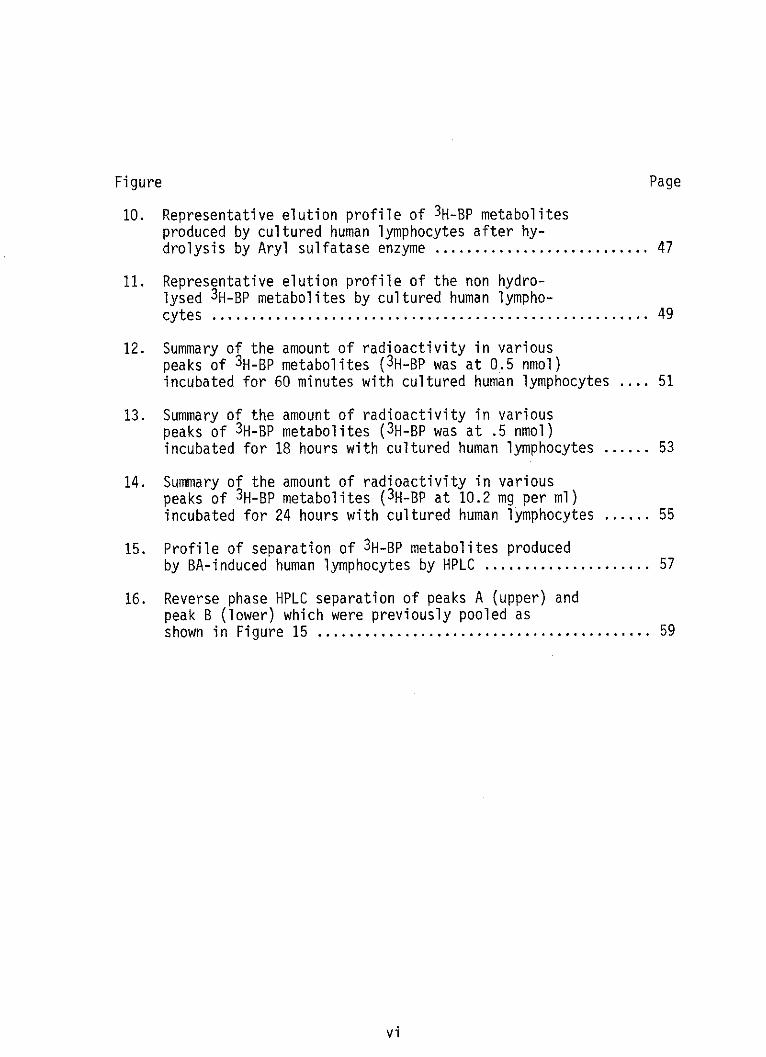
Figure Page
10. Representative elution profile of 3H-BP metabolitesproduced by cultured human lymphocytes after hy-drolysis by Aryl sulfatase enzyme....................... 47
11. Representative elution profile of the non hydro-lysed 3H-BP metabolites by cultured human lympho-cytes ....... ....................................... 49
12. Summary of the amount of radioactivity in variouspeaks of 3H-BP metabolites (3H-BP was at 0.5 nmol)incubated for 60 minutes with cultured human lymphocytes .... 51
13. Summary of the amount of radioactivity in variouspeaks of 3H-BP metabolites (3H-BP was at .5 nmol)incubated for 18 hours with cultured human lymphocytes......53
14. Summary of the amount of radioactivity in variouspeaks of 3H-BP metabolites (3H-BP at 10.2 mg per ml)incubated for 24 hours with cultured human lymphocytes......55
15. Profile of separation of 3H-BP metabolites producedby BA-induced human lymphocytes by HPLC...................57
16. Reverse phase HPLC separation of peaks A (upper) andpeak B (lower) which were previously pooled asshown in Figure 15 .................................... 59
vi

CHAPTER I
INTRODUCTION
Chemical carcinogenesis was first discovered in man in 1775 by
the British physician Percival Potts, when he correctly identified
the high incidence of scrotal cancer as an occupational disease among
the chimneysweeps of London. Prolonged exposure to soot and coal tar
was assumed to be the cause (52). Chemical carcinogens are non-viral
and non-radioactive substances, which have been shown to induce
cancer in a wide variety of animals and to cause cellular transforma-
tion in cultured tissues (12,21). The known chemical carcinogens
comprise a large and structurally diverse group of synthetic and
naturally occurring organic and inorganic compounds with various
species and tissue selectivities (10,29,30,57).
Epidemiologists consider that 70-90% of human cancer, excluding
skin cancers, are caused by environmental chemical pollutants such as
polycyclic aromatic hydrocarbons (PAH) (44). Among the chemically
induced cancers, lung cancer is one of the leading causes of death
and its incidence is increasing dangerously throughout the world.
The number of deaths in the United States from lung cancer in 1975
was estimated at 81,000 (13), and if current trends continue, it is
expected that 295,000 new cases of lung cancer will occur in the year
2000 (24).
1
2
Prospective and retrospective studies from all around the world
are in agreement that cigarette smoking is, by far, the most impor-
tant single contributing factor to the increased risk of developing
cancer of the respiratory tract in general, and the lung in particu-
lar (24). Statistical evidence from Finland (39), England (18),
U.S.A (28), and France (17), and histological evidence through the
systemic study of the bronchial epithelium of smokers and nonsmokers
(2,3,4), all supported the role of cigarette smoking in the causation
of lung cancer.
Accordingly, the Advisory Committee of the U.S. Surgeon General
has studied more than 100 prospective and retrospective reports, from
more than 15 countries, and summarized the data in the 1964, 1967 and
1972 reports (54,58,59) as follows:
Cigarette smoking is causally related to lung cancer, and themagnitude of the effect of cigarette smoking outweighs all otherfactors, with the risk of developing lung cancer increasing with theduration of smoking, is directly proportional to the number ofcigarettes smoked per day, and is diminished by discontinued smoking.In comparison with nonsmokers, the average male smokers of cigaretteshave approximately a 9-10 fold increased risk of developing lungcancer and heavy smokers at least a 20-fold increased risk. The riskof developing lung cancer for the combined groups of pipe smokers,cigar smokers and pipe and cigar is greater than for nonsmokers, butless than for cigarette smokers. With regard to cancer at othersites, the reports stated that pipe smoking appears to be causallyrelated to lip cancer, and cigarette smoking is a significant factorin the causation of cancer of the larynx.
The type of cigarettes smoked may also influence the risk
levels. The risk of lung cancer (squamous cell carcinoma and small
cell carcinoma) is less in smokers of filter cigarettes than in those
who smoke nonfilter cigarettes (62,63,64) probably due to the lower
yields of nicotine and tar in the filter cigarettes.

3
The following two figures, reproduced from Wynder et al. (63),
show that the relative risk of lung cancer correlates with the
number of cigarettes smoked per day (Figure 1) and that filter
cigarettes are less of a carcinogenic when compared to nonfilter
cigarettes (Figure 2).
7 0-
60k
501-
40
LUc 20 -
10-
CIGARETTES PER DAY:NEVER SMOKED
(NO CASES): (3)
_ F~1~LiAZ7Li1-9 10-20 21-40 41+
(7) (57) (74) (59)
(28)
ON FILTER (107)
FILTER (10+YRS) (83)
(NO. CASES)
(39)
() (34) (33)
(20)l il
1-9 10-20
CIGARETTES
21-40
PER DAY
Figure 1.
Figure 2.100KLU
x
V0
4l +k -& I I -Amum -, mmim I - -L-
w
E
(28)1

4
During tobacco smoking, organic matter burns incompletely
resulting in the formation of tobacco smoke. The smoke that emerges
from the tobacco product through the mouthpiece during puffing is
known as the mainstream smoke. The smoke that comes from the burn-
ing cone and from the mouthpiece during puff intermissions is known
as the sidestream smoke. Primarily, mainstream smoke is inhaled by
a smoker, and hence has gained most of the attention in studying
tobacco carcinogenesis.
Tobacco smoke is a two-phase system consisting of a vapor (gas)
phase and a disperse (particulate) phase. Cigarette smoke condensate
(CSC), which is called tar by some people, is the mixture of smoke
constituents (mostly the particulate phase) that is collected by
condensation at low temperatures.
The smoke condensate of cigarette, cigar and pipe smoke are
carcinogenic to the skin of mice, the bronchial epithelium of dogs
and rats and the connective tissue of rats, as demonstrated by more
than 100 studies from more than 10 countries (62,31). The dose-
response effects observed when CSC is applied to epithelial tissues
are quite comparable with those observed with pure carcinogenic
compounds (6).
Several investigators have fractionated CSC and bioassayed the
main fractions as well as the neutral and weakly acidic subfractions
(16,19,32,42,60,62). The CSC of the standard lAI low nicotine
cigarettes was fractionated by Swain et al. (56) into acidic, basic
and neutral fractions which were further divided into 12 subfractions.
Those 12 subfractions as well as the original and the reconstituted
material have been studied by different investigators (35,38,53).

5
Polycyclic aromatic hydrocarbons (PAH) which are products of
incomplete hydrocarbon combustion are found in polluted city air
(20), in smoked food (41), in water and in tobacco smoke (62),
where they represent the class of compounds credited with the highest
carcinogenic activity (32). Because CSC contains large amounts of
PAH, and because many of those PAH's present in CSC are powerful
carcinogens in experimental animals, they became logical suspects as
the carcinogens causing lung cancer in humans due to cigarette
smoking.
Xenobiotics including PAH's are metabolized in the body by a
membrane-bound mixed function oxygenase system known as aryl hydro-
carbon hydroxylase (AHH) or benzo-a-pyrene hydroxylase. AHH is part
of the endoplasmic reticulum and requires NADPH and molecular oxygen
to function. This system includes a complex of enzymes with NADPH
cytochrome C reductase, cytochrome P-450, cytochrome P-448, a lipid
factor and a cytochrome P-450 reductase (50).
AHH has been found in most mammalian tissues including liver,
skin, lung, intestine, kidney, placenta, lung alveolar macrophages,
monocytes, and lymphocytes (15). With regard to the activity of
this enzyme system, it varies in different tissues and is generally
very low in most tissues, with the highest activity being located in
the liver (15). AHH is inducible by a wide variety of exogenous
(PAH, CSC, phenobarbital, pesticides) as well as endogenous (steroidal
hormones) compounds.
There is a dose-response relationship between the chemical
inducers and the levels of AHH activity, and it is thought by some

6
that inducibility (the ratio of induced to basal activity) is
related to the susceptibility to lung cancer, or may be employed
in the assignment of individuals to various AHH genetic groups.
Kellerman et al. (36) reported a trimodal distribution of AHH in-
ducibility in the normal human population, with 53% in the low
inducibility group, 37% in the intermediate group and 10% in the
high inducibility group. Fifty patients with bronchogenic carcinoma
were subsequently compared with 46 patients having other types of
tumors and with 85 healthy controls (37). They concluded that a
person having the intermediate inducibility has a 16 times increased
risk and a person having the high inducibility has a 36 times in-
creased risk of developing lung cancer. Other claims of laryngeal
carcinoma (5), bronchogenic carcinoma (9), and renal tumors (33)
associated with the high AHH-inducibility have also been reported.
AHH activity has been implicated as playing a role in carcino-
genesis, teratogenesis, and mutagenesis by PAH and in drug activa-
tion and detoxification (1,12,14,23,34,50). Because PAH are found
in cigarette smoke and because the incidence of lung cancer parallels
the extent and duration of smoking, and the nicotine/tar yield of
the cigarettes smoked, AHH possibly plays an important role in the
development of cigarette smoke induced cancer.
The procarcinogen benzo-a-pyrene (BP) is a prototypic PAH,
which is a universal air pollutant, and it is estimated that 2,000
tons of BP are put into the air of the U.S. each year (44).

7
The metabolism of BP has been studied in both microsomal (endo-
plasmic reticulum) preparations and in cells and various organs from
several animal species. Polycyclic aromatic hydrocarbons such as BP
are generally considered to be carcinogenic in man and animals only
after metabolic activation by AHH enzyme to reactive electrophilic
intermediates. These are capable of covalent binding to protein and
nucleic acids, a step which apparently precedes tumor initiation
(7,22,25,51).
BP metabolism is a complex series of reactions, some of which
occur simultaneously. BP is initially oxygenated by AHH enzyme to
epoxides (26) and/or phenols and quinones (23). These metabolites
may be further metabolized to dihydrodiols by hydratases (49).
Epoxides and hydroxylated metabolites may also be further metabolized
to inactive, water-soluble, urine excretable conjugates of sulfates
(11,48) by sulfotransferases, glucuronides (46) by UDP-glucuronyl
transferase and glutathione (8,47) by glutathione-S-transferase
enzymes. Epoxides may bind to cell macromolecules where they can
cause alteration in cell metabolism and possibly induce malignant
transformation (27,40,43).
SULFATE Sulfo- PHENOL OR UDPG GLUCURONIDECONJUGATE T'ase ALCOHOL T'ase CONJUGATE
UDPG T'ase
PARENT ' OXYGENATED EH
Fi gure 3. COMPOUND ELECTROPHILE DIHYDROD
REDUCTASE GSH
S-T'ase DIOL-EPOXIDE
CRITICAL GLUTATHIONE TETROL CRITICALTARGET? CONJUGATE TARGET?

8
Generally, the secondary metabolism is a protective mechanism
which speeds the excretions of chemicals from the body. An excep-
tion is the reoxidation of diols (specifically, 7,8-dihydrodiol) to
diol-epoxides which are highly reactive and bind readily to DNA
(55). The 7,8-dihydrodiol-9,10-epoxides of BP are considered to be
the ultimate carcinogens (61,65) and their carcinogenic activity is
apparently related to their characteristic mutagenicity (65). A
general scheme of the metabolism of PAH's has been suggested by
Nebert et al. (45) and is illustrated in the following Figure (3).
Most investigators have employed animal systems (38,62),
animal tissue cultures (53) or bacterial systems (35) to study
tobacco carcinogenesis and mutagenesis and to relate that to humans.
This study, in part, has employed human tissue (lymphocytes) to
measure the AHH induction and cytotoxic action of CSC and CSC
fractions by studying the pattern of the dose-response relationship
in mitogen stimulated human lymphocytes, between CSC and CSC frac-
tions and AHH activity.
In another part of this study, we devised a means of measuring
the primary and secondary metabolite profiles of BP produced by the
mitogen stimulated human lymphocytes and effected the separation of
the BP conjugates, sulfates, glucuronides and glutathiones from the
primary metabolites and parent compound (BP) by the use of HPLC.
Use of these methods may provide data on individual persons relative
to their rate of activation/deactivation reactions.

CHAPTER BIBLIOGRAPHY
1. Ames, B.N., Durston, W.E., Yamasaki, E., et al. (1973). Carcinogensas mutagens: A simple test system combining liver homogenatefor activation and bacteria for detection. Proc. Nat. Acad.Sci. USA 70:2281.
2. Auerbach, 0., Stout, A.P., Hammond, E.C., and Garfinkel, L. (1961).
Changes in relation to lung cancer. N. Engl. J. Med. 265:253.
3. Auerbach, 0., Stout, A.P., Hammond, E.C., and Garfinkel, L. (1962).Changes in bronchial epithelium in relation to sex, age, residence, smoking and pneumonia. N. Engl. J. Med. 267:111.
4. Auerbach, 0., Stout, A.P., Hammond, E.C., and Garfinkel, L. (1962).Bronchial epithelium in former smokers. N. Eng. J. Med. 267:119.
5. Berwald, Y., and Sachs, L. (1965). In vitro transformation ofnormal cells to tumor cells by carcinogenic hydrocarbons.J.. Natl. Cancer Inst. 35:641.
6. Bock, F.G. (1968). Dose-response: Experimental carcinogenesis.Nat. Cancer Inst. Monogr. 28:57.
7. Brookes, P., and Lawley, P.D. (1964). Evidence for the binding ofpolynuclear aromatic hydrocarbons to the nucleic acids ofmouse skin: Relation between carcinogenic power of hydro-carbons and their binding to deoxyribonucleic acid. Nature202:781.
8. Boyland, E., and K. Williams (1965). An enzyme catalysing theconjugation of epoxides with glutathione. Biochem. J. 94:190.
9. Chen, T.T., and Heidelberger, C. (1969). Quantitative studies onthe malignant transformation of mouse prostate cells bycarcinogenic hydrocarbons in vitro. Intl. J. Cancer 4:166.
10. Clayson, D.B. (1962). Chemical Carcinogenesis, Little, Brown,Boston, Massachusetts.
9

10
11. Cohen, G.M., Haws, S.M., Moore, B.P., and Bridges, J.W. (1976).BP-3-yl hydrogen sulfate, a major ethyl acetate-extractable metabolite of BP in human, hamster and rat lungcultures. Biochem. Pharma y 25:2561.
12. Conney, A.H. (1967). Pharmacological implications of microsomal
enzyme induction. Pharmacol. Rev. 19:317.
13. Culter, S.J., Scotto, J., Devesa, S.S., et al. (1974). Third
National Cancer Survey - An overview of available information.
J. Natl. Cancer Inst. 53:1565.
14. Doly, J.W., Jerina, D.M., Witkop, B., Arene, P. (1972). Oxides
and the NIH shift: The metabolism, toxicity, and carcino-
genicity of aromatic compounds. Experientia 28:1129.
15. David, J.J., James, J., Robert, S., and Harri, V. (1977). Bio-
logical reactive intermediates, Plenum Press, New Yorkand London.
16. Davis, B.R., Witehead, Y.K., Gill, M.E., Lee, P.N., Butterworth,A.D., and Roe, F.J.C. (1975). Response of rat lung to in-
haled vapor phase constituents (VP) of TS alone or in
conjunction with SC given by intratracheal instillationBr. J. Cancer 31:462.
17. Denoix, P.F., Schwartz, D., and Anguera, G. (1958). L'enquitefrancaise sur 1'etiologue du cancer broncho-pulmonaire.Analyse detailee. Bull. Ass. Cancer 26:1085.
18. Doll, R., and Hill, A.B. (1964). Mortality in relation to smoking.
Ten years observations of British doctors. Brit. Med. J. 1:1399.
19. Dontewill, W., Elmenhorst, H., Harke, H.P., Reckzeh, G., Weber,
K.H., Misfeld, J., and Timm, J. (1970). ExperimentelleUntersuchungen uber die Tumorerzeugung von zigarettenranch-Kondensatem an der Mausehaut, I.Z., Krebsforsch 73:265.
20. Folk, H.L., and Kotin, P. (1963). Symposium on chemical car-
cinogenesis. Clin. Pharmacol. Ther. 4:88.
21. Gelboin, H.V. (1972). Studies on the mechanism of microsomal hy-
droxylase induction and its role in carcinogen action. Rev.Canc. Biol. 31:39.

11
22. Gelboin, H.V. (1969). Microsome-dependent binding of BP to DNA,Cancer Res. 29:1272.
23. Gelboin, H.V., Kinoshita, N., and Wiebel, F.J. (1972). Microsomalhydroxylazes: Induction and role in PAH carcinogenesis andtoxicity. Fed. Proc. 31:1298.
24. Gori, G.B., and Peters, J.A. (1975). Etiology and prevention ofcancer. Prev. Med. 4:235.
25. Grover, P.L., and Sims, P. (1968). Enzyme-catalyzed reactions ofPAH's with deoxyribonucleic acid proteins in vitro. Biochem.Pharmacol. 110:159.
26. Grover, P.L., Hewer, A., and Sims, P. (1972). Formation ofK-region epoxides as microsomal metabolites of pyrene andbenzo(a)pyrene. Biochem. Pharmacol. 21:2713.
27. Grover, P.L., Sims, P., Huberman, E., Marquardt, H., Kuroki, T.,and Heldelberger, C. (1971). In vitro transformation ofrodent cells by K-region derivatives of PAH's. Proc. Natl.Acad. Sci. 68:1098.
28. Hammond, E.L., and Horn, D. (1958). Smoking death-rates reporton forty-four months follow-up of 187,783 men. J. Amer. Med.Assn. 166:1159,1294.
29. Hartwell, J.L. (1951). Survey of compounds which have beentested for carcinogenic activity. U.S. Public Health ServicePubl. no. 149, 2nd ed., Washington, D.C.
30. Hartwell, J.L., and Shubik, P. (1957, 1959). Survey of compoundswhich have been tested for carcinogenic activity, suppl. 1and 2. U.S. Public Health Service Publ. no. 149, Washington,D.C.
31. Hoffman, D., Schmeltz, I., Hecht, S.S., and Wynder, E.L. (1976).On the identification of carcinogens, tumor promoters andcarcinogens in tobacco smoke. In "Smoking and Health", DHEWpubl. no. (NIH) 76-1221, p. 125, U.S. Gov. Printing Office,Washington, D.C.
32. Hoffman, D., and Wynder, E.C. (1971). A study of tobacco carcino-genesis. XI. Tumor initiators, tumor accelerators and tumorpromoting activity of condensate fractions. Cancer 27:848.

12
33. Huberman, E., Kuroki, T., Marquardt, H., Silkirk, J.K., Heildel-berger, C., Grover, P.L., and Sims, P. (1972). Transformationof hamster embryo cells by epoxides and other derivatives ofpolycyclic hydrocarbons. Cancer Res. 32:1391.
34. Jerina, D.M., and Daly, J.W. (1974). Arene oxides. A new aspectof drug metabolism. Science 185:574.
35. Kier, L.D., Yamasaki, E., and Ames, B.N. (1974). Detection ofmutagenic activity in cigarette smoke condensate. Proc. Natl.Acad. Sci. U.S.A. 71:4159,
36. Kellerman, G., Shaw, C.R., and Luyten-Kellermann, M. (1973).Genetic variation of AHH in human lymphocytes. Am. J. HumanGenet. 25:327.
37. Kellermann, G., Shaw, C.R., and Luyten-Kellerman, M. (1973).AHH inducibility and bronchogenic carcinoma. N. Engl. 3.Med. 289:934.
38. Kouri, R.E. (1974). Experimental lung cancer, carcinogenesisand bioassays. International symposium held at theBatelle Research Center, Seattle, WA., USA, June (1974),Springer-Verlag, Berlin, Heidelberg, New York (1974).
39. Kreyberg, L. (1968). Nonsmokers and geographic pathology oflung cancer. In: "The Lung", A.A. Liebon and D.E. Smith,eds.), pp. 273-283, Williams and Wilkins, Baltimore, MD.
40. Kuroki, T., Huberman, E., Marquardt, H., Selkirk, J.K., Heidel-berger, C., Grover, P.L., and Sims, P. (1972). Binding ofK-region epoxides and other derivatives of BA and dibenz-(a,b)-anthracene to DNA, RNA, and proteins of transformablecells. Chem - Biol. Interact 4:389.
41. Kuratsune, M. (1956). BP content of certain pyrogenic materials.J. Nat. Cancer Inst. 16:1485.
42. Lazar, P.H. (1974). Chouroulinkor, I., Izard, C., Moree-Testa,P., and Herman, D. (1974). Bioassays of carcinogenicityafter fractionation of cigarette smoke condensate.Biomedicine 20:214.
43. Marquardt, H., Kuroki, T., Huberman, E., Selkirk, J.K., Heidel-berger, C., Grover, P.L., and Sims, P. (1972). Malignanttransformation of cells derived from mouse prostate byepoxides and other derivatives of polycyclic hydrocarbons.Cancer Res. 32:716.

13
44. National Academy of Science Reports, USA (1972). Particulatepolycylic organic matter. Committee on Biologic Effectsof Atmospheric Pollutants, Division of Medical Sciences,National Research Council, Washington, D.C.
45. Nebert, W.D., Roberts, J., Adelman, C.R., and Critofalo, J.V.(1978). Pharmacological intervention in the aging process. Chapter 4, Genetic Control of Drug Metabolism,Plenum Press, N.Y.
46. Nemoto, N., and Gelboin, H.V. (1976).BP oxide phenols and dihydrodiolsacid. Biochem. Pharm. 25:1221.
Enzymatic conjugation ofwith UDP-glucuronic
47. Nemoto, N., Gelboin, H.V., Habig, W.H., Ketley, J.N., andJakoby, W.B. (1975). K-region BP-4,5-oxide in conjugatedby homogenous glutathione-s-transferase. Nature (London)255:512.
48. Nemoto, N., Takayama, S., and Gelboin, H.V. (1977). Enzymaticconversion of BP phenols, dihydrodiols and quinones tosulfate conjugates. Biochem. Pharmacol. 26:1825.
49. Oesch, F. (1972). Mammalian epoxide hydrases: Inducible en-zymes catalyzing the inactivation of carcinogenic andcytotoxic metabolites derived from aromatic and olefiniccompounds. Xenobiotica 3:305.
50. Paul , 0. P. , Tso, P.O. , and Dipaol o,Carcinogenesis - Part A, Marcel
J.P. (1974).Dekker, 1974.
51. Pietropaolo, C., and Weinstein, I.B. (1975).to natural and synthetic nucleic acids inmicrosomal system. Cancer Res. 35:2191.
52. Potts, P. (1775).and Collins.
Binding of 3H-BPa subcellular
Chirurgical Observations. London, Hawkes,
53. Rasmussen, R.E. (1975). Tobacco smoke condensate and DNArepair. Life Sciences 17:767.
54. Smoking and Health. Report of the Advisory Committee to theSurgeon General of the Public Health Service, U.S. PublicHealth Service Publication, No. 1103. Washington, D.C.,U.S. Government Printing Office (1964).
Chemi cal

14
55. Sims, P., Grover, P.L., Swaisland, A., Pal, K., and Hewer, A.(1974). Metabolic activation of BP proceeds by a diolepoxide. Nature (London) 252:326.
56. Swain, A.P., Cooper, J.E., and Stedman, R.L. (1969). Largescale fractionation of cigarette smoke condensate forchemical and biologic investigations. Cancer Res. 29:579.
57. Thompson, J.I. and Co. (1972). Survey of compounds which havebeen tested for carcinogenic activity, 1968-69 volume,Public Health Service, publ. no. 149, Washington, D.C.
58. The Health Consequences of Smoking (1968). A Public HealthService Review, U.S. Department of Health, Education andWelfare. Public Health Service publ. no. 1696, Washing-ton, D.C.
59. The Health Consequences of Smoking. A report to the SurgeonGeneral (1971). U.S. Department of Health, Educationand Welfare, U.S. Public Health Service, publ. (HSM)71-7513, p. 458.
60. Whitehead, J.K., and Rothwell, K. (1969). The mouse skincarcinogenicity of cigarette smoke condensate: Frac-tioned by solvent partition methods. British J. Cancer23:840.
61. Wood, A.W., Levin, W., Lu, A.Y.H., Yagi, H., Hernandez, 0.,Jerina, D.M., and Conney, A.H. (1976). Metabolism ofbenzopyrene derivatives to mutagenic products by highlypurified hepatic microsomal enzymes. J. Biol. Chem.251:4882.
62. Wynder, E.L., and Hoffmann, D. (1967). Tobacco and tobaccosmoke: Studies in experimental carcinogenesis. AcademicPress, N.Y.
63. Wynder, E.L., and Mabuchi, K. (1972). Etiology of lung cancer:Reflection of two decades of research. Cancer 30:1332.
64. Wynder, E.L., and Mabuchi, K. (1972). Etiological and preven-tive aspects of human cancer. Prev. Med. 1:300.
65. Wynder, E.L., and Stellman, S. (1978). The epidemiology ofthe less harmful cigarettes. JAMA (in press).
66. Yank, S.K., McCourt, D.W., Leutz, J.C., and Gelboin, H.V.(1977). Benzopyrene diol epoxides: Mechanisms ofenzymatic formation and optically active intermediates.Science 196:1199.

CHAPTER II
MATERIALS AND METHODS
Lymphocytes Isolation and Culturing
Venous blood was withdrawn from human volunteers and was added
to one-half volume of sterile heparinized saline solution (0.9%
sodium chloride solution containing 10 units/ml heparin sodium
salt). Sterile conditions were maintained throughout the cell
culture. Twenty to twenty-five ml aliquots of the blood heparinized
saline mixture were layered over 8 ml of a ficoll-hypaque solution
(6% ficoll, 10% sodium diatrizoate) and centrifuged at 1500 g for 15
minutes. The mononuclear leukocytes (lymphocytes and monocytes)
were recovered from the interface between the plasma and the ficoll-
hypaque. The lymphocytes were sedimented by centrifugation at 1200
g for 10 minutes. After removing the supernatant, the cell pellet
was resuspended in Joklick's modified Minimum Essential Medium
(JMEM) (Gibco F-13), containing 10-15% fetal calf serum (Gibco), 1%
pokeweed, 1% phytohemagglutinin (Gibco), and 50 units/ml heparin
(Sigma). The cell concentration was adjusted to 0.4 x 10⁶ cells/ml.
Five ml aliquots were placed into wells of 35 mm costar dishes and
the culture medium depth was 4-6 mm. The cell suspensions were
15

16
incubated at 37*C in a humidified 3-5% CO2 atmosphere for 96 hours.
Forty-four hours prior to harvest each costar dish, containing six
wells, received six different doses (a dose to each well) of each of
the tars or the lAl CSC fractions in a volume ranging from 6-30 pl
of DMSO or acetone: methanol mixture. Six other wells received the
inducer 1,2-benzoanthracene (BA) dissolved in 10 pl of methanol to a
final concentration of 10 M. Another six wells received the corres-
ponding solvent at the same volume to serve as control cultures.
Cell Harvest and AHH Assay
The AHH assay was that of Cantrell et al. (2).
The cells were harvested and sedimented by centrifugation at
1000 g then resuspended in 2 ml EMSAD buffer [25 mM 4-(2-hydroxyethyl,
1-1-piperazine propane sulfonic acid)] 0.3 mM magnesium chloride,
125 mM NaCl, 0.1% bovine serum albumin, and 10 mM dextrose] at
pH 8.0. The 2 ml EMASD cell suspension was divided into duplicates,
and each received 2 pg BP in a volume of 10 pl of methanol, and all
were incubated at 37C for 60 minutes. The reaction was stopped by
adding 0.5 ml of 20% neutral formalin with gentle shaking. The
cells were then allowed to stand in the dark for 10 minutes for
fixation.
Just prior to reading, each tube received one-half ml of 1N
NaOH and was mixed vigorously; then the fluorescence of the suspen-
sions was measured with excitation at 460 nm and emission at 522 nm.

17
Tar Preparation
Ten brands of cigarettes manufactured in the United States were
used. The ten brands were Camel, Chesterfield, Marlboro, Marlboro
Lights, Kent, Kent Lights, Winston, Winston Lights, True and Carlton.
Ten cigarettes of each brand were smoked by the smoking appara-
tus shown in Figure (1). Each cigarette was hooked to an aquafilter
attached to a glass bubbler immersed in 20 ml of a 1:1 acetone-
methanol mixture. The test tube that contained the organic solvent
mixture was placed in a conical flask filled with iced water.
Vacuum was created inside the flask by closing the syringe valve and
drawing back the syringe plunger. The smoke passed into the glass
filter and was trapped in the cold acetone-methanol mixture. On
opening the valve, the vacuum inside the flask was broken and the
cigarette smoking stopped while the plunger was pushed back in. The
average puff volume was 35 ml over 2-4 seconds. The cigarettes were
smoked to a butt length ranging from 20-25 mm. The tar trapped by
the aquafilter was extracted by elution with 20 ml of 1:1 acetone-
methanol, and pooled with the other 20 ml that was in the test tube.
The 40 ml tar solution was concentrated under nitrogen and each
fraction was brought to 20 ml. Ten (10) ml of each fraction was
transferred to preweighed vials, dried under nitrogen, and kept in a
vacuum dessicator for 48 hours. The vials were removed and weighed
and the amount of tar yield per cigarette was determined. Three
different concentrations of the remaining 10 ml tar solution was
made and used for the AHH induction study.

Syringe
Valve
- -t---
/ cooo
0000OoOO0
(4 0 '0 00-0 1D
Aquae-filter
Glass bubbler
Ice cold water
Acetonew-EtoH(1:1)
Figure 1
18
S.11,111,1000 - wwww"Nom
A

19
The fractions of the CSC from the standard 1A1 cigarettes were
generously provided by Dr. Richard E. Kouri (Microbiology Associates,
Bethesda, MD). Three different concentrations of each fraction were
prepared and kept frozen and later used for the AHH assay.
Assay of BP Secondary Metabolites
The method used was that of Cantrell et al. (3).
Human lymphocytes were isolated and cultured as described under
"lymphocytes isolation and culturing". The cultures received BA
(10 pM, during the period of culture, other cultures received no BA
and served as controls. The BA-induced cells and the control
cultures, respectively, were pooled, harvested as mentioned before
and resuspended at the end of the 96 hour incubation period in fresh
JMEM media at a concentration of 5 x 10⁶ per ml. The cells were
divided into 4 aliquots of 1 ml and marked AB,C and D. Tubes A,B
and C received 3H-BP at the start of the reaction and tube D received
3H-BP at the end of the AHH reaction. The amounts of BP and dura-
tion of incubation are indicated in the Results section. After the
reaction period, each tube received 1 ml of water and the tubes were
frozen and thawed twice to lyse the cells and terminate the AHH
activity. Additions were made to the tubes as follows: Tube A
received 10 pl of a mixture of beta-glucuronidase (0.07 I.U.) and
aryl sulfatase (0.1 I.U.) (Calbiochem). Tube B received the enzyme
mixture and 0.1 mg D-saccharic acid-1, 4-lactone, an inhibitor of
glucuronidase. Tubes C and D received only water. All tubes were
20
incubated for one hour at 37*C and the hydrolysis then terminated by
addition of 5 ml ethyl acetate to each tube with gentle mixing. The
tubes were extracted twice with ethyl acetate and the pooled organic
phases dried with sodium sulfate, then evaporated under nitrogen.
The extracts were brought up to 0.5 ml ethanol and 0.1 ml injected
into the high pressure liquid chromatograph for separation of meta-
bolites. A Hibar-II RP-18 reverse phase column was employed as a
separation medium. Elution of the column was with a linear 40-90%
ethanol in water gradient. Fractions were collected and counted by
liquid scintillation spectrometry.
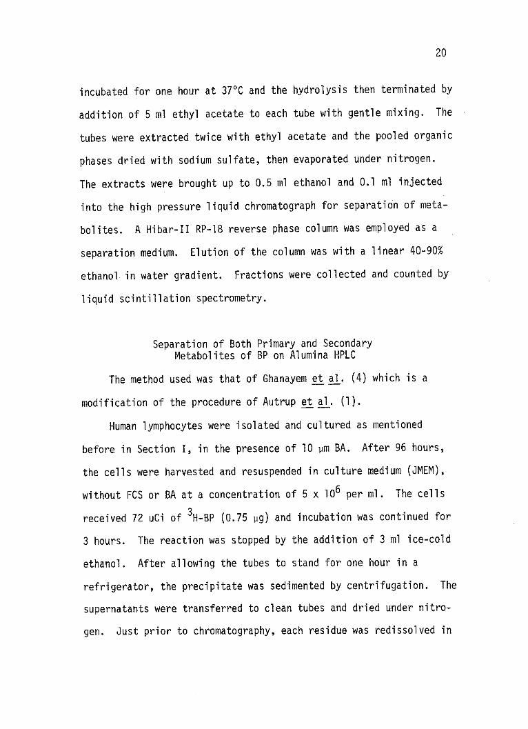
Separation of Both Primary and SecondaryMetabolites of BP on Alumina HPLC
The method used was that of Ghanayem et al. (4) which is a
modification of the procedure of Autrup et al. (1).
Human lymphocytes were isolated and cultured as mentioned
before in Section I, in the presence of 10 pm BA. After 96 hours,
the cells were harvested and resuspended in culture medium (JMEM),
without FCS or BA at a concentration of 5 x 10⁶ per ml. The cells
received 72 uCi of 3H-BP (0.75 Pg) and incubation was continued for
3 hours. The reaction was stopped by the addition of 3 ml ice-cold
ethanol. After allowing the tubes to stand for one hour in a
refrigerator, the precipitate was sedimented by centrifugation. The
supernatants were transferred to clean tubes and dried under nitro-
gen. Just prior to chromatography, each residue was redissolved in

2 ml 79% ethanol. A 500 pl aliquot was injected onto the prepacked
neutral alumina column for separation. A 4.6 x 250 mm steel HPLC
column was dry packed with neutral alumina, Brockmann activity 1,
80-200 mesh size. The column was equilibrated with hexane and the
500 pl sample was injected during a flow rate of 1.2 ml/minute.
Immediately the gradient programmer was started so as to form a
gradient of hexane to absolute ethanol over 30 minutes. Twenty (20)
2 ml fractions were collected over 34 minutes after which the
solvent was changed to water, ten fractions were collected and the
solvent was changed to 50 mM ammonium phosphate buffer, pH 3.0.
Fifteen (15) fractions were collected and the solvent was changed to
25% formic acid. Fifteen (15) fractions were collected. A total of
sixty 2 ml fractions were collected over the 100 minute elution
time.
A 200 1A aliquot of each fraction was taken and counted by
liquid scintillation spectrometry.
The hexane-alcohol gradient provided resolution of the non-
conjugated metabolites into two major peaks (Figure 15 in the
Results section). These were Peak A (3 fractions) and Peak B
(4 fractions). The two peaks were dried and subjected to reverse
phase HPLC on a Dupont Zorbax ODS 4.6 X 250 mm column. The peaks
were dissolved in 0.5 ml 70% ethanol, and a 100 pl aliquot injected
into the HPLC. Elution was with a 60-100% methanol gradient over 30
minutes with a flow rate of 1 ml/minute. The 200 pl fractions were

22
collected in minivials and the radioactivity was determined in a
Beckman LSC 7000. Identification of some peaks was made by com-
parison with authentic standards.

CHAPTER BIBLIOGRAPHY
1. Autrup, H. (1978). Separation of water-soluble metabolites ofbenzo-a-pyrene formed by cultured human colon. Bioch.Pharm. (in press).
2. Cantrell, E., Abreu, M., and Busbee, D. (1976). A simple assayof AHH in cultured human lymphocytes. Biochem. Biophys.Res. Commun. 70:474.
3. Cantrell, E.T., Marshall, M., McLemore, T., Ghanayem, B., andBusbee, D. (1979). Secondary metabolism of benzopyrenein human cells. Western Pharm. Proceedings.
4. Ghanayem, B., Cantrell, E., Busbee, D., Burleson, L. (1979).Separation of both primary and secondary metabolitesof benzo-a-pyrene on alumina HPLC chromatography.Abstracts, Texas Pharmacologist's Meeting, May, Abst. #42.
23
CHAPTER III
RESULTS
Quantitation of CSC in Cigarettes andEffect of Different CSC on AHH in Lymphocytes
The amount of CSC in mg per cigarette was quantitated (Table I).
The effect on AHH activity (BP hydroxylase) of exposure of human
lymphocytes to six different doses of CSC from two brands of unfiltered
first generation cigarettes (the first generation of manufactured
cigarettes to be delivered to the market; they are mostly unfiltered
with a high tar and nicotine yield), three brands of filtered second
generation cigarettes (the second generation of manufactured cigarettes,
mostly filtered, with a comparatively moderate tar and nicotine yield),
and five brands of the filtered third generation cigarettes (the third
generation cigarettes also called the less harmful cigarettes, mostly
have improved filtration systems like air filter and aqua filter with a
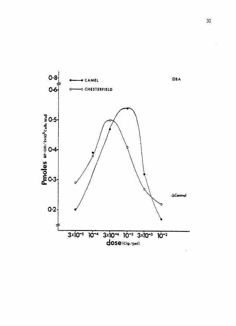
comparatively low tar and nicotine yield), demonstrates that a maximum
induction of 0.54 and 0.50 Pmoles 3-hydroxy beno-a-pyrene (BPOH) per
well per minute was reached at 3 x 10⁴ and 10-3 cig./well by Camel and
Chesterfield CSC, respectively, (Figure 1). A maximum induction of
0.53, 0.49 and 0.43 Pmoles BP-OH per well per min. was also reached by
Marlboro, Winston and Kent CSC's, respectively, at 10-3 cig./well dose
24

25
(Figure 2). At 10-3 cig./well dosage, Winston Lights produced a
maximum induction of 0.57 Pmoles per well per min., while Marlboro
Lights and True produced their maximal induction of 0.55 and 0.60
Pmoles BP-OH per well per min. at a 3 x 10⁻³ cig./well (Figure 3).
In Figure 1, 2, and 3 and with all CSC's tested, with the
exception of Kent Lights and Carlton, toxicity (decline in AHH
levels) was noticed at all doses higher than the maximal inducing
doses.
Under the same experimental conditions, the enzyme activity of
untreated control and BA (10 pm) induced human lymphocytes' response
are shown. The activities of control and BA treated cells were 0.25
and 0.80 Pmoles BPOH per well per min., respectively. With all
doses, duplicates have been used, and the mean of each duplicate was
represented in the figures.
Effect of CSC Subfractions on AHHin Lymphocytes
The effect of the standard lAl CSC and its fractions on AHH
activity in human lymphocytes was demonstrated in Figures 4-8. Each
point represents the mean of two duplicates from each dose.
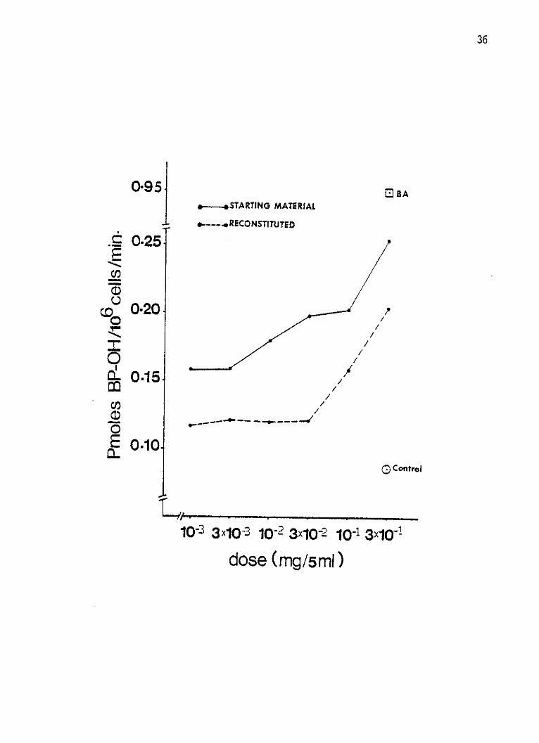
Whole lAl CSC and the reconstituted material demonstrated that
the response was proportional to the dose and at all six doses of
each of the starting and reconstituted materials neither a maximum
inducing nor a toxic dose could be reached or defined within the
26
limits of solubility (Figure 4). Exposure of cells to the weakly
acidic fractions of the lAl CSC resulted in a maximum response of
0.18 and 0.19 Pmoles BP-OH per 106 cells per min. This was reached
at 10~1 and 3 x 10⁻² mg/well, respectively (Figure 5). Doses
higher than 3 x 10⁻² produced toxicity by the WAE (weakly acidic
ether soluble fraction) (Figure 5).
The four basic fractions, with the exception of the BW (bases,
water soluble) produced an increased response with increasing dose.
The BIb (bases insoluble b) fraction was the most active AHH inducer
of all the lAl CSC fractions (Figure 6), which had a 0.66 Pmoles BP-
OH per 106 cells per minute at 3 x 10⁻¹ mg/well dose. From all the
basic fractions, BW was the only toxic, and its toxicity was observed
at doses higher than 3 x 10⁻² mg/well.
The strongly acidic fractions were weakly active with the SAI
fraction (strong acids, insoluble) was the most active (Figure 7).
All the strongly acidic fractions were toxic at one dose or another.
Neither a maximum response nor toxic effects were not achieved
by any of the neutral fractions rather a direct relationship was
observed between the dose and the response at all the dose levels
tested. NCH (neutrals, cyclohexane soluble) was the least active of
all the neutral fractions (Figure 8).
Estimation of Conjugation by EnzymeHydrolysis and HPLC
Human lymphocytes incubated for one hour with 3H-BP were found
to have approximately 20 milliunits of AHH by expressing radioactivity

27
in the peak reflecting formation of 3-OH-BP. The metabolite profile
was similar to that reported by others for human lymphocytes and the
identification of the peaks was assumed to be the same as reported
by Holder et al. (2).
Lymphocytes were incubated for 60 minutes with 12 pCi 3H-BP
(24 Ci/mM). Figure 9 presents the profiles of tube A which had both
aryl sulfatase and beta-glucurondiase enzymes and represents the
total of oxidized metabolites, sulfate, and glucuronide. Figure 10
presents the profiles of tube B which reflects the sum of free
metabolites plus sulfate only. Figure 11 presents the profiles of
tube C which represents only the unconjugated free metabolites.
The amount of radioactivity in the various peaks after hydro-
lysis is summarized in Figure 12, where 12 pCi 3H-BP was incubated
for 60 minutes reaction time. Figure 13 summarizes the results
after 12 pCi 3H-BP was incubated for 18 hours reaction time. Figure
14 represents similar data but with 10 pCi of 3H-BP which had been
diluted with cold BP to a specific activity of 10.2 pg per 10 pCi
(final concentration was 39.6 Pm in the tubes).
In Figures 12, 13, and 14, some of the peaks were identified.
Four diols were eluted first, followed by one quinone and two
phenolic metabolites.
Estimation of Conjugation by LC Separationon Alumina
Cultured human lymphocytes which had been mitogen activated and
induced with 10 pm BA for 96 hours were incubated after harvest for

28
3 hours with 0.75 pg 3H-BP. Figure 15 presents the elution profile.
Peak A represents mainly the parent compound and Peak B represents
mainly the oxidized metabolites. Both Peak A and Peak B were eluted
by the hexane-ETOH gradient.
H20 eluted the sulfates, phosphate eluted the glucuronides, and
formic acid eluted the glutathione conjugates (1).,
When fractions of peak A and peak B (from Figure 15) were
pooled separately, dried and fractionated by reverse phase HPLC, two
different patterns were obtained (Figure 16). The upper profile of
peak A shows mostly BP and some small amounts of the oxidized meta-
bolites. The lower profile of peak B shows small amounts of BP and
most of the oxidized metabolites.
29
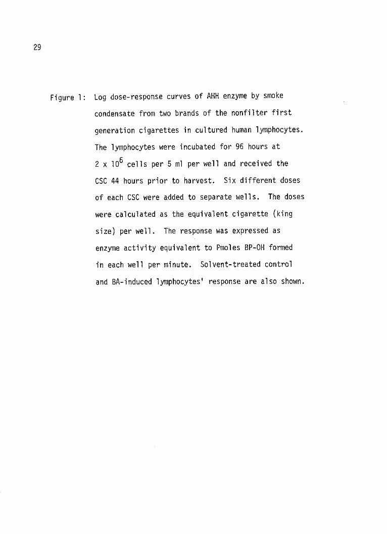
Figure 1: Log dose-response curves of AHH enzyme by smoke
condensate from two brands of the nonfilter first
generation cigarettes in cultured human lymphocytes.
The lymphocytes were incubated for 96 hours at
2 x 10⁶ cells per 5 ml per well and received the
CSC 44 hours prior to harvest. Six different doses
of each CSC were added to separate wells. The doses
were calculated as the equivalent cigarette (king
size) per well. The response was expressed as
enzyme activity equivalent to Pmoles BP-OH formed
in each well per minute. Solvent-treated control
and BA-induced lymphocytes' response are also shown.
30
0-81 1- CAMEL BA
0 o---o CHESTERFIELD
90*5
0-3
0
0
AControI
0-2
3x00 10 3x10⁻⁴ 10 3x0-3 10-2dose (Cig .0I

Figure 2:
31
Log dose-response curves of AHH enzyme by smoke
condensate from three brands of the second generation
filter cigarettes in cultured human lymphocytes.
The lymphocytes were incubated for 96 hours at
2 x 10⁶ cells per 5 ml per well and received the
CSC 44 hours prior to harvest. Six different doses
of each CSC were added to separate wells. The doses
were calculated as the equivalent cigarette (king
size) per well. The response was expressed as
enzyme activity equivalent to Pmoles BP-OH formed
in each well per minute. Solvent-treated control
and BA-induced lymphocytes' response are also shown.
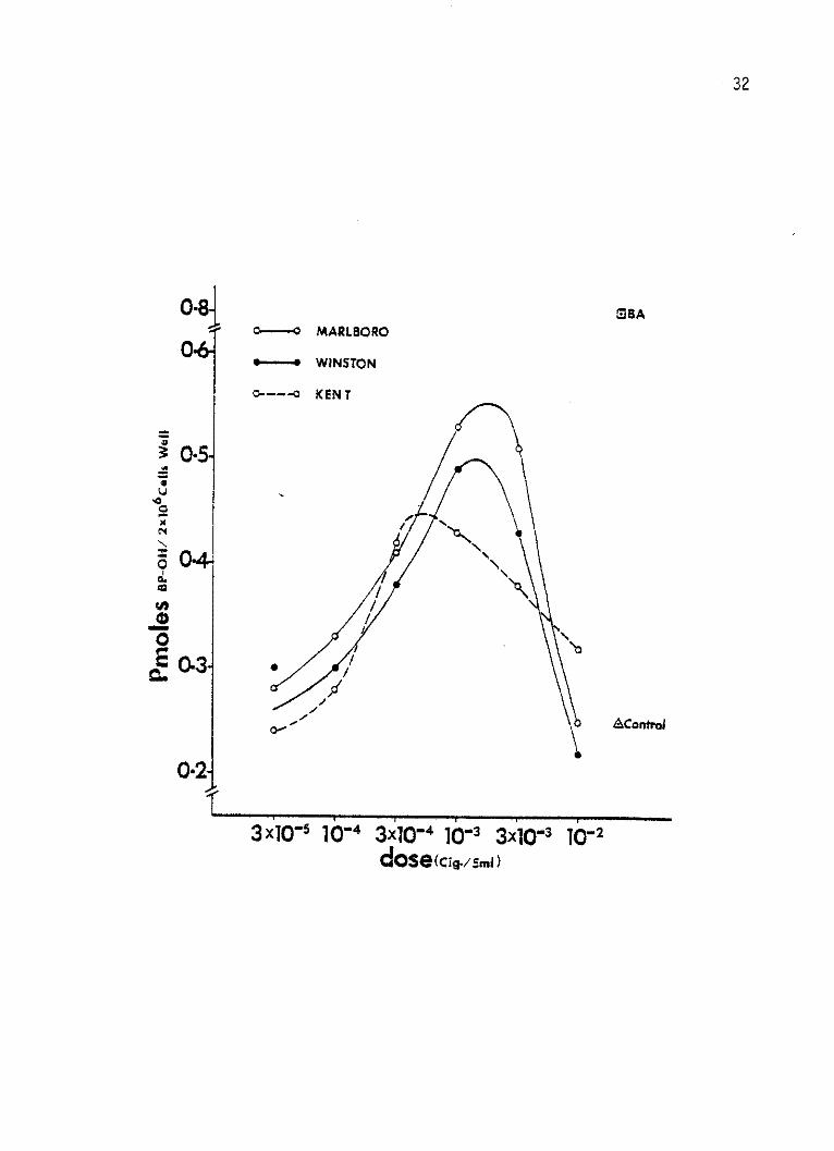
32
0-8-MBAaO--O MARLBORO
0-6*--- WINSTON
0--0 KEN T
04
x I
0-3
0.
0/E0*3
AControd
0021
3 x10⁵ 1b-4 3x10-n4 i-3 3x10⁻³ 10-n2dose(cig./5mo

33
Figure 3: Log dose-response curves of AHH enzyme by smoke con-
densate from five brands of the third (modern)
generation filter cigarettes in cultured human lym-
phocytes were incubated for 96 hours at 2 x 10⁶ cells
per 5 ml per well and received the CSC 44 hours prior
to harvest. Six different doses of each CSC were
added to separate wells. The doses were calculated
as the equivalent cigarette (king size) per well.
The response was expressed as enzyme activity equi-
valent to Pmoles (BP-OH) formed in each well per
minute. Solvent-treated control and BA-induced
lymphocytes' response are also shown.

--- MARLBORO L---- o WINSTON L-
o.0---- C A R LTON0---- KENT L-
&---- TRUE
34
S1BA
0.
~0-5
0-4
0.2
1x
zAControl
3x10 -4 1- 3xlQ4 1O-3 3x1O- 1&2
dose (ci../S.m)

35
Figure 4: Log dose-response curves of AHH enzyme by the whole
smoke condensate and the reconstituted smoke conden-
sate of the lAl standard cigarettes in cultured human
lymphocytes. The lymphocytes were incubated for
96 hours at 2 x 10⁶ cells per 5 ml per well and re-
ceived the smoke condensates 44 hours prior to harvest.
Six different doses of each the starting CSC and the
reconstituted material were added to separate wells.
The response was expressed as enzyme activity equi
valent to Pmoles BP-OH formed in 1 x 10⁶ cells per
minute. Solvent-treated control and BA-induced lym-
phocytes' response are also shown.
OBA.-- STARTING MATERIAL
*.---*RECONSTITUTED
OW." ANO .00"M oo 00 . 4
( Control
36
0-95j
0-25
0-20
0.15
0.10
E
CL
o
0O-
EC.
10-i 3x10⁻³ 10-2 3x10⁻² 10-1 3x10~1
dose (mg/5ml)
37
Figure 5: Log dose-response curves of AHH enzyme by the two
weakly acidic fractions of the lAl standard
cigarettes (WAE: weakly acidic ether soluble) in
cultured human lymphocytes. The lymphocytes were
incubated for 96 hours at 2 x 10⁶ cells per 5 ml
per well and received the CSC fractions 44 hours
prior to harvest. Six different doses of each
fraction were added to separate wells. The response
was expressed as enzyme activity equivalent to Pmoles
BP-OH per 1 x 10⁶ cells per minute. Solvent-treated
control and BA-induced lymphocytes' response are
also shown.

o-o WAE
38
- BA
0-95-
O-3040
~0
o 00200'.
U-o. 1
-rn-p
'011 No Omm mommO-e
400OOW
000.1--0400'
Spool' .0000 & Control
'I 3 3Xs-3 <6-2 3x 10 . 2 1
dose (g51
3x 11041

39
Figure 6: Log dose-response curves of AHH enzyme by the four
basic fractions of the lAI standard cigarettes
(BIa: bases insoluble a, BIb: bases insoluble b,
Bw: bases water soluble and Be: bases ether soluble)
in cultured human lymphocytes. The lymphocytes were
incubated for 96 hours at 2 x 10⁶ cells per 5 ml per
well and received the CSC fractions 44 hours prior to
harvest. Six different doses of each fraction were
added to separate wells. The response was expressed
as enzyme activity equivalent to Pmoles BP-OH per
1 x 10⁶ cells per minute. Solvent treated control
and BA-induced lymphocytes' response are also shown.

EJBA
0-95]
07
0.6
E0.5
0 0*41CM
QI)
E
0, 2w
10-3 3x1O- 3 102 3x10⁻²
dose (mg/5m1)
10.1 3x101
40
Blb
o-----o Bic
Bw
-/
- - --- ~0
- - -
0.1
I;
Control

41
Figure 7: Log dose-response curves of AHH enzyme by the three
strong acidic fractions of the IAl standard cigarettes.
(SAI: strong acids, insoluble, SAe: strong acids,
ether soluble, SAw: strong acids, ether soluble) in
cultured human lymphocytes. The lymphocytes were in-
cubated for 96 hours at 2 x 10⁶ cells per 5 ml well
and received the CSC fractions 44 hours prior to
harvest. Six different doses of each fraction were
added to separate wells. The response was expressed
as enzyme activity equivalent to Pmoles BP-OH per
1 x 10⁶ cells per minute. Solvent-treated control
and BA-induced response are also shown.

0*954
S0-30-
PO
C
0-2
0
ECL 0-101
SA1
SAE
SAW
i0-3 3x10- 3 0-2 3x0- 2
dose <(m/5
42
E BA
A Control
-1 3xIO-lI I
43
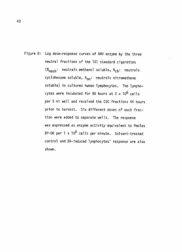
Figure 8: Log dose-response curves of AHH enzyme by the three
neutral fractions of the lAl standard cigarettes
(Nmeoh: neutrals methanol soluble, Nch: neutrals
cyclohexane soluble, Nnm: neutrals nitromethane
soluble) in cultured human lymphocytes. The lympho-
cytes were incubated for 96 hours at 2 x 10⁶ cells
per 5 ml well and received the CSC fractions 44 hours
prior to harvest. Six different doses of each frac-
tion were added to separate wells. The response
was expressed as enzyme activity equivalent to Pmoles
BP-OH per 1 x 10⁶ cells per minute. Solvent-treated
control and BA-induced lymphocytes' response are also
shown.

ElBA
O.O NC H.. NMEOH
e......NNM
0*95
~0.40zI
30
0
0-10
0
Eam1o
3x10⁻³ 10-2 3x10⁻² 16-
dose (mg/5m1)
44
.Apo, &lo00,
MOO/1001/MWO 46I
A/ oto
ra
3x10⁴-0-w3
45
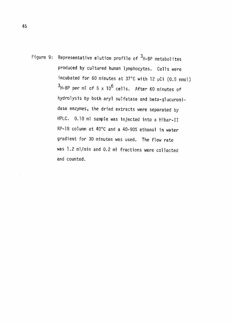
Figure 9: Representative elution profile of 3H-BP metabolites
produced by cultured human lymphocytes. Cells were
incubated for 60 minutes at 37'C with 12 PCi (0.5 nmol)3H-BP per ml of 5 x 10⁶ cells. After 60 minutes of
hydrolysis by both aryl sulfatase and beta-glucuroni-
dase enzymes, the dried extracts were separated by
HPLC. 0.10 ml sample was injected into a Hibar-II
RP-18 column at 40C and a 40-90% ethanol in water
gradient for 30 minutes was used. The flow rate
was 1.2 ml/min and 0.2 ml fractions were collected
and counted.

46
8 A
6
FRACTION

47
Figure 10: Representative elution profile of 3H-BP metabolites
produced by cultured human lymphocytes. Cells were
incubated for 60 minutes at 370C with 12 pCi (0.5 nmol)
3H-BP per ml of 5 x 10⁶ cells. After 60 minutes of
hydrolysis by aryl sulfatase enzyme, the dried ex-
tracts were separated by HPLC. 0.10 ml sample was
injected into a Hibar-II RP-18 column at 400C and a
40-90% ethanol in water gradient for 30 minutes was
used. The flow rate was 1.2 ml/min and 0.2 ml frac-
tions were collected and counted.

48
8 B
01 6 -
E4
2f
f raction

49
Figure 11: Representative elution profile of 3H-BP metabolites
produced by cultured human lymphocytes. Cells were
incubated for 60 minutes at 37*C with 12 pCi
(0.5 nmol) 3H-BP per ml of 5 x 10⁶ cells. The
dried extracts of the nonhydrolysed free metabolites
were separated by HPLC. 0.1 ml sample was injected
and a Hibar-II RP-18 column at 40*C and a 40-90%
ethanol in water gradient for 30 minutes was used.
The flow rate was 1.2 ml/min and 0.2 ml fractions
were collected and counted.

50
8 C
0x
2 l
11 all.If raction

Figure 12: Summary of amount of radioactivity in various peaks
after hydrolysis with beta-glucuronidase or aryl
sulfatase in cultured human lymphocytes incubated
for 60 minutes at 37'C with 12 pCi (0.5 nmol) 3H-BP
per ml of 5 x 10⁶ cells. a) both enzymes; b) both
enzymes and D-saccharic acid 1-4 - lactone previously
buffered to pH 7.5 (an inhibitor of beta-glucuronidase)
c) nonhydrolysed free metabolites. The background
plus blank counts are below the ordinate.
51

abc I IIIU~ ~ I I..J
1 2 3
II I I
LJ L1J
4 1
DIOLS Q PHEN
52
C,I0
x'm
0..
20
10o
0.1U
I a a 1 .1 1 , -- 1 2 , - ----
.J0

Figure 13: Summary of amount of radioactivity in various peaks
after hydrolysis with beta-glucuronidase or aryl
sulfatase in cultured human lymphocytes incubated
for 18 hours at 370C with 12 PCi (0.5 nmol) 3H-BP
per ml of 4 x 10⁶ cells . a) both enzymes; b) both
enzymes and D-saccharic acid 1,4-lactone previously
buffered to pH 7.5 (an inhibitor of beta-glucuronidase).
c) nonhydrolysed free metabolites. The background
plus blank counts are below the ordinate.
53
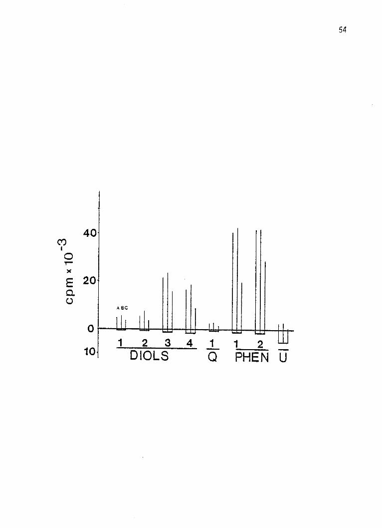
54
40Co
E 2 0
A 8C
1 2341 12DIOLS Q PHE N U

Figure 14: Summary of amount of radioactivity in various peaks
after hydrolysis with beta-glucuronidase or aryl
sulfatase in cultured human lymphocytes incubated
for 24 hours at 370C with 10 pCi of 3H-BP which had
been diluted with cold BP to a specific activity of
10.2 pg per 10 pCi per ml of 2.5 x 10⁶ cells.
a) both enzymes; b) both enzymes and D-saccharic acid
1,4- lactone previously buffered to pH 7.5 (an inhi-
bitor of beta-glucuronidase); c) nonhydrolysed free
metabolites. The background plus blank counts are
below the ordinate.
55

I -#
LL5L
2 3diols.
If!
LUW LUpLUn.
qa phen .U
56
151
C,
01o
E0.
10
5.
ABC
Ill0
29 1
----- --

57
Figure 15: Profile of separation of 3H-BP metabolites produced by
cultured human lymphocytes. The BA induced lymphocytes
were incubated for 3 hours at 370C with 72 pCi 3H-BP
(0.75 1GM) at a concentration of 5 x 10⁶ cells per ml.
The reaction was stopped by addition of 3 ml ice-cold
ethanol. The precipitate was sedimented, and the alco-
holic supernatant was taken and dried under nitrogen.
The residue was dissolved in 2.00 ml 70% ethanol.
0.5 ml the sample was injected onto a 4.6 x 2.50 nm
neutral alumina column during a flow rate of 1.2 ml/min.
A linear hexane-ethanol gradient was programmed for
30 minutes. Twenty 2 ml fractions were collected.
The solvent was changed to water and another ten frac-
tions were collected after which the solvent was
changed to phosphate buffer (0.05M, pH 3.0) . Fif-
teen fractions were collected then the solvent was
changed to 25% formic acid. Fifteen additional frac-
tions were collected. A total of sixty 2 ml fractions
were collected and 0.2 ml aliquots were taken and
counted.

58
8
6
x
2
vs
HfrEONIue o X~TM20a~n P04- FOR9MIC ACIDo
f ro ctiori

Figure 16: Reverse phase HPLC separation of peaks A and B (non-
conjugated metabolites) from the neutral alumina
column eluting with hexane-ethanol gradient (Figure 15).
Three fractions from peak A and 4 fractions from peak B,
respectively, were pooled, dried under nitrogen, and
each was dissolved in 0.5 ml 70% ethanol. A 0.10 ml
aliquot of each was injected into the HPLC. Elution
was with a 60-100% methanol gradient over 30 minutes
with a flow rate of 1.0 ml/min; 0.20 ml fractions
were collected and counted.
59

peak A
'3
peak B
fraction
60
KM
3,
2
C-
xo
0
2
1
0
0 0
I.
I'll- -

Summary of the quantitation of CSC yield of ten brands
of cigarettes. Ten ml of the CSC solution (equivalent
to 5 cigarettes) was dried under nitrogen in preweighed
vials and stored in a vacuum dessicator for 48 hours.
The vials were reweighed and the amount of CSC equiva-
lent to five cigarettes and the CSC yield in mg/cig.
was calculated.
61
Table I:

62
TABLE I
Brand
Camel
Chesterfield
Marlboro
Winston
Kent
Marlboro Lights
Winston Lights
Kent Lights
Carlton
True
Mg. Tar/5Ci.
168.4
184.3
073.4
054.2
058.2
052.1
076.4
005.6
004.7
013.9
Mg. Tar/Cig.
33.7
36.8
14.7
10.8
11.6
10.4
15.3
1.12
0.94
2.27
|_1|| 1. . ..... .. -... .. ... .,. .... ... _ .
1III .I I .KI] ] I IIII i II -.-. -- - --- s- - -.----. ., .
CHAPTER BIBLIOGRAPHY
1. Ghanayem, B., Cantrell, E., Busbee, D., and Burleson, L.(1979). Separation of both primary and secondarymetabolites of benzo-a-pyrene on alumina HPLC chroma-tography. Abstracts, Texas Pharmacologist's Mtg.,May, Abst. #42.
2. Holder, G.M., Yagi, H., Jerina, D.M., Levin, W., Lu, A.Y.H.,and Conney, A.H. (1975). Metabolism of BP: Effectsof substrate concentration and 3-methylcholanthreneon hepatic metabolism by microsomes from rats and mice.Arch. Biochem. Biophys. 170:557.
63

CHAPTER IV
DISCUSSION
The link between cigarette smoking and lung cancer was estab-
lished by the results of all prospective and retrospective investi-
gations from many countries. The first experiments to establish
tobacco smoke as a carcinogen in laboratory animals were done with
CSC (tar) applications to epithelial tissue of mice and rabbits
(4,19), which was followed by further confirmations of the same
results by a large number of investigations (3,5,17).
Since that time, part of the purpose of the tobacco carcinoge-
nesis' program has been to identify the carcinogenic tobacco smoke
components, and once such components have been identified attempts
might then be made for their reduction or removal. The identifica-
tion of carcinogenic tobacco smoke components was positively corre-
lated with the discovery of a lower risk of developing lung cancer
in the smokers of filter cigarettes than in the smokers of unfiltered
cigarettes (18).
In the present study, the CSC yield of what we call "third
generation" cigarettes (the less harmful cigarettes) such as Carlton
and Kent Lights was quantitatively lower than the CSC yield of both
64

65
the first (unfiltered) and the second generation (poorly filtered)
cigarettes. It was also demonstrated that the CSC yield of the
filter cigarettes was far lower than the yield of the unfiltered
cigarettes. These data, with the results of earlier reports (18)
that had attributed the diminished risk of developing lung cancer
in the smokers of filter cigarettes to the reduced yield of tar and
the suspected association between high AHH inducibility and suscep-
tibility to chemical carcinogenesis, led us to consider the rela-
tionship between AHH and the effects of CSC from different cigarettes
on mitogen stimulated cultured human lymphocytes. Cultured human
lymphocytes responded with increased levels of AHH enzyme upon
exposure to cigarette tars from all the brands of cigarettes tested.
A dose-response relationship was demonstrated. All the CSC's
tested were AHH inducers, but they differed in their potency,
efficacy and toxicity. A peak AHH induction (maximum response) was
reached at lower doses of CSC's derived from unfiltered cigarettes
than CSC's derived from filter cigarettes (the dose was expressed
as equivalent cigarettes per well). With the exception of Carlton
and Kent Lights, CSC's toxic effects were seen as reduction in
enzyme activity per well. Toxic effects were noticed at lower
doses of CSC's derived from unfiltered cigarettes than CSC's derived
from filter cigarettes. The extreme toxicity of unfiltered ciga-
rettes seemed to attenuate the course of the dose response before
maximum enzyme activity was induced. The less harmful cigarettes
were good inducers of AHH, with a maximum response reached at very

66
high doses, and toxicity was comparably low. Carlton and Kent
Lights' CSC's were effective inducers, but neither the maximum
response nor toxicity was reached by any of the doses tested under
our experimental conditions.
The data are consistent with the reports of AHH induction that
occurs in vivo following cigarette smoking (11,12,13). AHH induc-
tion by CSC's in human lymphocytes is also consistent with the
observations of Welch et al. (16), and Marcotte and Witschi (10)
who showed that pulmonary AHH was inducible in rats exposed to
regular or marijuana cigarettes' smoke and Kouri et al. (8) who
showed that B6 and C3H/fmai mice had responded by increased levels
of pulmonary AHH after their exposure to cigarettes' smoke. Our
data suggest that the first generation cigarettes (high-tar yield)
are highly toxic, which correlates with the high risk of developing
lung cancer in the smokers of such cigarettes. The third generation
cigarettes (lowest-tar yield) had very low toxicity, which also
correlates with the lower risk of developing lung cancer in the
smokers of such cigarettes. The data do not address the question
of possible relationships between AHH and high risk of developing
lung cancer, as all cigarettes were effective inducers of AHH and
all cells were derived from the same person.
Cultured human lymphocytes were induced by the whole CSC and
the reconstituted CSC derived from the standard lAl cigarettes. At
any given dose, the starting CSC was more efficacious than the

67
reconstituted CSC. This can be explained on the basis that some of
the actively inducing components of the CSC might be lost during
the fractionation processes. This is consistent with the finding
of Swain et al. (15) that the reconstitution of the 12 fractions of
the Al CSC recovered only 90.8% of the starting CSC activity.
Toxicity was not seen by either the starting or the reconstituted
materials under the experimental conditions. The data is consistent
with the findings of Kouri et al. (8) who described both the start-
ing and the reconstituted lAl materials as weak inducers of pulmo-
nary AHH activity in certain strains of mice. It was observed that
the starting CSC produced a lower reponse than certain fractions
(Bib), which suggests that CSC contains both inducing and inhibi-
tory components.
The weakly acidic fractions (WAI and WAE) showed a low effi-
cacy for inducing AHH enzyme in human lymphocytes. In fact, they
reduced enzyme activity of AHH at certain dose levels.
According to the findings of other investigators, WAI only,
was found to produce in vitro cellular transformation of the
C3H-lOT-1/2 cell lines (9). WAE was found to be an active Jn vivo
tumor promoter (2) and produced in vitro cellular transformation of
Swiss mouse cells (14). Both WAI and WAE were reported to be weak
inducers of AHH in C57/BL6 mouse in vivo (8). Regarding their
mutagenic activity, WAI was reported to be a good mutagenic agent
and WAE to be a weak mutagenic agent in bacteria (7).

68
All the strongly acidic fractions exhibited an inhibitory
action at doses lower than 10-2 mg/well and all had a weak activity
in inducing AHH in cultured human lymphocytes. All the three
fractions were toxic at some dose. The three fractions (SA, SAE,
and SAW) were reported to be weak inhibitors of pulmonary AHH
activity in vivo (8). Other investigators reported the three
fractions as being devoid of activity in the biological tests that
they used (2,7,9,14). With the report of Wynder and Hoffman (17)
that a significant tumor promoting activity was detected in the
acidic fraction of CSC, it is possible that some of the compounds
in WAI obtained from the lAl cigarettes are also present in the
WAE fraction.
The most potent and efficacious of the twelve fractions was
the basic fraction Bib. Bia fraction was a good AHH inducer, BE
was medium in its activity, while Bw was considered an inhibitor
and a toxic fraction. Our data is consistent with Kouri et al.
(8) who reported both Bjb and Bja as good inducers of pulmonary AHH
in vivo, and with Kier et al. (7) who reported both fractions as
mutagenic fractions in Salmonella. Bja as good inducers of pulmo-
nary AHH in vivo, and with Kier et al. (7) who reported both frac-
tions as mutagenic dractions in Salmonella. Bb was reported to
cause in vitro cell transformation in C3H-lOT 1/2 (9) and Swiss
mouse cells (9) and to cause tumor promotion in C3H-lOT-1/2 mouse
cells (2). BE was found to be a tumor promoter (2), a weak inducer

69
of pulmonary AHH (8), and a weak mutagen in Salmonella (7). Bw was
not active in all reported biological assays (2,7,9,14) but was a
weak inhibitor of pulmonary AHH (8).
None of the neutral fractions (Nch, Nnm, Nmeoh) was toxic
under these experimental conditions. With the exception of Nch,
they were potent and efficacious inducers of AHH. This data is
consistent with Kouri et al. (8) who described Nnm and Nmeoh as
good pulmonary AHH inducers and Nch as a weak inducer. Nmeoh was
a tumor promoter in C3H mouse cells (9) but was devoid of mutageni-
city in Salmonella (14). Nnm caused in vitro cell transformation
in Swiss mouse cells (14) and was devoid of mutagenicity (7).
The good response of AHH activity by certain basic fractions
and BIb and BIa) and by certain neutral fractions (Nnm and Nmeoh)
and their reported inhibitory action on BP metabolism by Kouri et
al. (8) suggest that these fractions contain certain PAH struc-
turally similar to BP which are capable of inducing AHH and are
capable of competitively inhibiting BP metabolism.
In view of the lack of information on the chemical composition
of the fractions, any correlation between the data from different
tests will be difficult. Until further information about the
chemical composition of those fractions and their mechanism of
carcinogenesis becomes available, the current data suggest united
efforts of the cancer researchers and the tobacco industries to
reduce the tar and nicotine yield of cigarettes as an emergent
action to face the high risks of lung cancer among smokers. We
70
have to admit progress has been made in this area, but more work is
imperative if we are to achieve the desired purpose and results.
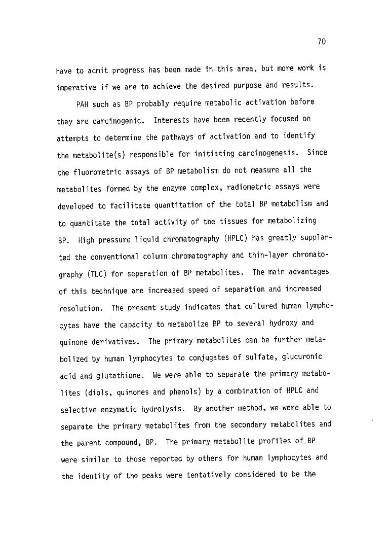
PAH such as BP probably require metabolic activation before
they are carcinogenic. Interests have been recently focused on
attempts to determine the pathways of activation and to identify
the metabolite(s) responsible for initiating carcinogenesis. Since
the fluorometric assays of BP metabolism do not measure all the
metabolites formed by the enzyme complex, radiometric assays were
developed to facilitate quantitation of the total BP metabolism and
to quantitate the total activity of the tissues for metabolizing
BP. High pressure liquid chromatography (HPLC) has greatly supplan-
ted the conventional column chromatography and thin-layer chromato-
graphy (TLC) for separation of BP metabolites. The main advantages
of this technique are increased speed of separation and increased
resolution. The present study indicates that cultured human lympho-
cytes have the capacity to metabolize BP to several hydroxy and
quinone derivatives. The primary metabolites can be further meta-
bolized by human lymphocytes to conjugates of sulfate, glucuronic
acid and glutathione. We were able to separate the primary metabo-
lites (diols, quinones and phenols) by a combination of HPLC and
selective enzymatic hydrolysis. By another method, we were able to
separate the primary metabolites from the secondary metabolites and
the parent compound, BP. The primary metabolite profiles of BP
were similar to those reported by others for human lymphocytes and
the identity of the peaks were tentatively considered to be the

71
same as reported by Holder et al. (6). This data suggest that
during a short time of reaction where the substrate concentration
does not saturate the enzyme complex, a substantial amount of
conjugation occurs via glucuronide formation. The phenols were
conjugated to an equal extent with glucuronide and sulfate. It
also suggested that the primary metabolites and conjugates vary in
amount between individuals, and even vary intraindividually depend-
ing upon time of incubation and BP concentration.
A revision of a method reported by Autrup et al. (1) led us to
separate the conjugates, the primary metabolites nd the parent com-
pound BP by a solvent gradient followed by pH step gradient elution
from a neutral alumina column. The advantage of our method over
the original method is that we were able to separate the primary
metabolites from the parent compound and reduce the total elution
volume.
If all of the intermediates of BP are enzymatically produced,
then a cell at great risk might either accelerate the production of
an activated intermediate (epoxide or diol epoxide) or have a
reduced ability to detoxify the activated carcinogen to conjugated,
water soluble, urine excretable products. Either action would lead
to intracellular accumulation of the active molecular species, with
increased probability of reacting with the target site to produce
malignant transformation. Conversely, a resistant (or a less sus-
ceptible) cell may be characterized by decelerated formation of the
activated carcinogen or increased conjugating enzymes' activity
yielding water soluble, urine excretable products.

72
If that assumption were true, then this assay protocol could
provide data on a persons relative rate of activation/inactivation
reactions, and hence reflect that person's relative risk to develop
cancer.

CHAPTER BIBLIOGRAPHY
1. Autrup, H. (1978). Separation of water-soluble metabolitesof benzo-a-pyrene formed by cultured human colon.Bioch. Pharm. (in press).
2. Bock, F.G., Swain, A.P., and Stedman, R.L. (1969). Bioassayof major fractions of cigarette smoke condensate by anaccelerated technique. Cancer Res. 29:584.
3. Dontewill, W. (1976). Biological evaluation of carcinogensin tobacco and tobacco smoke. In: Smoking and Health,DHEW, publ. no. (NIH) 76-1221, p. 209. U.S. Gov. PrintingOffice, Washington, D.C.
4. Graham, E.L., Croninger, A.B., and Wynder, E.L. (1957). Ex-perimental production of carcinoma with cigarette tar.IV. Successful experiments with rabbits. Cancer Res.13:855.
5. Hoffman, D., Schmeltz, I., Hecht, S.S., and Wynder, E.L.(190n the identification of carcinogens, tumor promotersand carcinogens in tobacco smoke. In: Smoking and Health,DHEW publ. no. (NIH) 76-1221, p. 125, U.S. Gov. PrintingOffice, Washington, D.C.
6. Holder, G.M., Yagi, H., Jerina, D.M., Levin, W., A.Y.H., Lu,and Conney, A.H. (1975). Metabolism of BP: Effects ofsubstrate concentration and 3-methylcholantrene pretreat-ment on hepatic metabolism by microsomes from rats andmice. Arch. Biochem. Biophys. 170:557.
7. Kier, L.D., Yamasaki, E., and Ames, B.N. (1974). Detectionof mutagenic activity in cigarette smoke condensate.Proc. Natl. Acad. Sci. U.S.A. 71:4159.
8. Kouri, R.E. (1974). Experimental lung cancer, carcinogenesisand bioassays. International symposium held at theBatelle Research Center, Seattle, Washington, U.S.A.,June (1974), Springer-Verlag, Berlin, Heidelberg,New York (1974).
73

74
9. Kouri, R.E., Whitmire, C., and Benedict, W. (1975). In vivoand in vitro effects of cigarette smoke condensatefractions. Proc. Amer. Assn. Cancer Res. 16:173.
10. Marcotte, J. and Witchi, H.P. (1972). Induction of pulmonaryAHH by marijuana. Res. Commun. Chem. Path. Pharmacol.4:561.
11. McLemore, T.L., Martin, R.R., Toppell, K.L., Busbee, D.L.,and Cantrell, E.T. (1977). Comparison of AHH inductionin cultured blood lymphocytes and pulmonary macrophages.J. Clin. Invest. 60:1017.
12. McLemore, T.L., Martin,R.R., Toppell, K.L., Cantrell, E.T.,and Busbee, D.L. (1978). In vitro induction of AHH;Dissociation between enzyme values in pulmonary macro-phages and blood lymphocytes from lung cancer patients.Cancer Res. (submitted).
13. McLemore, T.L., Martin, R.R., Busbee, D.L., Richie, R.C.,Springer, R.R., Toppell, K.L., and Cantrell, E.T.(1977). AHH activity in pulmonary macrophages andlymphocytes from lung cancer and noncancer patients.Cancer Res. 37:1175.
14. Rhim, J.S., and Huebner, R.J. (1973). In vitro transforma-tion assay of major fractions of cigarette smoke con-densate in mammalian cell lines. Proc. Soc. Exper.Biol. Med. 142:1003.
15. Swain, A.P., Cooper, J.E., and Stedman, R.L. (1969). Largescale fractionation of cigarette smoke condensate forchemical and biologic investigations. Cancer Res.29:579.
16. Welch, R.M., Loh, A., Conney, A.H. (1971). Cigarette smoke:Stimulatory effect on metabolism of 3,4-benzopyrene byenzymes in rat lung. Life Sciences 10:215.
17. Wynder, E.L., and Hoffmann, D. (1967). Tobacco and tobaccosmoke: Studies in experimental carcinogenesis. AcademicPress, N.Y.
18. Wynder, E.L., and Mabuchi, K. (1972). Etiological and preven-tive aspects of human cancer. Prev. Med. 1:300.
19. Wynder, E.L., Graham, E.L., and Croninger, A.B. (1953). Ex-perimental production of carcinoma with cigarette tar.Cancer Res. 13:885.

LITERATURE CITED
Ames, B.N., Durston, W.E., Yamasaki, E., et al. (1973). Carcino-gens as mutagens: A simple test system combining liver homo-genate for activation and bacteria for detection. Proc.Nat. Acad. Sci. USA 70:2281.
Auerbach, 0., Stout, A.P., Hammond, E.C., and Garfinkel, L.(1961). Changes in relation to lung cancer. N. Engl. J. Med.265:253.
Auerbach, 0., Stout, A.P., Hammond, E.C., and Garfinkel, L. (1962).Changes in bronchial epithelium in relation to sex, age, resi-dence, smoking and pneumonia. N. Engj. J. Med. 267:111.
Auerbach, 0., Stout, A.P., Hammond, E.C., and Garfinkel, L. (1962).Bronchial epithelium in former smokers. N. Engl. J. Med. 267:119.
Autrup, H. (1978). Separation of water-soluble metabolites ofbenzo-a-pyrene formed by cultured human colon. Bioch. Pharm.(in press).
Berwald, Y., and Sachs, L. (1965). In vitro transformation ofnormal cells to tumor cells by carcinogenic hydrocarbons.J. Natl. Cancer Inst. 35:641.
Bock, F.G. (1968). Dose-response: Experimental carcinogenesis.Nat. Cancer Inst. Monogr. 28:57.
Bock, F.G., Swain, A.P., and Stedman, R.L. (1969). Bioassay ofmajor fractions of cigarette smoke condensate by an accele-rated technique. Cancer Res. 29:584.
Brookes, P., and Lawley, P.D. (1964). Evidence for the bindingof polynuclear aromatic hydrocarbons to the nucleic acids ofmouse skin: Relation between carcinogenic power of hydro-carbons and their binding to deoxyribonucleic acid. Nature202:781.
75

76
Boyland, E., and K. Williams (1965). An enzyme catalysing theconjugation of epoxides with glutathione. Biochem. J. 94:190.
Cantrell, E., Abreu, M., and Busbee, D. (1976). A simple assay ofAHH in cultured human lymphocytes. Biochem. Biophys. Res.Commun. 70:474.
Cantrell, E.T., Marshall, M., McLemore, T., Ghanayem, B., andBusbee, D. (1979). Secondary metabolism of benzopyrene inhuman cells. Western Pharm. Proceedings.
Chen, T.T., and Heidelberger, C. (1969). Quantitative studies onthe malignant transformation of mouse prostate cells by car-cinogenic hydrocarbons in vitro. Intl. J. Cancer 4:166.
Clayson, D.B. (1962). Chemical Carcinogenesis, Little, Brown,Boston, Massachusetts.
Cohen, G.M., Moore, B.P., and Bridges, J.W. (1977). Organic solventsoluble sulfate ester conjugates of monohydroxy benzo-a-pyrenes.Biochem. Pharmacy 26:551.
Cohen, G.M., Haws, S.M., Moore, B.P., and Bridges, J.W. (1976).BP-3-yl hydrogen sulfate, a major ethyl acetate-extract-able metabolite of BP in human, hamster and rat lungcultures. Biochem. Pharmacology 25:2561.
Conney, A.H. (1967). Pharmacological implications of microsomalenzyme induction. Pharmacol. Rev. 19:317.
Culter, S.J., Scotto, J., Devesa, S.S., et al. (1974). ThirdNational Cancer Survey - An overview of available information.J. Natl. Cancer Inst. 53:1565.
Doly, J.W., Jerina, D.M., Witkop, B., Arene, P. (1972). Oxidesand the NIH shift: The metabolism, toxicity, and carcino-genicity of aromatic compounds. Experientia 28:1129.
David, J.J., James, J., Robert, S., and Harri, V. (1977). Bio-logical reactive intermediates, Plenum Press, New Yorkand London.
Davis, B.R., Witehead, Y.K., Gill, M.E., Lee, P.N., Butterworth,A.D., and Roe, F.J.C. (1975). Response of rat lung to in-haled vapor phase constituents (VP) of TS alone or inconjunction with SC given by intratracheal instillationBr. J. Cancer 31 :462.
77
Denoix, P.F., Schwartz, D., and Anguera, G. (1958). L'enquitefrancaise sur l'etiologue du cancer broncho-pulmonaire.Analyse detailee. Bull. Ass. Cancer 26:1085.
Doll, R., and Hill, A.B. (1964). Mortality in relation to smoking.Ten year's observations of British doctors. Brit. Med. J. 1:1399.
Dontewill, W., Elmenhorst, H., Harke, H.P., Reckzeh, G., Weber,K.H., Misfeld, J., and Timm, J. (1970). ExperimentelleUntersuchungen uber die Tumorerzeugung von zigarettenranch-Kondensatem an der Mausehaut, I.Z., Krebsforsch 73:265.
Folk, H.L., and Kotin, P. (1963). Symposium on chemical car-cinogenesis. Clin. Pharmacol. Ther. 4:88.
Gelboin, H.V. (1972). Studies on the mechanism of microsomal hy-droxylase induction and its role in carcinogen action. Rev.Canc. Biol. 31:39.
Gelboin, H.V. (1969). Microsome-dependent binding of BP to DNA,Cancer Res. 29:1272.
Gelboin, H.V., and Tso, P.O. (1977). Polycyclic aromatic hydro-carbons and cancer. I. Environment, chemistry and metabolism.Academic Press.
Gelboin, H.V., Kinoshita, N., and Wiebel, F.J. (1972). Microsomalhydroxylazes: Induction and role in PAH carcinogenesis andtoxicity. Fed. Proc. 31:1298.
Ghanayem, B., Cantrell, E., Busbee, D., Burleson, L. (1979). Sepa-ration of both primary and secondary metabolites of benzo(a)-pyrene on alumina HPLC chromatography. Abstracts, TexasPharmacologist's Meeting, May, Abst. #42.
Gori, G.B., and Peters, J.A. (1975). Etiology and prevention ofcancer. Prev. Med. 4:235.
Graham, E.L., Croninger, A.B., and Wynder, E.L. (1957). Experimen-tal production of carcinoma with cigarette tar. IV. Success-ful experiments with rabbits. Cancer Res. 13:855.
Grover, P.L., and Sims, P. (1968). Enzyme-catalyzed reactions ofPAH's with deoxyribonucleic acid proteins in vitro. Biochem.Pharmacol. 110:159.
Grover, P.L., Hewer, A., and Sims, P. (1972). Formation ofK-region epoxides as microsomal metabolites of pyrene andbenzo(a)pyrene. Biochem. Pharmacol. 21:2713.

78
Grover, P.L., Sims, P., Huberman, E., Marquardt, H., Kuroki, T.,and Heidelberger, C. (1971). In vitro transformation ofrodent cells by K-region derivatives of PAH's. Proc. Natl.Acad. Sci. 68:1098.
Hammond, E.L., and Horn, D. (1958). Smoking death-rates reporton forty-four months follow-up of 187,783 men. J. Amer. Med.Assn. 166:1159,1294.
Hartwell, J.L. (1951). Survey of compounds which have beentested for carcinogenic activity. U.S. Public Health ServicePubl. no. 149, 2nd ed., Washington, D.C.
Hartwell, J.L., and Shubik, P. (1957, 1959). Survey of compoundswhich have been tested for carcinogenic activity, supply. 1and 2. U.S. Health Service Publ. no. 149, Washington,D.C.
Hoffman, D., Schmeltz, I., Hecht, S.S., and Wynder, E.L. (1976).On the identification of carcinogens, tumor promoters andcarcinogens in tobacco smoke. In "Smoking and Health", DHEWpubl. no. (NIH) 76-1221, p. 125, U.S. Gov. Printing Office,Washington, D.C.
Hoffman, D., and Wynder, E.C. (1971). A study of tobacco carcinogenesis. XI. Tumor initiators, tumor accelerators and tumorpromoting activity of condensate fractions. Cancer 27:848.
Holder, G.M., Yagi, H., Jerina, D.M., Levin, W., A.Y.H., Lu, andConney, A.H. (1975). Metabolism of BP: Effects of substrate concentration and 3-methylcholantrene pretreatmenton hepatic metabolism by microsomes from rats and mice.Arch, Biochem. Biophys. 170:557.
Huberman, E., Kuroki, T., Marquardt, H., Silkirk, J.K., Heildelberger, C., Grover, P.L., and Sims, P. (1972). Transformationof hamster embryo cells by epoxides and other derivatives ofpolycyclic hydrocarbons. Cancer Res. 32:1391.
Jerina, D.M., and Daly, J.W. (1974). Arene oxides. A new aspectof drug metabolism. Science 185:574.
Kier, L.D., Yamasaki, E., and Ames, B.N. (1974). Detection ofmutagenic activity in cigarette smoke condensate. Proc. Natl.Acad. Sci. U.S.A. 71:4159.
Kellerman, G., Shaw, C.R., and Luyten-Kellermann, M. (1973).Genetic variation of AHH in human lymphocytes. Am. J. HumanGenet. 25:327.

79
Kellermann, G., Shaw, C.R., and Luyten-Kellerman, M. (1973).AHH inducibility and bronchogenic carcinoma. N. Engl. J.Med. 289:934.
Kouri, R.E. (1974). Experimental lung cancer, carcinogenesisand bioassays. International symposium held at theBatelle Research Center, Seattle, WA., USA, June (1974),Springer-Verlag, Berlin, Heidelberg, New York (1974).
Kouri, R.E., Whitmire, C., and Benedict, W. (1975). In vivo andin vitro effects of cigarette smoke condensate fractions.Proc. Amer. Assn. Cancer Res. 16:173.
Krahn, D.F., and Heidelberger, C. (1975). Microsomal mediatedmutagenesis in Chinese hamster cells by chemical oncogens.Proc. Am. Assn. Cancer Res. 16:74.
Kreyberg, L. (1968). Nonsmokers and geographic pathology oflung cancer. In: "The Lung", A.A. Liebon and D.E. Smith,eds., pp. 273-283, Williams and Wilkins, Baltimore, MD.
Kuroki, T., Huberman, E., Marquardt, H., Selkirk, J.K., Heidelberger, C., Grover, P.L., and Sims, P. (1972). Binding ofK-region epoxides and other derivatives of BA and dibenz-(a,b)-anthracene to DNA, RNA, and proteins of transformablecells. Chem - Biol. Interact 4:389.
Kuratsune, M. (1956). BP content of certain pyrogenic materials.J. Nat. Cancer Inst. 16:1485.
Lazar, P.H. (1974). Chouroulinkor, I., Izard, C., Moree-Testa,P., and Herman, D. (1974). Bioassays of carcinogenicityafter fractionation of cigarette smoke condensate.Biomedicine 20:214.
Marcotte, J., and Witchi, H.P. (1972). Induction of pulmonaryAHH by marijuana. Res. Commun. Chem. Path. Pharmacol.4:561.
Marquardt, H., Kuroki, T., Huberman, E., Selkirk, J.K., Heidel-berger, C., Grover, P.L., and Sims, P. (1972). Malignanttransformation of cells derived from mouse prostate byepoxides and other derivatives of polycyclic hydrocarbons.Cancer Res. 32:716.
McLemore, T.L., Martin, R.R., Toppell, K.L., Busbee, D.L., andCantrell, E.T. (1977). Comparison of AHH induction in culturedblood lymphocytes and pulmonary macrophages. J. Clin. Invest.60:1017.

80
McLemore, T.L., Martin, R.R., Toppell, K.L., Cantrell, E.T., andBusbee, D.L. (1978). In vitro induction of AHH: Dissocia-tion between enzyme values in pulmonary macrophages andblood lymphocytes from lung cancer patients. Cancer Res.(submitted).
McLemore, T.L., Martin, R.R., Busbee, D.L., Richie, R.C., Springer,R.R., Toppell, K.L., and Cantrell, E.T. (1977). AHH activityin pulmonary macrophages and lymphocytes from lung cancer andnoncancer patients. Cancer Res. 37:1175.
National Academy of Science Reports, USA (1972). Particulate poly-cylic organic matter. Committee on Biologic Effectsof Atmospheric Pollutants, Division of Medical Sciences,National Research Council, Washington, D.C.
Nebert, W.D., Roberts, J., Adelman, C.R., and Critofalo, J.V.(1978). Pharmacological intervention in the aging pro-cess. Chapter 4, Genetic Control of Drug Metabolism,Plenum Press, N.Y.
Nemoto, N., and Gelboin, H.V. (1976). Enzymatic conjugation ofBP oxide phenols and dihydrodiols with UDP-glucuronicacid. Biochem. Pharm. 25:1221.
Nemoto, N., Gelboin, H.V., Habig, W.H., Ketley, J.N., andJakoby, W.B. (1975). K-region BP-4,5-oxide in conjugatedby homogenous glutathione-s-transferase. Nature (London)255:512.
Nemoto, N., Takayama, S., and Gelboin, H.V. (1977). Enzymaticconversion of BP phenols, dihydrodiols and quinones tosulfate conjugates. Biochem. Pharmacol. 26:1825.
Oesch, F. (1972). Mammalian epoxide hydrases: Inducible en-zymes catalyzing the inactivation of carcinogenic andcytotoxic metabolites derived from aromatic and olefiniccompounds. Xenobiotica 3:305.
Paul, 0.P., Tso, P.O., and Dipaolo, J.P. (1974). ChemicalCarcinogenesis - Part A, Marcel Dekker, 1974.
Pietropaolo, C., and Weinstein, I.B. (1975). Binding of 3H-BPto natural and synthetic nucleic acids in a subcellularmicrosomal system. Cancer Res. 35:2191.
Potts, P. (1775). Chirurgical Observations. London, Hawkes andCollins.
81
Rasmussen, R.E. (1975). Tobacco smoke condensate and DNArepair. Life Sciences 17:767.
Rasco, M.A., Tamauchi, T., Johnston, D.A., Fischer, S.M., andShaw, C.R. (1978). AHH inducibility in normal and cancerpopulations. Meeting of the American Association forCancer Research, Washington, D.C. (abstract), April, 1978.
Rhim, J.S., and Huebner, R.J. (1973). In vitro transformationassay of major fractions of cigarette smoke condensatein mammalian cell lines. Proc. Soc. Exper. Biol. Med.142:1003.
Schmeltz, I., and Hoffman, D. (1977). Nitrogen containing com-pounds in tobacco smoke. Chem. Rev. 77:295.
Stedman, R.L. (1978). The chemical composition of tobacco andtobacco smoke. Chem. Rev. 68:153.
Smoking and Health. Report of the Advisory Committee to theSurgeon General of the Public Health Service, U.S. PublicHealth Service Publication, No. 1103. Washington, D.C.,U.S. Government Printing Office (1964).
Sims, P., Grover, P.L., Swaisland, A., Pal, K., and Hewer, A.(1974). Metabolic activation of BP proceeds by a diolepoxide. Nature (London) 252:326.
Swain, A.P., Cooper, J.E., and Stedman, R.L. (1969). Largescale fractionation of cigarette smoke condensate forchemical and biologic investigations. Cancer Res. 29:579.
Thompson, J.I. and Co. (1972). Survey of compounds which havebeen tested for carcinogenic activity, 1968-69 volume,Public Health Service, publ. no. 149, Washington, D.C.
The Health Consequences of Smoking (1968). A Public HealthService Review, U.S. Department of Health, Education andWelfare. Public Health Service publ. no. 1696, Washington, D.C.
The Health Consequences of Smoking. A report to the SurgeonGeneral (1971). U.S. Department of Health, Educationand Welfare, U.S. Public Health Service, publ. (HSM)71-7513, p. 458.

82
Welch, R.M., Loh, A., and Conney, A.H. (1971). Cigarette smoke:Stimulatory effect on metabolism of 3,4-benzopyrene byenzymes in rat lung. Life Sciences 10:215.
Whitehead, J.K., and Rothwell, K. (1969). The mouse skincarcinogenicity of cigarette smoke condensate: Frac-tioned by solvent partition methods. British J. Cancer23:840.
Wood, A.W., Levin, W., Lu, A.Y.H., Yagi, H., Hernandez, 0.,Jerina, D.M., and Conney, A.H. (1976). Metabolism ofbenzopyrene derivatives to mutagenic products by highlypurified hepatic microsomal enzymes. J. Biol. Chem.251:4882.
Wynder, E.L., and Hoffmann, D. (1967). Tobacco and tobaccosmoke: Studies in experimental carcinogenesis. AcademicPress, N.Y.
Wynder, E.L. (1972). Etiology of lung cancer: Reflection oftwo decades of research. Cancer 30:1332.
Wynder, E.L., and Mabuchi, K. (1972). Etiology of lung cancer:Reflection of two decades of research. Cancer 30:1332.
Wynder, E.L., and Mabuchi, K. (1972). Etiological and preven-tive aspects of human cancer. Prev. Med. 1:300.
Wynder, E.L., Graham, E.L., and Croninger, A.B. (1953). Experimental production of carcinoma with cigarette tar. CancerRes. 13:885.
Wynder, E.L., and Stellman, S. (1978). The epidemiology ofthe less harmful cigarettes. JAMA (in press).
Yang, S.K., Gelboin, H.V., Trump, B.F., Autrup, H., and Harris,C.C. (1977). Metabolic activation of BP and binding toDNA in cultured human bronchus. Cancer Res. 37:1210.
Yang, S.K., McCourt, D.W., Leutz, J.C., and Gelboin, H.V.(1977). Benzopyrene diol epoxides: Mechanisms ofenzymatic formation and optically active intermediates.Science 196:1199.