Embed Size (px)
Citation preview
Slide
1 of 38
Copyright Pearson Prentice Hall
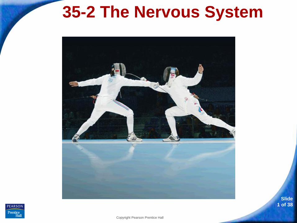
35-2 The Nervous System
35-2 The Nervous System
Slide
2 of 38
Copyright Pearson Prentice Hall
35-2 The Nervous System
The nervous system controls and
coordinates functions throughout the body
and responds to internal and external
stimuli.
35-2 The Nervous System
Slide
3 of 38
Copyright Pearson Prentice Hall
Neurons
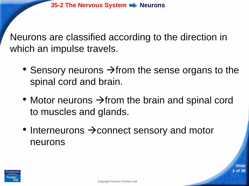
Neurons are classified according to the direction in
which an impulse travels.
• Sensory neurons from the sense organs to the
spinal cord and brain.
• Motor neurons from the brain and spinal cord
to muscles and glands.
• Interneurons connect sensory and motor
neurons
35-2 The Nervous System
Slide
4 of 38
Copyright Pearson Prentice Hall
Neurons
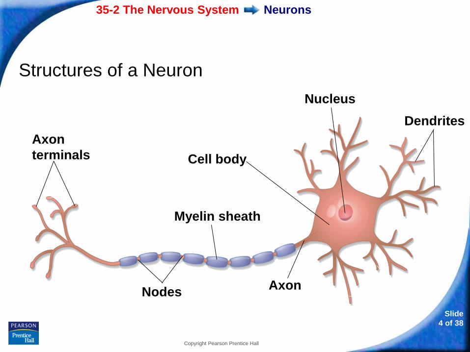
Structures of a Neuron
Axon
terminals
Myelin sheath
Cell body
Nodes Axon
Dendrites
Nucleus
35-2 The Nervous System
Slide
5 of 38
Copyright Pearson Prentice Hall
Neurons
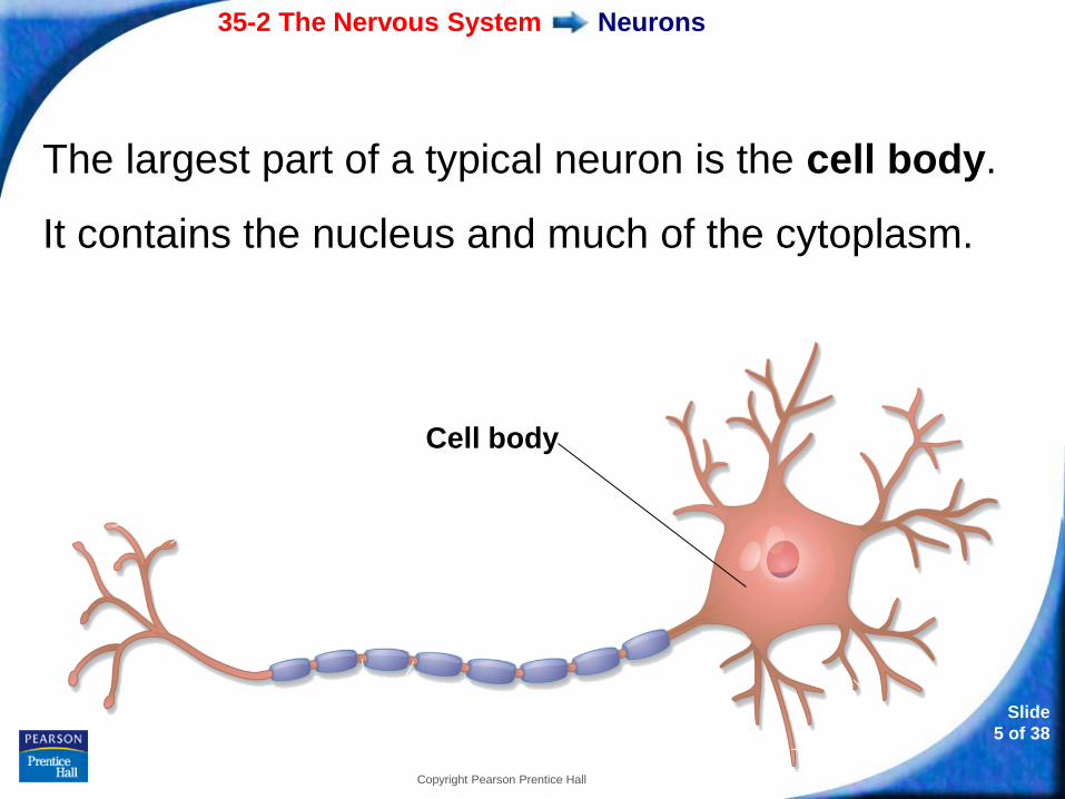
The largest part of a typical neuron is the cell body.
It contains the nucleus and much of the cytoplasm.
Cell body
35-2 The Nervous System
Slide
6 of 38
Copyright Pearson Prentice Hall
Neurons
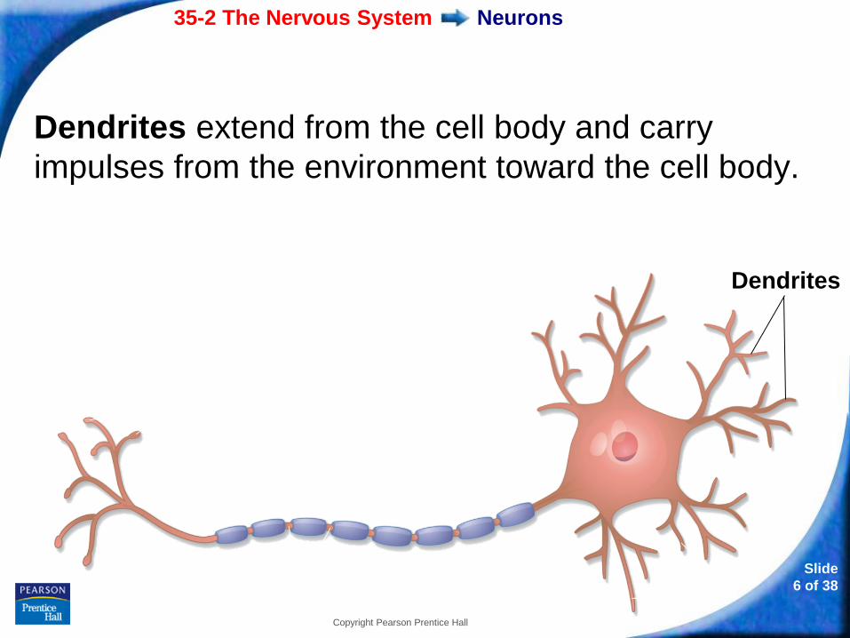
Dendrites extend from the cell body and carry
impulses from the environment toward the cell body.
Dendrites
35-2 The Nervous System
Slide
7 of 38
Copyright Pearson Prentice Hall
Neurons
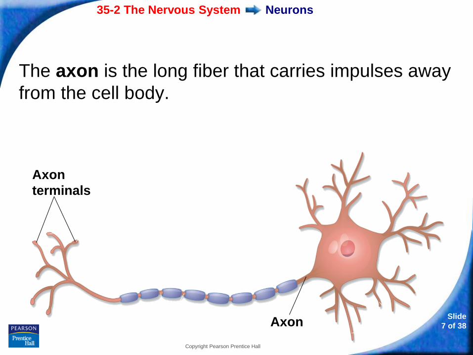
The axon is the long fiber that carries impulses away
from the cell body.
Axon
terminals
Axon
35-2 The Nervous System
Slide
8 of 38
Copyright Pearson Prentice Hall
Neurons
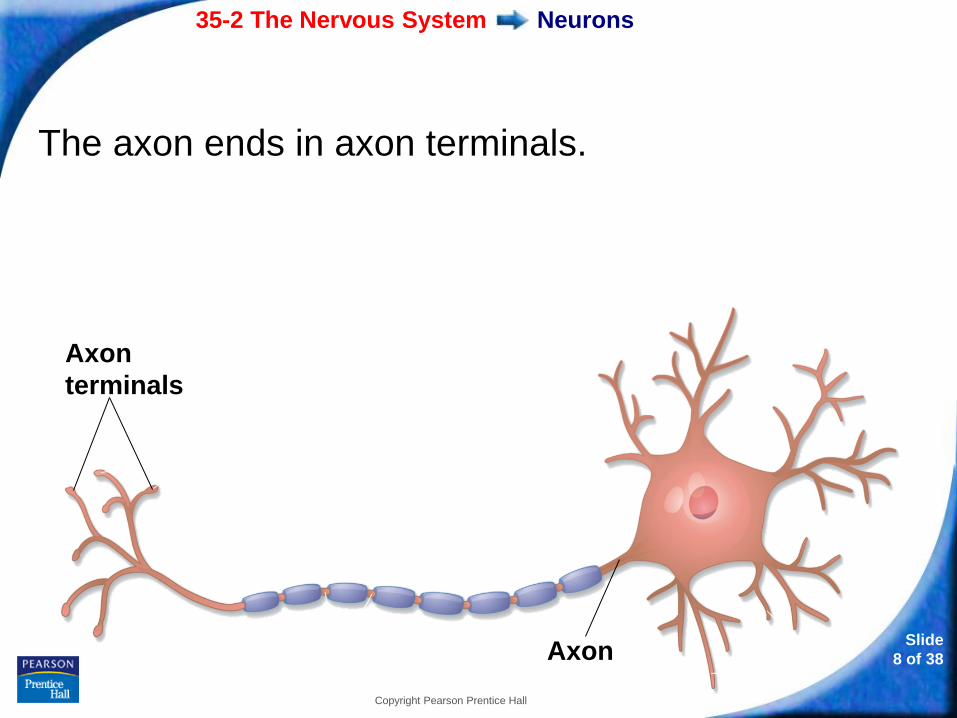
The axon ends in axon terminals.
Axon
terminals
Axon
35-2 The Nervous System
Slide
9 of 38
Copyright Pearson Prentice Hall
Neurons
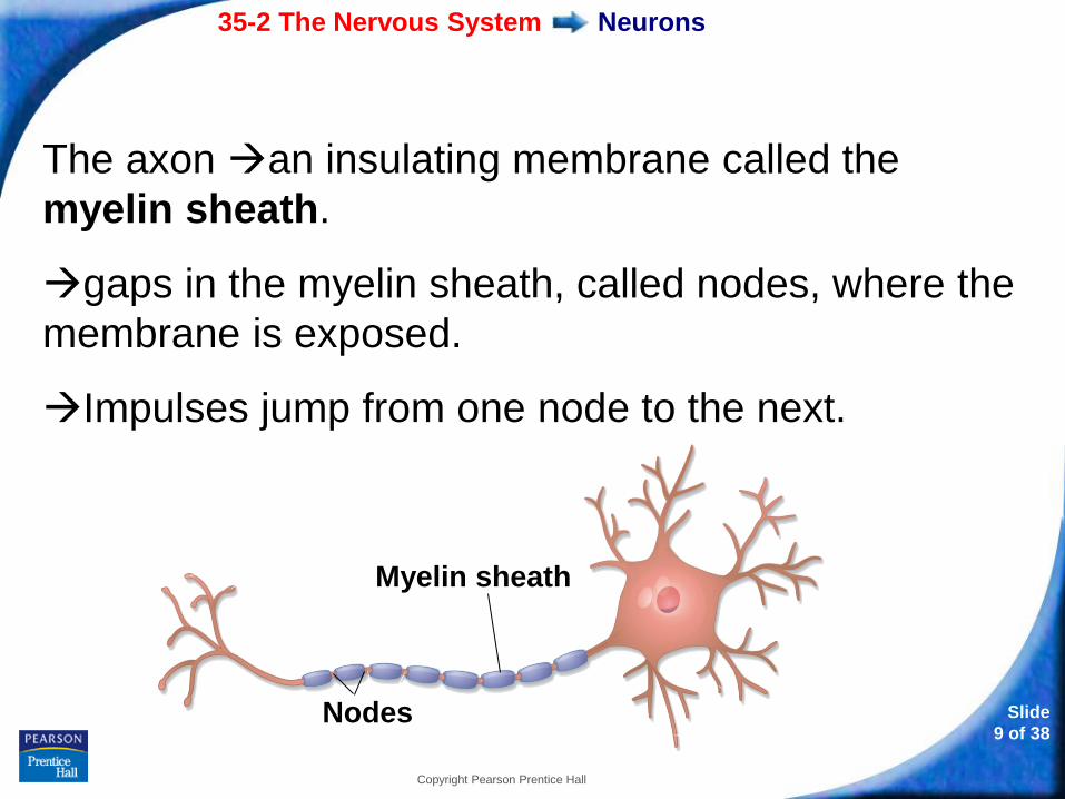
The axon an insulating membrane called the
myelin sheath.
gaps in the myelin sheath, called nodes, where the
membrane is exposed.
Impulses jump from one node to the next.
Myelin sheath
Nodes
35-2 The Nervous System
Slide
10 of 38
Copyright Pearson Prentice Hall
The Nerve Impulse
The Nerve Impulse
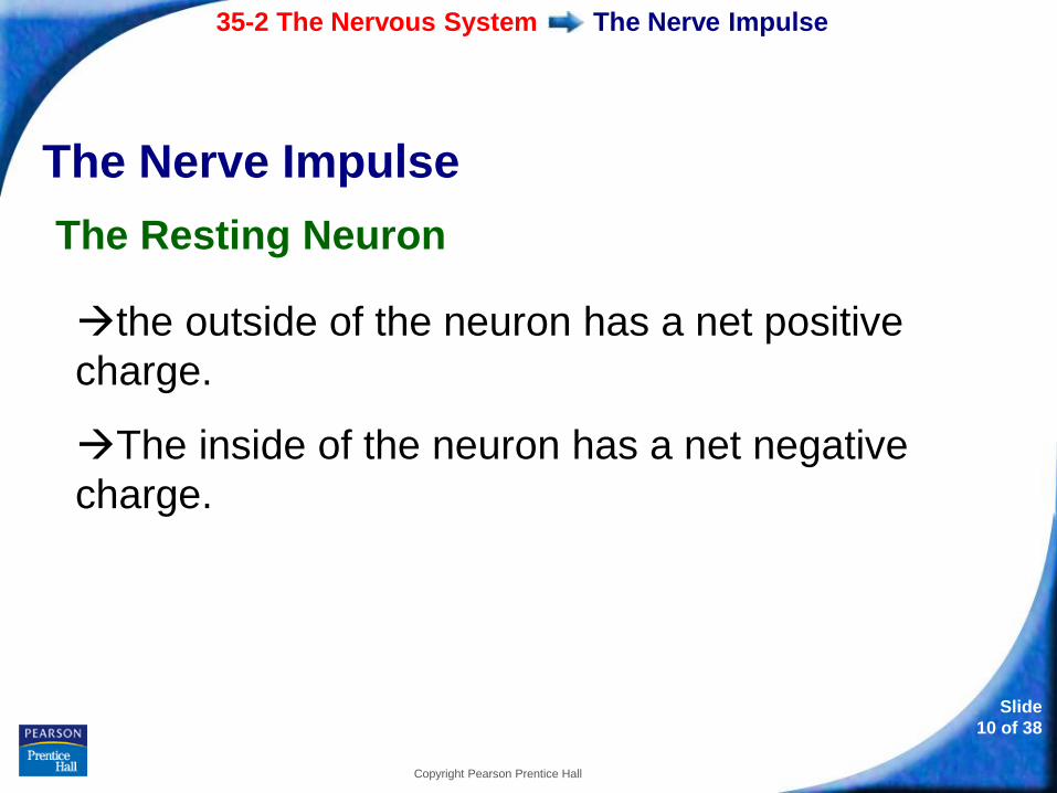
The Resting Neuron
the outside of the neuron has a net positive
charge.
The inside of the neuron has a net negative
charge.
35-2 The Nervous System
Slide
11 of 38
Copyright Pearson Prentice Hall
The Nerve Impulse
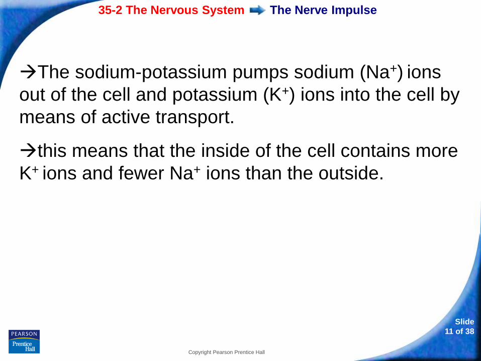
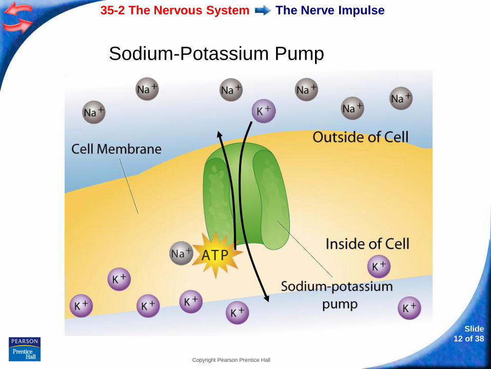
The sodium-potassium pumps sodium (Na+) ions
out of the cell and potassium (K+) ions into the cell by
means of active transport.
this means that the inside of the cell contains more
K+ ions and fewer Na+ ions than the outside.
35-2 The Nervous System
Slide
12 of 38
Copyright Pearson Prentice Hall
The Nerve Impulse
Sodium-Potassium Pump
35-2 The Nervous System
Slide
13 of 38
Copyright Pearson Prentice Hall
The Nerve Impulse
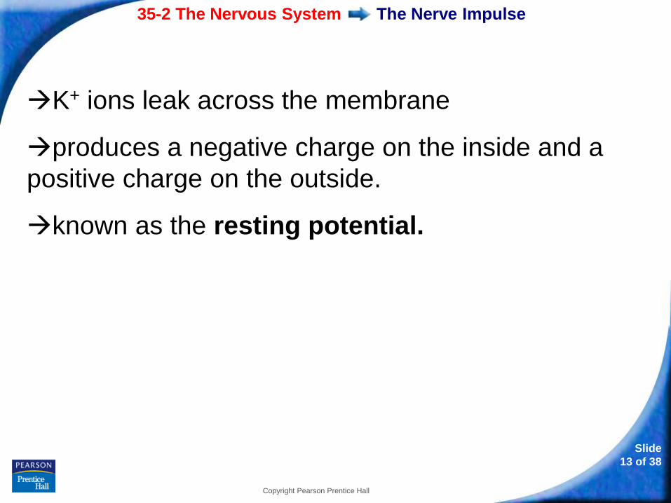
K+ ions leak across the membrane
produces a negative charge on the inside and a
positive charge on the outside.
known as the resting potential.
35-2 The Nervous System
Slide
14 of 38
Copyright Pearson Prentice Hall
The Nerve Impulse
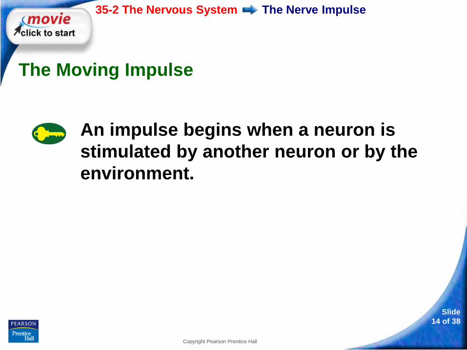
The Moving Impulse
An impulse begins when a neuron is
stimulated by another neuron or by the
environment.
35-2 The Nervous System
Slide
15 of 38
Copyright Pearson Prentice Hall
The Nerve Impulse
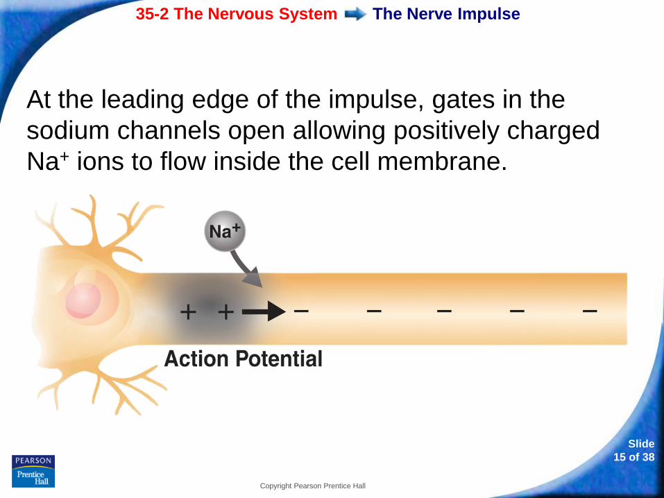
At the leading edge of the impulse, gates in the
sodium channels open allowing positively charged
Na+ ions to flow inside the cell membrane.
35-2 The Nervous System
Slide
16 of 38
Copyright Pearson Prentice Hall
The Nerve Impulse
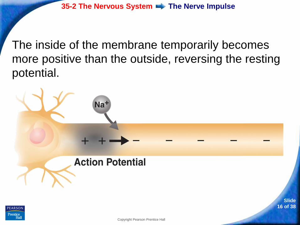
The inside of the membrane temporarily becomes
more positive than the outside, reversing the resting
potential.
35-2 The Nervous System
Slide
17 of 38
Copyright Pearson Prentice Hall
The Nerve Impulse
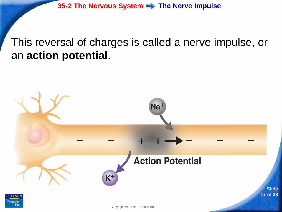
This reversal of charges is called a nerve impulse, or
an action potential.
35-2 The Nervous System
Slide
18 of 38
Copyright Pearson Prentice Hall
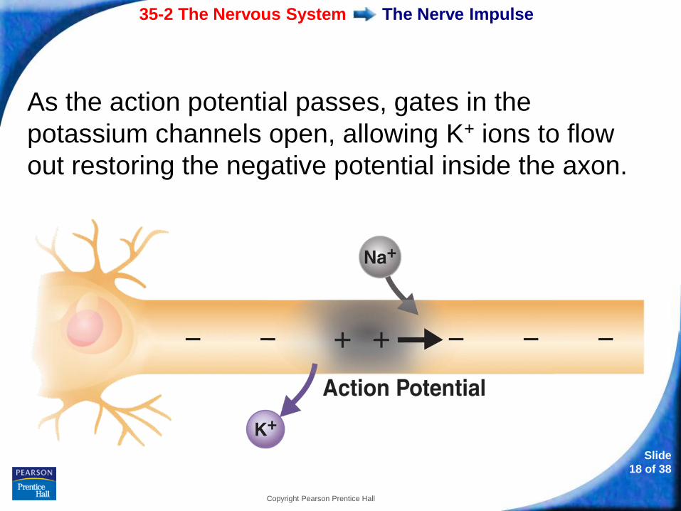
The Nerve Impulse
As the action potential passes, gates in the
potassium channels open, allowing K+ ions to flow
out restoring the negative potential inside the axon.
35-2 The Nervous System
Slide
19 of 38
Copyright Pearson Prentice Hall
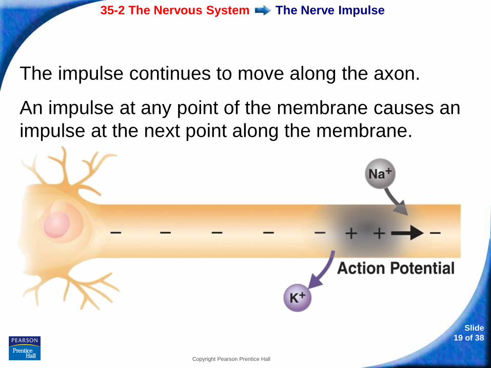
The Nerve Impulse
The impulse continues to move along the axon.
An impulse at any point of the membrane causes an
impulse at the next point along the membrane.
35-2 The Nervous System
Slide
20 of 38
Copyright Pearson Prentice Hall
The Nerve Impulse
Threshold
stimulus must be of adequate strength to cause
a neuron to transmit an impulse.
minimum level required is called the threshold.
35-2 The Nervous System
Slide
21 of 38
Copyright Pearson Prentice Hall
The Nerve Impulse
stronger than the threshold produces an
impulse.
weaker than the threshold produces no impulse.
35-2 The Nervous System
Slide
22 of 38
Copyright Pearson Prentice Hall
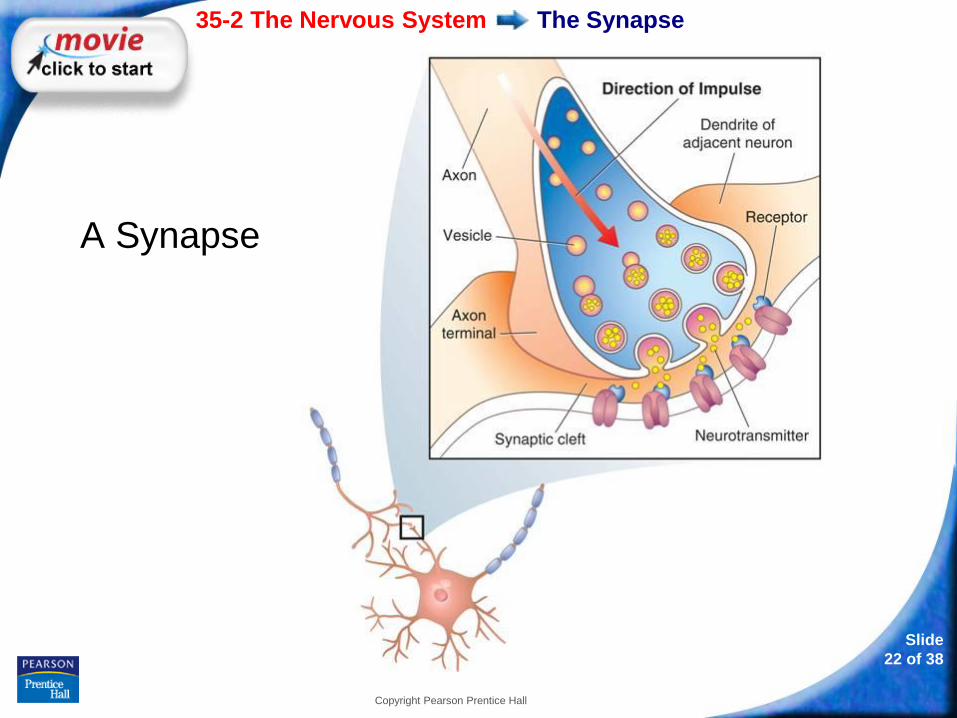
The Synapse
A Synapse
35-2 The Nervous System
Slide
23 of 38
Copyright Pearson Prentice Hall
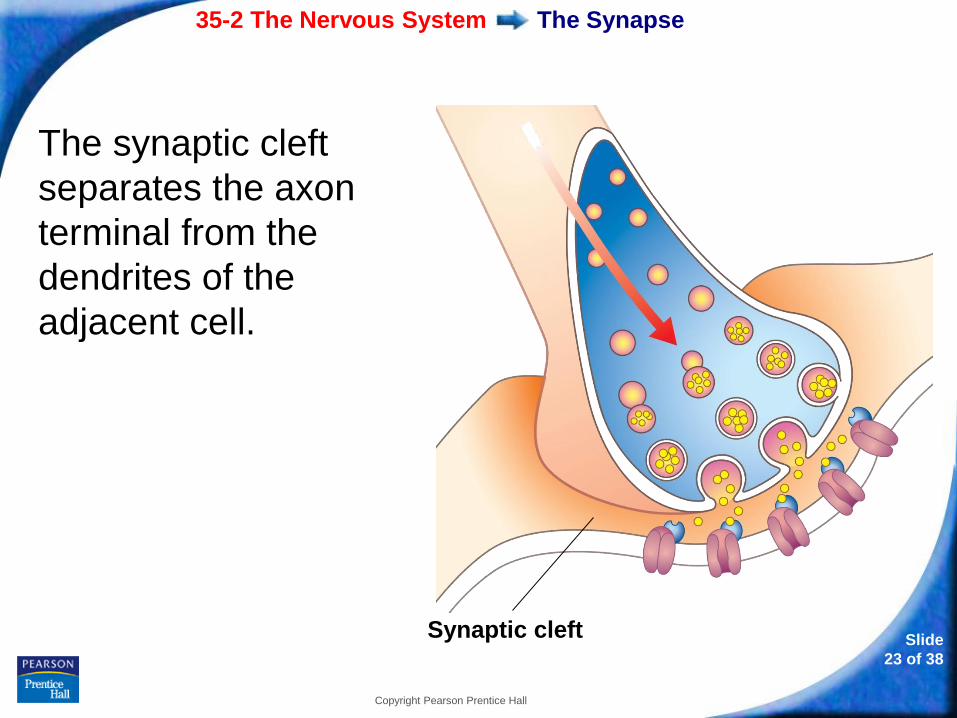
The Synapse
The synaptic cleft
separates the axon
terminal from the
dendrites of the
adjacent cell.
Synaptic cleft
35-2 The Nervous System
Slide
24 of 38
Copyright Pearson Prentice Hall
The Synapse
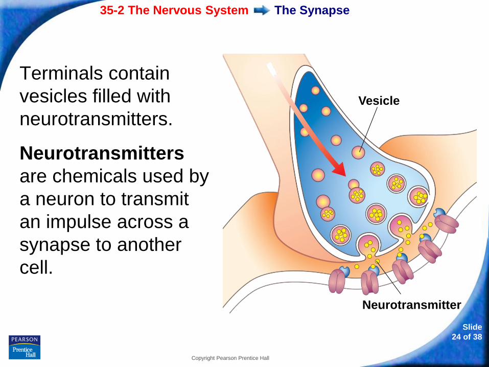
Terminals contain
vesicles filled with
neurotransmitters.
Neurotransmitters
are chemicals used by
a neuron to transmit
an impulse across a
synapse to another
cell.
Vesicle
Neurotransmitter
35-2 The Nervous System
Slide
25 of 38
Copyright Pearson Prentice Hall
The Synapse
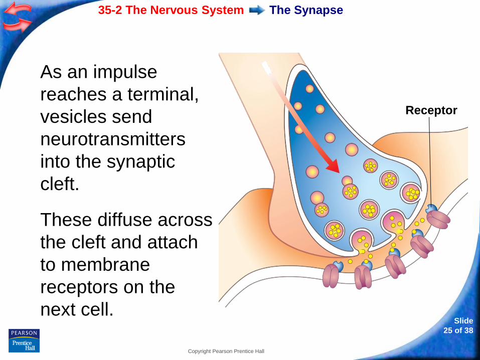
As an impulse
reaches a terminal,
vesicles send
neurotransmitters
into the synaptic
cleft.
These diffuse across
the cleft and attach
to membrane
receptors on the
next cell.
Receptor

- or -
Continue to: Click to Launch:
Slide
26 of 38
Copyright Pearson Prentice Hall
35-2
Slide
27 of 38
Copyright Pearson Prentice Hall
35-2
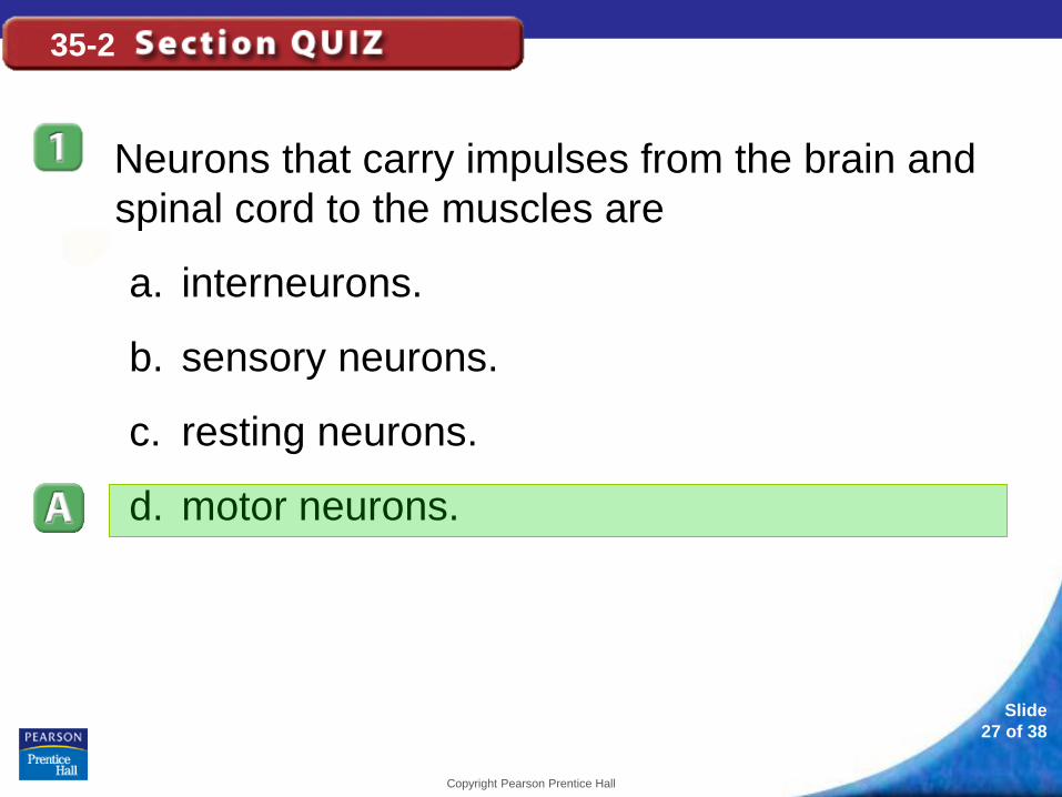
Neurons that carry impulses from the brain and
spinal cord to the muscles are
a. interneurons.
b. sensory neurons.
c. resting neurons.
d. motor neurons.
Slide
28 of 38
Copyright Pearson Prentice Hall
35-2
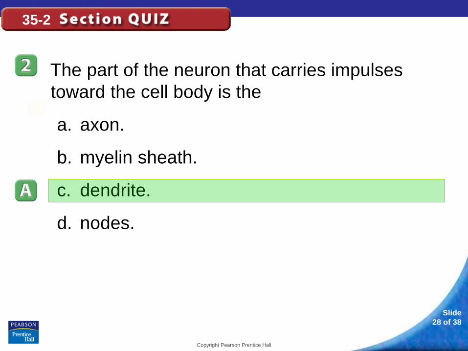
The part of the neuron that carries impulses
toward the cell body is the
a. axon.
b. myelin sheath.
c. dendrite.
d. nodes.
Slide
29 of 38
Copyright Pearson Prentice Hall
35-2
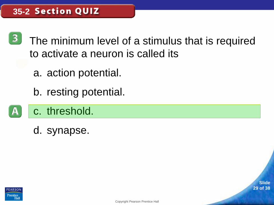
The minimum level of a stimulus that is required
to activate a neuron is called its
a. action potential.
b. resting potential.
c. threshold.
d. synapse.
Slide
30 of 38
Copyright Pearson Prentice Hall
35-2
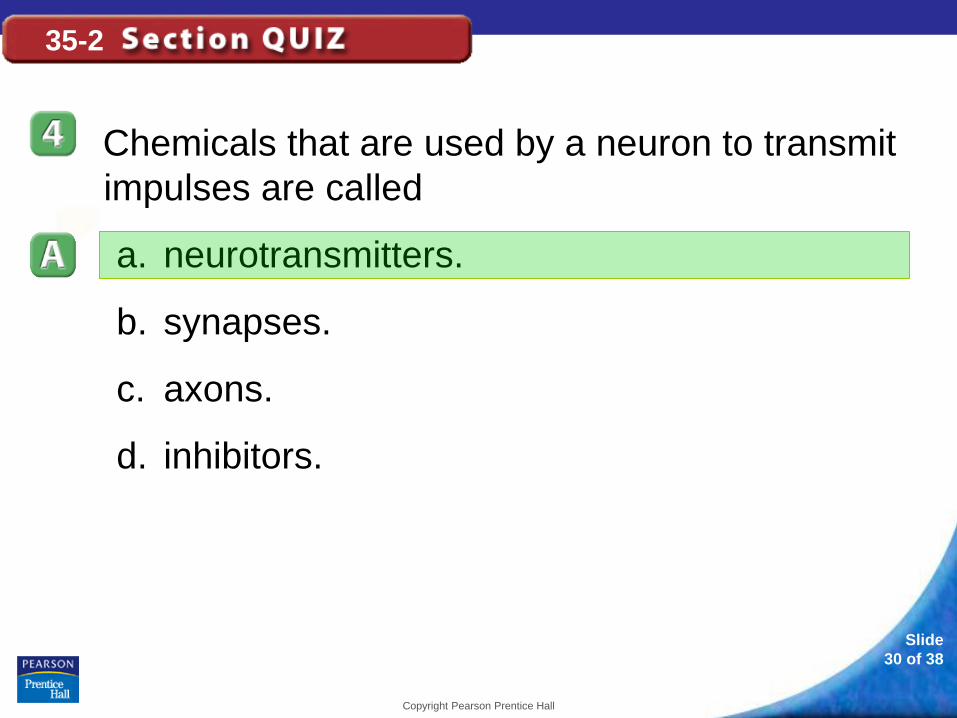
Chemicals that are used by a neuron to transmit
impulses are called
a. neurotransmitters.
b. synapses.
c. axons.
d. inhibitors.
Slide
31 of 38
Copyright Pearson Prentice Hall
35-2
An action potential begins when
a. sodium ions flow into the neuron.
b. potassium ions flow into the neuron.
c. sodium and potassium ions flow into the
neuron.
d. sodium and potassium ions flow out of the
neuron.
Slide
32 of 37
Copyright Pearson Prentice Hall
35-3 Divisions of the
Nervous System
35-3 Divisions of the Nervous
System
Slide
33 of 37
Copyright Pearson Prentice Hall
The human nervous system has two major divisions:
• central nervous system
• peripheral nervous system
35-3 Divisions of the Nervous
System
Slide
34 of 37
Copyright Pearson Prentice Hall
The Central Nervous System
The central nervous system relays
messages, processes information, and
analyzes information.
35-3 Divisions of the Nervous
System
Slide
35 of 37
Copyright Pearson Prentice Hall
The Central Nervous System
The central nervous system consists of the brain
and the spinal cord.
Both the brain and spinal cord are wrapped in
three layers of connective tissue known as
meninges.
35-3 Divisions of the Nervous
System
Slide
36 of 37
Copyright Pearson Prentice Hall
The Central Nervous System
Between the meninges and the central nervous
system tissue is a space filled with cerebrospinal
fluid.
Cerebrospinal fluid acts as a shock absorber that
protects the central nervous system.
Cerebrospinal fluid also permits exchange of
nutrients and waste products between blood and
nervous tissue.
35-3 Divisions of the Nervous
System
Slide
37 of 37
Copyright Pearson Prentice Hall
The Brain
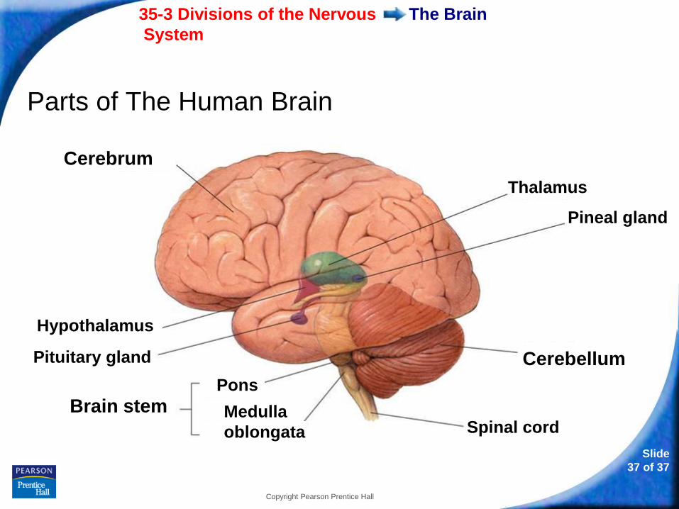
Parts of The Human Brain
Thalamus
Pineal gland
Cerebellum
Spinal cord
Hypothalamus
Pituitary gland
Cerebrum
Brain stem Pons
Medulla
oblongata
35-3 Divisions of the Nervous
System
Slide
38 of 37
Copyright Pearson Prentice Hall
The Brain
The Cerebrum
The largest and most prominent region of the
human brain is the cerebrum.
It controls the voluntary, or conscious, activities of
the body.
It is the site of intelligence, learning, and judgment.
35-3 Divisions of the Nervous
System
Slide
39 of 37
Copyright Pearson Prentice Hall
The Brain
A deep groove divides the cerebrum into
hemispheres, which are connected by a band of
tissue called the corpus callosum.
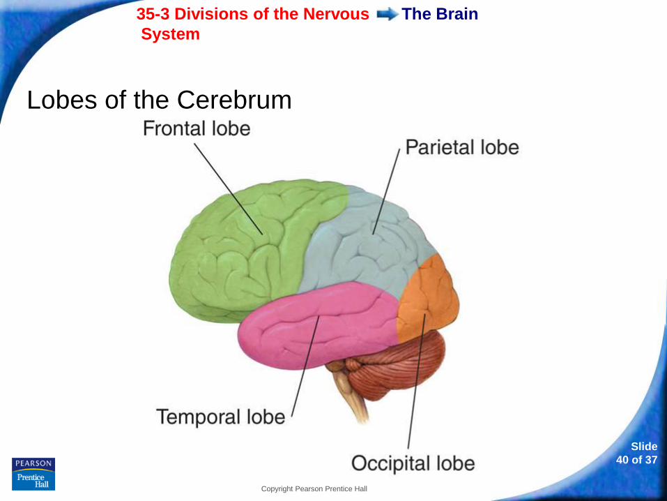
Each hemisphere is divided into regions called lobes.
35-3 Divisions of the Nervous
System
Slide
40 of 37
Copyright Pearson Prentice Hall
The Brain
Lobes of the Cerebrum
35-3 Divisions of the Nervous
System
Slide
41 of 37
Copyright Pearson Prentice Hall
The Brain
The outer layer of the cerebrum is called the cerebral
cortex and consists of gray matter.
The inner layer of the cerebrum consists of white
matter, which is made up of bundles of axons with
myelin sheaths.
35-3 Divisions of the Nervous
System
Slide
42 of 37
Copyright Pearson Prentice Hall
The Brain
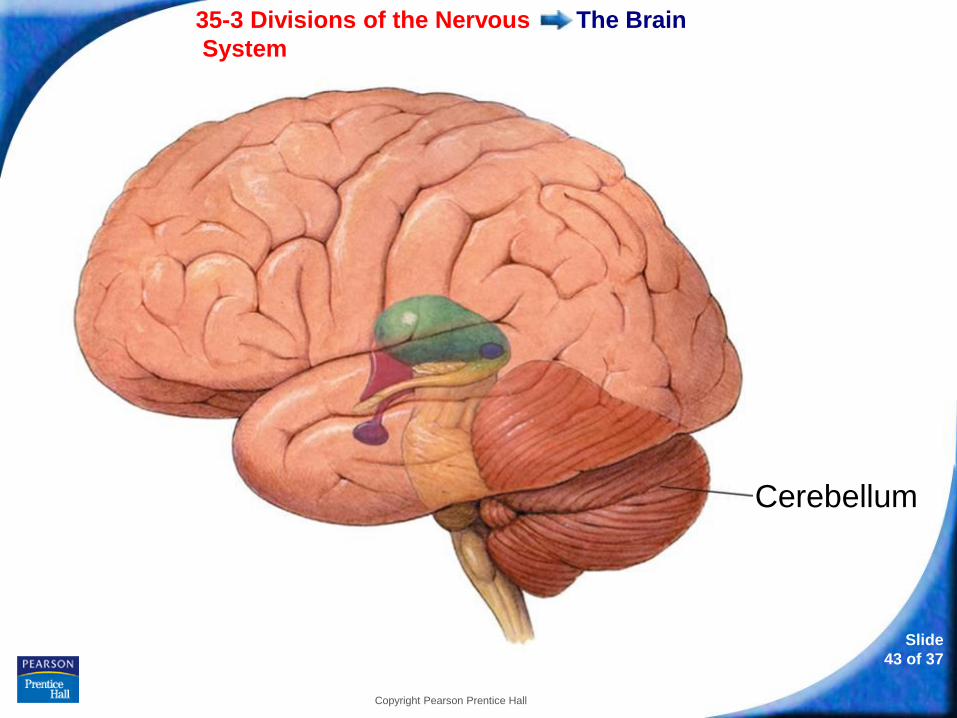
The Cerebellum
The second largest region of the brain is the
cerebellum.
It coordinates and balances the actions of the
muscles so that the body can move gracefully and
efficiently.
35-3 Divisions of the Nervous
System
Slide
43 of 37
Copyright Pearson Prentice Hall
The Brain
Cerebellum
35-3 Divisions of the Nervous
System
Slide
44 of 37
Copyright Pearson Prentice Hall
The Brain
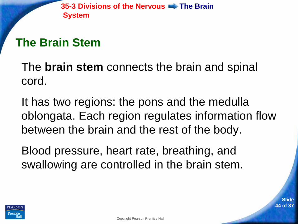
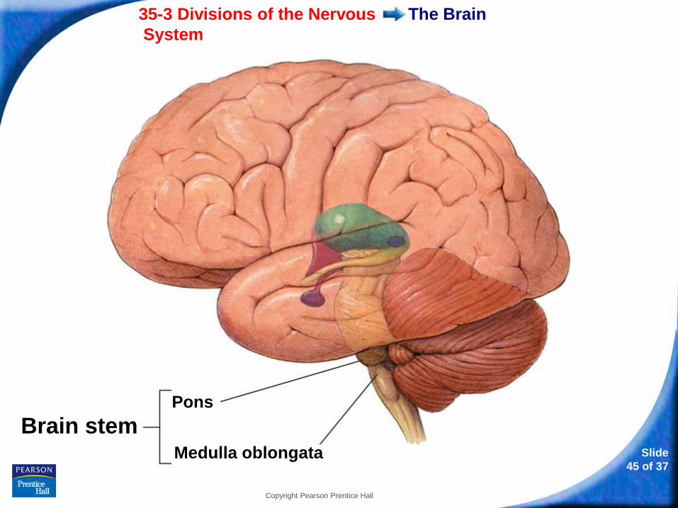
The Brain Stem
The brain stem connects the brain and spinal
cord.
It has two regions: the pons and the medulla
oblongata. Each region regulates information flow
between the brain and the rest of the body.
Blood pressure, heart rate, breathing, and
swallowing are controlled in the brain stem.
35-3 Divisions of the Nervous
System
Slide
45 of 37
Copyright Pearson Prentice Hall
The Brain
Pons
Medulla oblongata
Brain stem
35-3 Divisions of the Nervous
System
Slide
46 of 37
Copyright Pearson Prentice Hall
The Brain
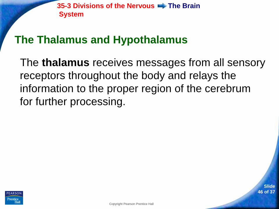
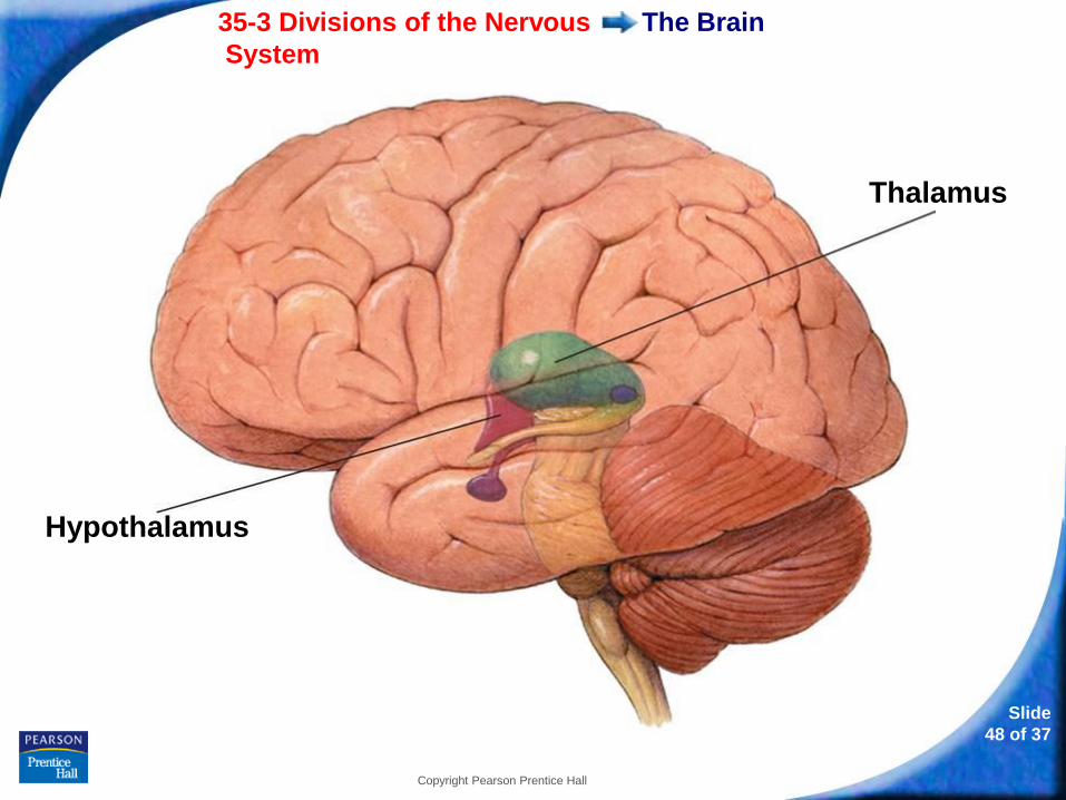
The Thalamus and Hypothalamus
The thalamus receives messages from all sensory
receptors throughout the body and relays the
information to the proper region of the cerebrum
for further processing.
35-3 Divisions of the Nervous
System
Slide
47 of 37
Copyright Pearson Prentice Hall
The Brain
The hypothalamus controls recognition and
analysis of hunger, thirst, fatigue, anger, and body
temperature.
It controls coordination of the nervous and
endocrine systems.
35-3 Divisions of the Nervous
System
Slide
48 of 37
Copyright Pearson Prentice Hall
The Brain
Thalamus
Hypothalamus
35-3 Divisions of the Nervous
System
Slide
49 of 37
Copyright Pearson Prentice Hall
The Spinal Cord
The Spinal Cord
The spinal cord is the main communications link
between the brain and the rest of the body.
Certain information, including some kinds of
reflexes, are processed directly in the spinal cord.
A reflex is a quick, automatic response to a
stimulus.
35-3 Divisions of the Nervous
System
Slide
50 of 37
Copyright Pearson Prentice Hall
The Peripheral Nervous System
The Peripheral Nervous System
The peripheral nervous system is all of the nerves
and associated cells that are not part of the brain
and the spinal cord.
The peripheral nervous system includes cranial
nerves, spinal nerves, and ganglia.
Ganglia are collections of nerve cell bodies.

35-3 Divisions of the Nervous
System
Slide
51 of 37
Copyright Pearson Prentice Hall
The Peripheral Nervous System
The sensory division of the peripheral nervous system transmits impulses from sense organs to the central nervous system.
The motor division transmits impulses from the central nervous system to the muscles or glands.
35-3 Divisions of the Nervous
System
Slide
52 of 37
Copyright Pearson Prentice Hall
The Peripheral Nervous System
The Somatic Nervous System
The somatic nervous system regulates activities
that are under conscious control, such as the
movement of skeletal muscles.
Some somatic nerves are involved with reflexes.
35-3 Divisions of the Nervous
System
Slide
53 of 37
Copyright Pearson Prentice Hall
The Peripheral Nervous System
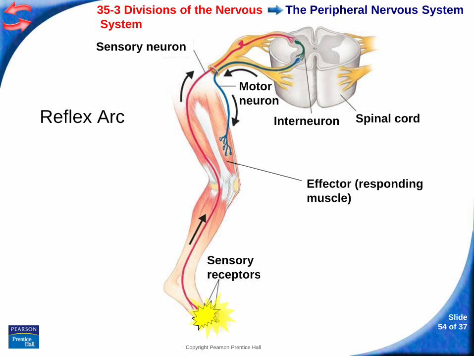
A reflex arc includes a sensory receptor, sensory
neuron, motor neuron, and effector that are involved
in a quick response to a stimulus.
35-3 Divisions of the Nervous
System
Slide
54 of 37
Copyright Pearson Prentice Hall
The Peripheral Nervous System
Reflex Arc
Sensory
receptors
Effector (responding
muscle)
Interneuron Spinal cord
Sensory neuron
Motor
neuron
35-3 Divisions of the Nervous
System
Slide
55 of 37
Copyright Pearson Prentice Hall
The Peripheral Nervous System
The Autonomic Nervous System
The autonomic nervous system regulates
involuntary activities.
The autonomic nervous system is subdivided into
two parts:
• sympathetic nervous system
• parasympathetic nervous system
35-3 Divisions of the Nervous
System
Slide
56 of 37
Copyright Pearson Prentice Hall
The Peripheral Nervous System
The sympathetic and parasympathetic nervous
systems have opposite effects on the same organ
system.
These opposing effects help maintain homeostasis.
- or -
Continue to: Click to Launch:
Slide
57 of 37
Copyright Pearson Prentice Hall
35-3
Slide
58 of 37
Copyright Pearson Prentice Hall
35-3
The brain stem functions as
a. a location for memory and learning.
b. the control site responsible for heart rate,
blood pressure, and breathing.
c. the location where all sensory information is
processed and delivered to the cerebrum.
d. an area that recognizes hunger, thirst, and
body temperature.
Slide
59 of 37
Copyright Pearson Prentice Hall
35-3
The left half of the cerebrum largely controls
a. the left side of the body.
b. both the right and left sides of the body.
c. the right side of the body.
d. the right half of the brain.
Slide
60 of 37
Copyright Pearson Prentice Hall
35-3
The part of the brain that is responsible for
coordination and balance is the
a. cerebellum.
b. cerebrum.
c. brain stem.
d. thalamus.
Slide
61 of 37
Copyright Pearson Prentice Hall
35-3
Reflex arcs are actions that are a part of the
peripheral nervous system's
a. sensory division.
b. somatic system.
c. autonomic system.
d. motor division.
Slide
62 of 37
Copyright Pearson Prentice Hall
35-3
Which of the following is NOT under the control
of the autonomic nervous system?
a. heartbeat
b. digestion
c. walking
d. sweating
END OF SECTION