Embed Size (px)
DESCRIPTION
breast
Citation preview
Asian Pacific Journal of Cancer Prevention, Vol 12, 2011 3101
High Ki-67 as a Poor Prognostic Indicator of Recurrence Free Survival in Breast Cancer Patients
Asian Pacific J Cancer Prev, 12, 3101-3104
Introduction
Breast cancer (BC) is the most frequently diagnosed cancer and the leading cause of cancer death in females worldwide, accounting for 23% (1.38 million) of the total new cancer cases and 14% (458,400) of the total cancer deaths in 2008 (Jemal et al., 2011). Understanding its biological behaviors and identifying its prognostic or predictive factors may help to improve the outcome. With the development of biological technology and medical science, some biological molecules such as human epidermal growth factor receptor 2 (HER2), estrogen receptor (ER) and progesterone receptor (PR) have been playing key roles in diagnosis, prognosis, monitoring and surveillance of BC in clinical practice (Goldhirsch et al., 2009; Ayoub et al., 2011). Many other molecules have also been assessed, including proliferation antigen Ki-67. Since it was identified as a nuclear antigen associated with cell proliferation in 1983 (Gerdes et al., 1983), Ki-67 has been studied with considerable enthusiasm. A detailed cell cycle analysis revealed that the antigen was present in the nuclei of cells in all phases as well as in mitosis, while quiescent or resting cells in the G0 phase did not express it (Gerdes et al., 1984; 1991). Despite having a much greater 1Department of Oncology, Zhongnan Hospital of Wuhan University, Hubei Key Laboratory on Tumor Biological Behavior and Hubei Cancer Clinical Study Center, Wuhan, 2Medical School of Jingchu University of Technology, Jingmen, China *For correspondence: [email protected]
Abstract
Objective: Ki-67 is a biomarker that reflects cell proliferation. Despite a clear understanding of the protein’s structure and properties, its functional role remains elusive. We conducted the present study to assess the prognostic value of Ki-67 in breast cancer (BC). Methods: We enrolled 164 individuals in this study: 30 patients with benign tumors and 134 with invasive BC. Immunohistochemistry (IHC) was used to detect Ki-67 expression The prognostic value of Ki-67 for 5-year recurrence-free survival (RFS) could be analyzed in 134 BC patients. Results: Ki-67 expression showed significant differences with the tumor grade, lymph node (LN) status, HER2 status and hormone receptor (HR) status (all P<0.05). When Ki-67 11% was used as cutoff to divide the 134 cases into two groups, with high and low expression, the patients in former had a significantly higher 5-year recurrence rate (37.1% vs 8.1%, P=0.001) and a worse RFS (log-rank test, P=0.0017) than those in low Ki-67 expression group. Ki-67 was an independent prognostic predictor of 5-year RFS in both univariate and multivariate analyses. Conclusions: Ki-67 can be used as a negative predictor of 5-year RFS of patients with invasive BC.
Keywords: Breast cancer - Ki-67 - recurrence-free survival - prognosis
RESEARCH COMMUNICATION
High Ki-67 Expression is a Poor Prognostic Indicator of 5-Year Survival in Patients with Invasive Breast Cancer
Xue-Qin Yang1,2, Fu-Bing Wang1, Chuang Chen1, Chun-Wei Peng1, Jing-Fang Zhang2, Yan Li1*
understanding of the protein’s structure and properties, its functional role remains elusive. Many studies investigated the clinical value of Ki-67 in BC and suggested it had some prognostic or predictive role in clinical practice (Pavelic et al., 1992; González-Vela et al., 2001; de Azambuja et al., 2007; Viale et al., 2008a; 2008b; Stuart-Harris et al., 2009; Karanikas et al., 2010; Santisteban et al., 2010). However, the guidelines of the American Society of Clinical Oncology did not recommend Ki-67 as a required routine biological marker of BC (Harris et al., 2007), probably because some uncertainty remains on the way for Ki-67 as a routine marker and further studies are needed. This study was conducted to explore the prognostic value of Ki-67 in BC patients.
Materials and Methods
Patients and specimens Formalin-fixed, paraffin-embedded complete tumor specimens from 134 patients with invasive BC from January 2002 to December 2006, aged from 31 to 78 years (median 49 yr), were collected from Hubei Cancer Hospital, China. Major pathological parameters were all available, including the tumor size, location and
Xue-Qin Yang et al
Asian Pacific Journal of Cancer Prevention, Vol 12, 20113102
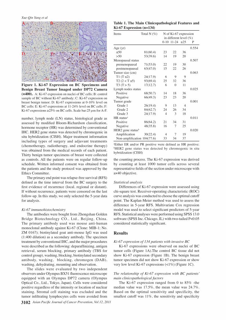
Table 1. The Main Clinicopathological Features and Ki-67 Expression (n=134)Items Total N (%) N of Ki-67 expression in different level (%) 0-10 11-24 ≥25 P
Age (yr) 0.554 ≤50 81(60.4) 23 22 36 >50 53(39.6) 14 19 20 Menopausal status 0.507 premenopausal 71(53.0) 22 19 30 postmenopausal 63(47.0) 15 22 26 Tumor size (cm) 0.063 T1 (T ≤2) 24(17.9) 6 9 9 T2 (2 < T ≤5) 93(69.4) 25 32 36 T3 (T > 5) 17(12.7) 6 0 11 Lymph nodes status 0.025 Positive 68(50.7) 14 18 36 Negative 66(49.3) 23 23 20 Tumor grade 0.001 Grade 1 26(19.4) 9 13 4 Grade 2 84(62.7) 24 26 34 Grade 3 24(17.9) 4 3 18 HR statusa 0.011 Positive 86(64.2) 21 34 31 Negative 48(35.8) 16 7 25 HER2 gene statusb 0.020 Amplification 30(22.4) 4 7 19 Non-amplification 104(77.6) 33 34 37 aEither ER and/or PR positive were defined as HR positive; bHER2 gene status was detected by chromogenic in situ hybridization (CISH)
number, lymph node (LN) status, histological grade as assessed by modified Bloom-Richardson classification, hormone receptor (HR) was determined by conventional IHC. HER2 gene status was detected by chromogenic in situ hybridization (CISH). Major treatment information including types of surgery and adjuvant treatments (chemotherapy, radiotherapy, and endocrine therapy) was obtained from the medical records of each patient. Thirty benign tumor specimens of breast were collected as controls. All the patients were on regular follow-up schedule. Written informed consent was obtained from the patients and the study protocol was approved by the Ethics Committee. The primary end point was relapse-free survival (RFS) defined as the time interval from the BC surgery to the first evidence of recurrence (local, regional or distant). If without recurrence, patients were censored on the last follow-up. In this study, we only selected the 5-year data for analysis.
Ki-67 immunohistochemistry The antibodies were bought from Zhongshan Golden Bridge Biotechnology CO., Ltd., Beijing, China. The primary antibody used was mouse anti-human monoclonal antibody against Ki-67 (Clone: MIB-1; No. ZM 0167); biotinylated goat anti-mouse IgG was used (1:400 dilution) as a secondary antibody. The specimen treatment by conventional IHC, and the major procedures were described as the following: deparaffinizing, antigen retrieval, serum blocking, primary antibody (TBS for control group), washing, blocking, biotinylated secondary antibody, washing, blocking, chromogen (DAB), washing, dehydrating, mounting and observation. The slides were evaluated by two independent observers under Olympus BX51 fluorescence microscope equipped with an Olympus DP72 camera (Olympus Optical Co., Ltd., Tokyo, Japan). Cells were considered positive regardless of the intensity or location of nuclear staining. Stromal cell staining was excluded and the tumor infiltrating lymphocytes cells were avoided from
the counting process. The Ki-67 expression was derived by counting at least 1000 tumor cells across several representative fields of the section under microscope with a×40 objective.
Statistical analysis Differences of Ki-67 expression were assessed using chi-square test. Receiver-operating characteristic (ROC) curve analysis was conducted to choose the optimal cutoff point. The Kaplan-Meier method was used to assess the difference in 5-year RFS. Multivariate Cox regression model was used to select significant predictors of 5-year RFS. Statistical analyses were performed using SPSS 13.0 software (SPSS Inc. Chicago, IL) with two-tailed P<0.05 considered statistically significant.
Results
Ki-67 expression of 134 patients with invasive BC Ki-67 expressions were observed on nuclei of BC tumor cells (Figure 1A).The control BC tissue did not show Ki-67 expression (Figure 1B). The benign breast tumor specimen did not show Ki-67 expression or show very low level Ki-67 expressions (<1%) (Figure 1C).
The relationship of Ki-67 expression with BC patients’ main clinicopathological factors The Ki-67 expression ranged from 0 to 85% -the median value was 17.5%, the mean value was 24.7%. Based on the optimal sensitivity and specificity, the smallest cutoff was 11%, the sensitivity and specificity
Figure 1. Ki-67 Expression on BC Specimens and Benign Breast Tumor Imaged under DP72 Camera (×400). A: Ki-67 expression on nuclei of BC cells; B: control sample of BC without Ki-67 antibody; C: Ki-67 expression on breast benign tumor; D: Ki-67 expressions at 0-10% level on BC cells; E: Ki-67 expressions at 11-24% level on BC cells; F: Ki-67 expressions ≥25% on BC cells. Scale bar:25 µm for A-F.

Asian Pacific Journal of Cancer Prevention, Vol 12, 2011 3103
High Ki-67 as a Poor Prognostic Indicator of Recurrence Free Survival in Breast Cancer Patients
0
25.0
50.0
75.0
100.0
New
ly d
iagn
osed
with
out
trea
tmen
t
New
ly d
iagn
osed
with
tre
atm
ent
Pers
iste
nce
or r
ecur
renc
e
Rem
issi
on
Non
e
Chem
othe
rapy
Radi
othe
rapy
Conc
urre
nt c
hem
orad
iatio
n
10.3
0
12.8
30.025.0
20.310.16.3
51.7
75.051.1
30.031.354.2
46.856.3
27.625.033.130.031.3
23.738.0
31.3
Table 2. Factors Correlated with 5-year RFS of BC Patients (n=134)Variables Univariate analysis Multivariate analysis Wald P RR 95% CI Wald P RR 95% CI Lower Upper Lower Upper
Age 0.007 0.933 0.973 0.510 1.854 NE NE NE NE NEMenopausal status 0.157 0.692 1.135 0.606 2.127 NE NE NE NE NETumor grade 29.548 0.000 4.919 2.769 8.736 7.984 0.005 2.704 1.356 5.389Tumor size 6.220 0.013 2.087 1.171 3.720 NE NE NE NE NELN status 14.696 0.000 4.588 2.106 9.998 5.824 0.016 2.858 1.218 6.707HR status 2.853 0.091 0.581 0.310 1.091 NE NE NE NE NEHER2 gene status 8.951 0.002 2.846 1.490 5.434 7.422 0.006 2.590 1.306 5.136Ki-67 status 18.509 0.000 2.002 1.459 2.748 .064 0.024 1.508 1.054 2.158
RR, relative risk; NE, not in the equation
Figure 2. The 5-year RFS of BC Patients with Different Levels of Ki-67 Expressionwere 71.8% and 52.6%, respectively; the largest cutoff was 25%, the sensitivity and specificity were 51.3% and 72.6%, respectively. Levels of Ki-67 expression in the study cases are shown in Figure 1 (D,E,F). According to Ki-67 expressions on BC specimens, the patients were divided into three subgroups: 0-10%, 11-24%, 25% and above. The relationships of Ki-67 expressions with patients’ main clinicopathological features were shown in table 1. The differences of Ki-67 expressions in different lymph nodes status, tumor grade, HR and HER2 status achieved statistical significance (p<0.05), whereas in other factors did not (p>0.05)
Ki-67 expression and 5-year RFS of BC patients In this study, the 5-year recurrent rate was 29.1% (39/134). Based on the cutoff of 11% of Ki-67 expression, the 134 BC patients were classified into two subgroups: those with a low Ki-67 expression (<11%, n=37) and those with a high Ki-67 expression (≥11%, n=97). The 5-year recurrent rate was 8.1% in patients with a low Ki-67 expression, and 37.1% in patients with a high Ki-67 expression. The difference between two groups was significant (P=0.001) as was that of the 5-year RFS (Log rank test, P=0.0017) (Figure 2).
Prognostic factors of 5-year RFS with invasive BC Among variables of patients’ age, menopausal status, tumor grade, tumor size, LN status, HR status, HER2 gene status and Ki-67 expression, univariate analysis indicated that LN status, tumor grade, tumor size, HER2, Ki-67 expression had significant correlations with 5-year RFS of BC (all P <0.05) (Table 2). Among all above factors, multivariate analysis with Cox regression model revealed that only LN status, tumor grade, HER2 gene status and Ki-67 expression were independent prognosticators.
Discussion
Ki-67 is a biomarker that can reflect cell proliferation state. Tumor cell proliferation state is an established predictor of prognosis. In BC, more and more data demonstrates that Ki-67 is a prognostic factor. However, Ki-67 as a prognostic marker in BC is still not yet conclusive. The acceptance of Ki-67 as a standard marker may require much more work. In this study, we investigated the prognostic value of Ki-67 expression for 5-year RFS of 134 patients with invasive BC and found that high Ki-67 expression was a poor prognostic factor of BC patients.
Since Ki-67 antigen was initially described in 1983 (Gerdes et al., 1983), the clinical usefulness were extensively explored. Pavelic et al. (1992) investigated the Ki-67 expression by IHC in 11 normal breast tissues and 42 invasive and 14 noninvasive BC, and found that the mean value of Ki-67-positive cells was 0.91±0.31% for normal breast tissue, 4.57±1.36% for noninvasive ductal carcinoma, and 12.76±2.18% for invasive BC, and also found that Ki-67 overexpression was associated with lymph node status. In order to better define the prognostic value of Ki-67, a study performed a meta-analysis including sixty-eight studies to evaluate the impact of Ki-67 on RFS and overall survival in early BC, found that higher Ki-67 expression was associated with higher probability of relapse and worse survival in BC patients (de Azambuja et al., 2007). Another study evaluated the prognostic and predictive value of Ki-67 labeling index (LI) in postmenopausal women with early BC, further confirmed Ki-67 as a prognostic factor, and also found that high Ki-67 LI levels identified a subgroup that particularly benefits from initial letrozole adjuvant therapy (Viale et al., 2008). In summary, these studies tended to suggest Ki-67 as a prognostic factor.
There are some controversial in the relationship between the main variables and Ki-67 expression in BC. A research investigated the association of Ki-67 with some biological prognostic factors of 102 BC patients and found that Ki-67(MIB-1) index was positively associated with the histological grade, tumor size and lymph node status but not associated with HER2 (González-Vela et al., 2001). Recently, another research retrospectively analyzed the archival pathology tissues of 356 patients with BC to evaluate the association between Ki-67 expression and the main clinicohistopathological parameters, and found that Ki-67expression was correlated with tumor grade, tumor
Xue-Qin Yang et al
Asian Pacific Journal of Cancer Prevention, Vol 12, 20113104
References
Ayoub N, Lucas C, Kaddoumi A (2011). Genomics and pharmacogenomics of breast cancer: current knowledge and trends. Asian Pac J Cancer Prev, 12, 1127-40.
Chen C, Peng J, Xia H, et al (2010). Quantum-dot-based immunofluorescent imaging of HER2 and ER provides new insights into breast cancer heterogeneity. Nanotechnology, 21, 095101.
Chen C, Peng J, Xia HS, et al (2009). Quantum dots-based immunofluorescence technology for the quantitative determination of HER2 expression in breast cancer. Biomaterials, 30, 2912-8.
Colozza M, Sidoni A, Piccart-Gebhart M (2010). Value of Ki67 in breast cancer: the debate is still open. Lancet Oncol, 11, 414-5.
de Azambuja E, Cardoso F, de Castro G Jr, et al (2007). Ki-67 as prognostic marker in early breast cancer: a meta-analysis of published studies involving 12,155 patients. Br J Cancer, 96, 1504-13.
Gerdes J, Lemke H, Baisch H, et al (1984). Cell cycle analysis of a cell proliferation-associated human nuclear antigen defined by the monoclonal antibody Ki-67. J Immunol, 133, 1710-5.
Gerdes J, Li L, Schlueter C, et al (1991). Immunobiochemical and molecular biologic characterization of the cell proliferation-associated nuclear antigen that is defined by monoclonal antibody Ki-67. Am J Pathol, 138, 867-73.
Gerdes J, Schwab U, Lemke H, Stein H (1983). Production of a mouse monoclonal antibody reactive with a human nuclear antigen associated with cell proliferation. Int J Cancer, 31, 13–20.
Goldhirsch A, Ingle JN, Gelber RD, al (2009). Thresholds for therapies: highlights of the St Gallen International Expert Consensus on the primary therapy of early breast cancer 2009. Ann Oncol, 20, 1319-29.
González-Vela MC, Garijo MF, Fernández F, Val-Bernal JF (2001). MIB1 proliferation index in breast infiltrating carcinoma: comparison with other proliferative markers and association with new biological prognostic factors. Histol Histopathol, 16, 399-406.
Harris L, Fritsche H, Mennel R, et al (2007). American Society of Clinical Oncology: American Society of Clinical Oncology 2007 update of recommendations for the use of tumor markers in breast cancer. J Clin Oncol, 25, 5287-312.
Jemal A, Bray F, Center MM, Ferlay J, Ward E, Forman D (2011). Global cancer statistics. CA Cancer J Clin, 61, 69-90.
Jonat W, Arnold N (2011). Is the Ki-67 labelling index ready for clinical use? Ann Oncol, 22, 500-5002.
Karanikas G, Koronakis N, Lagoudianakis EE, et al (2011). The value of proliferation indexes in breast cancer. Eur J Gynaecol Oncol, 31, 181-4.
Pavelic ZP, Pavelic L, Lower EE, Gapany M, Gapany S, et al (1992). c-myc, c-erbB-2, and Ki-67 expression in normal breast tissue and in invasive and noninvasive breast carcinoma. Cancer Res, 52, 2597-602.
Santisteban M, Reynolds C, Barr Fritcher EG, et al (2010). Ki67: a time-varying biomarker of risk of breast cancer in atypical hyperplasia. Breast Cancer Res Treat, 121, 431-7.
Stuart-Harris R, Caldas C, Pinder SE, Pharoah P (2009). Ki67 index, HER2 status, and prognosis of patients with luminal B breast cancer. J Natl Cancer Inst, 101, 736-50.
Viale G, Giobbie-Hurder A, Regan MM, et al (2008a). Prognostic and predictive value of centrally reviewed Ki-67 labeling
size, nodal status, and inversely associated with hormonal expression (Karanikas et al., 2010). In accordance with the results of theirs, our results found that Ki-67 expression was significantly different in different tumor grade, nodal status, and inversely associated with hormonal expression. Consisted with Karanikas’s study but contradicted with González-Vela’s study, Ki-67 expression showed significant difference in different HER2 status in our study. As to tumor size, Ki-67 expression in different tumor size did not have significant difference in present study, but it approached the border of achieving statistical significance. The relationship between them may need further study to define basing on standardized assessment of tumor size and enlarged research sample size.
Recently, Yerushalmi et al. (2010) have exhaustively reviewed the prognostic and predictive potential of Ki-67 in BC, and concluded that despite questions about its usefulness, there was increasing evidence that Ki67 was a valuable prognostic marker; the Ki-67 level above 10-14% was defined as high risk group in terms of prognosis. The acceptance of this definition might make comparisons of future studies more reliable (Jonat et al., 2011). Colozza et al expressed their opinions that only a complete standardisation of tissue handling and processing could improve the value of Ki67 as a clinically useful and widely accepted marker (Colozza et al., 2010). However, despite the absence of standardisation of Ki67 pathological assessment, the experts considered the Ki67 labelling index important for selecting the addition of chemotherapy to endocrine therapy in hormone receptor-positive BC, and classified tumors as low, intermediate, and highly proliferating according to the value of Ki67 labelling index of less than or equal to 15%, 16-30%, and more than 30%, respectively (Goldhirsch et al., 2009; Colozza et al., 2010).
We agree with the above experts’ opinions on the standardization of Ki67 pathological assessment and cutoff. In addition, improving the detection method from semi-quantitative to quantitative technology may shorten the distance to make a conclusion. For example, recently, Chen et al reported that quantum dots-based probes have advantages such as higher fluorescent efficiency over organic fluorescent dyes, better clarities and sensitivities and accuracies than conventional IHC techniques; notably, it can quantify the biomarkers, which provides a potentially important new method for Ki-67 detection in clinical practice (Chen et al., 2009; 2010).The relatively small sample size and retrospective research with a nonrandomized database collected were two main limitations of the present study.
In conclusion, Ki-67 expressions showed significant difference in different lymph nodes status, tumor grade, HR and HER2 status, and Ki-67 can be used as a negative prognosticator of 5-year RFS of patients with invasive BC.
Acknowledgements
This study was supported by the grants from the Science Fund for Creative Research Groups of the National Natural Science Foundation of China (No. 20621502 and 20921062), the Science and Research Program of Health Department of Hubei Province (No. JX5B69), and the
Science and Research Program of Jingchu University of Technology (No.ZR201002).
Asian Pacific Journal of Cancer Prevention, Vol 12, 2011 3105
High Ki-67 as a Poor Prognostic Indicator of Recurrence Free Survival in Breast Cancer Patients
index in postmenopausal women with endocrine-responsive breast cancer: results from Breast International Group Trial 1-98 comparing adjuvant tamoxifen with letrozole. J Clin Oncol, 26, 5569-75.
Viale G, Regan MM, Mastropasqua MG, et al (2008b). Predictive value of tumor Ki-67 expression in two randomized trials of adjuvant chemoendocrine therapy for node-negative breast cancer. J Natl Cancer Inst, 100, 207-12.
Yerushalmi R, Woods R, Ravdin PM, Hayes MM, Gelmon KA (2010). Ki67 in breast cancer: prognostic and predictive potential. Lancet Oncol, 11, 174-83.