Embed Size (px)
Citation preview

30. Genetics and recombination in bacteria

Lecture Outline 11/16/05
• Replication in bacteria• Types of recombination in bacteria
– Transduction by phage– Conjugation (“mating”)
• F+ plasmids• Hfr strains
– Transformation of raw DNA
• Evidence for recombination in nature– Resistance plasmids
The Bacterial Genome and Its Replication
• The bacterial chromosome– Is usually a circular DNA molecule with few associated proteins
• In addition to the chromosome– Many bacteria have plasmids, smaller circular DNA molecules that can replicate independently of the bacterial chromosome

Genetics fo Bacteria• Use huge numbers of individuals (billions) To find very rare events
• Few morphological traits– Antibiotic resistance– “Auxotrophs” cannot synthesize essential nutrients (arg- or trp-)
– “Prototrophs” have normal synthesis (arg+, trp+)
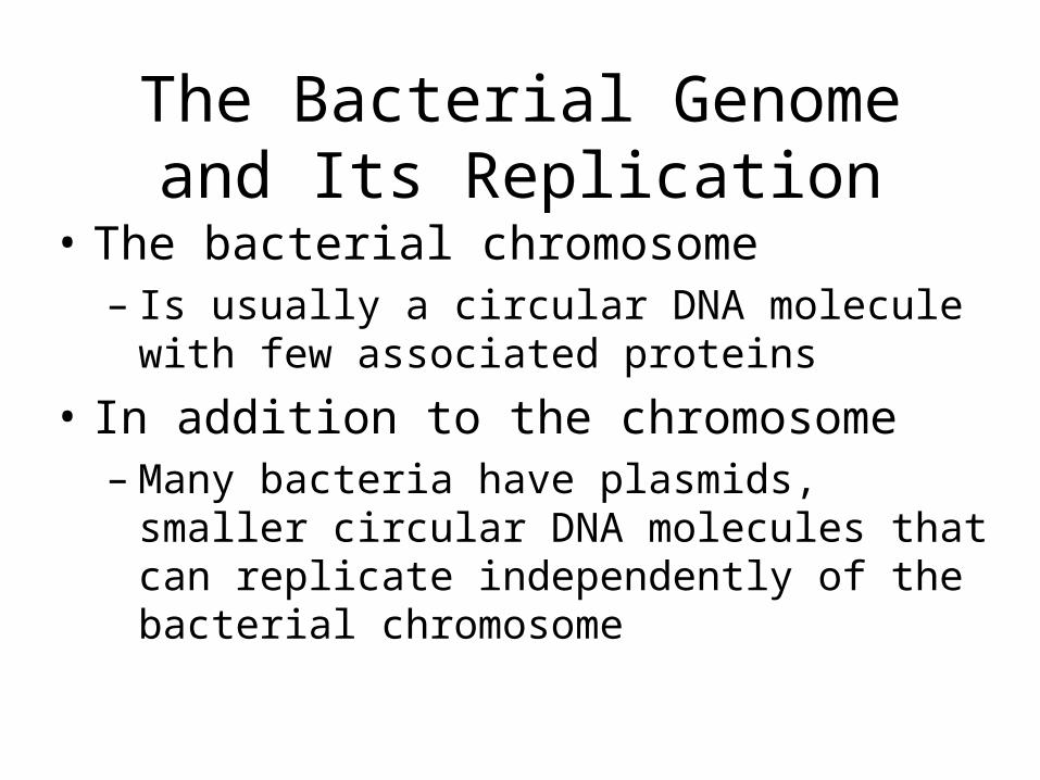
Replication of the circular chromosome
Replicationfork
Origin of replication
Termination of replication
Figure 18.14
Replication always starts at a certain place
Normal replication fork for DNA synthesis

Mutantstrainarg trp+
EXPERIMENT
Figure 18.15
Mix two mutant strains: Arg+ Trp- and Arg- Trp+. Grown them on complete media. After a short while, test them on culture medium without Trp or Arg.
Mutantstrainarg+ trp–
Mixture
Bacterial cells usually divide asexually by binary fission
but they can occasionally exchange
genes:

Coloniesgrew
Mutantstrainarg+ trp–
Mutantstrainarg– trp+
No colonies(control)
No colonies(control)
Mixture
To grow on minimal medium, the cell must be able to make both Arginine and Tryptophan (Arg+, Trp+).
--> Evidence for genetic transfer of one of those genes to the other strain.
CONCLUSION
RESULTS
Now test them on minimal culture mediumWhy do they need the control plates?
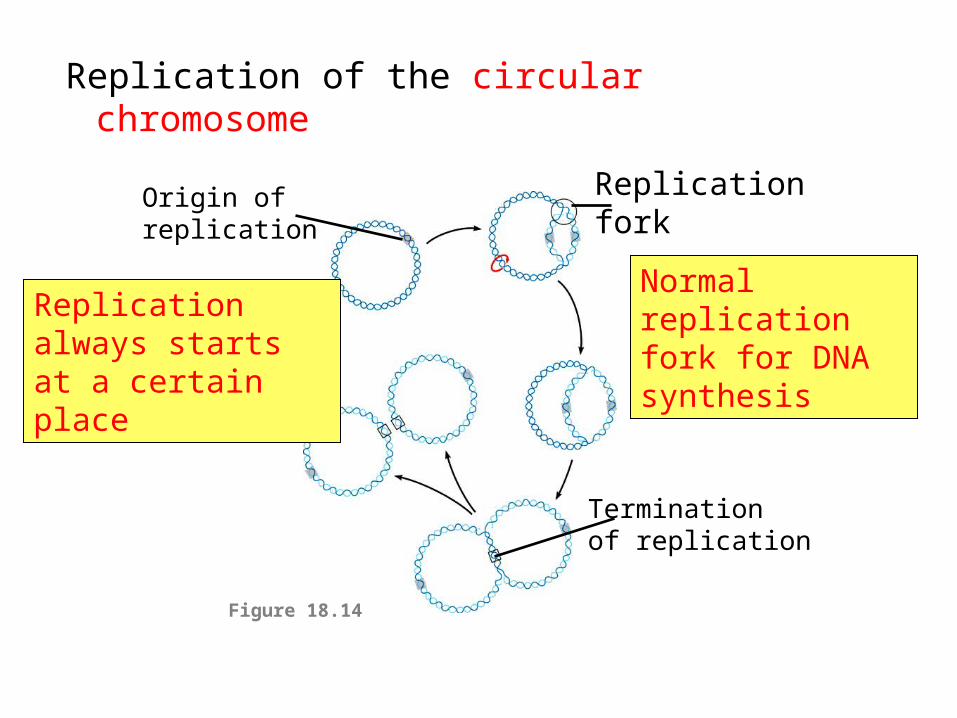
Four ways bacteria can exchange genes

1. Transduction
Phage can transfer bacterial genes between cells

1. Phage virus infects A+B+ cell
2. Reproduction and lysis
Once in a while host DNA is mistakenly packaged in a capsid
3. Transfer of a+ DNA from phage to new cell

2. Conjugation– direct transfer of genetic material between bacterial cells that are temporarily joined
Figure 18.17 Sex pilus 1 m
Recipient cell is F-(has no plasmid)
Donor cell contains F+ plasmid
One way transfer

Conjugation and transfer of an F plasmid from an F+ donor to
an F recipient
Figure 18.18a
F+ cell can form amating bridge with an F– celland transfer its F plasmid.
Single strand of the F plasmid breaks at a specific point and moves into the recipient cell.
Both cells are now F+.
Bacterial chromosomes
F+ cell
F+ cell
F+ cell
Mating bridge
F– cell
F Plasmid

Donor F+ cell
Synthesis of complementary strand in recipient

Copyright ©The McGraw-Hill Companies, Inc. Permission required for reproduction or display
Structure of F plasmid
These genes play a role in the transfer of DNAThey are thus designated tra and trb followed by a capital letter

3. High Frequency Recombination(Hfr cells)
F factor can sometimes become integrated in to a bacterial chromosome.
Cell is F+ because it has all of the F factor genes
F+
MUCH more likely to transfer chromosomal genes to F- cell during conjugation

Usually carries some chromosomal DNA along with it when it is transferred to an F– cell
See this in action
Conjugation of Hfr cell with F- cell)

Conjugation and transfer of part of the bacterial chromosome from an Hfr donor to an F– recipient
F+ cell Hfr cell
F factorThe circular F plasmid in an F+ cellcan be integrated into the circularchromosome by a single crossoverevent (dotted line).
1The resulting cell is called an Hfr cell (for High frequency of recombination).
2
Since an Hfr cell has all the F-factor genes, it can form a mating bridge with an F– cell and transfer DNA.
3 A single strand of the F factorbreaks and begins to move through the bridge. DNA replication occurs in both donor and recipient cells, resulting in double-stranded DNA
4 The location and orientation of the F factor in the donor chromosome determine the sequence of gene transfer during conjugation. In this example, the transfer sequence for four genes is A-B-C-D.
5 The mating bridgeusually breaks well before the entire chromosome andthe rest of the F factor are transferred.
6
Two crossovers can result in the exchange of similar (homologous) genes between the transferred chromosome fragment (brown) and the recipient cell’s chromosome (green).
7 The piece of DNA ending up outside thebacterial chromosome will eventually be degraded by the cell’s enzymes. The recipient cell now contains a new combination of genes but no F factor; it is a recombinant F– cell.
8
Temporarypartialdiploid
Recombinant F–
bacterium
A+B+ C+
D+
F– cell A–B–
C–D–
A–B–
C–D– D–
A–C–
B–
A+B+C+D+A+B+
D+C+
A+
A+
B+
A–B–
C–D–
A–B+
C–D–
A+B+ B–
A+
Hfr cell
D–A–C–
B–
A+B+C+D+
A+B+
Figure 18.18b

Integration of F+ plasmid into a chromosome

Transformation
• Transformation– uptake of naked, foreign DNA from the surrounding environment
•Remember Griffith’s experiment with heat killed bacteria and mice?

Does this happen in nature?
• In E. coli and Salmonella, roughly 17% of their genes have been acquired from other species (over 100 million years . . . )
• Such “horizontal transfer” is an important issue for the spread of antibiotic resistance


Spread of Atrizine decomposing bacteria
• A few bacterial species are capable of metabolizing the synthetic herbicide Atrizine
• All have nearly identical genes.

Atrizine catabolism plasmid
Transposons flank these genes
Dispersed atrizine catabolism genes (ABC) acquired separately?
Genes DEF in an operon
Martinez et al. J Bact. Oct 2001

Resistance mechanismsAntibiotic Method of resistance
------------------------------------------------------------------------
Chloramphenicol reduced uptake into cellTetracycline active efflux from the cellB-lactams, Erythromycin, eliminates or reduces binding of antibiotic to targetB-lactams, Erythromycin hydrolysisAminoglycosides, Chloramphenicol, inactivation of antibiotic by enzymatic modificationB-lactams, Fusidic Acid sequestering of the antibiotic by protein bindingSulfonamides, Trimethoprim metabolic bypass of inhibited reactionSulfonamides, Trimethoprim overproduction of antibiotic target (titration)Bleomycin binding of specific immunity protein to antibiotic
http://www.bioteach.ubc.ca/Biodiversity/AttackOfTheSuperbugs/