Embed Size (px)
Citation preview

The Department of Immunology and Oncology studies the molecular and cellular basis of immune system function and tumour development. Our intention is to generate knowledge that can help develop new and improved approaches to immune response modulation during inflammatory reactions, infection and tumour development, and to identify targets for the prevention, diagnosis and treatment of cancer.
This is an exciting time for research in immunology and oncology. In the last two years, the department’s work has addressed many aspects of innate and adaptive immunity, which without doubt will have an impact on immunotherapeutic applications. We continue at the front line of immunology and oncology research, characterising the molecular mechanisms that underlie inflammation, the processes that drive tissue-specific tumour development, as well as tumour immunology and the relationships among stem cells, inflammation and cancer.
In 2013-2014, Dr Yolanda Carrasco was established as a Staff Scientist in the department, and we have welcomed Dr Antonio Bernad’s group, which reinforces the department’s activity in stem cell research. The molecular and cellular mechanisms that underlie the immune response and tumour development often overlap, providing many opportunities for collaboration among the groups in the department as well as with other labs in the CNB and at other institutes. An example is the nanobiomedicine initiative to develop nanotechnology-based biomedical applications.
3 / Immunology and Oncology

40 / IMMUNOLOGY & ONCOLOGY
Our research explores the development and functional specialisation of inflammatory dendritic cells and macrophages during the immune response to bacterial (Klebsiella pneumoniae, Citrobacter rodentium) and fungal (Candida albicans) infections, allergic reactions induced by acaridae-derived allergens, and sterile inflammation caused by peritoneal injury.
The methodology designed for addressing these objectives involves the following experimental approaches:
Development of in vitro and in vivo infection models, allergic reactions and peritoneal injury in different mouse strains, including mice deficient in cytokines, cytokine/chemokine receptors, or molecules involved in dendritic cell and macrophage differentiation or activation.
Cell biology techniques designed for the purification or isolation of defined cell populations from mouse bone marrow, lung, kidney, lymph nodes, spleen and peritoneum involving magnetic bead and FACS cell separation methods.
Analysis of monocytes, dendritic cells and macrophages in cell suspensions or tissue sections, by electron microscopy or confocal microscopy after immunofluorescent staining.
Analysis of gene expression at the mRNA level by real-time quantitative PCR, whole mouse genome microarray analyses and chromatin immunoprecipitation.
Del Fresno C, Soulat D, Roth S, Blazek K, Udalova I, Sancho D,
Ruland J, Ardavín C. Interferon-b production via Dectin-1-Syk-IRF5
signaling in dendritic cells is crucial for immunity to C. albicans.
Immunity 2013; 38:1176-186
López-Bravo M, Minguito M, Domínguez P, del Fresno C, Martín
P, González-Cintado L, Martínez del Hoyo G, Ardavín C. IL-4 blocks
Th1-polarizing/inflammatory cytokine gene expression during monocyte-derived dendritic cell
differentiation through histone hypoacetylation. J Allergy Clin
Immunol 2013; 132:1409-1419
Sierra-Filardi E, Nieto C, Domínguez-Soto A, Barroso R,
Sánchez-Mateos P, Puig-Kroger A, López-Bravo M, Ardavín C,
Rodríguez-Fernández JL, Sánchez-Torres C, Mellado M, Corbí AL.
CCL2 shapes macrophage polarisation by GM-CSF and
M-CSF: identification of CCL2/CCR2-dependent gene expression profile. J Immunol 2014; 192:3858-
3867
SELECTEDPUBLICATIONS
PRINCIPAL INVESTIGATOR:
Carlos Ardavín
SENIOR SCIENTIST:
María López-Bravo
PREDOCTORAL SCIENTISTS:
María MinguitoJorge Domínguez
Lidia Feo
TECHNICIAN:
Leticia González
VISITING SCIENTISTS:
Fernanda Sánchez Vallecillo (CONICET, Argentina)
Daria Kamzol (Wrocław University of Technology, Poland)
Differentiation and functional specialisation of dendritic cells during inflammatory, infectious and allergic processes
1 Characterisation of spleen macrophage subpopulations during Listeria monocytogenes infection
2 Six-colour FACS analysis of lung dendritic cell and macrophage subsets during HDM-induced allergy
1
2

IMMUNOLOGY & ONCOLOGY / 41
Daszkiewicz L, Vazquez-Mateo C, Rackov C, Ballesteros-Tato
A, Weber K, Madrigal-Aviles A, Di Pilato M, Fotedar A,
Fotedar R, Flores JM, Esteban M, Martinez-A C, Balomenos D. Distinct p21 requirements
for regulating normal and self-reactive T cells through IFN-a
production. Sci Rep 2014; 5:7691
SELECTEDPUBLICATIONS
PRINCIPAL INVESTIGATOR:
Dimitrios Balomenos
PREDOCTORAL SCIENTISTS:
Gorjana RackovRahman Shokri
Ana Belén Saccone García Kathrin Weber
STUDENTS:
Adrián Madrigal AvilésJorge de Andrés Anaya
Novel p21 and Fas functions in normal and autoimmune T memory responses
T cell responses are necessary to react to and eliminate external microorganisms and protect humans from infection. Genetic or environmental factors can cause aberrant responses of T cells to the organism they are meant to protect, generating autoreactive memory T cells. Compared to normal memory T cells, autoreactive T cells have unique features due to their repeated encounters with autoantigens. We work to identify differences between normal and autoimmune memory T cells.
Enhanced p21 expression suppresses autoimmune but not normal T cell memory responses
We previously identified p21 as a regulator of autoimmune manifestations. Although several later studies from our group and other laboratories addressed the effect of p21 in T cells and autoimmunity, the mechanism by which p21 directs its autoimmunity-suppressing effect has been a point of debate. We showed that p21 overexpression by T cells of autoimmune lupus-prone Fas-deficient (lpr) mice reduces autoimmunity independently of the p21 effect as a cell cycle inhibitor; instead, p21 limited activation of autoreactive B6-lpr memory T cells and their ability to produce IFN-a. We are addressing the therapeutic potential of p21, since it reduces autoreactive T cell activation but does not affect normal protective immunity.
An alternative function for the Fas/FasL apoptosis system
Our analysis of memory T cell activation, apoptosis and cell cycle regulation events in immunity and autoimmune disease have identified a previously unreported role for Fas, and establish an attenuating role in the response of persistently activated T cells. We now focus on the mechanistic aspects of this new role of the Fas/FasL system on preactivated T cells.
p21 regulates macrophage activation and polarisation
Independently of its cell cycle inhibitory capacity, p21 regulates macrophage activation by controlling the NF-gB pathway, and decreases sensitivity to septic shock. We also found that p21 enables polarisation of M1 to M2 macrophages by promoting p50/p50 formation, and thus inhibits IFN-` production, STAT1 phosphorylation and iNOS production after persistent inflammation. We are studying how p21 controls macrophage activation and polarisation.
Identification of activation and apoptosis regulators specific for controlling autoimmune T cell memory and inflammation
1 Decreased infiltration of destructive CD4+ T cells (red) in the kidneys of p21-overexpressing MRL/lpr-p21tg compared to MRL/lpr mice.
2 p21 regulates macrophage activation through NF-gB inhibition. During persistent inflammation, p21 promotes p50/p50 formation and M1 to M2 macrophage polarisation by suppressing IFN-` production and downstream events that define M1 responses.
1
2

42 / IMMUNOLOGY & ONCOLOGY
The group's research lines are organised around the study of the mechanisms that regulate the immune response and immune tolerance, as modulation of these mechanisms might be a treatment strategy for autoimmune diseases and in cancer immunotherapy.
We studied the role of phosphoinositol-3-kinase p110a in the regulation of in situ apoptosis, macrophage proliferation and polarisation towards M1 or M2 phenotypes in atherosclerotic lesions. Atherosclerotic plaques in fat-fed LDLR-/-p110a-/- mice were smaller than controls, with less immune cell infiltration and less macrophage proliferation in atherosclerotic lesions. This was associated with higher intracellular cAMP levels, suggesting that higher cAMP levels and the ensuing inhibition of macrophage proliferation contribute to atheroprotection in LDLR-null mice lacking p110a.
We observed increased expression of the NKG2D ligand MICA in systemic lupus erythematosus (SLE) patient kidneys but not healthy subjects. We found glomerulus-specific expression of the NKG2D ligands Rae-1 and Mult-1 in various murine SLE models, which correlated with a larger number of glomerular-infiltrating NK cells. MRL/MpJ and MRL/MpJlpr SLE-like mice showed phenotypically immature NK cells accumulate in the spleen, but not in bone marrow or kidneys of diseased mice. These findings and the presence of glomerulus-specific NKG2D ligands in lupus-prone mice identify a role for NK cells and NKG2D ligands in the lupus nephritic process.
We also develop nanoparticle-based nanomedicines that allow efficient, specific targeting of drugs, biomolecules or cell types to a desired site of action for antitumour and immunosuppressive therapies. This objective is divided into five sub-objectives:
1. Comparative analysis of approaches to efficient nanoparticle targeting in cancer therapies, and development of new-generation polymeric magnetic nanoparticles.
2. Direct specific magnetic nanoparticle-loaded cells by applying an external magnetic field to the target tissue for immunomodulatory lymphoid cell transfer therapies.
3. Development and therapeutic study of magnetic nanoparticles functionalised with RNA interference.
4. Study of the cell interactome induced by magnetic nanoparticle and metabolic degradation of iron oxide nanoparticles to reduce systemic toxicity and increase effectiveness.
5. Study of the biological effects of intracellular hyperthermia-mediated nanoparticles.
Zotes TM, Spada R, Mulens V, Pérez-Yagüe S, Sorzano CO, Okkenhaug
K, Carrera AC, Barber DF. PI3K p110 is expressed by gp38(-)CD31(+) and gp38(+)CD31(+) spleen stromal cells
and regulates their CCL19, CCL21, and LT`R mRNA levels. PLoS One
2013; 8:e72960
Zotes TM, Arias CF, Fuster JJ, Spada R, Pérez-Yagüe S, Hirsch E, Wymann
M, Carrera AC, Andrés V, Barber DF. PI3K p110g deletion attenuates
murine atherosclerosis by reducing macrophage proliferation but not
polarisation or apoptosis in lesions. PLoS One 2013; 8:e72674
Mejías R, Gutiérrez L, Salas G, Pérez-Yagüe S, Zotes TM, Lázaro FJ, Morales MP, Barber DF. Long
term biotransformation and toxicity of dimercaptosuccinic acid-coated
magnetic nanoparticles support their use in biomedical applications. J
Control Release 2013; 171:225-233Ruiz A, Salas G, Calero M,
Hernández Y, Villanueva A, Herranz F, Veintemillas-Verdaguer S,
Martínez E, Barber DF, Morales MP. Short-chain PEG molecules strongly bound to magnetic nanoparticle for
MRI long circulating agents. Acta Biomater 2013; 9:6421-6430
P201431531. Dispositivopara generar hipertermia en
células mediante nanopartículas magnéticas (MNPs)
SELECTEDPUBLICATIONS
PRINCIPAL INVESTIGATOR:
Domingo F. Barber
POSTDOCTORAL SCIENTISTS:
José Manuel Rojas Carrasco Marinn Talelli
Raquel Mejías Laguna
PREDOCTORAL SCIENTISTS:
Patricia Hernández Flores Laura Sanz Ortega
Vladimir MulensLaura Morillas Navas
Roberto SpadaTeresa M. Zotes Ciprés
TECHNICIAN:
Sonia Pérez-Yagüe
TECHNOLOGY ADVISOR:
José Luis Tajada Herráiz
Lymphocytes in physiological and pathological processes: autoimmune diseases, cancer immunotherapy, and nanobiomedicine
PATENT
1
1 Matrix (red) degradation by THP-1 derived macrophages (green) associated with superparamagnetic ion oxide nanoparticles (SPIONs). (Photo: L Sanz/JM Rojas)
2 Uptake of magnetic nanoparticles by pancreatic adenocarcinoma (Pan02) cells, observed by transmission electron microscopy (TEM). (Photo: M Talelli)
2

IMMUNOLOGY & ONCOLOGY / 43
Myocardial infarction is one of the major challenges facing health care systems in developed countries. Its treatment, despite recent advances, remains a serious problem and requires multidisciplinary approaches. Adult organ function depends on the concerted, regulated action of specialised stem cells. Our group focusses on the study of multipotent cardiac progenitor cells (CPC) isolated from adult heart, although precise understanding of cardiac precursor and stem cell biology is still lacking. This is a critical step toward developing more rational strategies to fight against cardiovascular disease.
Human CPC (also mouse and pig) have been characterised as MSC (mesenchymal stem cell)-like populations. Partial hCPC membrane proteomic analysis allowed the definition of more than 30 relevant functions especially associated with adult stem cell biology. We demonstrated that podocalyxin-like protein 1 (PODXL) regulates hCPC activation, migration and differentiation, and also modulates their local immunoregulatory capacity (Moscoso et al., 2013). In the last two years, we have been involved in an intensive collaborative European effort (CAREMI) for the molecular characterisation of hCPC using complementary high-throughput platforms (global proteomics, ITRAQ, RNAseq). In summary, we have identified 9,943 proteins, of which 25.3% are exclusive to or preferentially expressed in hCPC.
In 2014, an international clinical trial (Ia-IIb) was launched to evaluate the therapeutic potential of hCPC in acute myocardial infarct. At the moment, the escalation phase has concluded (6 patients) and the final phase (49 additional patients) is under way.
In mouse models, we demonstrated that the transcriptional factor Bmi-1+ (a member of the polycomb family) is an important marker of mouse CPC (mCMC). The Bmi-1+ CMC population (B-CPC) contributes to homeostatic cardiomyocyte turnover and repair after acute injury, and fulfils the criteria for denomination as long-term cardiac resident stem cells. B-CPC are distributed throughout the entire myocardium, forming small groups of cells found preferentially in perivascular locations or embedded in sarcomers. We are currently studying the distinct stimuli that affect the biological responses of B-CPC in vivo, as well as its importance in heart turnover and functional demonstration of candidate pathways identified by differential RNAseq analysis.
SELECTEDPUBLICATIONS
Estrada JC, Torres Y, Benguría A, Dopazo A, Roche E, Carrera-
Quintanar L, Acín-Pérez R, Enríquez JA, Torres R, Ramírez JC, Samper
E, Bernad A. Human mesenchymal stem cell replicative senescence and oxidative stress are closely linked to
aneuploidy. Cell Death Ther 2013; 4:e691
Tomé M, Sepúlveda JC, Delgado M, Andrades JA, Campisi J, González MA, Bernad A.. miR-335 correlates with senescence/aging in human
mesenchymal stem cells and inhibits their therapeutic actions through
inhibition of AP-1 activity. Stem Cells 2014; 32:2229-2244
Izarra A, Moscoso I, Levent E, Cañón S, Cerrada I, Díez-Juan A, Blanca V,
Núñez-Gil IJ, Valiente I, Ruíz-Sauri A, Sepúlveda P, Tiburcy M, Zimmermann
WH, Bernad A. miR-133a enhances the protective capacity of cardiac progenitor cells after myocardial
Stem Cell Reports 2014; 3:1-14
Izarra A, Moscoso I, Carreiro C, Fondevila D, Martín-Caballero J,
Blanca V, Valiente I, Díez-Juan A, Bernad A. miRNA-1 and miRNA-133a
are involved in early commitment of pluripotent stem cells and
demonstrate antagonistic roles in regulation of cardiac differentiation. J Tissue Engineer Regen Med 2014;
doi: 10.1002/term.1977
PRINCIPAL INVESTIGATOR:
Antonio Bernad Miana
POSTDOCTORAL SCIENTISTS:
José Luis Torán GarcíaSusana Cañón Sánchez
María Tomé Pizarro
PREDOCTORAL SCIENTISTS:
Diego Herrero Alonso
TECHNICAL ASSISTANCE:
Rosa María Carmona CanoreaSusana Aguilar García
VISITOR:
Francisco Miguel Cruz Uréndez (CNIC, Madrid)
Cardiac stem cells
1 Spontaneous differentiation of B-CPC in vitro. Endothelial cells (CD31, blue) and smooth muscle cells (SMA; red); nuclei (green).
2 Mature binucleated cardiomyocyte derived in vivo from B-CPC (YFP+). Sarcomeric _-actinin(red); nuclei (blue).
1
2

44 / IMMUNOLOGY & ONCOLOGY
Lymphocyte dynamic plasticity is intrinsic to lymphocyte function and is crucial for adaptive immune protection. Better knowledge of the molecular mechanisms that govern lymphocyte behaviour will reveal essential aspects of the immune response. It will also help to understand the ability of certain cancer cells to acquire lymphocyte-like dynamics in order to migrate and invade other tissues (metastasis). Using B lymphocytes as a model system and state-of-the-art technology, we study (1) the molecular mechanisms that underlie the switch between two opposite cell behaviours (stopped/synapse vs motile/kinapse) in the presence of chemokine and antigen, and (2) the functional relevance of the synapse/kinapse status balance for B cell fate; (3) we also study the importance of pathogen-mediated effects on B cell dynamics in influencing and their influence on the synapse/kinapse balance. Our recent results can be summarised in two main points.
Molecular mechanisms involved in the regulation of B cell dynamics by antigen and CXCL13. We identified a major role for the scaffold protein vinculin in governing B cell adhesion and motility. BCR signalling triggers vinculin recruitment to the immune synapse, where vinculin co-localises with F-actin at the peripheral supramolecular activation cluster (pSMAC). Lack of vinculin at the synapse impairs appropriate pSMAC assembly, enabling B cells to move in response to CXCL13 while displacing the antigen cluster to the cell uropod. Syk and actomyosin regulate vinculin recruitment and stability at the synapse. Vinculin activation and translocation from cytosol to adhesion sites requires its interaction with the lipid PIP2. Accordingly, PIP2 and the enzyme that produces it, PIPKIgamma, localise at the immune synapse pSMAC, and vinculin recruitment coincides with a PIPKIgamma-mediated PIP2 wave at early stages of synapse formation.
Modulation of B cell dynamics by innate signals. We found that TLR4 stimulation by lipopolysaccharide (LPS; a cell wall component of gram-negative bacteria) modulates B cell behaviour in dose- and exposure time-dependent manners. TLR4-stimulated B cells have enhanced cell polarisation and migration, as well as increased directionality in their movement compared to unstimulated B cells; the MyD88-dependent signalling cascade and Rac GTPases are involved in these changes.
Barrio L, Delgado Cuevas V, Menta R, Mancheño-Corvo
P, delaRosa O, Dalemans W, Lombardo E, Carrasco YR.
Human adipose tissue-derived mesenchymal stem cells
promote B cell motility and chemoattraction. Cytotherapy
2014; 16:1692-1699
Barrio L, Saez de Guinoa J, Carrasco YR. TLR4 shapes B cell dynamics via MyD88-dependent
pathways and Rac GTPases. J Immunol 2013; 191:3867-3875
Sáez de Guinoa J, Barrio L, Carrasco YR. Vinculin arrests
motile B cells by stabilizing integrin clustering at the immune
synapse. J Immunol 2013; 191:2742-2751
Bañon-Rodríguez I, Sáez de Guinoa J, Bernardini A, Ragazzini
C, Fernández E, Carrasco YR, Jones GE, Wandossel F,
Antón IM. Ruffle formation and directional persistence during
mesenchymal and ameboid cell migration rely on WIP expression.
PLoS One 2013; 8:e70364
Perez-Rivero G, Cascio G, Fernandez Soriano S, Gil A, Sáez
de Guinoa J, Rodriguez-Frade JM, Gomariz RP, Holgado BL,
Cabañas C, Carrasco YR, Stein JV, Mellado M. Janus kinases 1 and 2 regulate chemokine-mediated integrin activation
and naïve T cell homing. Eur J Immunol 2013; 43:1745-1757
SELECTEDPUBLICATIONS
GROUP LEADER:
Yolanda R. Carrasco
PREDOCTORAL SCIENTISTS:
Julia Sáez de Guinoa CorralLaura Barrio Cano
MASTER’S DEGREE STUDENTS:
Sara Violeta Merino CortésSara Román García
Adrián Tirado
B cell dynamics
1 TLR4 stimulation enhances B cell polarisation, migration and straightness
2 Syk regulates vinculin recruitment to the B cell immune synapse
1 2

IMMUNOLOGY & ONCOLOGY / 45
We focus on the molecular mechanisms by which kinases control cell behaviour and, when altered, human disease. In recent years, we have concentrated especially on phosphoinositide 3-kinase (PI3-kinase), an enzyme that generates the PIP3 product, which is increased in cancer and autoimmunity as it triggers cell survival, invasion and division.
PI3-kinase mouse models and biochemical approach
PI3-kinases are heterodimers composed of a p110 catalytic and a p85 regulatory subunit. p110alpha and beta, and associated p85alpha and p85beta, are ubiquitous and are altered in cancer, whereas p110delta and the closely-related p110gamma isoform are more abundant in haematopoietic cells; when deregulated, they are involved in development of chronic inflammation/autoimmunity. In animal models, we study the physiological function of PI3K and the consequences of its deregulation in cancer and autoimmunity.
Mechanism of PI3-kinase beta action on DNA/chromatin remodelling
The PI3-kinase pathway is frequently altered in cancer. Many laboratories and companies have focussed on PI3-kinase alpha; we study the less well-known PI3-kinase beta isoform. We showed that PI3-kinase beta (p110beta) localises to the nucleus and regulates DNA replication, segregation and repair. We aim to determine the molecular basis of p110beta action in DNA homeostasis and chromatin remodelling in cancer and stem cells to understand why certain tumour types increase p110beta expression.
Alternative cancer treatment based on interfering molecules
Metastasis remains the leading cause of death from many tumours. The PIP3 product enables cell survival; for this reason, cells with altered regulation of PI3-kinase alpha or beta, or with mutations in the phosphatase PTEN (which reduces PIP3 levels), are present in ~50% of human tumours, most often at metastatic phases.
PI3-kinase is a key target in cancer, but only broad-spectrum inhibitors or p110alpha inhibitors are being tested clinically. Our aim is to develop a strategy for the treatment of tumours accessible by endoscopy (or orally), based on delivery of interfering molecules (RNA and peptides). The first target selected is p85beta. p85 subunits regulate the activity and localisation of this enzyme. Normal cells express mainly p85alpha, while some tumour types show predominant p85beta expression that correlates with metastasis. In parallel to defining p85beta mechanism of action in metastasis, we study the effect of interfering with p85beta action in an established tumour in the mouse.
Redondo-Muñoz J, Pérez-García V, Carrera AC. Phosphoinositide 3-kinase beta: when a kinase is more than a kinase. Trends Cell
Mol Biol 2014; 8:83-92
Cariaga AE, Cortés I, García E, Pérez-García V, Pajares MJ,
Idoate MA, Redondo-Muñóz J, Antón IM, Carrera AC.
Phosphoinositide 3-kinase p85beta regulates invadopodium
formation Biol Open 2014; pii:BIO20148185
Suárez-Fueyo A, Rojas JM, Cariaga AE, García E, Steiner
BH, Puri KD, Barber DF, Carrera AC. Inhibition of PI3Kb�
reduces kidney infiltration by macrophages and ameliorates systemic lupus in the mouse. J
Immunol 2014; 193:544-554
Pérez-García V, Redondo-Muñóz J, Kumar A, Carrera AC. Cell activation-induced
phosphoinositide 3-kinase alpha/beta dimerization regulates
PTEN activity. Mol Cell Biol 2014; 34:3359-3373
Redondo-Muñoz J, Pérez-García V, Rodríguez MJ, Valpuesta JM,
Carrera AC. Phosphoinositide 3-kinase beta protects nuclear
envelope integrity by controlling RCC1 localization and Ran
activity. Mol Cell Biol 2014; doi: 10.1128/MCB.01184-14
SELECTEDPUBLICATIONS
PRINCIPAL INVESTIGATOR:
Ana C. Carrera
POSTDOCTORAL SCIENTISTS:
Ana María González-GarcíaAriel E. Cariaga Martínez
Rosa Varona ValleRaúl Jiménez Pérez
Javier Redondo Muñoz
PREDOCTORAL SCIENTISTS:
Jesús Vallejo DíazVicente Pérez García
TECHNICIANS:
Carmen Hernández AgüeroLorena Sanz González
Molecular targets in health and disease: focus on PI3-kinase
1 2
1 Localisation of p85beta (green) at invadopodia identified by costaining of actin or cortactin (blue) and by its capacity to degrade gelatin (red). Depletion of p85beta with interfering RNA impairs cell invasion analysed as the degradation of gelatin (red) in controls but not in cells lacking p85beta expression (bottom).
2 Passive diffusion of GFP outside the nucleus, enhanced after p110beta depletion.

46 / IMMUNOLOGY & ONCOLOGY
Aguilera-Montilla N, Chamorro S, Nieto C, Sánchez-Cabo F,
Dopazo A, Fernández-Salguero PM, Rodríguez-Fernández JL,
Pello OM, Andrés V, Cuenda A, Alonso B, Domínguez-Soto A,
Sánchez-Ramón S, Corbí AL. Aryl hydrocarbon receptor contributes
to the MEK/ERK-dependent maintenance of the immature state
of human dendritic cells. Blood 2013; 121:e108-17
Criado G, Risco A, Alsina-Beauchamp D, Pérez-Lorenzo MJ, Escós A, Cuenda A (2014).
Alternative p38 mitogen-activated protein kinases are essential for
collagen-induced arthritis. Arthritis Rheumatol 2014; 66:1208-1217
del Reino P, Alsina-Beauchamp D, Escós A, Cerezo-Guisado MI, Risco
A, Aparicio N, Zur R, Fernandez-Estévez M, Collantes E, Montans J,
Cuenda A. Pro-oncogenic role of alternative p38 mitogen-activated
protein kinases p38g and p38d. Cancer Res 2014; 74:6150-6160
Arechederra M, Priego N, Vazquez-Carballo A, Sequera C, Gutierrez-
Uzquiza A, Cerezo-Guisado MI, Ortiz-Rivero S, Roncero C, Cuenda
A, Guerrero C, Porras A. p38 MAPK down-regulates fibulin 3
expression through methylation of gene regulatory sequences. Role in migration and invasion. J Biol Chem
2014; pii:jbc.M114.582239
SELECTEDPUBLICATIONS
Our group studies the physiological and pathological functions of the p38MAPK family of mitogen-activated protein kinases. Our research focusses on
study of their physiological roles using mice transgenic for distinct p38 isoforms, and
In the 2013-2014 period, we analysed the role of p38g and p38d, two of the less well-known
g and p38d deficiency markedly reduced arthritis severity and suppressed clinical disease, synovial
severity in p38g/dand anti-collagen antibody responses than in controls, indicating that p38g and p38d are crucial
g and p38d as potential therapeutic targets in complex diseases, such as rheumatoid arthritis, that involve innate and adaptive immune responses.
g and p38d in colitis-associated colon cancer using the azoxymethane/dextran sodium sulphate
g/d deficiency decreased tumour
chemokines. Analysis of leukocyte populations in p38g/d-null mouse colon showed less macrophage and
p38g/d-/- bone marrow (BM) had fewer
increased significantly in p38g/d-/-
to p38g/d-/- mice that received p38g/d-/- BM. Our results establish that p38g and p38dinduced tumour formation by regulating haematopoietic cell response to injury, and validate p38g and p38d as potential targets for cancer therapy.
PRINCIPAL INVESTIGATOR:
Ana Cuenda
ASSOCIATE SCIENTIST:
Lourdes Planelles
POSTDOCTORAL SCIENTISTS:
Ana Risco
PREDOCTORAL SCIENTISTS:
Rafal Zur Dayanira Alsina-Beauchamp
Alejandra Escós
TECHNICIAN:
Ruth Gómez-CaroCarla Argüello Pilco
MASTER’S DEGREE STUDENTS:
Angel Nuñez-BuizaLaura García-Ibañez
Esther Blanco
VISITING SCIENTISTS:
Noelia Aparicio(Universidad Francisco de Vitoria, Spain),
Carmen Vela (Universidad Europea de Madrid, Spain)
Role of stress-activated protein kinase p38MAPK in human diseases
1 Reduced incidence of colitis-associated tumours in p38g/d-deficient mice
2 New p38g/p38d-dependent pathway of cytokine production in response to bacteria lipopolysaccharide (LPS)
1
2

IMMUNOLOGY & ONCOLOGY / 47
The cancer epigenetics laboratory is interested in the study of epigenetic mechanisms during cell development and differentiation as well as their alterations in ageing and cancer. To address these issues, we carried out genome-wide analyses of DNA methylation and histone modification in adult stem and terminally differentiated cells and in different tumour types. Using these approaches, we identified a chromatin signature (H3K4me1) associated with DNA hypomethylation in human ageing. We also identified DNA methylation patterns associated with specific thyroid cancer subtypes and with the expression of glycine N-methyltransferase in human hepatocellular carcinoma.
Using autologous and heterologous experimental systems in mice, we demonstrated that the aberrant DNA demethylation-dependent expression of the cytokine GM-CSF and its receptor by colon cancer cells has strong an Antitumoural activity of cytokine GM-CSF in colon cancer ti-tumour effects and is an independent marker of good prognosis in patients with colorectal tumours. Our laboratory also had the opportunity to collaborate in a study that, using a genetic progression model of Braf(V600E), identified new targets for therapeutic intervention in intestinal cancers. We also contributed to the first reconstruction of the Denisovan and the Neanderthal DNA methylomes.
Gokhman D, Lavi E, Prüfer K, Fraga MF, Riancho JA, Kelso J, Pääbo S, Meshorer
E, Carmel L. Reconstructing the DNA methylation maps of the Neanderthal
and the Denisovan. Science 2014; 344:523-527
Rodríguez-Rodero S, Fernández AF, Fernández-Morera JL, Castro-Santos
P, Bayon GF, Ferrero C, Urdinguio RG, Gonzalez-Marquez R, Suarez C,
Fernández-Vega I, Fresno Forcelledo MF, Martínez-Camblor P, Mancikova
V, Castelblanco E, Perez M, Marrón PI, Mendiola M, Hardisson D, Santisteban
P, Riesco-Eizaguirre G, Matías-Guiu X, Carnero A, Robledo M, Delgado-Álvarez E, Menéndez-Torre E, Fraga
MF. DNA methylation signatures identify biologically distinct thyroid cancer
subtypes. J Clin Endocrinol Metab 2013; 98:2811-2821
Rad R, Cadiñanos J, Rad L, Varela I, Strong A, Kriegl L, Constantino-Casas
F, Eser S, Hieber M, Seidler B, Price S, Fraga MF, Calvanese V, Hoffman G,
Ponstingl H, Schneider G, Yusa K, Grove C, Schmid RM, Wang W, Vassiliou G, Kirchner T, McDermott U, Liu P, Saur D, Bradley A. A Genetic Progression
Model of Braf(V600E)-Induced Intestinal Tumorigenesis Reveals Targets for
Therapeutic Intervention. Cancer Cell 2013; 24:15-29
Urdinguio RG, Fernandez AF, Moncada-Pazos A, Huidobro C, Rodriguez RM,
Ferrero C, Martinez-Camblor P, Obaya AJ, Bernal T, Parra-Blanco A, Rodrigo L, Santacana M, Matias-Guiu X, Soldevilla
B, Dominguez G, Bonilla F, Cal S, Lopez-Otin C, Fraga MF. Immune dependent
and independent anti-tumor activity of GM-CSF aberrantly expressed by
mouse and human colorectal tumors. Cancer Res 2013; 73:395-405
SELECTEDPUBLICATIONS
PRINCIPAL INVESTIGATOR:
Mario Fernández Fraga
POSTDOCTORAL SCIENTISTS:
Agustín Fernández FernándezRocío González UrdinguioGustavo Fernández BayónSandra Rodríguez Rodero
PREDOCTORAL SCIENTISTS:
Estela García TorañoMaría García García
Antonella Carrella
STUDENTS:
Álvaro del Real BoltThalia Belmonte García
TECHNICIANS:
Cristina Mangas AlonsoCristina Bravo Mendoza
Cancer epigenetics
1
2
2 Antitumour activity of cytokine GM-CSF in colon cancer.
1 Epigenetic changes during ageing of human adult stem cells.

48 / IMMUNOLOGY & ONCOLOGY
Anti-tumour activity of chemokine receptor-specific antibodies
Chemokines, small cytokines that direct cell movement, have a central role in the maintenance of innate and acquired immunity. These proteins interact with specific G protein-coupled receptors and participate in the pathogenesis of inflammatory and infectious diseases as well as in cancer. Chemokines can help limit tumour development by increasing leukocyte migration to a tumour site and by inducing long-term antitumour immunity. In contrast, they can also facilitate tumour cell survival, proliferation, and metastatic potential. Expression of a chemokine receptor by a tumour preferentially directs metastasis to the organs in which the chemokine ligand is secreted, suggesting chemokine receptors as promising therapeutic targets.
Our main research interest is to determine how chemokines participate in the control of tumour growth and progression, and to evaluate their potential as antitumour targets. The use of specific monoclonal antibodies for chemokine receptor targeting allows us to block signalling by preventing ligand binding to its receptor, and also to tag the tumour cells and trigger a host immune response to them.
The chemokine receptor CCR9 is expressed primarily on thymocytes and a small subset of intraepithelial lymphocytes. Its overexpression increases the migratory and invasive capacity of prostate cancer cells, directs ovarian cancer and melanoma metastases to the small intestine, and activates anti-apoptotic pathways that lead to survival and increased proliferation of leukaemia cell lines. We recently generated and characterised mouse anti-human CCR9 monoclonal antibodies that reduced human T lymphoblastic cell tumours transplanted into mice by >85%. Tumour size reduction was concomitant with an increase in the fraction of apoptotic tumour cells and in tumour necrotic areas, as well as a decrease in the fraction of proliferating cells and in tumour vascularisation. Our results suggest that CCR9-expressing tumours such as acute and chronic T cell lineage leukaemia, prostate cancer, breast cancer and melanoma might be targeted with these antibodies.
Chamorro S, Vela M, Franco-Villanueva A, Carramolino L, Gutiérrez J, Gómez L, Lozano M, Salvador B, García-Gallo M, Martínez-A C, Kremer L. Antitumor
effects of a monoclonal antibody to human CCR9 in leukemia cell xeno-
grafts. MAbs 2014; 6:1000-1012
González-Magaldi M, Martín-Acebes MA, Kremer L, Sobrino F. Membrane
topology and cellular dynamics of foot-and-mouth disease virus 3A pro-
tein. PLoS One 2014; 9:e106685
Goñi GM, Epifano C, Boskovic J, Ca-macho-Artacho M, Zhou J, Bronowska A, Martín MT, Eck MJ, Kremer L, Gräter
F, Gervasio FL, Perez-Moreno M, Lietha D. Phosphatidylinositol 4,5-bis-phosphate triggers activation of focal adhesion kinase by inducing cluster-
ing and conformational changes. Proc Natl Acad Sci USA 2014; 111:E3177-86
Aranda JF, Reglero-Real N, Mar-cos-Ramiro B, Ruiz-Sáenz A, Fernán-
dez-Martín L, Bernabé-Rubio M, Krem-er L, Ridley AJ, Correas I, Alonso MA, Millán J. MYADM controls endothelial
barrier function through ERM-depend-ent regulation of ICAM-1 expression.
Mol Biol Cell 2013; 24:483-494
Sánchez-Martín D, Martínez-Torrec-uadrada J, Teesalu T, Sugahara KN,
Alvarez-Cienfuegos A, Ximénez-Em-bún P, Fernández-Periáñez R, Martín
MT, Molina-Privado I, Ruppen-Cañás I, Blanco-Toribio A, Cañamero M, Cues-ta AM, Compte M, Kremer L, Bellas C,
Alonso-Camino V, Guijarro-Muñoz I, Sanz L, Ruoslahti E, Alvarez-Vallina L. Proteasome activator complex PA28 identified as an accessible target in
prostate cancer by in vivo selection of human antibodies. Proc Natl Acad Sci
USA 2013; 110:13791-13796
PCT/EP2014/075578. Anticuerpos frente a CCR9 y sus aplicaciones
SELECTEDPUBLICATIONS
PRINCIPAL INVESTIGATOR:
Leonor Kremer
POSTDOCTORAL SCIENTIST:
Sonia Chamorro
PREDOCTORAL SCIENTISTS:
María VelaMercedes Llorente
TECHNICIAN:
María Lozano
PATENT
1 Effects of the treatment with the anti-CCR9 mAb 91R on xenograft MOLT-4 tumours. 91R promotes apoptosis and necrosis and reduces cell proliferation and angiogenesis in CCR9+ tumour xenografts. (Left) Necrosis and apoptosis was detected by haematoxylin/eosin staining of tissue sections (Bar = 25 +m). (Centre) Apoptosis level of tumour cells was analysed by TUNEL assays (Bar = 50 +m). (Right) Blood vessels were detected by CD31 staining (Bar = 50 +m).
1
2
2 91R-induced reduction of leukaemia xenograft growth. Luminescent MOLT-4 cells were inoculated subcutaneously into each flank of BALB/c Rag2-/- mice on day 0. Experimental groups received intraperitoneal inoculations of 91R or control IgG2b mAb on days 1 and 6. (Left) Luminescence image of a representative mouse from each group, at day 19 post-cell inoculation. (Right) Tumour weights per mouse at experiment endpoint (day 62), after sacrifice and tumour removal.

IMMUNOLOGY & ONCOLOGY / 49
Tardáguila M, Mira E, García-Cabezas MA, Feijoo AM, Quintela-Fandino M, Azcoitia I, Lira SA, Mañes S. CX3CL1
promotes breast cancer via transacti-vation of the EGF pathway. Cancer Res
2013; 73:4461-4473
Mira E, Carmona-Rodríguez L, Tardágui-la M, Azcoitia I, González-Martín A,
Almonacid L, Casas J, Fabriás G, Mañes S. A lovastatin-elicited genetic program
inhibits M2 macrophage polarisation and enhances T cell infiltration into
spontaneous mouse mammary tumors. Oncotarget 2013; 4:2288-2301
Tardáguila M, Mañes S. CX3CL1 at the crossroad of EGF signals: Relevance
for the progression of ERBB2+ breast carcinoma. Oncoimmunology 2013;
2:e25669
Tardáguila M, Mañes S. 2014. The complex role of chemokines in cancer: the case of the CX3CL1/CX3CR1 axis. in
Oncology - Theory & Practice, pp. 181-211 ISBN: 978-1-922227-80-5. iConcept
Press (Australia)
Gómez-Moutón C, Fischer T, Peregil RM, Jiménez-Baranda S, Stossel TP, Naka-mura F, Mañes S. Filamin A interaction with the CXCR4 third intracellular loop
regulates endocytosis and signaling of WT and WHIM-like receptors. Blood
2014; doi: 10.1182/blood-2014-09-601807
SELECTEDPUBLICATIONS
GROUP LEADER:
Santos Mañes
SENIOR SCIENTISTS:
Emilia MiraRosa Ana Lacalle
POSTDOCTORAL SCIENTIST:
Manuel Tardáguila
PREDOCTORAL SCIENTISTS:
Juan Carlos de KaramLorena Carmona-Rodríguez
Jesús OgandoAna Martín Leal
TECHNICIAN:
Rosa M. Peregil
VISITING STUDENTS:
Verena Haage (Konstanz University, Germany)
Jez Marston (Yale University, New Haven CT, USA)
Alexandros Karabatzakis (University of Thessaly, Greece)
Álvaro San Hipólito Luengo (Univ Europea de Madrid, Spain)
Toni Bauzá Massanet (Universidad de Lleida, Spain)
Gabriel García Molina (Univ de Alcalá de Henares, Spain)
Recent clinical and experimental evidence indicates that solid tumours exacerbate inflammation to favour their own progression. This leads to a tumour microenvironment orchestrated largely by inflammatory cells, which alters the metabolic needs of the tissue, fostering proliferation, survival, mutagenesis, migration and metastasis of malignant cells. In addition, tumour-induced inflammation usually leads to angiogenesis (the process by which new blood vessels are formed from pre-existing vasculature) and immunosuppression, which impedes immune-mediated tumour clearance. The immune system is nonetheless able to identify and delete neoplastic cells, and clinical practice now indicates that reprogramming of immune cells is a therapeutically relevant strategy for certain tumours. Successful restriction of the progression or even the eradication of advanced neoplasias using passive immunotherapy with antibodies that block immune checkpoints (CTLA4, PD1/PD-L1/2) or adoptive transfer of T cells genetically engineered to express chimaeric antigen receptors has had such an extraordinary impact on clinical oncology that the journal Science chose cancer immunotherapy as the major scientific breakthrough in 2013.
Harnessing the maximum therapeutic potential of the immune system requires detailed comprehension of the cellular and molecular networks in the tumour microenvironment, as well as of the mechanisms that regulate immune responses. We hope that knowledge of the mechanisms that balance pro- and anti-tumour immunity will lead to the design of more effective anti-cancer therapeutics. The main achievements of our group in these two years include:
I. Identification of the pro-inflammatory chemokine CX3CL1 (fractalkine) as a tumour promoter of ErbB2+ breast carcinomas through its crosstalk with the ErbB signalling pathway.
II. Determination of the genetic program elicited by lovastatin (a mevalonate pathway inhibitor) in the tumour microenvironment, which leads to normalisation of tumour-associated vasculature and anti-tumour polarisation of innate immune cells.
III. Characterisation of the interaction between the chemokine receptor CXCR4, which is implicated in tumour metastasis, with the actin-binding protein filamin-A, and the consequences of this bond in receptor signalling and internalisation.
Signalling networks in inflammation and cancer
1 CX3CL1 staining of in situ (A) and invasive (B) human ductal carcinomas; adjacent non-tumour breast tissue (a) is also indicated. Higher magnification insets are also shown (see numbers). Although CX3CL1 expression is downregulated in tumour tissues, CX3CL1 paradoxically promotes the growth of new neoplastic lesions.
2 Scanning electron micrographs showing the ultrastructure of blood vessels of sporadic breast tumours in mice treated with vehicle (A,B) or lovastatin (C,D). Note the irregular borders and discontinuities or gaps in the vessels of vehicle-treated mice, which suggest endothelial cell hyperactivity. This contrasts with the regular, continuous, tightly-packed endothelium in lovastatin-treated tumours, which lends an appearance of “smoothness” to the vessel lumen.
1
2

50 / IMMUNOLOGY & ONCOLOGY
Bolós V, Mira E, Martínez-Poveda B, Luxán G, Cañameros
M, Martínez-A C, Mañes S, de la Pompa JL. Notch activation stimulates migration of breast
cancer cells and promotes tumor growth. Breast Canc Res 2013;
15:R54
Sánchez Sorzano CO, Pascual-Montano A, Sanchez de Diego
A, Martínez-A C, van Wely KHM. Chromothripsis: Breakage-fusion-bridge over and over again. Cell
Cycle 2013; 12:2016-23
Gatchalian I, Fütterer A, Rothbart SB, Tong Q, Rincon-Arano H,
Sánchez de Diego A, Groudine M, Strahl BD, Martínez-A C,
van Wely KHM, Kutateladze TG. Dido3 PHD modulates cell
differentiation and division. Cell Rep 2013; 4:148-58
Villares R, Kakabadse D, Juarranz Y, Gomariz RP, Martinez-A C,
Mellado M. Growth hormone prevents the development
of autoimmune diabetes. Proc Natl Acad Sci USA 2013;
110:E4619-E4627
Sanchez de Diego A, Alonso Guerrero A, Martínez-A C, van Wely KHM. Dido3-dependent
HDAC6 targeting controls cilium size. Nat Commun 2014; 5:3500
SELECTEDPUBLICATIONS
PRINCIPAL INVESTIGATOR:
Carlos Martínez-A
SENIOR SCIENTIST:
Karel van Wely
POSTDOCTORAL SCIENTISTS:
Thierry FischerCristina Pacios Bras
Ricardo VillaresJesús de Celis
Julio Gutiérrez HernándezMaría Ángeles Garcia
PREDOCTORAL SCIENTIST:
Amaia Talavera
TECHNICIANS
Ainhoa SánchezAgnes Fütterer
Astrid Alonso Guerrero
We study the connections between embryonic stem cell (ESC) differentiation and chromosome instability. Most sporadic tumours have a combination of genetic defects termed chromosomal instability (CIN), found in ~85% of non-hereditary carcinomas. We study aneuploidy (whole-chromosome CIN) and a combination of translocations, duplications and deletions (structural CIN).
We focus on the Dido locus, a gene complex strongly expressed in ESC, that encodes three splice variants (Dido1, Dido2 and Dido3). Whereas Dido2 and Dido3 have the PHD, TF2SM and SPOC protein domains, Dido1 only encompasses PHD. Dido3, the largest, most broadly expressed isoform, is a nuclear protein that interacts with centrosomes and the synaptonemal complex in somatic and germ cells, respectively.
N-terminal truncation of Dido3 (Dido36NT) provokes centrosome amplification and CIN. Dido3 interacts with several proteins, including histone deacetylase 6 (HDAC6). We showed that Dido3-dependent targeting of HDAC6 is a key determinant of cilium size in growth-arrested cells. Dido3 availability at the centrosome governs ciliary HDAC6 levels, and redistribution of the two proteins controls tubulin acetylation. Increased Dido36NT expression and interaction with HDCA6 results in ciliopathies (Figure 1). We used this mutation to interpret classical features of CIN, including frequency of chromosome breakage during cell growth. We consider that, by generating substrates for the non-homologous end-joining system, CIN-associated centromere fission helps to initiate breakage-fusion-bridge cycles and acquisition of additional copies of chromothriptic chromosomes.
C-terminal truncation of the Dido3DCT gene causes early embryonic lethality. ESC derived from blastocysts of Dido36CT mutants do not differentiate in vitro, in contrast to wild-type cells. Differentiation is restored by ectopic expression of wild-type Dido3 and constructs bearing different Dido domains, including the PHD domain. We showed that Dido3 PHD binding to histone H3K4me3 is disrupted by mitotic phosphorylation of an adjacent histone threonine, which triggers Dido3 translocation from chromatin to spindle microtubules. Our crystal structure of Dido3 PHD in complex
with H3K4me3 revealed an aromatic-cage-like binding site. Biochemical, structural, and mutational analyses of the binding mechanism identified specificity and affinity determinants, and explained the affinity of other PHD domains for H3K4me3 (Figure 2A). Our findings link transcriptional control in embryonic development and regulation of cell division (Figure 2B).
Stem cells and immunity
1 Working model of Dido-dependent histone deacetylase 6 (HDAC6) targeting. (A) Basal levels of Dido (blue) and HDAC6 (green) maintain a steady state in which cilium growth and resorption are in equilibrium. (B) Increased Dido recruitment to the basal body, through build-up of actin fibres (red) or liberation from the nucleus, for example, results in higher local HDAC6 concentrations. Subsequent intraciliary transport directs HDAC6 to the apical tip, where tubulin deacetylation promotes gradual reduction in cilium size.
2 The crystal structure of the PHD finger of Dido in complex with the H3K4me3 peptide and a model of the biological effects. (A) The Dido PHD finger is depicted as a solid surface with the peptide shown in a stick model. Protein residues involved in interactions with peptide Ala1 (wheat), Arg2 (orange), Thr3 (light green), Lys4me3 (salmon), Gln5 (light blue), and Thr6 (yellow). (B) A model for Dido3 translocation from chromatin to the mitotic spindle in mitosis.
1
2

IMMUNOLOGY & ONCOLOGY / 51
PRINCIPAL INVESTIGATOR:
Mario Mellado
SENIOR SCIENTIST:
José Miguel Rodríguez Frade
POSTDOCTORAL SCIENTISTS:
Ricardo VillaresLaura Martínez Muñoz
Rubén Rodríguez Barroso
PREDOCTORAL SCIENTISTS:
Dimitri KakabadseGraciela Cascio
TECHNICIAN:
Mª Pilar Lucas
Chemokine receptors: new targets for therapeutic intervention
Although originally described as specific mediators of leukocyte directional movement, the chemokines are involved in a much wider variety of physiological and pathological processes including tumour cell growth and metastasis, angiogenesis, chronic inflammatory diseases and HIV-1 infection. Chemokine biology is more complex than was initially predicted, as several studies suggest that chemokines can dimerise and that their receptors are found as dimers and/or higher order oligomers at the cell surface.
Using fluorescence resonance energy transfer (FRET) techniques, we have confirmed the functional relevance and regulation of homo- and heterodimeric receptor complexes. HIV-1 entry into a target cell requires interaction between viral envelope glycoprotein gp120 and host cell membrane chemokine receptors CXCR4 or CCR5 in combination with CD4. We observed oligomerisation between membrane receptors involved in HIV-1 infection, and demonstrate that CD4/CCR5/CXCR4 complexes modify the gp120 binding, actin polymerisation and cytoskeleton rearrangement necessary for HIV-1 entry.
We also studied the functional relevance of several chemokine-triggered signalling events, and reported that the JANUS kinases (JAK) participate in chemokine-induced integrin activation. We showed that reduced JAK1 and JAK2 expression impair naïve T cell migration in response to gradients of the chemokines CXCL12 and CCL21, and that the in vivo homing of these naïve T cells to lymph nodes is decreased. In addition, we found that the JAK pathway is activated following CXCL12 stimulation in neural precursor cells (NPC), but that JAK activity is not necessary for NPC migration. Immature interneuron migration to the cerebral cortex is nonetheless dependent on CXCL12-mediated PI3K p110` activation.
As chemokines play an essential role in inflammatory and autoimmune disease, we also initiated the study of a murine model of type 1 diabetes, a T cell-mediated autoimmune disease characterised by immune cell infiltration of pancreatic islets and destruction of insulin-producing `-cells. We observed that sustained growth hormone expression reduced prodromal disease symptoms and eliminated progression to overt diabetes. The effect involves several GH-mediated mechanisms; GH altered the cytokine environment, triggered anti-inflammatory macrophage (M2) polarisation, maintained activity of the suppressor T cell population, and limited Th17 cell plasticity.
Martínez-Muñoz L, Barroso R, Dyrhaug SY, Navarro G, Lucas
P, Soriano SF, Vega B, Costas C, Muñoz-Fernández MA, Santiago C, Rodríguez-Frade JM, Franco
R, Mellado M. CCR5/CD4/CXCR4 oligomerisation prevents HIV-1
gp120IIIB binding to the cell surface. Proc Natl Acad Sci USA
2014; 111:E1960-E1969
Sierra-Filardi E, Nieto C, Domínguez-Soto A, Barroso R,
Sánchez-Mateos P, Puig-Kroger A, López-Bravo M, Joven J, Ardavín
C, Rodríguez-Fernández JL, Sánchez-Torres C, Mellado M, Corbí
AL. CCL2 shapes macrophage polarisation by GM-CSF and M-CSF:
Identification of CCL2/CCR2-dependent gene expression profile.
J Immunol 2014; 192:3858-3867
Villares R, Kakabadse D, Juarranz Y, Gomariz RP, Martínez-A C, Mellado
M. Growth hormone prevents the development of autoimmune
diabetes. Proc Natl Acad Sci USA 2013; 110:E4619-E4627
Holgado BL, Martínez-Muñoz L, Sánchez-Alcañiz JA, Lucas
P, Pérez-García V, Pérez G, Rodríguez-Frade JM, Nieto M, Marín O, Carrasco YR, Carrera
AC, Álvarez-Dolado M, Mellado M. CXCL12-mediated murine neural
progenitor cell movement requires PI3K` activation. Mol Neurobiol
2013; 48:217-231
Pérez-Rivero G, Cascio G, Fernández Soriano S, Gil Sanz
A, Sáez de Guinoa J, Rodríguez-Frade JM, Gomariz RP, Holgado BL, Cabañas C, Carrasco YR, Stein JV,
Mellado M. Janus kinases 1 and 2 regulate chemokine-mediated
integrin activation and naïve T-cell homing. Eur J Immunol 2013;
43:1745-1757
SELECTEDPUBLICATIONS
1
21 CCR5 expression on CD4+ T cells inhibits
formation of the X4 gp120-induced virological synapse by blocking F-actin recruitment (green) and ERM phosphorylation (red).
2 Ischaemic stroke model in mice. Representative images of murine brains isolated at various days post-induction (dpi) of the lesion (6, 10 and 21 days; top panel). Nuclear staining of coronal brain sections from mice with ischemia at distinct dpi (bottom). Images show sections of equivalent position on the axial axis. The hippocampus (H) and third ventricle (V) are labelled. Arrows indicate the glial scar, dashed line marks the damaged area.

52 / IMMUNOLOGY & ONCOLOGY
SELECTEDPUBLICATIONS
Highly transformed solid tumours acquire invasive features and the ability to escape from antitumour immunity. The therapies that help to overcome immune evasion by tumours are an important new strategy for cancer treatment. We study the molecular mechanisms by which tumours couple the development of malignant traits to the impairment of T cell responses. We showed that diacylglycerol metabolism by two enzymes of the diacylglycerol kinase (DGK) family limit T cell responses. We are delimiting the contribution of diacylglycerol kinases to T cell functions and oncogenic transformation. Targeting these enzymes, alone or in combination with other inhibitors in wide clinical use, could help in the treatment of aggressive forms of cancer.
We work in two different albeit complementary areas:
1. DGK_ and c as negative regulators of the adaptive immune response
The DGK are a conserved family of lipid kinases that phosphorylate diacylglycerol (DAG), catalysing its conversion into phosphatidic acid (PA). In T lymphocytes, DGKalpha and zeta limit Ras guanyl-releasing protein (RasGRP1)-dependent activation of Ras and activation of certain PKC isoforms. DGKalpha is abundantly expressed in quiescent T lymphocytes and we have demonstrated its transcriptional repression by antigens and IL2-mediated activation of the PI3K/AKT/FoxO axis. This negative regulation is necessary for adequate T cell functions, and its failure results in induction of a non-responsive state known as anergy. DGKalpha expression is elevated in tumour-infiltrating T cells, and we are developing genetic and chemical tools that help us to better understand the mechanisms by which tumour cells induce upregulation of DGKalpha in infiltrating T cells. DGKzeta is also expressed in T lymphocytes and we have shown its interaction with sorting nexin 27 (SNX27).
2. DGK_�and�c: lipid restraint on tumour metabolism
DGKalpha sustains tumour cell survival, migration and invasion, and its pharmacological targeting abolishes tumour growth, with no effect on untransformed cell survival, suggesting its potential as a cancer-specific target. We described DGKalpha upregulation in tumour 3D cultures as part of
the transcriptional program that helps sustain Src activation. Src regulation by DGKalpha limits the effect of Src inhibitors, and DGKA transcriptional upregulation in response to PI3K/Akt inhibitors contributes to development of pharmacological resistance.
Ghai R, Tello-Lafoz M, Norwood SJ, Yang Z, Clairfeuille T,
Teasdale RD, Mérida I, Collins BM. Phosphoinositide binding
by the SNX27 FERM domain regulates localisation at the
immune synapse of activated T-cells. J Cell Sci 2014;
pii:jcs.158204
Torres-Ayuso P, Daza-Martín M, Martín-Pérez J, Ávila-Flores A,
Mérida I. Diacylglycerol kinase _�promotes 3D cancer cell growth
and limits drug sensitivity through functional interaction with Src. Oncotarget 2014; 5:9710-9726
Almena M, Andrada E, Liebana R, Merida I. Diacylglycerol
metabolism attenuates T-cell receptor signaling and alters
thymocyte differentiation. Cell Death Dis 2013; 4:e912
Gharbi SI, Avila-Flores A, Soutar D, Orive A, Koretzky GA, Albar JP, Mérida I. Transient PKC_�shuttling
to the immunological synapse is governed by DGKc and regulates
L-selectin shedding. J Cell Sci 2013; 126:2176-2186
PRINCIPAL INVESTIGATOR:
Isabel Mérida
POSTDOCTORAL SCIENTISTS:
Juana Antonia ÁvilaPedro Torres
PREDOCTORAL SCIENTISTS:
Denise Soutar Elena Andrada
María TelloGonzalo Martínez
STUDENTS:
Manuel DazaMartí Duran
TECHNICIANS:
Raquel ArcosRosa Liébana
Role of diacylglycerol kinases in the control of immune response and cancer progression
1
2
1 DGK_ is necessary for 3D tumour growth. Left panels show 3D cultures of control or DGK_�-depleted colon cancer cells stained with a fluorescent DNA marker (4',6-diamidino-2-phenylindole; DAPI) for total cell analysis and for DGK�expression. Right panels show control and DGK_�-silenced colon cancer cells stained with DAPI and proliferation inferred by staining with EdU (5-ethynyl-2'-deoxyuridine).
2 DGK upregulation contributes to tumour immune evasion. Enhanced tumour metabolism generates a hypoxic, acidic microenvironment, and tumour-secreted cytokines recruit cells with immunosuppressive functions (e.g., Treg cells). This microenvironment facilitates tumour-infiltrating lymphocyte switching from an active to a hypofunctional state. Increased expression of negative receptors (such as CTLA4, PD-1 or LAG-3) and negative regulators such as DGK attenuates TCR signalling and fosters tumour immune evasion.
IMMUNOLOGY & ONCOLOGY / 53
SELECTEDPUBLICATIONS
Lee YY, Moujalled D, Doerflinger, M, Gangoda L, Weston R, Rahimi A, Moreno de Alboran I, Herold
M, Bouillet P, Xu Q. CREB-binding protein (CBP) regulates beta-
adrenoceptor (beta-AR)-mediated apoptosis. Cell Death Differ 2013;
20:941-952
Fernandez D, Ortiz M, Rodriguez L, Garcia A, Martinez D, Moreno
de Alboran I. The Proto-Oncogene c-myc Regulates
Antibody Secretion and Ig Class Switch Recombination. J Immunol
2013; 190:6135-6144
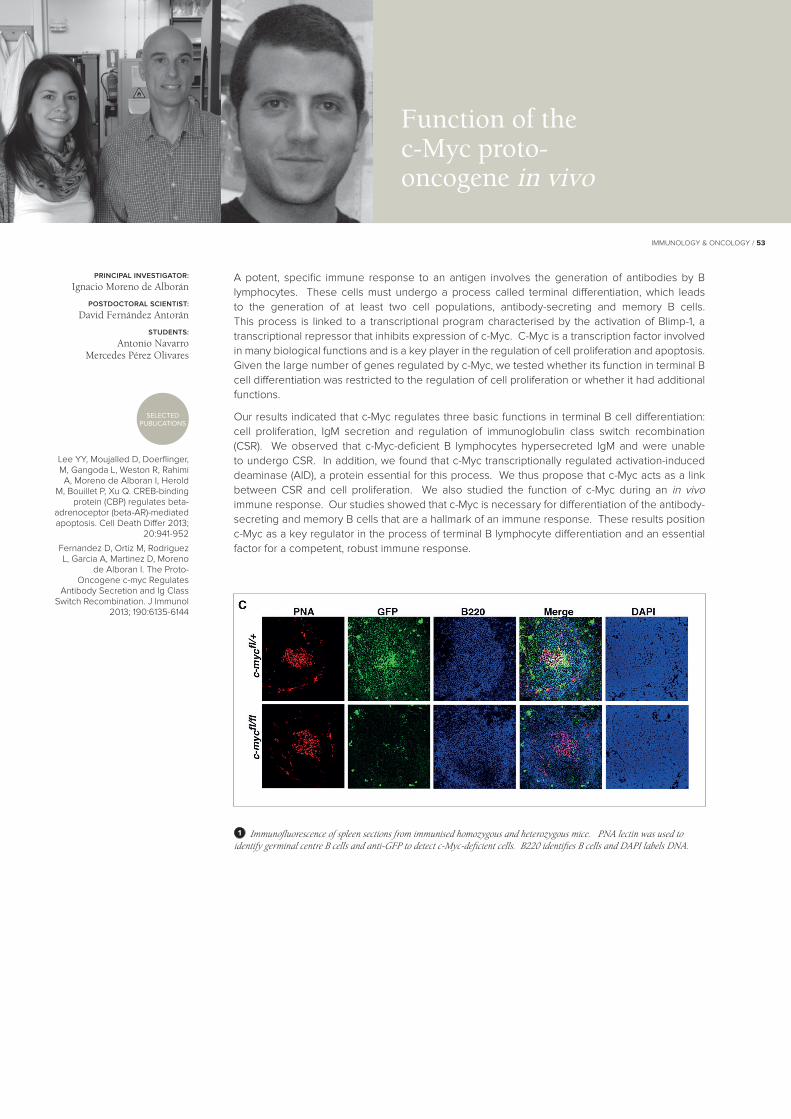
A potent, specific immune response to an antigen involves the generation of antibodies by B lymphocytes. These cells must undergo a process called terminal differentiation, which leads to the generation of at least two cell populations, antibody-secreting and memory B cells. This process is linked to a transcriptional program characterised by the activation of Blimp-1, a transcriptional repressor that inhibits expression of c-Myc. C-Myc is a transcription factor involved in many biological functions and is a key player in the regulation of cell proliferation and apoptosis. Given the large number of genes regulated by c-Myc, we tested whether its function in terminal B cell differentiation was restricted to the regulation of cell proliferation or whether it had additional functions.
Our results indicated that c-Myc regulates three basic functions in terminal B cell differentiation: cell proliferation, IgM secretion and regulation of immunoglobulin class switch recombination (CSR). We observed that c-Myc-deficient B lymphocytes hypersecreted IgM and were unable to undergo CSR. In addition, we found that c-Myc transcriptionally regulated activation-induced deaminase (AID), a protein essential for this process. We thus propose that c-Myc acts as a link between CSR and cell proliferation. We also studied the function of c-Myc during an in vivo immune response. Our studies showed that c-Myc is necessary for differentiation of the antibody-secreting and memory B cells that are a hallmark of an immune response. These results position c-Myc as a key regulator in the process of terminal B lymphocyte differentiation and an essential factor for a competent, robust immune response.
PRINCIPAL INVESTIGATOR:
Ignacio Moreno de Alborán
POSTDOCTORAL SCIENTIST:
David Fernández Antorán
STUDENTS:
Antonio NavarroMercedes Pérez Olivares
Function of the c-Myc proto-oncogene in vivo
1 Immunofluorescence of spleen sections from immunised homozygous and heterozygous mice. PNA lectin was used to identify germinal centre B cells and anti-GFP to detect c-Myc-deficient cells. B220 identifies B cells and DAPI labels DNA.

54 / IMMUNOLOGY & ONCOLOGY
García-Castro A, Zonca M, Florindo-Pinheiro D, Carvalho-Pinto CE, Cordero A, Gutiérrez Del Burgo B, García-Grande A, Mañes S, Hahne M, González-
Suárez E, Planelles L. APRIL promotes breast tumor growth
and metastasis and is associated with aggressive basal breast
cancer. Carcinogenesis 2015; doi: 10.1093/carcin/bgv020
APRIL (a proliferation-inducing ligand) is a cytokine of the tumour necrosis factor family described to be involved in B cell survival, proliferation and antibody production. APRIL transgenic (Tg) mice show an expanded peritoneal B-1 B cell population, increased natural antibody levels, and extended T-independent humoural responses compare to controls. In our laboratory, we are studying the molecular programmes activated by the APRIL pathway on B cells, as well as its potential clinical use.
The APRIL pathway is also associated with haematologic malignancies. In addition, APRIL is overexpressed in breast carcinoma tissue lesions, although neither its role in breast tumourigenesis nor the underlying molecular mechanism is known. To study the participation of the APRIL pathway in breast carcinoma, we have addressed this question using cell lines, mouse tumour models and primary samples. We observed that several breast cancer cell lines express APRIL and both of its receptors, BCMA (B cell maturation antigen) and TACI (transmembrane activator and CAML-interactor), independently of luminal or basal tumour cell phenotype. We found that the mitogen-activated protein kinases p38, ERK1/2 and JNK1/2 are activated in response to APRIL. Silencing experiments decreased cell proliferation, demonstrating that APRIL is a critical autocrine factor for breast tumour growth. Studies of 4T1 orthotopic breast tumours in APRIL-Tg mice showed that an APRIL-enriched environment increased tumour growth and promoted lung metastasis associated with enhanced tumour cell proliferation; BCMA and TACI expression suggests that both participate in these processes. We also detected APRIL, BCMA and TACI in luminal, TNBC and HER2 primary breast carcinomas, with higher levels in more aggressive basal tumours. APRIL was observed near Ki67+ nuclei and was distributed heterogeneously in the cancer cells, in the leukocyte infiltrate, and in the myoepithelial layer adjacent to the tumour area; these results imply that APRIL provides proliferation signals to tumour cells through paracrine and autocrine signalling. Our study identifies the participation of APRIL signalling in breast cancer promotion. We propose impairment of this pathway as a potential therapeutic strategy.
SELECTEDPUBLICATION
PRINCIPAL INVESTIGATOR:
Lourdes Planelles
POSTDOCTORAL SCIENTIST:
Manuela Zonca
STUDENT:
Fernando Gutiérrez del Burgo
APRIL signalling pathway regulation and function
1 4T1 tumour growth and lung metastasis are enhanced in an APRIL-enriched environment. 4T1 cells were transplanted into the mammary fat pad of syngeneic WT and APRIL-Tg female mice. (A) Mean tumour volume ± SD measured twice weekly (left) and individual weight at different times post-sacrifice (right) of 4T1 tumours from WT (white circles) and APRIL-Tg (black circles) mice; n = 6/group. (B) Representative images of lung macrometastases (day 39) in WT and APRIL-Tg mice. Asterisks mark some metastases. One representative experiment of three is shown.
2 Human basal breast carcinomas express APRIL protein. Immunohistochemistry analysis of APRIL expression in three paraffin-embedded basal invasive ductal breast carcinomas. Representative images of APRIL staining (brown) are shown. Squared areas indicate APRIL positive cells in b) “normal” ducts near tumour cells and c) tumour cell cytoplasm.
1
2

IMMUNOLOGY & ONCOLOGY / 55
We study the interactions between pathogens and cells of the immune system to gain insight into functionally important features of the immune system, with the objective of applying this knowledge to studies of human disease.
The main research project is the identification of genes expressed by tumour cells and cells infected by viruses whose expression is associated with activation of NK cell cytotoxicity and cytokine secretion. We also study NK receptors that modulate lymphocyte activation, in particular the cell biology of these receptors, to understand their regulated expression and function. Recently we began to study patients with primary T cell immunodeficiencies and have identified defects in NK cell development and function as well as aberrant expression of NK receptors on the defective T cells of these patients.
1. We showed that the shedding of molecules such as MICA and ICAM-1 is increased after HCMV infection both in vitro and in vivo. This phenomenon depends on increased activity of the metalloproteases ADAM17 and MMP14 due to decreased expression of the endogenous inhibitor of metalloproteases TIMP3. Assay of the sheddase activity of ADAM17 may be of use as a biomarker in patients at risk of developing CMV disease.
2. We discovered that innate immune recognition of double-stranded RNA produced during viral infection plays a key role in the induction of NKG2D ligand expression. These data suggest interesting parallels between the production of type I interferons and the induction of expression of NKG2D ligands. We identified proteins in both vaccinia virus and influenza virus whose expression markedly reduces the induction of both IFN-` and NKG2D ligands after infection.
3. Our recent analyses of patients with primary immunodeficiency diseases (PID) characterised by T cell lymphopaenia suggest defects other than simply the absence of T cell function. Specifically, the NK cells of these patients, although normal in number, were immature. These observations raise the exciting hypothesis that disruption of the normal crosstalk between immune cells can negatively affect the differentiation and function of the remaining cells of the immune system, and so contribute to the spectrum of pathology in these patients.
Esteso G, Luzón E, Sarmiento E, Gómez-Caro R, Steinle A,
Murphy G, Carbone J, Valés-Gómez M, Reyburn H. Human
cytomegalovirus infection leads to altered micro-RNA expression,
TIMP3 downregulation and metalloprotease substrate
release. J Immunol 2014; 193:1344-1352
Royo S, Sainz B Jr, Hernández-Jiménez E, Reyburn H, Lopez-
Collazo E, Guerra S. Study of primary human macrophages
infected with different poxvirus-derived vaccine vectors J Virol
2014; 88:5511-5523
Ashiru O, López-Cobo S, Fernández-Messina L, Pontes-
Quero S, Pandolfi R, Reyburn HT, Valés-Gómez M. A GPI anchor explains the unique biological
features of the common NKG2D-ligand allele MICA*008. Biochem
J 2013; 454:295-302
SELECTEDPUBLICATIONS
PRINCIPAL INVESTIGATOR:
Hugh Reyburn
PREDOCTORAL SCIENTISTS:
Gema Romera CardenasDaniela Dukovska
Miriam Agúndez LlacaAlfonso Blázquez Moreno
TECHNICIAN:
Ruth Gómez-Caro Gil
Receptor ligand interactions in immune responses to cancer and viruses
1 Deposition of NKG2D-containing lytic granules from an NK cell onto a tumour cell induced to express ligands of NKG2D.
1

56 / IMMUNOLOGY & ONCOLOGY
Our group studies the biological functions of the Gadd45 and p38 MAPK (mitogen-activated protein kinase) families in the suppression and development of autoimmunity and cancer. Gadd45 proteins are characterised mainly as classical tumour suppressors that induce cell cycle arrest and apoptosis in response to DNA damage or oncogenic stimuli. They play key roles in a range of other physiological processes, including DNA demethylation and repair, maintenance of genomic stability through mitosis and immunological regulation and activation, although the molecular mechanisms involved in these functions are still under study.
We found an important role for Gadd45a in suppression of autoimmunity through regulation of CD4+ T cell functions. Whereas Gadd45 is typically associated with growth arrest in most cell types, p38 activation has a key stimulatory role in lymphocytes. p38 is necessary for T cell activation and Gadd45a is a major modulator of p38 in this process. Gadd45 acts as an autoimmune suppressor in vivo by negatively regulating T cell proliferation in response to TCR activation. Unlike Gadd45a, the Gadd45b and Gadd45g isoforms potentiate p38 signalling in Th1 and CD8+ cytotoxic T cells, which is necessary for full effector function. Gadd45b is necessary for full expression of the Th1 lineage-inducing protein T-bet. Gadd45 family members thus appear to work synergistically to promote full maturation and function of Th1 and CD8+ cells.
In addition to the role in suppression of autoimmunity, we identified an unanticipated function for Gadd45g, but not Gadd45a or Gadd45b, during embryonic development. Gadd45g expression is central to male fertility, testis development and sex determination. Gadd45g-deficient mice showed an unexpected male-to-female sex reversal phenotype. We found that Gadd45g is necessary for SRY expression and that lack of Gadd45g blocks SOX9, resulting in ovary development. The genetic basis of human male-to-female sex reversal remains unexplained in the majority of cases. Our results identify Gadd45g as a candidate gene in human non-syndromic male infertility and in partial or complete male-to-female primary sex reversal in 46, XY individuals.
Salvador JM, Brown-Clay JD, Fornace AJ. Gadd45 in stress
signalling, cell cycle control and apoptosis. Adv Exp Med Biol
2013; 793:1-19
Johnen H, González-Silva L, Carramolino L, Flores JM, Torres
M, Salvador JM. Gadd45g is essential for primary sex
determination, male fertility and testis development. PLoS One
2013; 8:e58751
SELECTEDPUBLICATIONS
PRINCIPAL INVESTIGATOR:
Jesús María Salvador
POSTDOCTORAL SCIENTISTS:
Heiko JohnenMaría Salvador Bernáldez
PREDOCTORAL SCIENTISTS:
Laura González SilvaUmberto Rosato
TECHNICIANS:
Vanesa Cano DaganzoCarmen Mireya Martínez García
T cell signalling in autoimmune diseases and cancer
1 Upstream regulators of Gadd45 and its downstream inducers. Gadd45 controls crucial functions, including proliferation, apoptosis, cell cycle arrest, tumour angiogenesis, genomic stability and gonad development.
1

IMMUNOLOGY & ONCOLOGY / 57
Tumour immune activation and evasion
Our laboratory studies different aspects of the cell biology of various ligands for the activating receptor NKG2D. In addition to the importance of this receptor-ligand system for immune response activation, the release of NKG2D ligands by tumour cells modulates receptor expression on effector T and NK cells and thus impairs cytotoxicity. Moreover, elevated serum levels of NKG2D ligands correlate with a poorer prognosis in cancer patients. The apparent complexity of the regulation of NKG2D ligand cell surface expression and shedding impedes understanding of the interactions between NKG2D-expressing immune effectors and target cells. The existence of two ligand families encoded in distinct regions of chromosome 6 was initially thought to be responsible for this biological diversity, but we have shown that the NKG2D ligands share properties that do not fit this simple genetic classification.
MICA and MICB (major histocompatibility complex class I-related chain A/B) are two very polymorphic genes that make up one of the two human NKG2D ligand families. We reported that the human MICA allele, MICA*008, is attached to the membrane through a GPI anchor. The importance of this unanticipated finding resides in the fact that the MICA*008 allele shows the highest frequency worldwide, and is not affected by various viral immune evasion mechanisms that target other MICA alleles. We also found that, at difference from other MICA alleles, MICA*008 is released in exosomes (Ashiru et al. Cancer Res 2010; 70:481). That both NKG2D ligand families have transmembrane and GPI-anchored members explains why pathogen immunoevasins are able to recognise some but not all MICA alleles, and suggests that this structural dichotomy permits, in evolutionary terms, the pathogen-driven blockade of cellular pathways.
Our research aims to better understand the association of these molecules with cancer progression.
Esteso G, Luzón E, Sarmiento E, Gómez-Caro R, Steinle A, Murphy
G, Carbone J, Valés-Gómez M, Reyburn HT. Altered micro-
RNA expression after infection with human cytomegalovirus
leads to TIMP3 downregulation and increased shedding of
metalloprotease substrates, including MICA. J Immunol 2014;
193:1344-1352
Ashiru O, López-Cobo S, Fernández-Messina L, Reyburn
HT, Valés-Gómez M. The human NKG2D-ligand MICA*008 is attached to the plasma
membrane through a GPI-anchor. Biochem J 2013; 454:295-300
SELECTEDPUBLICATIONS
PRINCIPAL INVESTIGATOR:
Mar Valés Gómez
POSTDOCTORAL SCIENTIST:
Gloria Esteso Tornero
PREDOCTORAL SCIENTISTS:
Eva M. García CuestaSheila López CoboDaniela Dukovska
VISITORS:
José Ramón Vidal Castiñeira Daria Gajewska
Myriam Oliveira Rodríguez
1 Possibilities for NKG2D ligand processing
1