Embed Size (px)
Citation preview
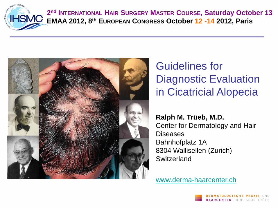
Guidelines for
Diagnostic Evaluation
in Cicatricial Alopecia
Ralph M. Trüeb, M.D.
Center for Dermatology and Hair
Diseases
Bahnhofplatz 1A
8304 Wallisellen (Zurich)
Switzerland
www.derma-haarcenter.ch
2nd INTERNATIONAL HAIR SURGERY MASTER COURSE, Saturday October 13
EMAA 2012, 8th EUROPEAN CONGRESS October 12 -14 2012, Paris
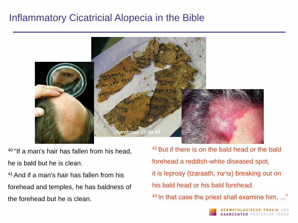
42 But if there is on the bald head or the bald
forehead a reddish-white diseased spot,
it is leprosy (tzaraath, צרעת) breaking out on
his bald head or his bald forehead.
43 In that case the priest shall examine him, ...”
Inflammatory Cicatricial Alopecia in the Bible
40 "If a man's hair has fallen from his head,
he is bald but he is clean.
41 And if a man's hair has fallen from his
forehead and temples, he has baldness of
the forehead but he is clean.
Leviticus 13:40-43
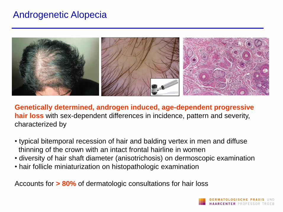
Androgenetic Alopecia
Genetically determined, androgen induced, age-dependent progressive
hair loss with sex-dependent differences in incidence, pattern and severity,
characterized by
• typical bitemporal recession of hair and balding vertex in men and diffuse
thinning of the crown with an intact frontal hairline in women
• diversity of hair shaft diameter (anisotrichosis) on dermoscopic examination
• hair follicle miniaturization on histopathologic examination
Accounts for > 80% of dermatologic consultations for hair loss
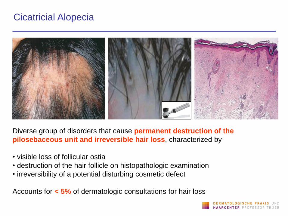
Cicatricial Alopecia
Diverse group of disorders that cause permanent destruction of the
pilosebaceous unit and irreversible hair loss, characterized by
• visible loss of follicular ostia
• destruction of the hair follicle on histopathologic examination
• irreversibility of a potential disturbing cosmetic defect
Accounts for < 5% of dermatologic consultations for hair loss
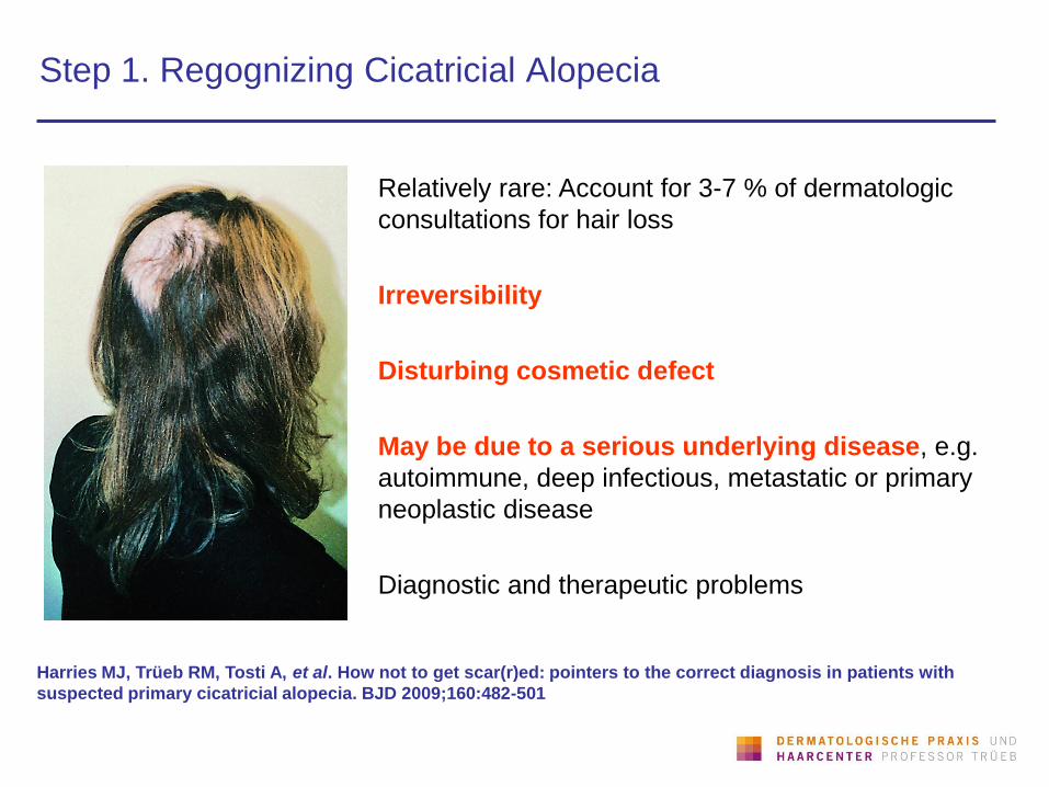
Step 1. Regognizing Cicatricial Alopecia
Relatively rare: Account for 3-7 % of dermatologic
consultations for hair loss
Irreversibility
Disturbing cosmetic defect
May be due to a serious underlying disease, e.g.
autoimmune, deep infectious, metastatic or primary
neoplastic disease
Diagnostic and therapeutic problems
Harries MJ, Trüeb RM, Tosti A, et al. How not to get scar(r)ed: pointers to the correct diagnosis in patients with
suspected primary cicatricial alopecia. BJD 2009;160:482-501
Cicatricial Alopecia
Sperling LC, Solomon AR, Whiting DA. A new look at scarring alopecia.
Arch Dermatol 2000;136:235-242
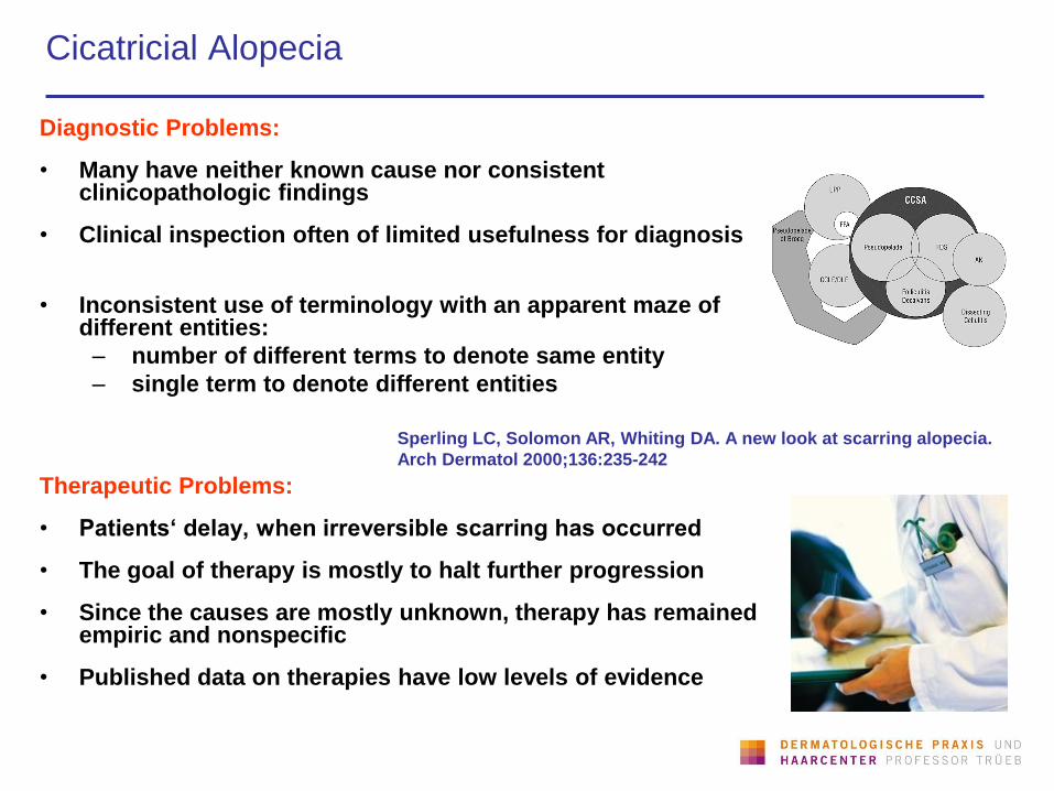
Diagnostic Problems:
• Many have neither known cause nor consistent clinicopathologic findings
• Clinical inspection often of limited usefulness for diagnosis
• Inconsistent use of terminology with an apparent maze of different entities:
– number of different terms to denote same entity
– single term to denote different entities
Therapeutic Problems:
• Patients‘ delay, when irreversible scarring has occurred
• The goal of therapy is mostly to halt further progression
• Since the causes are mostly unknown, therapy has remained empiric and nonspecific
• Published data on therapies have low levels of evidence
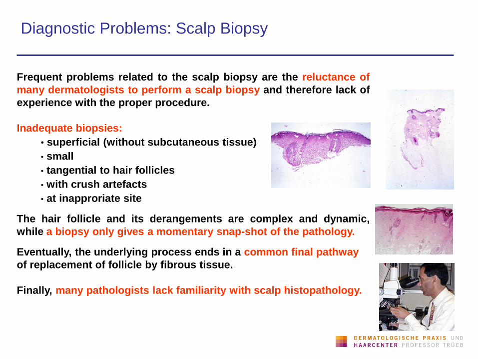
Diagnostic Problems: Scalp Biopsy
Frequent problems related to the scalp biopsy are the reluctance of
many dermatologists to perform a scalp biopsy and therefore lack of
experience with the proper procedure.
Inadequate biopsies:
• superficial (without subcutaneous tissue)
• small
• tangential to hair follicles
• with crush artefacts
• at inapproriate site The hair follicle and its derangements are complex and dynamic,
while a biopsy only gives a momentary snap-shot of the pathology.
Eventually, the underlying process ends in a common final pathway
of replacement of follicle by fibrous tissue.
Finally, many pathologists lack familiarity with scalp histopathology.
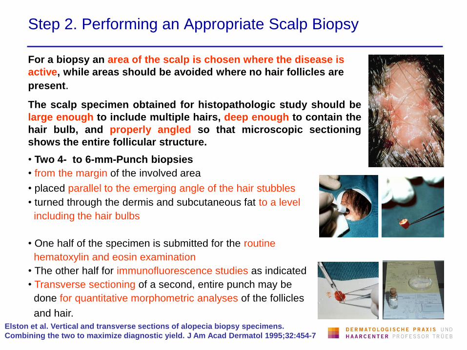
Step 2. Performing an Appropriate Scalp Biopsy
For a biopsy an area of the scalp is chosen where the disease is
active, while areas should be avoided where no hair follicles are
present.
The scalp specimen obtained for histopathologic study should be
large enough to include multiple hairs, deep enough to contain the
hair bulb, and properly angled so that microscopic sectioning
shows the entire follicular structure.
• Two 4- to 6-mm-Punch biopsies
• from the margin of the involved area
• placed parallel to the emerging angle of the hair stubbles
• turned through the dermis and subcutaneous fat to a level
including the hair bulbs
• One half of the specimen is submitted for the routine
hematoxylin and eosin examination
• The other half for immunofluorescence studies as indicated
• Transverse sectioning of a second, entire punch may be
done for quantitative morphometric analyses of the follicles
and hair.
Elston et al. Vertical and transverse sections of alopecia biopsy specimens.
Combining the two to maximize diagnostic yield. J Am Acad Dermatol 1995;32:454-7
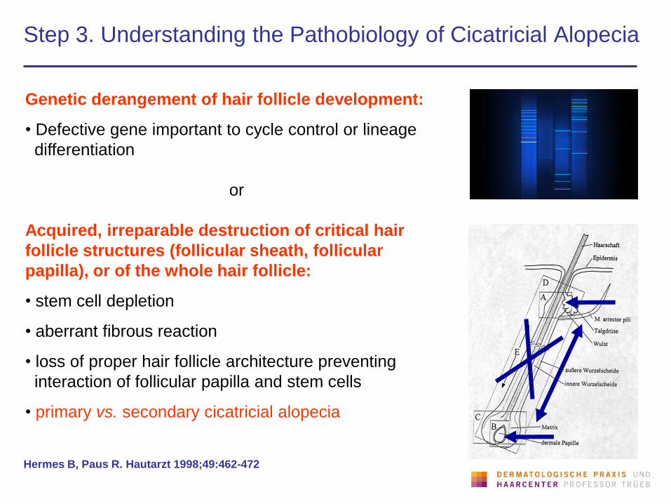
Step 3. Understanding the Pathobiology of Cicatricial Alopecia
Genetic derangement of hair follicle development:
• Defective gene important to cycle control or lineage
differentiation
or
Acquired, irreparable destruction of critical hair
follicle structures (follicular sheath, follicular
papilla), or of the whole hair follicle:
• stem cell depletion
• aberrant fibrous reaction
• loss of proper hair follicle architecture preventing
interaction of follicular papilla and stem cells
• primary vs. secondary cicatricial alopecia
Hermes B, Paus R. Hautarzt 1998;49:462-472

Genetic/Developmental Defects (Rare)
Aplasia cutis
congenita
Epidermal
nevus
Incontinentia pigmenti
Bloch-Sulzberger
Alopecia in patient
with GABEB Alopecia ichthyotica
(in lamellar ichthyosis)

Step 4. Making a Distinction between Primary and Secondary
Cicatrical Alopecia
Templeton SF, Solomon AR. Scarring alopecia: a classification based on microscopic criteria.
J Cutan Pathol 1994;21:97-109
Secondary cicatricial alopecia:
Results from destructive cutaneous disease in which the follicle is
destroyed in a non-specific manner:
- trauma (chemical, physical)
- infection (fungal, bacterial, viral)
- infiltration (granulomatous, neoplastic)
- autoimmune (circumscribed scleroderma, cicatricial pemphigoid,
temporal arteritis)
*
Primary cicatricial alopecia:
Target of inflammation and destruction is the follicle
Cause mostly unknown, classification on the basis of inflammatory
infiltrate:
- lymphocytic
- neutrophilic
- mixed
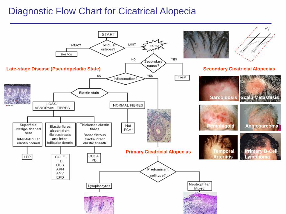
Diagnostic Flow Chart for Cicatrical Alopecia
Scalp Metastasis
Secondary Cicatricial Alopecias
Sarcoidosis
Pemphigoid
Temporal
Arteriitis
Scalp Metastasis
Angiosarcoma
Primary B-Cell
Lymphoma
Late-stage Disease (Pseudopeladic State)
Primary Cicatricial Alopecias
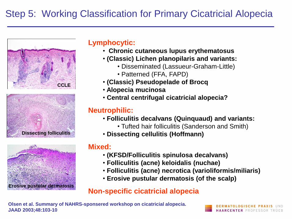
Step 5: Working Classification for Primary Cicatricial Alopecia
Lymphocytic: • Chronic cutaneous lupus erythematosus
• (Classic) Lichen planopilaris and variants:
• Disseminated (Lassueur-Graham-Little)
• Patterned (FFA, FAPD)
• (Classic) Pseudopelade of Brocq
• Alopecia mucinosa
• Central centrifugal cicatricial alopecia?
Neutrophilic: • Folliculitis decalvans (Quinquaud) and variants:
• Tufted hair folliculitis (Sanderson and Smith)
• Dissecting cellulitis (Hoffmann)
Mixed: • (KFSD/Folliculitis spinulosa decalvans)
• Folliculitis (acne) keloidalis (nuchae)
• Folliculitis (acne) necrotica (varioliformis/miliaris)
• Erosive pustular dermatosis (of the scalp)
Non-specific cicatricial alopecia
Olsen et al. Summary of NAHRS-sponsered workshop on cicatricial alopecia.
JAAD 2003;48:103-10
CCLE
Dissecting folliculitis
Erosive pustular dermatosis
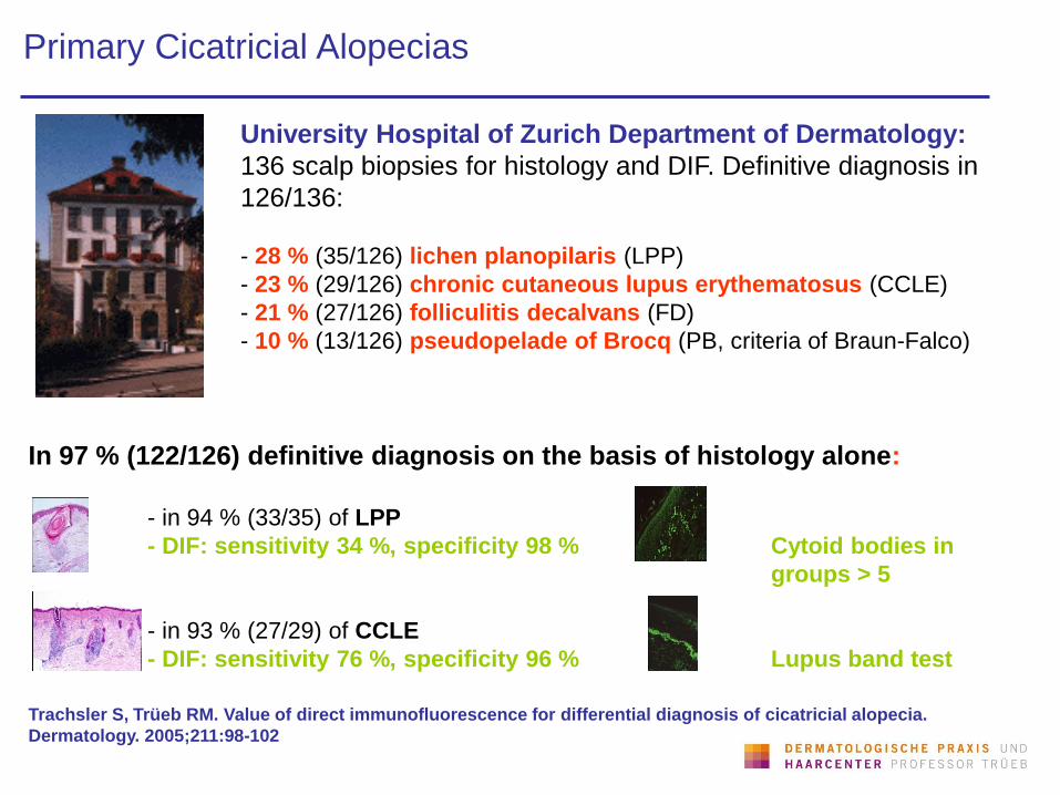
Primary Cicatricial Alopecias
University Hospital of Zurich Department of Dermatology:
136 scalp biopsies for histology and DIF. Definitive diagnosis in
126/136:
- 28 % (35/126) lichen planopilaris (LPP)
- 23 % (29/126) chronic cutaneous lupus erythematosus (CCLE)
- 21 % (27/126) folliculitis decalvans (FD)
- 10 % (13/126) pseudopelade of Brocq (PB, criteria of Braun-Falco)
In 97 % (122/126) definitive diagnosis on the basis of histology alone:
- in 94 % (33/35) of LPP
- DIF: sensitivity 34 %, specificity 98 % Cytoid bodies in
groups > 5
- in 93 % (27/29) of CCLE
- DIF: sensitivity 76 %, specificity 96 % Lupus band test
Trachsler S, Trüeb RM. Value of direct immunofluorescence for differential diagnosis of cicatricial alopecia.
Dermatology. 2005;211:98-102
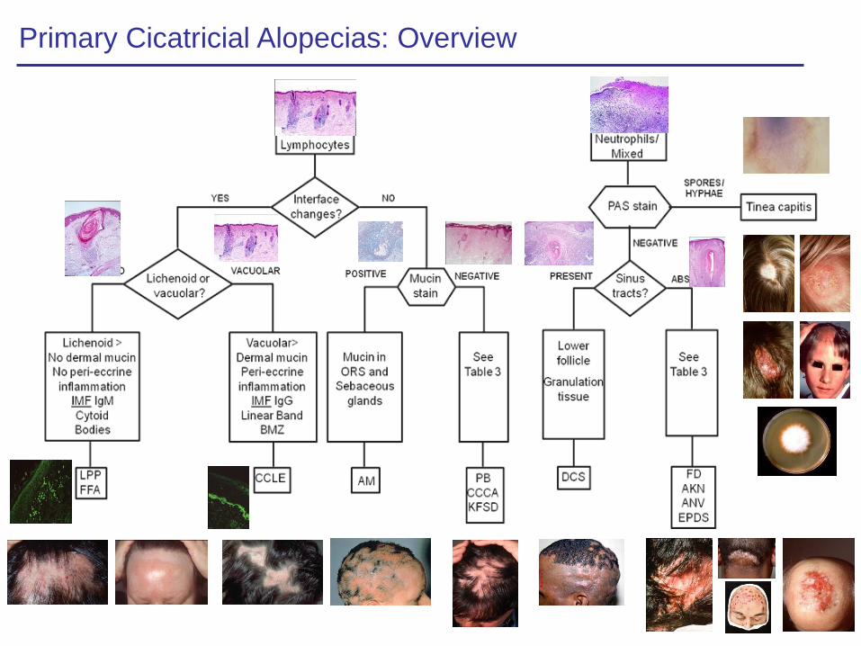
Primary Cicatricial Alopecias: Overview
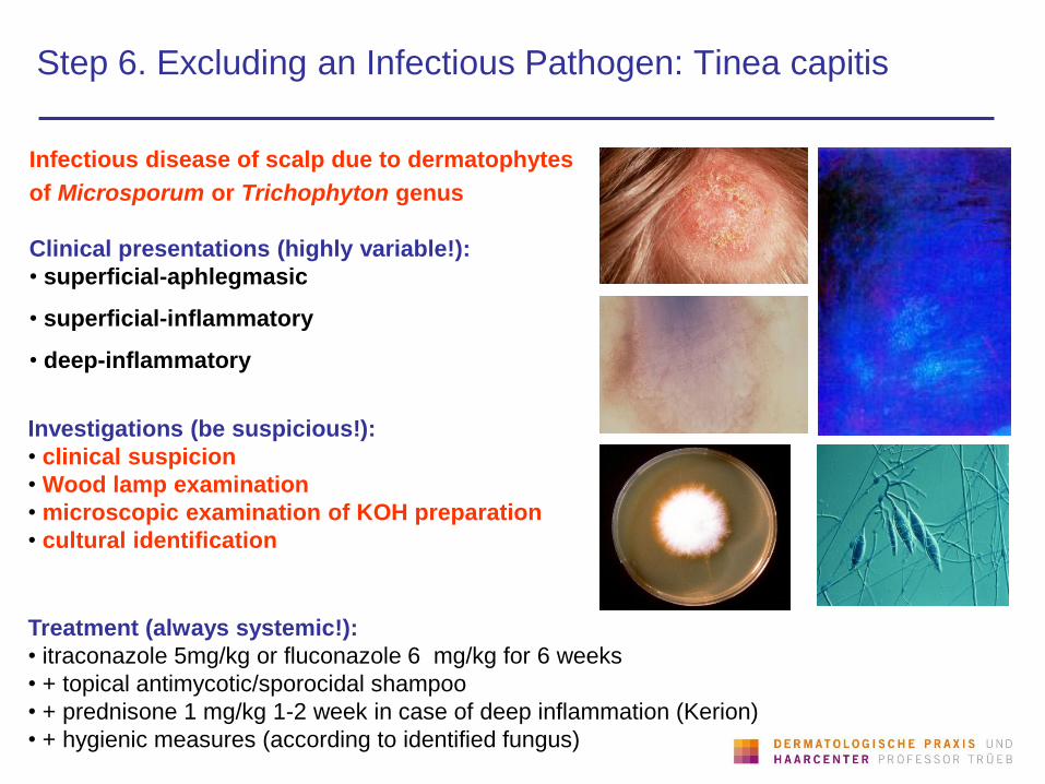
Step 6. Excluding an Infectious Pathogen: Tinea capitis
Infectious disease of scalp due to dermatophytes
of Microsporum or Trichophyton genus
Clinical presentations (highly variable!):
• superficial-aphlegmasic
• superficial-inflammatory
• deep-inflammatory
Investigations (be suspicious!):
• clinical suspicion
• Wood lamp examination
• microscopic examination of KOH preparation
• cultural identification
Treatment (always systemic!):
• itraconazole 5mg/kg or fluconazole 6 mg/kg for 6 weeks
• + topical antimycotic/sporocidal shampoo
• + prednisone 1 mg/kg 1-2 week in case of deep inflammation (Kerion)
• + hygienic measures (according to identified fungus)
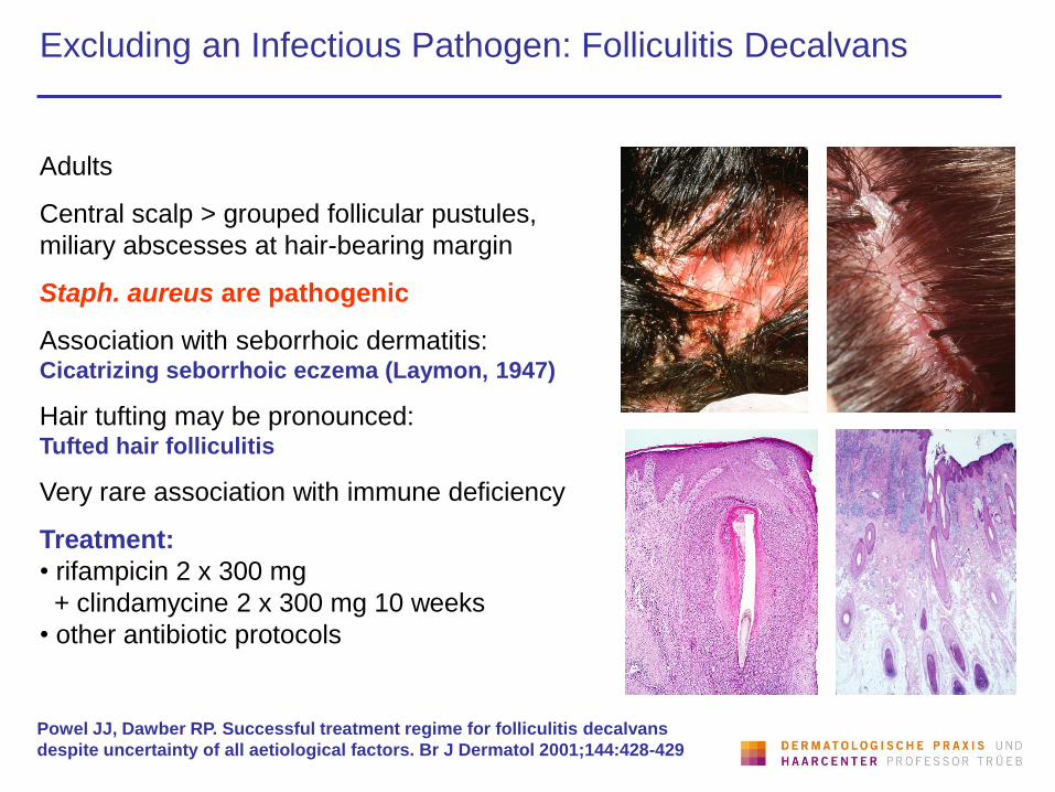
Excluding an Infectious Pathogen: Folliculitis Decalvans
Adults
Central scalp > grouped follicular pustules,
miliary abscesses at hair-bearing margin
Staph. aureus are pathogenic
Association with seborrhoic dermatitis:
Cicatrizing seborrhoic eczema (Laymon, 1947)
Hair tufting may be pronounced: Tufted hair folliculitis
Very rare association with immune deficiency
Treatment:
• rifampicin 2 x 300 mg
+ clindamycine 2 x 300 mg 10 weeks
• other antibiotic protocols
Powel JJ, Dawber RP. Successful treatment regime for folliculitis decalvans
despite uncertainty of all aetiological factors. Br J Dermatol 2001;144:428-429
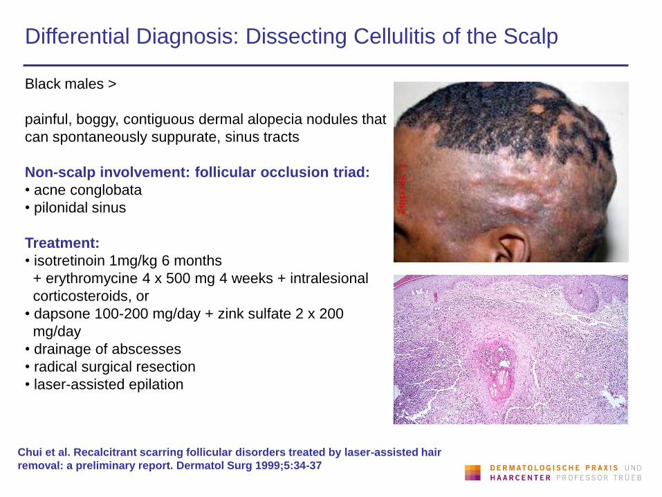
Differential Diagnosis: Dissecting Cellulitis of the Scalp
Black males >
painful, boggy, contiguous dermal alopecia nodules that
can spontaneously suppurate, sinus tracts
Non-scalp involvement: follicular occlusion triad:
• acne conglobata
• pilonidal sinus
Treatment:
• isotretinoin 1mg/kg 6 months
+ erythromycine 4 x 500 mg 4 weeks + intralesional
corticosteroids, or
• dapsone 100-200 mg/day + zink sulfate 2 x 200
mg/day
• drainage of abscesses
• radical surgical resection
• laser-assisted epilation
Chui et al. Recalcitrant scarring follicular disorders treated by laser-assisted hair
removal: a preliminary report. Dermatol Surg 1999;5:34-37
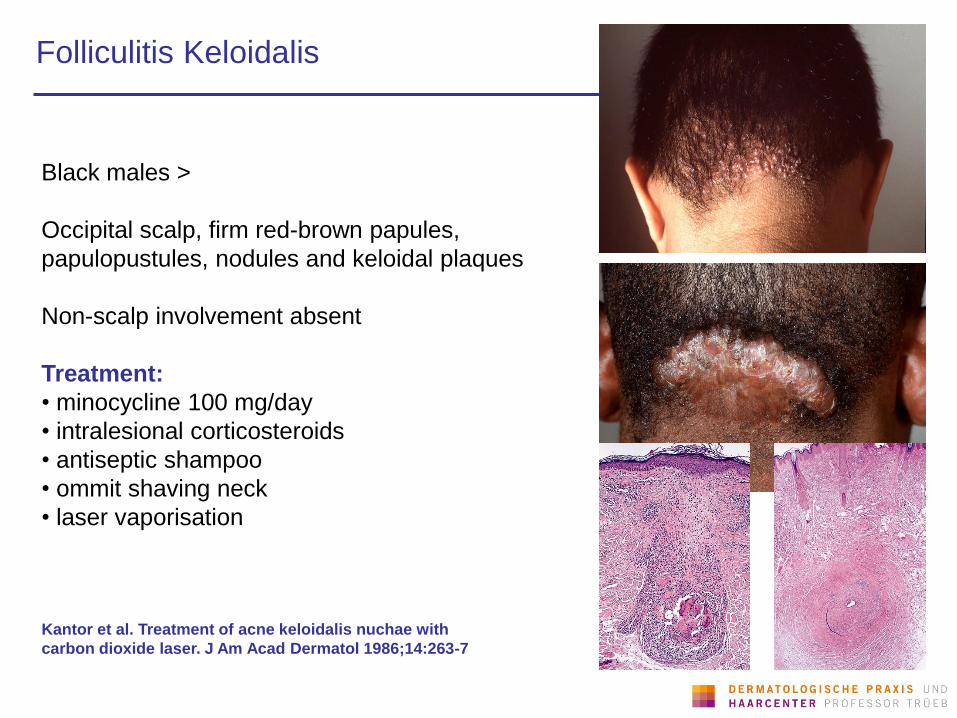
Folliculitis Keloidalis
Black males >
Occipital scalp, firm red-brown papules,
papulopustules, nodules and keloidal plaques
Non-scalp involvement absent
Treatment:
• minocycline 100 mg/day
• intralesional corticosteroids
• antiseptic shampoo
• ommit shaving neck
• laser vaporisation
Kantor et al. Treatment of acne keloidalis nuchae with
carbon dioxide laser. J Am Acad Dermatol 1986;14:263-7
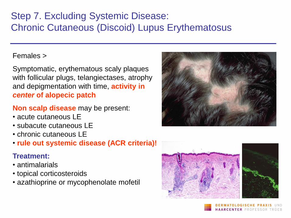
Step 7. Excluding Systemic Disease:
Chronic Cutaneous (Discoid) Lupus Erythematosus
Females >
Symptomatic, erythematous scaly plaques
with follicular plugs, telangiectases, atrophy
and depigmentation with time, activity in
center of alopecic patch
Non scalp disease may be present:
• acute cutaneous LE
• subacute cutaneous LE
• chronic cutaneous LE
• rule out systemic disease (ACR criteria)!
Treatment:
• antimalarials
• topical corticosteroids
• azathioprine or mycophenolate mofetil
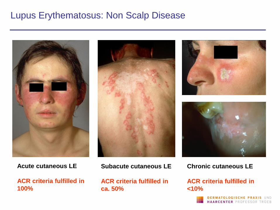
Lupus Erythematosus: Non Scalp Disease
Acute cutaneous LE
ACR criteria fulfilled in
100%
Subacute cutaneous LE
ACR criteria fulfilled in
ca. 50%
Chronic cutaneous LE
ACR criteria fulfilled in
<10%
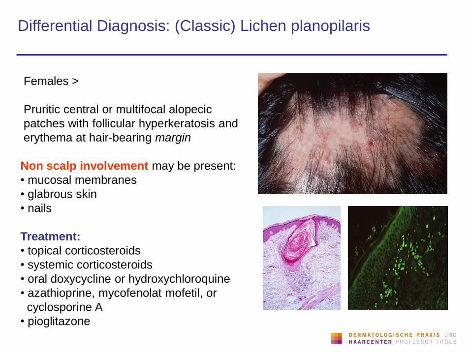
Differential Diagnosis: (Classic) Lichen planopilaris
Females >
Pruritic central or multifocal alopecic
patches with follicular hyperkeratosis and
erythema at hair-bearing margin
Non scalp involvement may be present:
• mucosal membranes
• glabrous skin
• nails
Treatment:
• topical corticosteroids
• systemic corticosteroids
• oral doxycycline or hydroxychloroquine
• azathioprine, mycofenolat mofetil, or
cyclosporine A
• pioglitazone
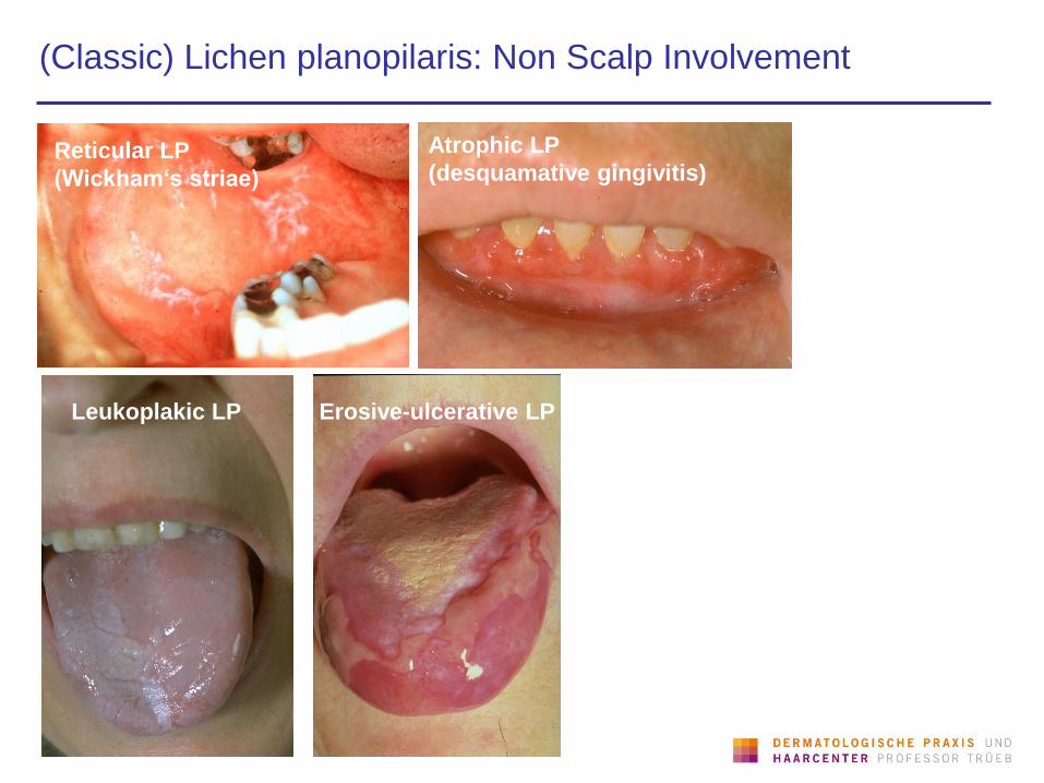
(Classic) Lichen planopilaris: Non Scalp Involvement
Atrophic LP
(desquamative gingivitis)
Erosive-ulcerative LP
Reticular LP
(Wickham‘s striae)
Leukoplakic LP
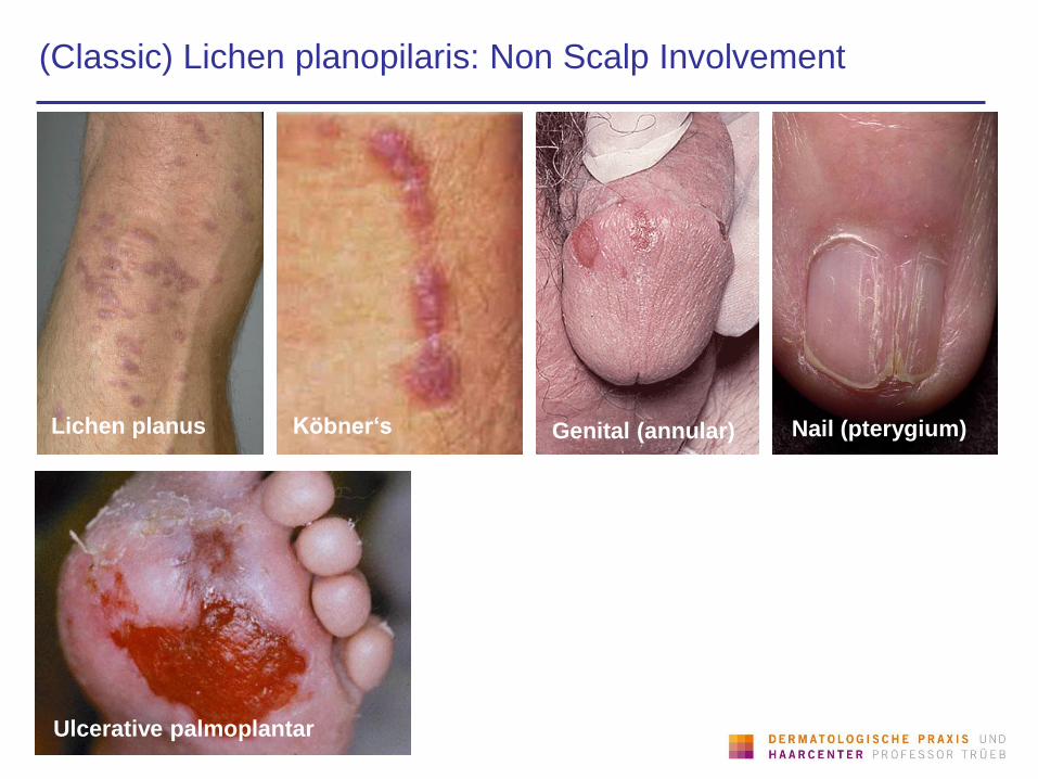
(Classic) Lichen planopilaris: Non Scalp Involvement
Lichen planus Köbner‘s Genital (annular) Nail (pterygium)
Ulcerative palmoplantar
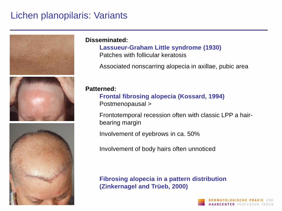
Lichen planopilaris: Variants
Disseminated:
Lassueur-Graham Little syndrome (1930)
Patches with follicular keratosis
Associated nonscarring alopecia in axillae, pubic area
Patterned:
Frontal fibrosing alopecia (Kossard, 1994)
Postmenopausal >
Frontotemporal recession often with classic LPP a hair-
bearing margin
Involvement of eyebrows in ca. 50%
Involvement of body hairs often unnoticed
Fibrosing alopecia in a pattern distribution
(Zinkernagel and Trüeb, 2000)
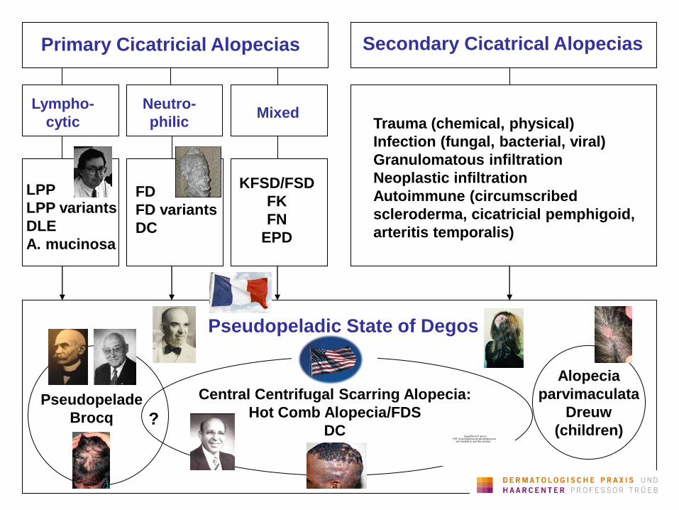
Primary Cicatricial Alopecias Secondary Cicatrical Alopecias
Lympho-
cytic
Neutro-
philic Mixed
LPP
LPP variants
DLE
A. mucinosa
FD
FD variants
DC
KFSD/FSD
FK
FN
EPD
Trauma (chemical, physical)
Infection (fungal, bacterial, viral)
Granulomatous infiltration
Neoplastic infiltration
Autoimmune (circumscribed
scleroderma, cicatricial pemphigoid,
arteritis temporalis)
Pseudopelade
Brocq
Alopecia
parvimaculata
Dreuw
(children)
Central Centrifugal Scarring Alopecia:
Hot Comb Alopecia/FDS
DC
Pseudopeladic State of Degos
? QuickTime™ and a
TIFF (Uncompressed) decompressorare needed to see this picture.
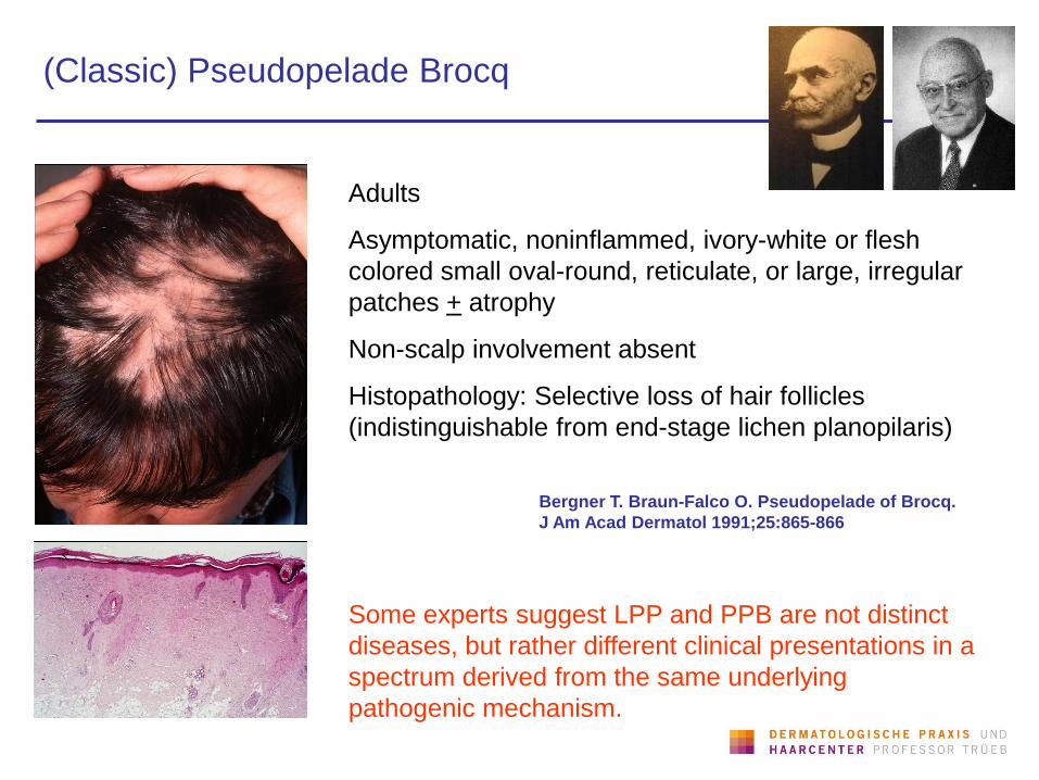
(Classic) Pseudopelade Brocq
Adults
Asymptomatic, noninflammed, ivory-white or flesh
colored small oval-round, reticulate, or large, irregular
patches + atrophy
Non-scalp involvement absent
Histopathology: Selective loss of hair follicles
(indistinguishable from end-stage lichen planopilaris)
Some experts suggest LPP and PPB are not distinct
diseases, but rather different clinical presentations in a
spectrum derived from the same underlying
pathogenic mechanism.
Bergner T. Braun-Falco O. Pseudopelade of Brocq.
J Am Acad Dermatol 1991;25:865-866
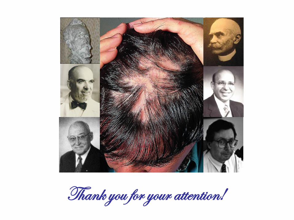
Thank you for your attention!