Embed Size (px)
Citation preview

TECHNICAL REPORT AD ________________ NATICK/TR-17/014
ISOLATION, CHARACTERIZATION AND
IDENTIFICATION OF ENVIRONMENTAL BACTERIAL ISOLATES WITH SCREENING FOR ANTAGONISM
AGAINST THREE BACTERIAL TARGETS
by Robert Stote
Jennifer M. Rego and
Romy Kirby
April 2017
Final Report October 2012 – September 2013
Approved for public release; distribution is unlimited
U.S. Army Natick Soldier Research, Development and Engineering Center Natick, Massachusetts 01760-5020


REPORT DOCUMENTATION PAGE Form Approved OMB No. 0704-0188
Public reporting burden for this collection of information is estimated to average 1 hour per response, including the time for reviewing instructions, searching existing data sources, gathering and maintaining the data needed, and completing and reviewing this collection of information. Send comments regarding this burden estimate or any other aspect of this collection of information, including suggestions for reducing this burden to Department of Defense, Washington Headquarters Services, Directorate for Information Operations and Reports (0704-0188), 1215 Jefferson Davis Highway, Suite 1204, Arlington, VA 22202-4302. Respondents should be aware that notwithstanding any other provision of law, no person shall be subject to any penalty for failing to comply with a collection of information if it does not display a currently valid OMB control number.
PLEASE DO NOT RETURN YOUR FORM TO THE ABOVE ADDRESS. 1. REPORT DATE (DD-MM-YYYY)
21-04-2017 2. REPORT TYPE
Final 3. DATES COVERED (From - To)
October 2012 – September 20134. TITLE AND SUBTITLE
ISOLATION, CHARACTERIZATION AND IDENTIFICATION OF ENVIRONMENTAL BACTERIAL ISOLATES WITH SCREENING FOR ANTAGONISM AGAINST THREE BACTERIAL TARGETS
5a. CONTRACT NUMBER
5b. GRANT NUMBER
5c. PROGRAM ELEMENT NUMBER
6. AUTHOR(S)
Robert Stote, Jennifer M. Rego, and Romy Kirby
5d. PROJECT NUMBER
13-115b 5e. TASK NUMBER
5f. WORK UNIT NUMBER
7. PERFORMING ORGANIZATION NAME(S) AND ADDRESS(ES)
8. PERFORMING ORGANIZATION REPORT NUMBER
NATICK/TR-17/014
9. SPONSORING / MONITORING AGENCY NAME(S) AND ADDRESS(ES) 10. SPONSOR/MONITOR’S ACRONYM(S)
11. SPONSOR/MONITOR’S REPORT NUMBER(S)
12. DISTRIBUTION / AVAILABILITY STATEMENT
Approved for public release; distribution is unlimited 13. SUPPLEMENTARY NOTES
14. ABSTRACT Current antimicrobial treatments exhibit a broad range of killing power, promoting the increase of multi-drug resistant organisms. This has led to the urgent need to develop targeted antimicrobials as an alternative to today’s treatments. This report summarizes work conducted to identify microorganisms that exhibit narrow-spectrum activity through the secretion of antimicrobials, termed bacteriocins, from a pool of environmental isolates collected at Fort Devens. The environmental isolates were characterized and found to be comprised mostly of microorganisms from the genus Bacillus and Staphylococcus. The environmental isolates were screened for bacteriocin-induced activity against three target strains of interest to the DoD: Bacillus anthracis Sterne, Staphylococcus aureus and Pseudomonas aeruginosa. The percentage of environmental isolates that demonstrated activity against Bacillus anthracis Sterne was 15% (9 of 62 isolates screened), while 2% of the isolates (2 of 114 isolates screened) exhibited activity against Staphylococcus aureus. No isolates were active against Pseudomonas aeruginosa. The active isolates were screened further against additional targets to confirm their narrow-spectrum activity. This work successfully identified environmental microorganisms that exhibit bacteriocin-driven activity to produce narrow-spectrum antimicrobials that target DoD relevant microorganisms.
15. SUBJECT TERMS
16. SECURITY CLASSIFICATION OF: 17. LIMITATION OF ABSTRACT
SAR
18. NUMBER OF PAGES
24
19a. NAME OF RESPONSIBLE PERSON
Robert Stote a. REPORT
U
b. ABSTRACT
U
c. THIS PAGE
U 19b. TELEPHONE NUMBER (include area code)
508-233-4629
Standard Form 298 (Rev. 8-98)Prescribed by ANSI Std. Z39.18
SPORES ISOLATION BACILLUS ANTHRACIS ENVIRONMENTAL ISOLATES CODING SELECTION RESISTANCE(BIOLOGY) TARGETED ANTIMICROBIALS ISOLATES BACTERIOCINS ANTIMICROBIAL AGENTS PSEUDOMONAS AERUGINOSA TARGETS IDENTIFICATION MULTI-DRUG RESISTANCE NARROW SPECTRUM ANTIBIOTICS ASSAYING NOMENCLATURE STAPHYLOCOCCUS AUREUS BACTERIA MICROORGANISMS MULTI-DRUG RESISTANT ORGANISMS BIOASSAY CHARACTERIZATION NARROW-SPECTRUM ANTIMICROBIALS
U.S. Army Natick Soldier Research, Development and Engineering Center ATTN: RDNS-TMS General Green Avenue, Natick, MA 01760-5020

This page intentionally left blank

iii
Table of Contents
List of Tables ................................................................................................................................. iv
Preface ............................................................................................................................................. v
1 Introduction ............................................................................................................................ 1
2 Materials and Methods .......................................................................................................... 3
2.1 Isolation of Environmental Pool ............................................................................................... 3
2.2 Colony characterization ............................................................................................................ 3
2.3 Screening Isolates for Activity of Bacteriocins Against Targets ........................................... 4
3 Results and Discussion .......................................................................................................... 5
3.1 Isolation and Identification of Environmental Bacterial Isolates ......................................... 5
3.2 Characterization of Activity of Environmental Bacterial Isolates Against the Target Bacteria .................................................................................................................................................... 7
4 Conclusions ............................................................................................................................ 8
5 References .............................................................................................................................. 9
Appendix: Results of Physical and Biochemical Analysis to Determine Identity of Environmental Isolates ................................................................................................................ 11
iv
List of Tables Table 1. Summary of Surfaces Probed for Environmental Bacterial Isolates ................................ 5 Table 2. Summary of Genus Identification of Environmental Bacterial Isolates in Relation to
Sample Collection Location .................................................................................................... 7 Table 3. Summary of Activity of Isolates Against Target Microorganisms .................................. 8 Table 4. Detailed Summary of Active Isolates .............................................................................. 8

v
Preface This report documents work by the Biological Sciences and Technology Team (BSTT) of the Warfighter Directorate at the Natick Soldier Research, Development and Engineering Center (NSRDEC) during the period from October 2012 to September 2013. This work involved initial investigations of narrow-spectrum antimicrobial agents, termed bacteriocins, to facilitate their future incorporation into materials and textiles. The objective of this work was to isolate a pool of bacteria from the environment and subsequently evaluate the bacteriocin-induced activity against three target microorganisms of interest to the military: Bacillus anthracis Sterne, Pseudomonas aeruginosa, and Staphylococcus aureus. Of the over 100 environmental isolates collected, 11 environmental isolates were identified as possessing activity against Bacillus anthracis Sterne and Staphylococcus aureus while no isolates were identified as being active against Pseudomonas aeruginosa.

vi
This page intentionally left blank

1
ISOLATION, CHARACTERIZATION AND IDENTIFICATION OF ENVIRONMENTAL BACTERIAL
ISOLATES WITH SCREENING FOR ANTAGONISM AGAINST THREE BACTERIAL TARGETS
1 Introduction The Biological Sciences and Technology Team (BSTT), Warfighter Directorate, of the Natick Soldier Research, Development and Engineering Center (NSRDEC) is investigating the use of narrow-spectrum antimicrobials in non-traditional textiles to protect the Warfighter from pathogenic bacteria. This investigation has several parts, one of which is to identify environmental bacterial isolates that produce bacteriocins, a class of narrow-spectrum antimicrobials, against specific targets. The objective of this technical report, is to describe the generation and characterization of a bacterial pool of environmental isolates and the subsequent screen of the pool for bacteriocin-driven antimicrobial activity against military relevant pathogenic target strains of bacteria. The work was performed from October 2012 to September 2013. As background, drug resistant strains of bacteria are on the rise, while the number of drugs being developed against them is declining (Cooper and Shlae, 2011). This creates a situation where infections that are currently treatable may not be in the very near future. Additionally, many of these drug resistant strains demonstrate resistance to the antimicrobials used in textiles, polymer surfaces, wipes, and ointments, making the agent ineffective (Tattawasart et al. 1999, Thomas et al. 2000, McDonnell and Russell, 1999, Silver, 2003). Though a number of hypotheses have been offered to explain this increase in resistance, one of the leading causes has been identified as the use of broad-spectrum antimicrobials that unintentionally promote the selection of bacterial strains resistant to the antimicrobial (Levy and Marshall, 2004). When broad-spectrum antimicrobials are employed, not all of the bacteria are killed. Those that have developed a resistance to the antimicrobial will survive. Since the more benign microorganisms that contribute to keeping drug-resistant pathogenic strains under control are killed off, the resistant strains are allowed to grow unfettered (Levy and Marshall, 2004, Gulberg et al. 2011). After a few exposures to a broad-spectrum antimicrobial, the collection of bacteria becomes primarily composed of resistant strains, making the broad-spectrum antimicrobial ineffective. When this occurs, the current strategy is to replace an ineffective antimicrobial agent with another broad-spectrum antimicrobial agent. This provides an opportunity for the bacterial pool to be further screened for those bacteria that are now resistant to the new antimicrobial agent as well as the previous antimicrobial, leading to the development of multi-drug resistant bacteria, or “super bugs”. As concern about the emergence of multi-drug resistant bacteria increases, new strategies are being investigated to inhibit the development of resistant strains. One promising new strategy is to use narrow-spectrum antimicrobials, such as bacteriocins, as alternatives to broad-spectrum agents. Bacteriocins are narrow-spectrum bacteria toxins secreted by bacteria to kill other closely related bacteria that are competing for the same resources. Many bacteriocins have very specific

2
activity spectrums, as they only target one or two species. This selectivity offers a paradigm shift from the typical application of conventional antimicrobials where instead of broadly killing everything, specific pathogens could be targeted, leaving beneficial bacteria unaffected and capable of thriving (Abt and Pamer, 2014). Generally considered a green alternative to the currently used antimicrobials, few bacteriocins have demonstrated adverse effects on eukaryote cells (Cotter et. al., Cox et. al., Galvz et al.). These and other characteristics make bacteriocins an appealing option to supersede broad-spectrum antimicrobials. Selecting bacteriocins for Army-specific needs requires the screening of large pools of environmental isolates to obtain two pieces of information: (1) is the activity against the desired target observed, and (2) how broad is the activity toward other organisms? From this information, environmental isolates that display bacteriocin-generating activity against pathogenic bacteria can be identified for further characterization. This would lead to the eventual purification and application of the bacteriocin itself. In this reported study, a pool of environmental isolates was generated, identified to the genus level, and screened for bacteriocin activity. Identification of the environmental isolates was achieved via phenotypical analysis as well as analysis by the Omnilog system. To evaluate activity, three military relevant microorganisms were selected as targets: Bacillus anthracis Sterne, Pseudomonas aeruginosa and Staphylococcus aureus. The environmental isolates were screened against these targets for bacteriocin-like activity.

3
2 Materials and Methods
2.1 Isolation of Environmental Pool
A pool of bacterial isolates was collected from several locations at Fort Devens (Ayer, MA). 12 sites of interest were wiped with sterile cotton swabs, including air conditioning (AC) ducts, a door handle, a cot, a cutting board, a kitchen sink, and the handle of a washing machine. The swiped cotton swabs were then used to inoculate nutrient agar plates. The plates were incubated at 37 o C for 24 h. Individual colonies were picked from each plate using a sterile loop, and were streaked onto a new nutrient agar plate to obtain a pure isolate. The initial plates were then incubated again for another 24 h to allow any slow-growing colonies to develop. Newly developed colonies were picked and streaked onto a new nutrient agar plate to obtain a pure isolate.
2.2 Colony characterization
Identification of environmental isolates followed the flowchart from “Bergey’s Manual of Determinative Bacteriology” (Holt et al. 1994), which utilizes physical and biochemical analysis to identify the genus, and sometimes species, of unknowns. These tests are well-established methods utilized to identify unknown bacteria samples and have been well-documented in previous literature. These physical and biochemical tests include: (1) the Gram stain test, which is used to determine if the organism is Gram-negative or Gram-positive; (2) the acid-fast test, which identifies Mycobacterium; (3) the oxidase test, which identifies organisms that produce the enzyme cytochrome oxidase; (4) the catalase test, which differentiates Staphylococci (a catalase positive organism) from Streptococci (a catalase negative organism); (5) the starch hydrolysis test, which evaluates the organism’s ability to synthesize and secrete enzymes to break down starch into smaller subunits to be used by the organism; (6) the mannitol and glucose fermentation test, which investigates how an organism metabolizes sugars; (7) the Voges-Proskauer test, which tests the organism’s ability to produce non-acidic end products from the metabolism of glucose and is helpful in differentiating the Enterobacteriaceae; (8) the citrate test, which studies the organism’s ability to apply citrate as a carbon source and is also helpful in differentiating the Enterobacteriaceae; (9) the nitrate reductase test, which investigates bacteria’s ability or inability to reduce nitrate to nitrite; (10) the novobiocin sensitivity test, which evaluates an organism’s susceptibility to novobiocin, an antibiotic that obstructs DNA replication and is helpful in differentiating among Gram-positive cocci; (11) the diffusible pigment test using king agar, which differentiates among species of Pseudomonas by testing the secretion of pigments by the organism. Additionally, the bacteria isolates were viewed under a standard microscope to characterize their shape. The Omnilog Identification system, which uses a colorimetric assay based upon the consumption of different carbon sources, was employed to further characterize the pool of environmental microorganisms. Materials for physical and biochemical analysis were sourced from several different companies:
Gram stain from ENG Scientific, Clifton, NJ Acid-fast from Electron Microscopy Sciences, Hatfield, PA Oxidase from Becton, Dickinson and Company (BD), Sparks, MD Starch hydrolysis from ENG Scientific, Clifton, NJ Mannitol and glucose fermentation from Sigma Aldrich, St Louis, MO

4
Voges-Proskar test kit from BD, Sparks, MD Citrate from Sigma Aldrich, St Louis, MO Novobiocin sensitivity from Remel, Lenexa, KS Diffusible pigment test using king agar from Sigma-Aldrich, St Louis, MO Gen III Omnilog system and any associated supplies from Biolog, Inc., Hayward, CA
2.3 Screening Isolates for Activity of Bacteriocins Against Targets
Selected isolates were screened for bacteriocin activity against three targets: Bacillus anthracis Sterne (surrogate for Bacillus anthracis), Staphylococcus aureus (ATCC 27217), or Pseudomonas aeruginosa (ATCC 15692) using soft agar overlays. A culture containing the target microorganism and a culture of the environmental isolate to be tested were inoculated into nutrient broth and incubated at 37 oC until an Optical Density (OD) of 1 (~108 cfu/mL) was achieved. Soft agar for overlay experiments was prepared with 7% agar in nutrient broth. Overlays were prepared by adding 60 µL of the target organism with 1.35 µL of a 100 mg/mL stock mitomycin C solution (from Amresco Inc., Solon, OH) to a 7 mL aliquot of soft agar. Since some bacteriocin are produced as a threat response, mitomycin C was added to induce their production. The isolate was tested by dropping 6 L of culture onto each plate in duplicate. The plates were incubated at 37 oC overnight. Positive activity was determined by the presence of a zone of clearing.

5
3 Results and Discussion
3.1 Isolation and Identification of Environmental Bacterial Isolates
Table 1 lists the surfaces swabbed at Fort Devens, their specified sample name, and the total number of unique bacteria colonies isolated. Four of the surfaces investigated did not yield any growth of bacteria colonies. These surfaces were the tent canvas (FD1 and FD4), a door handle (FD5) and a scarcely used AC duct (FD9). The lack of bacteria colonies isolated from this door handle and AC duct may be due to the infrequent use of both surfaces, which limited the interactions necessary for bacteria transfer from the environment to the surface. The absence of bacteria colonies isolated from the tent fabric may be due in part to the fact that the shelter was assembled only a month before the samples were collected. This short period of time may not have allowed sufficient exposure of the surface to the elements and people for microorganisms to be detected. Additionally, tent materials are treated with a water repellant and an antifungal treatment, which may inhibit the adhesion and/or growth of bacteria. Because the kitchen was recently set up and had not yet been used, it was expected that not many bacterial isolates would be present, as the results depict. It is anticipated that re-visiting the kitchen to swab surfaces after the kitchen has been used for meal preparation would yield different results, including the isolation of more microorganisms. Table 1. Summary of Surfaces Probed for Environmental Bacterial Isolates
Sample Name
Surface Number of Colonies Isolated
FD1 tent canvas 0
FD2 tent 6 cot metal handle 1
FD3 tent 6 AC duct return 2
FD4 tent canvas 0
FD5 door handle 0
FD6 tent 8 AC duct return 39
FD7 rigid shelter door handle 31
FD8 rigid shelter air return 24
FD9 AC duct return (barely used) 0
FD10 laundry 47 washer handle 9
FD11 kitchen sink and handles 7
FD12 kitchen cutting board 1
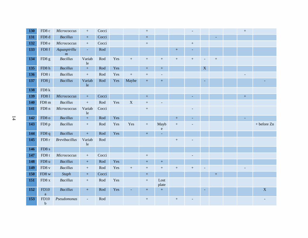
Following the isolation of environmental bacterial isolates, they were characterized. The Appendix depicts the results of the physical and biochemical tests carried out following Bergey’s Manual of Determinative Bacteriology to identify each microorganism. Using this methodology, all but nine of the isolates were identified to their genus. A total of 114 unique isolates were identified.

6
To confirm the genus identification results of the isolates as determined by the physical and biochemical tests, and to also assist with identifying the species of the isolated organisms, the Omnilog Identification system (Omnilog) was used. The Omnilog was able to positively identify the genus of 54% of the isolates (61 of 114 isolates), with a few to the species level (Appendix). The Omnilog was unable to identify any of the nine unknowns. The failure of the Omnilog system to identify the genus or species for a greater number of environmental isolates may be due to the limitations of the Omnilog database. The Omnilog database consists of information for primarily clinical microorganisms and not environmental microorganisms, like the bacterial isolates used in this study. Though not done in this work, an alternative method, such as genomic sequencing, would be more effective to confirm the genus identification results obtained from biochemical and physical analysis. This should be considered for future efforts. Table 2 summarizes the results of genus identification through physical and biochemical analysis as well as Omnilog studies for each of the 114 unique environmental isolates collected. Excluding the unidentified organisms, seven distinctive genera were identified. Bacteria from the genus Bacillus and Staphylococcus were present in the greatest amount at 47% and 25% respectively. Conversely, only one isolate was identified from the genus Aquaspirillum and Brevibacillus. The greatest number of isolates were obtained from two of the AC ducts (57%) and the door handle (27%). Of the total number of isolates collected from the two AC ducts, 60% were identified as the genus Bacillus. These results are to be expected as AC ducts contain a significant amount of debris on their interior surfaces and the spore forming bacteria Bacillus can easily become embedded in the interior of the ducts. Of the total number of isolates collected from the door handle, 84% were identified as the genus Staphylococcus. As Staphylococcus make up a significant portion of the skin microbiota, it is reasonable to infer that the large quantity collected from the door handle is due to the frequent interaction of people’s hands with the door handle. Table 2. Summary of Genus Identification of Environmental Bacterial Isolates in Relation to Sample Collection Location
Frequency
All
Surfaces Laundry Kitchen sink
Cutting board
Tent 6 cot
Tent 6 AC
Tent 8 AC
Rigid shelter door handle
Rigid shelter AC
Organism Genus Aquaspirillum 1 1
Bacillus 53 4 7 1 1 2 23 1 14
Brevibacillus 1 1
Cupriavidus 11 11
Micrococcus 7 2 5
Pseudomonas 3 1 2
Staphylococcus 29 2 26 1
Unknown 9 2 3 2 2
Total 114 9 7 1 1 2 39 31 24

7
3.2 Characterization of Activity of Environmental Bacterial Isolates Against the Target Bacteria
Table 3 shows a summary of the number of isolates that exhibited activity against the target strains evaluated. Since bacteriocins typically only demonstrate activity against close competitors, initial screens of bacteriocin activity against the target microorganisms focused on the same genus. Bacillus isolates and the nine unknowns were screened for activity against a Bacillus anthracis Sterne target; Staphylococcus isolates and the nine unknowns were screened for activity against a Staphylococcus aureus target (ATCC 27217), and Pseudomonas isolates and the nine unknowns were screened for activity against a Pseudomonas aeruginosa target (ATCC 15692). None of the unknowns showed activity against any of the three targets. Of the 53 Bacillus isolates, 15% or 9 of the 62 isolates tested showed activity against the Bacillus anthracis Sterne target. Only one Staphylococcus isolate showed activity against the Staphylococcus aureus target. None of the Pseudomonas isolates exhibited activity against the Pseudomonas aeruginosa target. Secondary activity screens were performed to determine if any of the isolates produced bacteriocins against bacteria from the other target genera. All the isolates were screened against the Staphylococcus aureus and Pseudomonas aeruginosa targets. Only one of the isolates, a Bacillus, showed activity against the Staphylococcus aureus target, while no isolates showed activity against the Pseudomonas aeruginosa target. Thus, only 2% or 2 out of the 114 isolates tested were found to be active against the Staphylococcus aureus target. Table 4 summarizes in greater detail the results of the isolates that were found to be active against the tested target strains. Table 3. Summary of Activity of Isolates Against Target Microorganisms
Target Organism Total isolates
screened Number of isolates positive for activity
Bacillus anthracis Sterne 62 9
Staphylococcus aureus 114 2
Pseudomonas aeruginosa 114 0
Table 4. Detailed Summary of Active Isolates
# Name Organism Identity Target Active
Against
62 FD6 b Bacillus subtilis BA
65 FD6 e Bacillus BA
142 FD8 o Bacillus BA
148 FD8 u Bacillus thuringensis BA
151 FD8 x Bacillus licheniformis BA
157 FD10 f Bacillus thuringensis BA
162 FD11 b Bacillus BA
164 FD11 d Bacillus BA
165 FD11 e Bacillus BA
60b FD3 b Bacillus SA
105 FD7 g Staph SA BA: Bacillus anthracis Sterne, SA: Staphlocooccus aureus (ATCC 27217)

8
4 Conclusions A low percentage of environmental isolates displayed bacteriocin activity against the tested targets, but this finding was expected. When interpreting the bacteriocin activity results, one should keep in mind that the isolates were initially screened against a single target strain for each genera. Though most bacteria produce bacteriocins, generally they only produce bacteriocins against bacteria strains that exist as competitors in their environment, which contributes to the narrow activity spectrum of bacteriocins. Thus, a low number of isolates were anticipated to be active against the target bacteria strains. Within that context, the Bacillus sp. bacteria strains isolated from environmental sources were expected to yield activity against the Bacillus anthracis Sterne target since Bacillus anthracis is a soil-borne bacteria. Nine isolates of Bacillus did in fact produce bacteriocins against the Bacillus anthracis Sterne target. Additionally, characterizing a single strain of Staphylococcus with activity against the target strain of Staphylococcus was not surprising given that most of the Staphylococcus isolates were from the same door handle and may be the same species/strain. What was more interesting was the unique finding of one Bacillus isolate that was active against the Staphylococcus target, yet inactive against the Bacillus anthracis Sterne target. The results from the spectrum screen expand this uniqueness in that the isolate did not show activity toward any of the Bacillus strains tested. Further study of this isolate is warranted to understand why it is active against the Staphylococcus target and not its Bacillus competitors. The narrow-spectrum activity of the 11 environmental isolates found to demonstrate bacteriocin activity against the target strains was further confirmed with the assistance of a collaborator at the University of Massachusetts, Amherst. The 11 active isolates identified in the initial screening (Table 4) were tested for activity against additional targets of different genera and strains to characterize the extent of activity displayed by the isolates. The results of these additional activity screens showed that the 11 isolates exhibited no activity against unrelated genus strains and only minimal activity against varying strains of the same genus (results not shown). These findings provide additional support to infer that the bacteriocin activity of the environmental isolates is specific to a narrow-spectrum range of activity. Thus, the nine isolates identified as active against Bacillus anthracis Sterne and the two isolates that demonstrated activity against Staphylococcus resulting from this study are excellent candidates for development as narrow-spectrum antimicrobials to specifically target pathogens of interest to the DoD. The low number of environmental isolates found to be active against the target strains evaluated illustrates the narrow-spectrum activity of bacteriocins. Moreover, screening of the active isolates against additional organisms revealed minimal activity, which further validates the specific bacteriocin-driven activity of the environmental isolates. The active environmental bacteria isolates provide a valuable resource for narrow-spectrum antimicrobial agents. Future studies will build upon this work by isolating, purifying, and characterizing bacteriocins produced from these active environmental bacteria isolates.
17/014

9
5 References Abt M.C. and E.G Pamer. “Commensal bacteria mediated defenses against pathogens”. Curr. Opin. Immunol. 29C: 16-22. (2014) Cooper, M.A. and D Shlae. “Fix the Antibiotic Pipeline”. Nature 472:32. (2011) Cotter, P.A., C. Hin, and R.P. Ross. “Bacteriocin Developing Innate Immunity for Food”. Nature Reviews Microbiology. 3:777-78 (2005) Cox, C.P., P.S. Coburn, and M.S. Gilman. “Enterococcal Cytolysis: a Novel Two Component Peptide System that Serves as a Bacterial Defence Against Eukaryote and Prokaryote Cells”. Current Protein Peptide Science 6:77-84 (2005) Galvz, A., H. Ahriovel, R.L. Lopez, and N.B. Omar. “Bacteriocin-based Strategies for Food Biopreservation”. International Journal of Food Microbiology 120:51-70 (2007) Gulberg, E., S. Cao, O.G. Berg, C. IIback, L. Sandegren, D. Hughes, and D.L. Andersson. “Selection of resistant bacteria have very low antibiotic concentrations”. PLOS pathogens 7(7):1-9. (2011) Holt, J. G., N.R. Krieg, P.H. Sneath, J.T. Staley, and S.T. Williams. “Bergey’s manual of determinative bacteriology”. Baltimore: Williams and Wilkins, 75, 121. (1994) Levy, S.B. and B. Marshall. “Antibacterial resistance worldwide: causes, challenges and response”. Nature medicine. 10(12):S122-S129. (2004) McDonnell, G. and A.D. Russell. Antiseptics and disinfectants: activity, action and resistance. Clin. Microbiol. Rev. 12:147—179. (1999) Silver, S. “Bacterial silver resistance: molecular biology and uses and misuses of silver compounds”. FEMS Microbial Rev. 27(2-3):341-53. (2003) Tattawasart, U., J-Y Maillard, J.R Furr, and A.D. Russell. “Development of resistance to chlorhexidine diacetate and cetylpyridinium chloride in Pseudomonas stutzeri and changes in antibiotic susceptibility” J. Hosp. Infect. 42(2):219-229. (1999) Thomas, L., J-Y Maillard, R.J.W. Lambert, and A.D. Russell. “Development of resistance to chlorhexidine diacetate in Pseudomonas aeruginosa and the effect of a 'residual' concentration”. J. Hosp. Infect. 46(4):297-303. (2000)

10
This page intentionally left blank

11
Appendix: Results of Physical and Biochemical Analysis to Determine Identity of Environmental Isolates
#
Name Organism Identity
Gram Shape
Spore Swollen
Catalase
Starch
Hyd
Oxidase
Glucose
Ferm
VP
Novo-
biocin
Citrate
Growth in
6.5% Na
Nitrate Reduction
59 FD2 a Bacillus + Rod Yes + + + X -
60 FD3 a Bacillus + Rod Yes + + + - -
61 FD6 a Bacillus Variable
Rod Yes + + + + - X
62 FD6 b Bacillus + Rod Yes + +
63 FD6 c Bacillus + Rod Yes - + + - X
64 FD6 d Cupriavidus - Rod + -
65 FD6 e Bacillus + Rod Yes + +
66 FD6 f Bacillus + Rod Yes - + -
67 FD6 g Bacillus + Rod Yes - + -
68 FD6 h Cupriavidus - Rod + -
69 FD6 i Bacillus + Rod Yes + + -
70 FD6 j Cupriavidus - Rod + - +
71 FD6 k Bacillus + Rod Yes + + beforeZn
72 FD6 l Bacillus + Rod Yes + + -
73 FD6 m Bacillus + Rod Yes - + + - X
74 FD6 n Cupriavidus - Rod + -
75 FD6 o Pseudomonas - Rod Maybe
+ - -
76 FD6 p Cupriavidus - Rod + - X
77 FD6 q Cupriavidus Variable
Rod Yes + -
78 FD6 r Cupriavidus - Rod + + -
79 FD6 s Bacillus + Rod Yes + + -

12
80 FD6 t1 Bacilus + Rod Yes - + -
81 FD6 t2 Bacillus Variable
Rod Yes Yes + + - -
82 FD6 u Bacillus + Rod Yes Yes + + -
83 FD6 v Bacillus + Rod Yes - + - -
84 FD6 w Bacillus + Rod Yes - + - -
85 FD6 x1
Bacillus + Rod Yes + + + X
86 FD6 x1
Bacillus Variable
Rod Yes - - + - + before Zn
87 FD6 y Bacillus Variable
Rod Yes + + -
88 FD6 z Cupriavidus - Rod
89 FD6 aa Cupriavidus - Rod Maybe
+ + -
90 FD6 bb
Bacillus + Rod Yes + - +
91 FD6 cc Bacillus Variable
Rod Yes - + - + - -
92 FD6 dd
Variable
rod Yes Yes + X
93 FD6 ee Bacillus + Rod Yes + + +
94 FD6 ff Bacillus Variable
Rod No X + - + before Zn; frothy
95 FD6 gg
- Rod + -
96 FD6 hh
Bacillus + Rod Yes X + - -
97 FD6 ii Cupriavidus - Rod + + -
98 FD6 jj Cupriavidus - Rod + -
99 FD7 a Staph + Cocci +
100 FD7 b Staph + Cocci + X + after Zn
101 FD7 c Staph + Cocci + +
102 FD7 d Staph + Cocci + - +

13
103 FD7 e Staph + Cocci + +
104 FD7 f Staph + Cocci + +
105 FD7 g Staph + Cocci + X
106 FD7 h Staph + Cocci + +
107 FD7 i Staph + Cocci + +
108 FD7 j Staph + Cocci + +
109 FD7 k Staph + Cocci +
110 FD7 l Staph + Cocci + +
111 FD7 m Staph + Cocci + X
112 FD7 n Staph + Cocci +
113 FD7 o Staph + Cocci + + + w/ gas
114 FD7 p Micrococcus + Cocci + - -
115 FD7 q Staph + Cocci + + + after Zn; frothy
116 FD7 r Staph + Cocci + +
117 FD7 s Staph + Cocci + +
118 FD7 t1 Staph + Cocci + +
119 FD7 u Staph + Cocci + -
120 FD7 v Staph + Cocci + +
121 FD7 w Staph + Cocci + +
122 FD7 x Staph + Cocci + +
123 FD7 y Staph + Cocci + +
124 FD7 z Staph + Cocci + +
125 FD7 aa Staph + Cocci + +
126 FD7 bb
+
127 FD7 cc Micrococcus + Cocci + -
128 FD8 a Bacillus + Rod Yes + +
129 FD8 b Bacillus Variable
Rod Yes + + -
14
130 FD8 c Micrococcus + Cocci + - +
131 FD8 d Bacillus + Cocci + -
132 FD8 e Micrococcus + Cocci + +
133 FD8 f Aquaspirillum
- Rod + -
134 FD8 g Bacillus Variable
Rod Yes + + + + + - +
135 FD8 h Bacillus + Rod Yes + + X
136 FD8 i Bacillus + Rod Yes + + - -
137 FD8 j Bacillus Variable
Rod Yes Maybe + + - -
138 FD8 k
139 FD8 l Micrococcus + Cocci + - +
140 FD8 m Bacillus + Rod Yes X + -
141 FD8 n Micrococcus Variable
Cocci + -
142 FD8 o Bacillus + Rod Yes + - -
143 FD8 p Bacillus + Rod Yes Yes + Maybe
+ - + before Zn
144 FD8 q Bacillus + Rod Yes + -
145 FD8 r Brevibacillus Variable
Rod + -
146 FD8 s
147 FD8 t Micrococcus + Cocci + -
148 FD8 u Bacillus + Rod Yes + +
149 FD8 v Bacillus + Rod Yes + + + + + - -
150 FD8 w Staph + Cocci + +
151 FD8 x Bacillus + Rod Yes + Lost plate
152 FD10 a
Bacillus + Rod Yes - + + - X
153 FD10 b
Pseudomonas - Rod + + - -

15
154 FD10 c
Bacillus + Rod Yes + +
155 FD10 d
Bacillus + Rod + -
156 FD10 e
Staph + Cocci + +
157 FD10 f Bacillus + Rod Yes + +
158 FD10 g
- Rod + +
159 FD10 h
Staph + Cocci + + ?
160 FD10 i Nocardia? - Mixed
Yes Not swolle
n
+ Positive
161 FD11 a
Bacillus + Rod Yes X + + - + after Zn
162 FD11 b
Bacillus + Rod Yes - + -
163 FD11 c
Bacillus + Rod Yes + - + -
164 FD11 d
Bacillus + Rod Yes - + + -
165 FD11 e
Bacillus + Rod Yes Maybe + + -
166 FD11 f Bacillus Variable
Rod Yes - + + + -
167 FD11 g
Bacillus + Rod Yes + -
168 FD12 a
Bacillus + Rod Yes + + + - X
116a
FD7ra Bacillus X X Yes + +
118b
FD7 t2 + Cocci +
60b FD 3b Bacillus + Rod Yes Yes + - +
71b FD6Kb
+ Cocci or rod
No ? +


![WOR8294 Assaying and Refining of Gold[1]](https://img.pdfslide.us/doc/110x75/5571fa7e497959916992597b/wor8294-assaying-and-refining-of-gold1.jpg)