Embed Size (px)
Citation preview
TERMS OF USEThis pdf is provided by Magnolia Press for private/research use. Commercial sale or deposition in a public library or website is prohibited.
ZOOTAXAISSN 1175-5326 (print edition)
ISSN 1175-5334 (online edition)Copyright © 2011 · Magnolia Press
Zootaxa 2866: 39–49 (2011) www.mapress.com/zootaxa/ Article
Ultrastructural analysis of Coarazuphium formoso (Coleoptera: Carabidae, Zuphiini), a new Brazilian troglobitic beetle
THAÍS GIOVANNINI PELLEGRINI1 & RODRIGO LOPES FERREIRA1Laboratório de Ecologia Subterrânea, Setor de Zoologia, Departamento de Biologia, Universidade Federal de Lavras, Lavras, MG. CEP 37200-000, Brazil. E-mail: [email protected] author. E-mail: [email protected]
Abstract
It is described Coarazuphium formoso sp. n., from male and female specimens collected in Barriguda cave and Calor de Cima cave, both in the municipality of Campo Formoso (Bahia, Brazil). The most striking difference between C. formosoand other species of the genus is the presence of three pairs of setae on the dorsal surface of the head close to the posterior margin, while the other species have only one or two pairs. Species from this genus showed advanced troglobiomorphic characters in comparison to other Brazilian cave beetles. Characters as increased extra-optic sensory structures, and the presence of particular sensilla, and sensory and gustatory receptors were not detected under routine microscopy but re-quired ultrastructural scrutiny. Similar analyses are needed in other epigean Zuphiini species for better interpreting their functional meaning.
Key words: Coarazuphium, ground beetle, cave dwelling, sensilla, antenna, eyes, mouthparts, legs
Introduction
Members from the Zuphiini tribe are normally winged epigean organisms (Casale, 1998). The first record of a tro-globiotic Zuphiini was that of Parazuphium tessai (Godoy & Vanin, 1990), found in Padre Cave (municipality of Santana, Bahia, Brazil). This species has common traits with two other Zuphiini genera, Parazuphium Jeannel, 1942 and Zuphium Latreille, 1806. Later on, Moore (1994) recorded two new genera of troglobiotic Zuphiini, Speozuphium and Speothalpius, each with a new species, Speozuphium poulteri and Speothalpius grayi, respec-tively. Both were found in the cave systems of the Nullarbor Plain in Australia. Gnaspini et al. (1998), then, have proposed a new genus, Coarazuphium, which included Parazuphium tessai and two other new species, C. cessaima and C. bezerra. These three species differ from other zuphiinids in relation to their relatively elongated first anten-nomeres (although this is still shorter than the second to fourth together), rounded head margins, and a pair of setae lateral to the eyes (rather than anterior to them). Another important characteristic of this genus is the presence of typical troglobiomorphisms, such as the reduction or absence of pigmentation, eyes, and wings and the elongation of appendages and antennae.
To date, all Coarazuphium specimens have been found only in caves in the Bambuí Speological Province (Gnaspini et al, 1998; Álvares & Ferreira, 2002). C. tessai was found in Gruta do Padre Cave, in the municipality of Santana and C. cessaima was found in the Lapa do Bode Cave, in the municipality of Itaetê, both in Bahia. C. bezerra was found in the Lapa do Bezerra Cave, in the municipality of São Domingos in Goiás, and C. pains was found in the Tabocas III cave, in the municipality of Pains in Minas Gerais.
Our objective is to describe a new species of troglobitic beetle, Coarazuphium formoso, from the Una Speleo-logical Province. This description is focused on an ultrastructural analysis of the antennae, eyes, mouthparts, and legs. According to Moldovan et al. (2004), these structures indicate the degree of adaptation to life in caves. They also provide adequate criteria to identify cave species with otherwise homogeneous morphologies.
Accepted by J. Serrano: 28 Feb. 2011; published: 4 May 2011 39
TERMS OF USEThis pdf is provided by Magnolia Press for private/research use. Commercial sale or deposition in a public library or website is prohibited.
Methods and material
All specimens were collected from two caves: Barriguda Cave and Calor de Cima Cave, both in Campo Formoso, Bahia, Brazil. Carabids were thoroughly searched visually throughout the base and walls of the caves. Special attention was also paid to decaying vegetation (e.g. leaves and tree bark), animal carcasses, and vertebrate feces, though this material was only common near the cave entrances, where no individuals were found. Carabids were captured with a fine brush and placed in vials containing 70% ethanol. Measurements and drawings were made under a stereomicroscope and a camera lucida microscope. To dissect male and female genitalia, fine entomologi-cal pins were used. Both specimens were prepared in Kayser glycerol gelatin; drawings were made under a Leica MDLS phase contrast microscope. The ultrastructural analyses were conduced through use of a scanning electron microscope. Parts from paratype 1 were placed on aluminum support stubs, placed over a film of aluminum foil with carbon tape, sputter-covered with gold (Baltec SCD 050), and observed in a LEO EVO 40 XVP scanning electron microscope (Leo Electron Microscopy).
We followed the criteria of Schneider (1964), McIver (1975), Zacharuk (1980), Kim & Yamasak (1995) and Merivee et al. (2000) for naming ultrastructures and assessing their function.
Type specimens are deposited in the Zoology Collection, Seção de Invertebrados Subterrâneos (ISLA 1057 male holotype; ISLA 1058 female paratype 1; ISLA 1059 male paratype 2 and ISLA 1060 male paratype 3), at the Universidade Federal de Lavras (UFLA), Campus Universitário de Lavras, Minas Gerais, Brazil.
Taxonomy
Family CARABIDAE Latreille, 1802
Tribe Zuphiini Bonelli, 1810
Genus Coarazuphium Gnaspini, Vanin & Godoy, 1998
Coarazuphium formoso sp. n. (Fig. 1–11).
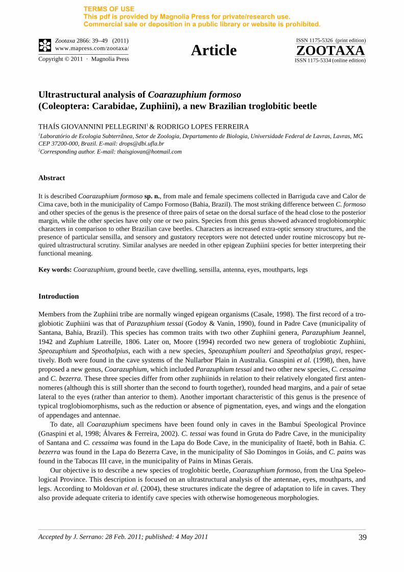
Description. Morphometric data from paratypes are given in parenthesis .Holotype male (Fig. 1A). Total length, from the apex of the mandible to the apex of the elytra: 5.41 mm (5.16–
5.53), width, from at the widest region of the elytra: 1.51 mm (1.32–1.45). Body pale reddish brown, two paratypes are yellowish to pale brown, dorsal integument of the elytra covered with short recumbent hairs.
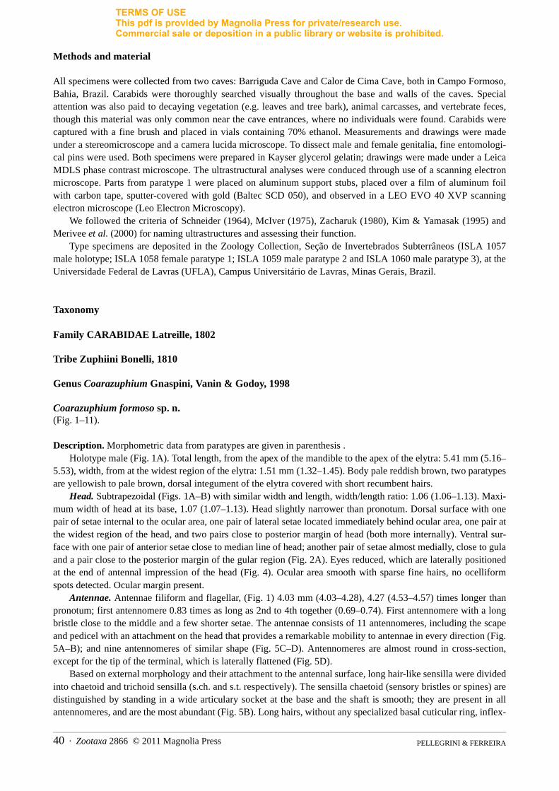
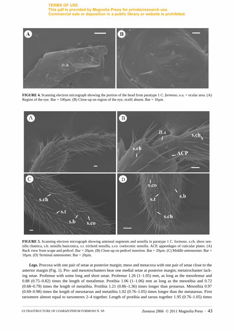
Head. Subtrapezoidal (Figs. 1A–B) with similar width and length, width/length ratio: 1.06 (1.06–1.13). Maxi-mum width of head at its base, 1.07 (1.07–1.13). Head slightly narrower than pronotum. Dorsal surface with one pair of setae internal to the ocular area, one pair of lateral setae located immediately behind ocular area, one pair at the widest region of the head, and two pairs close to posterior margin of head (both more internally). Ventral sur-face with one pair of anterior setae close to median line of head; another pair of setae almost medially, close to gula and a pair close to the posterior margin of the gular region (Fig. 2A). Eyes reduced, which are laterally positioned at the end of antennal impression of the head (Fig. 4). Ocular area smooth with sparse fine hairs, no ocelliform spots detected. Ocular margin present.
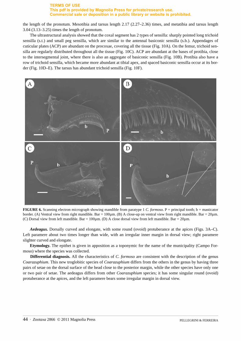
Antennae. Antennae filiform and flagellar, (Fig. 1) 4.03 mm (4.03–4.28), 4.27 (4.53–4.57) times longer than pronotum; first antennomere 0.83 times as long as 2nd to 4th together (0.69–0.74). First antennomere with a long bristle close to the middle and a few shorter setae. The antennae consists of 11 antennomeres, including the scape and pedicel with an attachment on the head that provides a remarkable mobility to antennae in every direction (Fig. 5A–B); and nine antennomeres of similar shape (Fig. 5C–D). Antennomeres are almost round in cross-section, except for the tip of the terminal, which is laterally flattened (Fig. 5D).
Based on external morphology and their attachment to the antennal surface, long hair-like sensilla were divided into chaetoid and trichoid sensilla (s.ch. and s.t. respectively). The sensilla chaetoid (sensory bristles or spines) are distinguished by standing in a wide articulary socket at the base and the shaft is smooth; they are present in all antennomeres, and are the most abundant (Fig. 5B). Long hairs, without any specialized basal cuticular ring, inflex-
PELLEGRINI & FERREIRA 40 · Zootaxa 2866 © 2011 Magnolia Press
TERMS OF USEThis pdf is provided by Magnolia Press for private/research use. Commercial sale or deposition in a public library or website is prohibited.
ible in their sockets, were classified as trichoid sensilla (sensory hairs) also distinguished by the relatively greater
length; they can be found on the 5th to 11th antennomeres.Short pegs and cones, which reach above the socket, were classified as basiconic sensilla (s.b.) (sensory pegs or
cones) irrespective of whether they sit in a wide or tight socket; they are present on the 4th to 11th antennomeres. Some BÖhm sensilla (B.s.) (sensory pit-pegs) typical bristle are also present found in areas opposite the interseg-mental membrane between head and scape, as well as between scape and pedicel on the scape and pedicel bases, respectively (Fig. 5B). Coeloconic sensilla (s.co.), (sensory pit-pegs) are small pit organs, on the floor of depres-sions in the antennal cuticle, these organs ending at the very tip of the cone; they can be found on the 4th to 11th
antennomers (Fig. 5C–D). Appendages of cuticular plates (ACP), which are small cuticular process, are abundant at the bases of all antennomeres, close to the intersegmental joints (Fig. 5C).
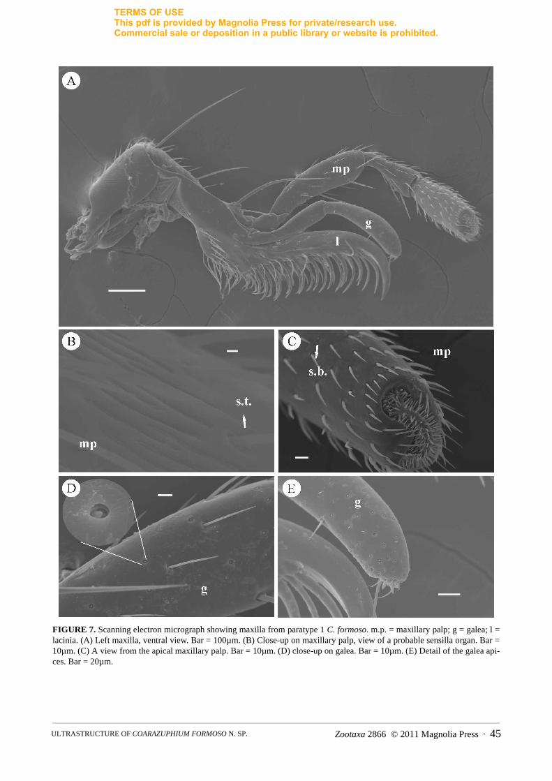
Mouthparts. Sensilla on the mandible, maxilla, labial palpus, labrum, and clypeus of the paratype 1 were examined. The mandible is acutely bent inwardly at its tip. On the ventral side, longitudinal rows of setae are pres-ent (Fig. 6A–B). On the dorsal surface, a series of hair sensilla projects from the submolar region to near the cutic-ular processes (Fig. 6C–D).
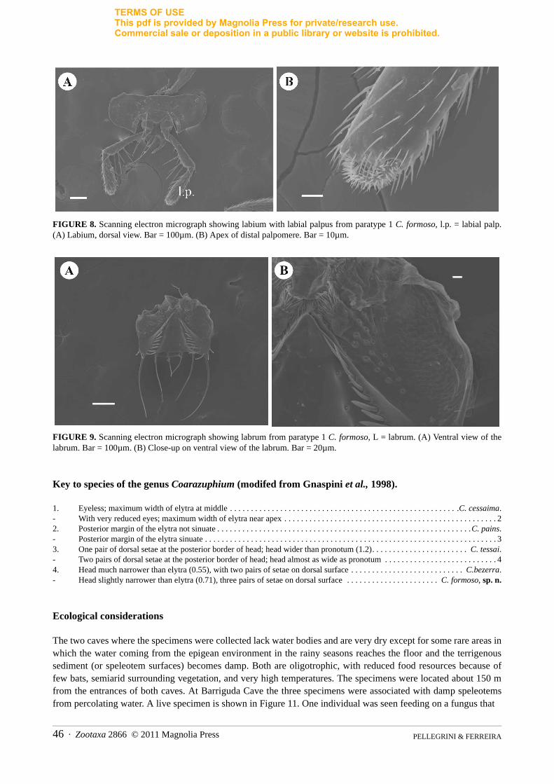
The maxilla basically consists of the lacinia, maxillary palp, and galea (Fig. 7A). The lacinia is shorter than the galea, with an acute and curved end, with rows of long setae and cuticular process. The four-segmented maxillary palp is long and filiform with spaced basiconic sensilla present on the surfaces of the segments. Trichoid sensilla are distributed along the maxillary palp, and they become more abundant and smaller on the last segment (Fig. 7C). There are also grooves in this segment that may indicate a sensory organ or gustatory receptors (Fig. 7B).
The galea is biarticulated, composed of 2 segments, with different types of basiconic sensilla. These sensilla become more abundant near the apex of the last segment (Fig. 7D–E).
FIGURE 1. Coarazuphium formoso. A) Habitus from male holotype. B) Habitus from female holotype.
Zootaxa 2866 © 2011 Magnolia Press · 41ULTRASTRUCTURE OF COARAZUPHIUM FORMOSO N. SP.
TERMS OF USEThis pdf is provided by Magnolia Press for private/research use. Commercial sale or deposition in a public library or website is prohibited.
FIGURE 2. A) Head and pronotum lateral view.B) Pronotum, ventral view.
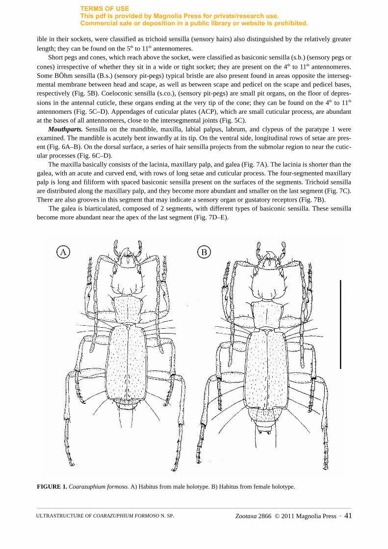
FIGURE 3. Aedeagus, left lateral view, dorsal view and right lateral view, respectively.
The labium has two pairs of small setae at its base and one pair of long setae near the labial palpi (Fig. 8A), which has some long hairs within. The types of sensilla on the three-segmented labial palpomeres are the same as those on the maxillary palpomeres (Fig. 8B). The labrum is quadrangular (Fig. 9). On the ventral side confluent rows of long setae are present.
Pronotum. Shape trapezoidal, 1.17 (1.13–1.29) times wider than long (Figs. 1, 2B). Maximum width close to anterior angle and as wide as head. Anterior and posterior angles are acute. Dorsal surface (Figs. 1A–B) with two pairs of erect setae: one close to the anterior angle of the pronotum and the other, shorter, close to the posterior angle. Ventral surface with one pair of anterior setae medially located (Fig. 2A).
Elytra. Elytra are free (Fig. 1), together 1.78 (1.67–1.86) times longer than wide. Maximum width nearly one third the distance from the apex and 2.73 (2.60–2.79) times longer than pronotum. Apex of elytra sinuous. Seven large setae in each elytron: 3 close to the anterior angle, 2 marginal in posterior half, and 2 on posterior margin. There is also one smaller seta on the posterior internal margin of each elytra. Wings absent. Abdominal sterna 1–5, glabrous, sixth sternum with a small pair of setae close to its posterior margin.
PELLEGRINI & FERREIRA 42 · Zootaxa 2866 © 2011 Magnolia Press
TERMS OF USEThis pdf is provided by Magnolia Press for private/research use. Commercial sale or deposition in a public library or website is prohibited.
FIGURE 4. Scanning electron micrograph showing the portion of the head from paratype 1 C. formoso, o.a. = ocular area. (A) Region of the eye. Bar = 100μm. (B) Close-up on region of the eye, ocelli absent. Bar = 10μm.
FIGURE 5. Scanning electron micrograph showing antennal segments and sensilla in paratype 1 C. formoso. s.ch. show sen-silla chaetica, s.b. sensilla basiconica, s.t. trichoid sensilla, s.co. coeloconic sensilla. ACP, appendages of cuticular plates. (A) Back view from scape and pedicel. Bar = 20µm. (B) Close-up on pedicel insertion. Bar = 20µm. (C) Middle antennomer. Bar = 10µm. (D) Terminal antennomer. Bar = 20µm.
Legs. Procoxa with one pair of setae at posterior margin; meso and metacoxa with one pair of setae close to the anterior margin (Fig. 1). Pro- and mesotrochanters bear one medial setae at posterior margin; metatrochanter lack-ing setae. Profemur with some long and short setae. Profemur 1.26 (1–1.05) mm, as long as the mesofemur and 0.88 (0.75–0.82) times the length of metafemur. Protibia 1.06 (1–1.06) mm as long as the mesotibia and 0.72 (0.68–0.79) times the length of metatibia. Protibia 1.21 (0.86–1.36) times longer than protarsus. Mesotibia 0.97 (0.69–0.98) times the length of mesotarsus and metatibia 1.02 (0.76–1.05) times longer than the metatarsus. First tarsomere almost equal to tarsomeres 2–4 together. Length of protibia and tarsus together 1.95 (0.76–1.05) times
Zootaxa 2866 © 2011 Magnolia Press · 43ULTRASTRUCTURE OF COARAZUPHIUM FORMOSO N. SP.
TERMS OF USEThis pdf is provided by Magnolia Press for private/research use. Commercial sale or deposition in a public library or website is prohibited.
the length of the pronotum. Mesotibia and tarsus length 2.17 (2.27–2.36) times, and metatibia and tarsus length 3.04 (3.13–3.25) times the length of pronotum.
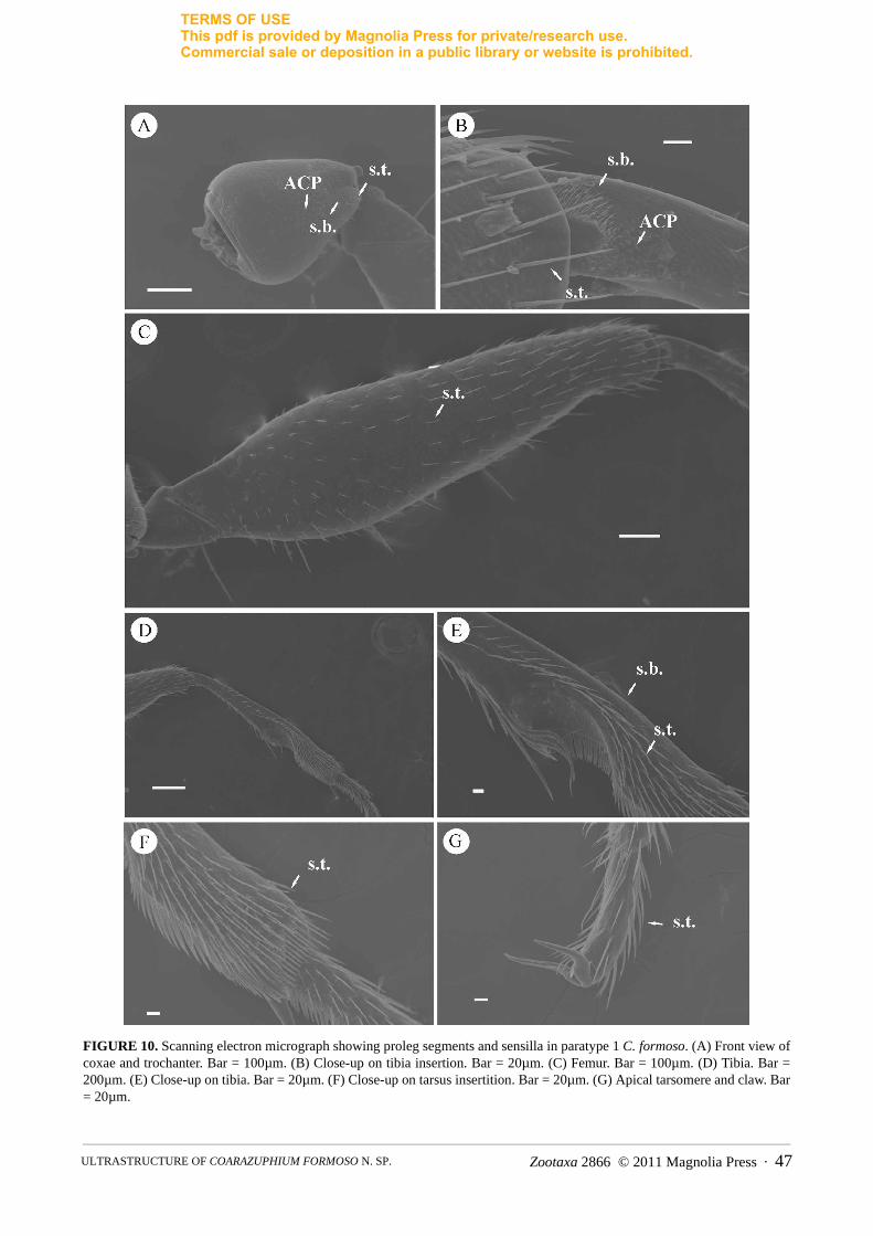
The ultrastructural analysis showed that the coxal segment has 2 types of sensilla: sharply pointed long trichoid sensilla (s.t.) and small peg sensilla, which are similar to the antennal basiconic sensilla (s.b.). Appendages of cuticular plates (ACP) are abundant on the procoxae, covering all the tissue (Fig. 10A). On the femur, trichoid sen-silla are regularly distributed throughout all the tissue (Fig. 10C). ACP are abundant at the bases of protibia, close to the intersegmental joint, where there is also an aggregate of basiconic sensilla (Fig. 10B). Protibia also have a row of trichoid sensilla, which became more abundant at tibial apex, and spaced basiconic sensilla occur at its bor-der (Fig. 10D–E). The tarsus has abundant trichoid sensilla (Fig. 10F).
FIGURE 6. Scanning electron micrograph showing mandible from paratype 1 C. formoso. P = principal tooth; b = masticator border. (A) Ventral view from right mandible. Bar = 100µm. (B) A close-up on ventral view from right mandible. Bar = 20µm. (C) Dorsal view from left mandible. Bar = 100µm. (D) A close dorsal view from left mandible. Bar = 20µm.
Aedeagus. Dorsally curved and elongate, with some round (ovoid) protuberance at the apices (Figs. 3A–C). Left paramere about two times longer than wide, with an irregular inner margin in dorsal view; right paramere slighter curved and elongate.
Etymology. The epithet is given in apposition as a toponymic for the name of the municipality (Campo For-moso) where the species was collected.
Differential diagnosis. All the characteristics of C. formoso are consistent with the description of the genus Coarazuphium. This new troglobitic species of Coarazuphium differs from the others in the genus by having three pairs of setae on the dorsal surface of the head close to the posterior margin, while the other species have only one or two pair of setae. The aedeagus differs from other Coarazuphium species; it has some singular round (ovoid) protuberance at the apices, and the left paramere bears some irregular margin in dorsal view.
PELLEGRINI & FERREIRA 44 · Zootaxa 2866 © 2011 Magnolia Press
TERMS OF USEThis pdf is provided by Magnolia Press for private/research use. Commercial sale or deposition in a public library or website is prohibited.
FIGURE 7. Scanning electron micrograph showing maxilla from paratype 1 C. formoso. m.p. = maxillary palp; g = galea; l = lacinia. (A) Left maxilla, ventral view. Bar = 100µm. (B) Close-up on maxillary palp, view of a probable sensilla organ. Bar = 10µm. (C) A view from the apical maxillary palp. Bar = 10µm. (D) close-up on galea. Bar = 10µm. (E) Detail of the galea api-ces. Bar = 20µm.
Zootaxa 2866 © 2011 Magnolia Press · 45ULTRASTRUCTURE OF COARAZUPHIUM FORMOSO N. SP.
TERMS OF USEThis pdf is provided by Magnolia Press for private/research use. Commercial sale or deposition in a public library or website is prohibited.
FIGURE 8. Scanning electron micrograph showing labium with labial palpus from paratype 1 C. formoso, l.p. = labial palp. (A) Labium, dorsal view. Bar = 100µm. (B) Apex of distal palpomere. Bar = 10µm.
FIGURE 9. Scanning electron micrograph showing labrum from paratype 1 C. formoso, L = labrum. (A) Ventral view of the labrum. Bar = 100µm. (B) Close-up on ventral view of the labrum. Bar = 20µm.
Key to species of the genus Coarazuphium (modifed from Gnaspini et al., 1998).
1. Eyeless; maximum width of elytra at middle . . . . . . . . . . . . . . . . . . . . . . . . . . . . . . . . . . . . . . . . . . . . . . . . . . . . . . .C. cessaima.- With very reduced eyes; maximum width of elytra near apex . . . . . . . . . . . . . . . . . . . . . . . . . . . . . . . . . . . . . . . . . . . . . . . . . . . 22. Posterior margin of the elytra not sinuate . . . . . . . . . . . . . . . . . . . . . . . . . . . . . . . . . . . . . . . . . . . . . . . . . . . . . . . . . . . . .C. pains.- Posterior margin of the elytra sinuate . . . . . . . . . . . . . . . . . . . . . . . . . . . . . . . . . . . . . . . . . . . . . . . . . . . . . . . . . . . . . . . . . . . . . . 33. One pair of dorsal setae at the posterior border of head; head wider than pronotum (1.2) . . . . . . . . . . . . . . . . . . . . . . . C. tessai.- Two pairs of dorsal setae at the posterior border of head; head almost as wide as pronotum . . . . . . . . . . . . . . . . . . . . . . . . . . . 44. Head much narrower than elytra (0.55), with two pairs of setae on dorsal surface . . . . . . . . . . . . . . . . . . . . . . . . . . . C.bezerra.- Head slightly narrower than elytra (0.71), three pairs of setae on dorsal surface . . . . . . . . . . . . . . . . . . . . . . C. formoso, sp. n.
Ecological considerations
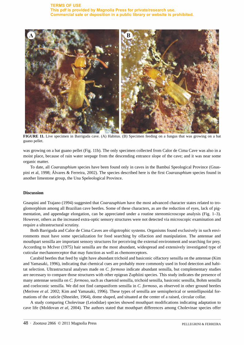
The two caves where the specimens were collected lack water bodies and are very dry except for some rare areas in which the water coming from the epigean environment in the rainy seasons reaches the floor and the terrigenous sediment (or speleotem surfaces) becomes damp. Both are oligotrophic, with reduced food resources because of few bats, semiarid surrounding vegetation, and very high temperatures. The specimens were located about 150 m from the entrances of both caves. At Barriguda Cave the three specimens were associated with damp speleotems from percolating water. A live specimen is shown in Figure 11. One individual was seen feeding on a fungus that
PELLEGRINI & FERREIRA 46 · Zootaxa 2866 © 2011 Magnolia Press
TERMS OF USEThis pdf is provided by Magnolia Press for private/research use. Commercial sale or deposition in a public library or website is prohibited.
FIGURE 10. Scanning electron micrograph showing proleg segments and sensilla in paratype 1 C. formoso. (A) Front view of coxae and trochanter. Bar = 100µm. (B) Close-up on tibia insertion. Bar = 20µm. (C) Femur. Bar = 100µm. (D) Tibia. Bar = 200µm. (E) Close-up on tibia. Bar = 20µm. (F) Close-up on tarsus insertition. Bar = 20µm. (G) Apical tarsomere and claw. Bar = 20µm.
Zootaxa 2866 © 2011 Magnolia Press · 47ULTRASTRUCTURE OF COARAZUPHIUM FORMOSO N. SP.
PELLEGRINI & FERREIRA48 · Zootaxa 2866 © 2011 Magnolia Press
FIGURE 11. Live specimen in Barriguda cave. (A) Habitus. (B) Specimen feeding on a fungus that was growing on a batguano pellet.
was growing on a bat guano pellet (Fig. 11b). The only specimen collected from Calor de Cima Cave was also in amoist place, because of rain water seepage from the descending entrance slope of the cave; and it was near someorganic matter.
To date, all Coarazuphium species have been found only in caves in the Bambuí Speological Province (Gnas-pini et al, 1998; Álvares & Ferreira, 2002). The species described here is the first Coarazuphium species found inanother limestone group, the Una Speleological Province.
Discussion
Gnaspini and Trajano (1994) suggested that Coarazuphium have the most advanced character states related to tro-glomorphism among all Brazilian cave beetles. Some of these characters, as are the reduction of eyes, lack of pig-mentation, and appendage elongation, can be appreciated under a routine stereomicroscope analysis (Fig. 1–3).However, others as the increased extra-optic sensory structures were not detected via microscopic examination andrequire a ultrastructural scrutiny.
Both Barriguda and Calor de Cima Caves are oligotrophic systems. Organisms found exclusively in such envi-ronments must have some specialization for food searching by olfaction and manipulation. The antennae andmouthpart sensilla are important sensory structures for perceiving the external environment and searching for prey.According to McIver (1975) hair sensilla are the most abundant, widespread and extensively investigated type ofcuticular mechanoreceptor that may function as well as chemoreceptors.
Carabid beetles that feed by sight have abundant trichoid and basiconic olfactory sensilla on the antennae (Kimand Yamasaki, 1996), indicating that chemical cues are probably more commonly used in food detection and habi-tat selection. Ultrastructural analyses made on C. formoso indicate abundant sensilla, but complementary studiesare necessary to compare those structures with other epigean Zuphiini species. This study indicates the presence ofmany antennae sensilla on C. formoso, such as chaetoid sensilla, trichoid sensilla, basiconic sensilla, Bohm sensillaand coeloconic sensilla. We did not find campaniform sensilla in C. formoso, as observed in other ground beetles(Merivee et al. 2002; Kim and Yamasaki, 1996). These types of sensilla are semispherical or semiellipsoidal for-mations of the cuticle (Shneider, 1964), dome shaped, and situated at the center of a raised, circular collar.
A study comparing Cholevinae (Leiodidae) species showed mouthpart modifications indicating adaptation tocave life (Moldovan et al, 2004). The authors stated that mouthpart differences among Cholevinae species offer
TERMS OF USEThis pdf is provided by Magnolia Press for private/research use. Commercial sale or deposition in a public library or website is prohibited.
TERMS OF USEThis pdf is provided by Magnolia Press for private/research use. Commercial sale or deposition in a public library or website is prohibited.
useful material for taxonomists, especially for cave species with otherwise homogeneous morphology. Because of the lack of information in the literature it was not possible to compare those structures of C. formoso with other Coarazuphium species.
Hair sensilla are present in C. formoso mandibles. According to Kim & Yamasaki (1995), these sensilla exter-nal features, aporous, tapered and without any sculptures on their surface, suggest that they are typical mechanore-ceptors or basiconic sensilla.
Although one specimen was seen on fungus growing on a bat guano pettlet, other food items consumed by C. formoso are unknown. According to Evans and Forsythe (1985), the function of long setae and cuticular processes in maxilla lacinia, when present by zoophagous fluid-feeding beetles, is to distribute the digestive juices on to the prey and to deal with a fluid or semi-fluid food intake; those lacinia structures were observed in the study speci-men. Kim and Yamasaki (1995) detected two types of sensilla on the lacinia from a ground beetle, trichoid sensilla and small peg sensilla, distributed on the mesal part and on the proximal region, respectively. According to them, these sensilla are mechanosensilla judging by their external appearance.
Bland (1983) stated that the variety of basiconic sensilla on the maxilla galea, compared with those on the palp apex, indicate a more diverse receptive ability and probably a more important role in food rejection or acceptance. Like the beetle Fiduciarius saishutoicus (Kim and Yamasaki, 1995), the sensilla act as chemoreceptors and/or mechanoreceptors. In this study, it was possible to see mouthpart structures that likely have tactile, olfactory or gustative function.
In conclusion, the ultrastructural analysis of C. formoso showed the presence of subtle troglomorphic charac-ters proper of cave dwelling species. These analyses should be appplied to other Coarazuphium taxa to test their degree of adaptation to the hypogean environment.
Acknowledgements
We thank Fernando Vaz de Mello for help with genitalia dissection, Leopoldo B. Oliveira and Maysa F. Vilella for help with the drawings, Xavier Prous for help with collections, and Robert M. Hughes for English editing and review. We are also grateful to Dr. Paulo Rebelles Reis (EPAMIG – CTSM/ EcoCentro Lavras) for use of the phase contrast microscope. We thank Dr. Eduardo Alves (Laboratório de Microscopia – Departamento de Fitopatologia – UFLA) for the use of the scanning microscope. Finally, we would like to thank the National Counsel of Technolog-ical and Scientific Development (CNPq) (Process nº 477712/2006-1) for the financial support.
References
Álvares, E.S.S. & Ferreira, R.L. (2002) Coarazuphium pains, a new species of troglobitic beetle from Brazil (Coleoptera, Cara-bidae, Zuphiini). Lundiana, 3(1), 41–43.
Bland, R.G. (1983) Sensilla on the antennae, mouthparts, and body of the larva of the alfalfa weevil, hypera postica (gyllenhal) (Coleoptera : Curculionidae). International Journal of Insect Morphology and Embryology, 12(5–6), 261–272.
Evans, M.E.G. & Forsythe, T.G. (1985) Feeding mechanisms and their variation in form of some adult grond-beetles (Coleoptera: Caraboidea). Journal of Zoology, 206, 113–143.
Gnaspini, P. & Trajano, E. (1994) Brazilian cave invertebrates, with a checklist of troglomorphic taxa. Revista Brasileira de Entomologia, 38, 549–584.
Gnaspini, P; Vanin, S.A. & Godoy, N.M. (1998) A new genus of troglobitic carabid beetles from Brazil (Coleoptera, Carabidae, Zuphiini). Papéis Avulsos de Zoologia, S. Paulo, 40, 297–309.
Godoy, N.M. & Vanin, S.A. (1990) Parazuphium tessai, a new cavernicolous beetle from Bahia, Brazil (Coleoptera, Carabidae, Zuphiini). Revista Brasileira de Entomologia, 34, 795–799.
Kim, J.L. & Yamasaki, T. (1996) Sensilla of Carabus (Isiocarabus) fiduciarius saishutoicus Csiki (Coleoptera: Carabidae).International Journal of Insect Morphology and Embryology, 25, 153–172.
McIver, S.B. (1975) Structure of cuticular mechanoreceptors of arthropods. Annual Review of Entomology. 20, 381–397.Merivee, E; Ploomi, A; Rahi, M; Bresciani, J; Ravn, H.P; Luik, A. & Sammelselg, V. (2000). Antennal sensilla of the ground
beetle Bembidion properans Steph (Coleoptera: Carabidae). Micron, 33, 429–440. Moldovan, O.T; Branko, J. & Erichsen, E. (2004) Adaptation of the mouthparts in some subterranean Cholevinae (Coleoptera,
Leiodidae). Natura Croatica, 13(1), 1–18.Moore, B.P. (1995) Two remarkable new genera and species of troglobitic Carabidae (Coleoptera) from Nullarbor Caves. Jour-
nal of the Australian Entomological Society 34, 159–161.Schneider, D. (1964) Insect antennae. Annual Reviews of Entomology, 9, 103–22.Zacharuk, R.Y. (1980) Ultrastructure and function of insect chemosensilla. Annual Reviews of Entomology, 25, 27–47.
Zootaxa 2866 © 2011 Magnolia Press · 49ULTRASTRUCTURE OF COARAZUPHIUM FORMOSO N. SP.


![THE CUTICULAR PATTERN IN AN INSECT, RHODNIUS ...[ 45 ]9 THE CUTICULAR PATTERN IN AN INSECT,RHODNIUS PROLIXUS STAL BY M. LOCKE Department of Zoology, University College of the West](https://img.pdfslide.us/doc/110x75/60d8dfdd6bafa25aa5444dad/the-cuticular-pattern-in-an-insect-rhodnius-45-9-the-cuticular-pattern-in.jpg)