Embed Size (px)
Citation preview
Accepted by J. Martin: 8 Feb. 2011; published: 22 Mar. 2011
ZOOTAXAISSN 1175-5326 (print edition)
ISSN 1175-5334 (online edition)Copyright © 2011 · Magnolia Press
Zootaxa 2797: 25–44 (2011) www.mapress.com/zootaxa/ Article
25
Proposal of new specific status for tea-infesting populations of the nominal citrus spiny whitefly Aleurocanthus spiniferus (Homoptera: Aleyrodidae)
KENKICHI KANMIYA1, SHIGENORI UEDA2, ATSUSHI KASAI3, KOJI YAMASHITA4, YASUSHI SATO5 & YUTAKA YOSHIYASU6
1Institute of Comparative Studies of International Cultures and Societies, Kurume University, Mii-machi 1635, Kurume, Fukuoka 839-0851, Japan. E-mail k_kanmiya @nifty.com2 National Agricultural Research Center for Kyushu Okinawa Region, Mii-machi 1823-1, Kurume, Fukuoka 839-8503, Japan. E-mail [email protected] Laboratory of Applied Entomology, Graduate School of Life and Environmental Sciences, Kyoto Prefectural University, Shimogamo,Kyoto 606-8522, Japan. E-mail [email protected] Tea Industry Research Division, Agriculture and Forestry Technology Department, Kyoto Prefectural Agriculture, Forestry and Fish-eries Technology Center, Nakanosono, Shirakawa, Uji, Kyoto 611-0022, Japan. E-mail k-yamashita13 @pref.kyoto.lg.jp5 National Institute of Vegetable and Tea Science, National Agriculture and Food Research Organization, Kanaya, Shimada, Shizuoka428-8501, Japan. E-mail [email protected] Laboratory of Applied Entomology, Graduate School of Life and Environmental Sciences, Kyoto Prefectural University, Shimogamo,Kyoto 606-8522, Japan. E-mail [email protected]
Abstract
The citrus spiny whitefly Aleurocanthus spiniferus (Quaintance) is a pest of citrus plants that is native to South-East Asia.Although serious outbreaks of the tea-infesting whitefly in China, Taiwan and Japan have been attributed to this speciesover the last 20 years, recent research has shown different host preferences between the two whiteflies. Hence, the twopests have tentatively been differentiated as tea-infesting and citrus-infesting populations. We further compared morpho-logical, acoustic and genomic features between the two populations in Japan. Morphological differences were recognisedin the arrangement of spines, porettes and papillae on the dorsal disc and number of marginal crenulations and marginalwaxy fringe of 4th-instar nymphs, as well as wing maculation and genitalic organs of adults. In courtship behaviour, theacoustic properties of male vibratory signals also differed between the two. Furthermore, genetic analysis of mtCOI se-quences (759 bp) showed that the tea-infesting population was clearly distinct from the citrus-infesting group, with highbootstrap values. The mtCOI sequence identities were 76.2% between the two populations. Genetic differentiation be-tween the two populations was shown by the high value (0.99650) of pairwise Fst, indicating the sexual isolation of thetwo populations. Consequently, these two populations are regarded as different representatives, consisting of a sibling re-lationship, but clearly distinguished from each other as independent genomic populations. Here, we describe the tea-in-festing population and propose a new scientific name, Aleurocanthus camelliae Kanmiya & Kasai sp. nov., and a newcommon name, camellia spiny whitefly, thus distinguishing it from A. spiniferus (Quaintance), the citrus spiny whiteflythat constitutes the citrus-infesting population.
Key words: Aleyrodidae, citrus spiny whitefly, host preference, mating signals, mtCOI sequences, new species, tea pest
Introduction
The citrus (or orange) spiny whitefly Aleurocanthus spiniferus (Quaintance) is among the most serious pests of cit-rus plants (Byrne et al. 1990). It originated in tropical Asia and has spread to Africa, Australia, the Pacific Islandsand Italy (Nguyen et al. 1993; Gyeltshen et al. 2010). A. spiniferus was recognised to be an invasive pest of citrusplants planted in Nagasaki, Japan, in 1915; thereafter, it spread rapidly to Kyushu, becoming an exceedinglydestructive pest (Clausen 1978). However, A. spiniferus was fully controlled on citrus by an introduced parasitoidwasp (Encarsia smithi) from China, and heavy infestations decreased to a low level (Kuwana & Ishii 1927;Ohgushi 1969). In addition to being a citrus pest, A. spiniferus has also been thought to damage tea plants (Camel-
KANMIYA ET AL.26 · Zootaxa 2797 © 2011 Magnolia Press
lia sinensis) in temperate China over the past 20 years. Han and Cui (2003) reviewed several prominent outbreakssaid to involve A. spiniferus in the main tea regions of China since the 1960s.
The distributional range of A. spiniferus appeared to have fully expanded to the tea-growing regions of Chinaby 1989. The presence of A. spiniferus in tea gardens eventually spread to Taiwan (Suh 1994) and Japan (Yamash-ita et al. 2005), although no records of A. spiniferus occurring as a pest of Ca. sinensis, Camellia sasanqua or Cley-era japonica and Eurya japonica existed at that time in Japan (Japanese Society of Applied Entomology andZoology 2006). The tea-infesting A. spiniferus population in Japan quickly spread to four prefectures in Kinki dis-trict by 2008, and subsequent outbreaks followed in another eight prefectures during 2009–2010 (Kasai et al.2010). Kasai et al. (2010) investigated the host plant suitability of some Camellia and Citrus plant species for tea-and citrus-infesting A. spiniferus populations in Japan. They recognised significant differences in host preferencebetween two populations based on oviposition and larval feeding behaviour. The citrus-infesting population laid noeggs on Camellia leaves, whereas the tea-infesting population laid a few eggs on Citrus but no nymphs settled onCitrus leaves. Kasai et al. (2010) suggested that the Japanese tea-infesting population was derived from that ofChina or Taiwan. In this study, we extensively compared all adult and nymphal stages of the tea-infesting popula-tion, reared from five theaceous plants, and the citrus-infesting population, reared from three rutaceous plants, toascertain morphological differences between the two.
In a preliminary investigation of male mating signals of both populations using tea-infesting whitefliesobtained from Uji, Kyoto, and citrus-infesting whiteflies from Okabe, Shizuoka, Japan, we recognised differencesin acoustic properties between the host types (Kanmiya et al. 2009). In this study, we added acoustic data fromspecimens obtained from tea plants in four different prefectures and citrus plants from Shizuoka Prefecture, distin-guishing them as the tea- and citrus-infesting populations.
Material and methods
Taxonomy. For microscopic examination, nymphs and puparia were mounted using the following method: Livenymphal materials were treated with 10% KOH solution for 1–2 nights at room temperature or heated gently belowthe boiling point for about 5 min; removed from the KOH solution and placed in 70% acetic acid for 30 min atroom temperature; placed in 2.5–3.5% hydrogen peroxide to bleach the black cuticle to a brownish colour; rinsedwith 85% ethanol; placed in lactophenol and heated at 70°C for 30 min; removed from lactophenol and placed in70% acetic acid for 30 min at room temperature; placed in glacial acetic acid for 30 min; removed from glacial ace-tic acid, treated with acetosalicylate and heated at 70°C for 30 min; transferred to carboxylol for 20 min at roomtemperature and finally mounted in Canada balsam. The morphological terminology for nymphs and adults wasthat of Bink-Moenen (1983), Martin (1985) and Gill (1990).
Morphological observation. A digital HF microscope ( VH-8000 or VHX-1000, KEYENCE, Tokyo, Japan)with a zoom lens (VH-Z450 or VH-Z75, KEYENCE) and a scanning electron microscope (VE-9800, KEYENCE)were used at the Institute of Comparative Studies of International Cultures and Societies, Kurume University,Kurume, Japan. A scanning electron microscope (JSM-5510LV; JEOL, Tokyo, Japan) and optical stereomicro-scope (DM2500; Leica, Wetzlar, Germany) were also used at the Graduate School of Life and Environmental Sci-ences, Kyoto Prefectural University, Kyoto, Japan. Body parts were measured using a calibrated ocularmicrometer.
Morphometric analysis. To compare the quantitative characters of the two pest populations, we collected 4th-instar nymphs of tea- and citrus-infesting populations. Table 1 lists the specimens used in this study. In 2010, 45female 4th-instar nymphs of a tea-infesting population were collected in Kyoto City and Yame district, Japan, and
53 female 4th-instar nymphs of a citrus-infesting population were collected in Shizuoka City, Shimizu City andSeiyo City, Japan. The live individuals were measured (width of fringe wax) under a stereomicroscope. In 2009, 39female 4th-instar nymphs of a tea-infesting population were collected in Uji City, Ohchi district and Yame district,
Japan, and 40 female 4th-instar nymphs of a citrus-infesting population were collected in Kyoto City, Chikugo Cityand Karatsu City, Japan. These individuals (after preservation in 99% ethanol) were measured (number of marginalcrenulations, body length and body width) under a stereomicroscope. The width of fringe wax and the number ofmarginal crenulations were analysed at the p = 0.05 level of significance using Holm's sequentially rejective Bon-ferroni tests after Wilcoxon’s rank-sum tests (Holm 1979). The body length and body width data were pooled foreach species and analysed at the p = 0.05 level of significance using Wilcoxon’s rank-sum test.
Zootaxa 2797 © 2011 Magnolia Press · 27NEW STATUS FOR CITRUS SPINY WHITEFLY
Acoustic analysis. We compared acoustic properties of male mating signals in tea- and citrus-infesting popula-tions. The adult males used in this study originated from the following: Sayama, Saitama Prefecture (recorded Oct.2009, 3 individuals), Uji, Kyoto Prefecture (Aug. 2008, 3 individuals), Ohchi, Shimane Prefecture (July 2009, 3individuals), Yame, Fukuoka Prefecture (Sept. 2009, 4 individuals) reared on tea plants, plus Okabe, Fujieda, Shi-zuoka Prefecture (May 2008, 8 individuals) reared on the citrus plant Citrus unshiu. Host plants infested withnymphs were collected from the tea or citrus fields and reared in the laboratory at Kurume University (light:dark,14:8 h; 25 ± 2°C room temperature) to adult emergence. The emerged adults used for mating in acoustic experi-ments were separated by sex in the early morning (08:00–09:30) each day, and all males were discarded in the lateevening. Virgin male and female adults more than 6 h old were paired and released into a small cylindrical plasticcase that contained a piece of live host leaflet. Acoustic recordings were conducted in an anechoic room or ananechoic chamber at 25 ± 2°C. The signal recording and analysing systems were as shown in Kanmiya (1996). Theapparatus used to detect substrate transmitted signals in courtship was illustrated in Kanmiya (2006).
Molecular phylogenetic and population genetic analyses. Aleurocanthus populations for mitochondrialcytochrome oxidase I (mtCOI) gene sequence analysis were collected from 10 regions (7 prefectures) in Japan(Table 2). DNA extraction was performed as described in Ueda and Brown (2006). Whiteflies were ground inextraction buffer (50 mM Tris-HCl, pH 7.0; 100 mM NaCl; 10 mM EDTA; 1% SDS) and treated with a TE-satu-rated phenol:chloroform:isoamyl alcohol (25:24:1) solution. After centrifugation, the supernatant was mixed with0.1 volume of 3 M sodium acetate (pH 5.2) and 3 volumes of ethanol. Nucleic acids were precipitated by centrifu-gation (14,000 RPM, 10 min) at 4°C and dissolved in distilled water. The mtCOI gene sequence was amplified bypolymerase chain reaction (PCR) using Ex Taq polymerase (Takara, Shiga, Japan) with the primers 5'-TTGATTTTTTGGTCATCCAGAAGT -3' and 5'- TCCAATGCACTAATCTGCCATATTA -3' (Frohlich et al.1999).
The PCR products were cloned into a pGEM-T Easy Vector (Promega, Madison, WI, USA). DNA sequencesfor deposit in the DDBJ/EMBL/GenBank databases were determined for cloned inserts from independent plasmidsusing a BigDye Terminator Cycle Sequencing Kit v3.1 (Applied Biosystems, Foster City, CA, USA) and resolvedusing an ABI PRISM 3100-Avant Genetic Analyser (Applied Biosystems). Phylogenetic relationships were analy-sed using 759-bp mtCOI sequences from Aleurocanthus populations, including Aleurotrachelus camelliae Kuwanaas an outgroup (Table 2). Its relatedness to genus Aleurocanthus is highlighted by features held in common, includ-ing similar wing maculation and pulsed mating signals in adult males, black puparium metallic and broadly elliptic,pointed frontal and rounded hind end, adhesive secretion and distinctly sclerotised outer margin and operculumbroadly occupying the vasiform orifice. Phylogenetic relationships were determined using the maximum-likeli-hood (ML) method of PhyML 3.0 (Guindon & Gascuel 2003). Bootstrap values were calculated using the Hase-gawa–Kishino–Yano (HKY) model of nucleotide substitution, with 1000 replicates. Genetic distances betweenmtCOI sequences of Aleurocanthus populations were estimated using Kimura's two-parameter model in MEGA4.0 (Tamura et al. 2007). DnaSP v.5.10 (Librado & Rozas 2009) was used to evaluate the number of haplotypes,haplotype diversity values, nucleotide diversity and the average number of differences within populations, alongwith pairwise estimates of Fst between populations.
Taxonomy
Genus Aleurocanthus Quaintance & Baker, 1914
Aleurocanthus Quaintance & Baker, 1914: 102. Type species: Aleurodes spinifera Quaintance, 1903: 63–64, by original desig-nation.
Aleurocanthus Quaintance and Baker:Quaintance and Baker, 1917 (15 spp. worldwide). —Corbett, 1935 (8 spp. from Malay-sia). —Takahashi, 1942 (8 spp. from Thailand). —Takahashi, 1956 (3 spp. from Micronesia). —David and Subramaniam,1976 (14 spp. from India). —Dubey and Sundararaj, 2005 (31 spp. from India). —Martin, 1987 (5 spp. of worldwidepests). —Martin, 1999, 2005 (generic diagnosis).
Generic diagnosis. The genus Aleurocanthus Quaintance & Baker is readily recognised in puparium by manystout glandular spines on the dorsal disc and submargin, and the usual carriage of exuviae of earlier instars in astack on the dorsum, as well as white marginal waxy fringes. Martin (1987) prepared a key to a few species of this
KANMIYA ET AL.28 · Zootaxa 2797 © 2011 Magnolia Press
genus which infest economically important plants. The key included detailed figures of five species, including A.spiniferus.
Puparium medium in size, subelliptical or oblong in outline, colour usually dark brown to black and oftenfringed with white waxy secretion marginally. Margin distinctly crenulate or truncate-lobulate; submarginal areanot separated from dorsum by suture. Thoracic and caudal tracheal folds and combs not discernible from dorsalview; dorsal disc and/or submargin covered with stout glandular spines of various length with acute or fimbriateapices; cephalic, 8th abdominal and caudal setae present; caudal furrow absent. Vasiform orifice small, subcircularor subcordate in outline, highly elevated as a tubercle-like projection of dorsum; operculum elliptical, almost fillingorifice; lingula visible or concealed. Adult forewing usually dusky having several paler maculae, with radius andcubitus, and a prominent flexure present at the branch of R1 (vestigial) and Rs suddenly curving posteriorly at thebranch beyond mid-wing length; hind wing with only radius, prominent maculation absent.
Remarks. The genus currently contains around 80 described species worldwide (Martin & Mound 2007;Evans 2008). It is well represented in the Oriental region, with about 50 described species. In Japan, only two spe-cies, A. cinnamomi Takahashi, 1931 and A. spiniferus (Q., 1903) are currently distributed (Miyatake 1980). Theoutbreak population currently infesting tea plants and that on citrus plants, which has been long established, havehitherto both been called A. spiniferus in Japan, China and Taiwan. The first observation that the host plant prefer-ence of Japanese tea-infesting population differs from that of the citrus-infesting population of A. spiniferus (Kasaiet al. 2010) led to this investigation of possible differences in species recognition between them based on morpho-metric, bioacoustic and genome analyses, as discussed below.
TABLE 1. Geographic origin, collection date and host plants of sample populations used for morphometric analysis.
*Width of fringe wax measured from live samples. †Number of marginal crenulations, body length and body width measuredfrom samples in ethanol.
Description of species
Aleurocanthus camelliae Kanmiya & Kasai, sp. nov.
Puparium. (Figs. 1F, H, I, 3E, 6A) Length: (female) 1084.8 ± 51.1 µm (mean ± SD), range: 988–1237 µm (n =42); (male) 796.3 ± 23.2 µm, range: 650–858 (n = 41); width: (female) 751.5 ± 45.9 µm, range: 624–858 µm;(male) 491.3 ± 29.5 µm, range: 390–572 µm. Dorsum highly sclerotised, oval-shaped, convex on submedian areasof cephalothorax and abdomen; middle length of puparium located at abdominal segments II (75%) or III (25%) inthe female and abdominal segments I (71%) or II (29%) in the male. Length/width ratio of puparium: (female) 1.45± 0.1 µm (n = 18); (male) 1.62 ± 0.1 µm (n = 24). Cephalic eyespot ovoid, clearly defined with a distinct rim,located laterally and close to base of 3rd submarginal spine. Dorsal abdominal sutures distinct on segments III/VIII,especially depressed as a deep suture on VII/VIII. Tergite VIII 49.0 ± 6.9 µm long and 0.74 ± 0.11 times as long asthe width of the vasiform orifice (female, n = 10). Vasiform orifice distinctly elevated, obtuse, 1.28 ± 0.08 times
Species Acronym Geographic origin (prefecture) Host Plant Date n
A. camelliae Uji† Uji (Kyoto) Ca. sinensis 24 July 2009 21
Kyoto* Kyoto (Kyoto) Ca. sasanqua 4 Apr. 2010 22
Ohchi† Ohchi (Shimane) Ca. sinensis 15 July 2009 6
Yame† Yame (Fukuoka) Ca. sinensis 23 Sept. 2009 12
Yame* Yame (Fukuoka) Ca. sinensis 18 Dec. 2009 23
A. spiniferus Shizuoka* Shizuoka (Shizuoka) Ci. unshiu 22 Feb. 2010 12
Shimizu* Shizuoka (Shizuoka) Ci. unshiu 16 Apr. 2010 24
Kyoto† Kyoto (Kyoto) Ci. unshiu 25 Apr. 2009 12
Seiyo* Seiyo (Ehime) Ci. unshiu 29 Apr. 2010 17
Chikugo† Chikugo (Fukuoka) Ci. natsudaidai 23 Nov. 2009 10
Karatsu† Karatsu (Saga) Ci. unshiu 20 July 2009 18
Zootaxa 2797 © 2011 Magnolia Press · 29NEW STATUS FOR CITRUS SPINY WHITEFLY
longer than wide, 84.3 ± 4.1 µm long, 65.9 ± 2.3 µm wide (female, n = 10), inset from posterior puparial margin byits own width, fully occupied by the operculum, which obscures lingula unless operculum is raised. Operculum indorsal view 58.9 ± 8.9 µm long, 56.1 ± 6.4 µm wide (female, n = 10), with posterior margin roundly depressed and
fringed by thick, microscopic hairs. Lingula usually not visible in the final pupal stage, but always prominent in 4th
nymphal stage, seemingly bi-segmented, with dense microscopic hairs and a pair of long setae apically (Fig. 4B);its length subequal to length of operculum when protruding to excrete.
Margin. Outline oblong, widest across abdominal segment II/III in the female, and across abdominal segmentsI/II in the male; marginal crenulation rather tightly arranged with 1.1–1.3-µm gap between the teeth (Fig. 5E): eachtooth 20–22 µm long, 12.5–14 µm wide, total number of marginal crenulations 174.6 ± 10 (n = 21) in female; num-ber of teeth within 0.1 mm: 6–8 in female, 7–10 in male. Microscopic submarginal papillae present roughly in arow outside of submarginal spines that are approximately 2 μm long, 3–5 in number between spines (Figs. 1H, 4D).
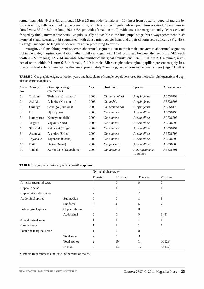
TABLE 2. Geographic origin, collection years and host plants of sample populations used for molecular phylogenetic and pop-ulation genetic analysis.
TABLE 3. Nymphal chaetotaxy of A. camelliae sp. nov.
Numbers in parentheses indicate the number of males.
Code No.
Acronym Geographic origin (prefecture)
Year Host plant Species Accession no.
1 Toshima Toshima (Kumamoto) 2008 Ci. natsudaidai A. spiniferus AB536792
2 Ashikita Ashikita (Kumamoto) 2008 Ci. unshiu A. spiniferus AB536793
3 Chikugo Chikugo (Fukuoka) 2009 Ci. natsudaidai A. spiniferus AB558172
4 Uji Uji (Kyoto) 2008 Ca. sinensis A. camelliae AB536794
5 Kameyama Kameyama (Mie) 2009 Ca. sinensis A. camelliae AB536795
6 Yagyuu Yagyuu (Nara) 2009 Ca. sinensis A. camelliae AB536796
7 Shigaraki Shigaraki (Shiga) 2009 Ca. sinensis A. camelliae AB536797
8 Asamiya Asamiya (Shiga) 2009 Ca. sinensis A. camelliae AB536798
9 Toyonaka Toyonaka (Osaka) 2009 Ca. sinensis A. camelliae AB536799
10 Daito Daito (Osaka) 2009 Ca. japonica A. camelliae AB536800
11 Tsubaki Kurinodake (Kagoshima) 2009 Ca. japonica Aleurotrachelus camelliae
AB536801
Nymphal chaetotaxy
1st instar 2nd instar 3rd instar 4th instar
Anterior marginal setae 4 0 0 0
Cephalic setae 0 1 1 1
Cephalo-thoratic spines 2 6 7 9
Abdominal spines Submedian 0 0 1 3
Subdorsal 0 4 6 7
Submarginal spines Cephalothorax 0 0 0 5
Abdominal 0 0 0 6 (5)
8th abdominal setae 1 1 1 1
Caudal setae 1 1 1 1
Posterior marginal setae 1 0 0 0
Total setae 7 3 3 3
Total spines 2 10 14 30 (29)
In total 9 13 17 33 (32)
KANMIYA ET AL.30 · Zootaxa 2797 © 2011 Magnolia Press
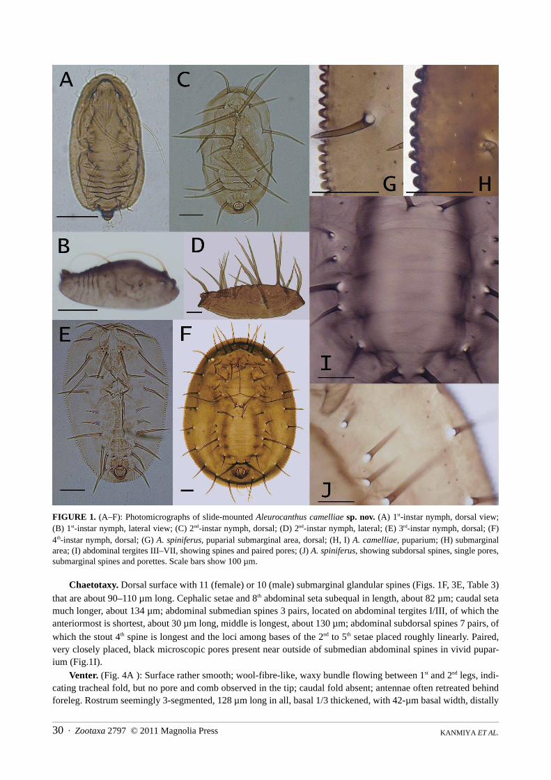
FIGURE 1. (A–F): Photomicrographs of slide-mounted Aleurocanthus camelliae sp. nov. (A) 1st-instar nymph, dorsal view;(B) 1st-instar nymph, lateral view; (C) 2nd-instar nymph, dorsal; (D) 2nd-instar nymph, lateral; (E) 3rd-instar nymph, dorsal; (F)4th-instar nymph, dorsal; (G) A. spiniferus, puparial submarginal area, dorsal; (H, I) A. camelliae, puparium; (H) submarginalarea; (I) abdominal tergites III–VII, showing spines and paired pores; (J) A. spiniferus, showing subdorsal spines, single pores,submarginal spines and porettes. Scale bars show 100 µm.
Chaetotaxy. Dorsal surface with 11 (female) or 10 (male) submarginal glandular spines (Figs. 1F, 3E, Table 3)that are about 90–110 µm long. Cephalic setae and 8th abdominal seta subequal in length, about 82 µm; caudal setamuch longer, about 134 µm; abdominal submedian spines 3 pairs, located on abdominal tergites I/III, of which theanteriormost is shortest, about 30 µm long, middle is longest, about 130 µm; abdominal subdorsal spines 7 pairs, ofwhich the stout 4th spine is longest and the loci among bases of the 2nd to 5th setae placed roughly linearly. Paired,very closely placed, black microscopic pores present near outside of submedian abdominal spines in vivid pupar-ium (Fig.1I).
Venter. (Fig. 4A ): Surface rather smooth; wool-fibre-like, waxy bundle flowing between 1st and 2nd legs, indi-cating tracheal fold, but no pore and comb observed in the tip; caudal fold absent; antennae often retreated behindforeleg. Rostrum seemingly 3-segmented, 128 µm long in all, basal 1/3 thickened, with 42-µm basal width, distally
Zootaxa 2797 © 2011 Magnolia Press · 31NEW STATUS FOR CITRUS SPINY WHITEFLY
narrowed, with a needlelike stylet bundle nearly 56 µm long. Pair of fine ventral abdominal setae 20–23 μm long;spinules scattered around the area of setae. Row of waxy projections produced along inner side of marginal teeth,which line up at slightly wider intervals than marginal teeth, comprising approximately 70% of total marginalteeth; each projection 20–30 µm long, mushroom-like, with basal stalk and flat top, which may serve as larvaladhesive to leaf surface.
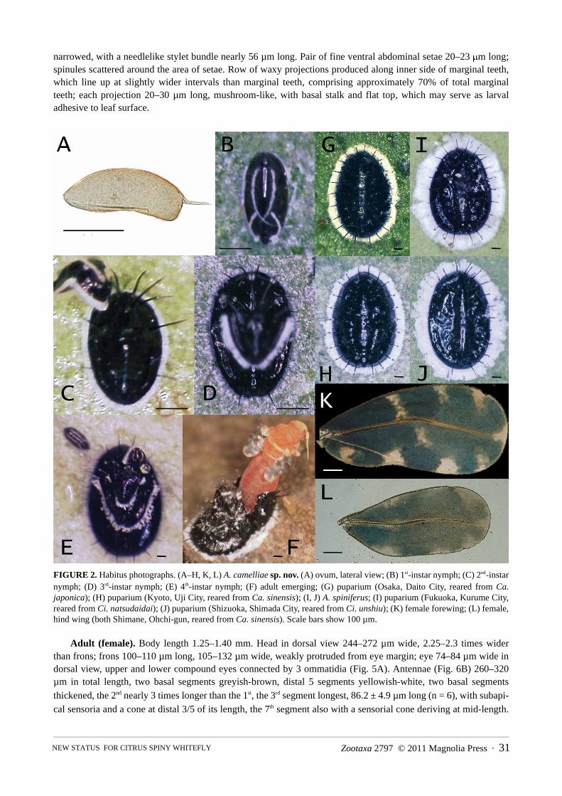
FIGURE 2. Habitus photographs. (A–H, K, L) A. camelliae sp. nov. (A) ovum, lateral view; (B) 1st-instar nymph; (C) 2nd-instarnymph; (D) 3rd-instar nymph; (E) 4th-instar nymph; (F) adult emerging; (G) puparium (Osaka, Daito City, reared from Ca.japonica); (H) puparium (Kyoto, Uji City, reared from Ca. sinensis); (I, J) A. spiniferus; (I) puparium (Fukuoka, Kurume City,reared from Ci. natsudaidai); (J) puparium (Shizuoka, Shimada City, reared from Ci. unshiu); (K) female forewing; (L) female,hind wing (both Shimane, Ohchi-gun, reared from Ca. sinensis). Scale bars show 100 µm.
Adult (female). Body length 1.25–1.40 mm. Head in dorsal view 244–272 µm wide, 2.25–2.3 times widerthan frons; frons 100–110 µm long, 105–132 µm wide, weakly protruded from eye margin; eye 74–84 µm wide indorsal view, upper and lower compound eyes connected by 3 ommatidia (Fig. 5A). Antennae (Fig. 6B) 260–320µm in total length, two basal segments greyish-brown, distal 5 segments yellowish-white, two basal segmentsthickened, the 2nd nearly 3 times longer than the 1st, the 3rd segment longest, 86.2 ± 4.9 µm long (n = 6), with subapi-cal sensoria and a cone at distal 3/5 of its length, the 7th segment also with a sensorial cone deriving at mid-length.

KANMIYA ET AL.32 · Zootaxa 2797 © 2011 Magnolia Press
Rostrum 136–155 µm total length, seemingly 3-segmented, basal segments 92 µm long, 44 µm wide, distal seg-ments 66 µm long, 24 µm wide, apically browned. Forewing (Figs. 2K, 3A, D): 1.1–1.2 mm long, 413–550 µm
wide at widest width (across Fig. 3A, ��� ) and 320–450 µm wide at middle of wing (across Fig. 3A, ��� ),with 9 greyish-white maculae (Figs. 2K, 3A), their maculation most distinct soon after emergence (Fig. 3D), thenturning largely brownish-green or brownish-blue, with maculae somewhat obscured by waxy powders. Hind wing(Figs. 2L, 3A) 0.95–1.02 mm long, 0.41–0.4 mm wide, evenly greyish-white, or with several blurred maculaedepending on age. Abdomen with two ventral wax plates.
Adult (male). Body length 0.90–1.10 mm. Wing maculation almost identical to that of female. In dorsal view,tergal sclerite I vestigial, II invisible, III/VIII and subgenital plate distinct (Fig. 6E), highly darkened on tergites VI/VII, subgenital plate and claspers; each III/V tergite subequal in length, about 44 µm; tergite VI longest, 64 µm
long; tergite VII reduced, 15 µm long, laterally extended and enclosing 7th spiracle; tergite VIII a small square, dis-tally leaning on subgenital plate. Forewing 0.84–0.9 mm long, about 0.37 mm wide. Vasiform orifice in dorsalview about 45–59 µm long, nearly 1.2–1.3 times longer than wide; operculum rounded with distal incision, 23–35µm long and wide; lingual 23–28 µm long, 8–11 µm wide. In lateral view, subgenital plate about 85–100 µm deepand 77–85 µm long, widely concave on anterior margin and gently depressed on ventral margin. Four distinct ven-tral abdominal wax plates (Fig. 6F).
Genitalia. (Figs. 4E, F, 6C) Aedeagus 108–120 µm long, gradually broadened basally to 23–27-µm basalwidth, upcurved toward apex and with slender distal half, apex extending near distal 3/4 length of clasper; clasperin dorsal view 108–123 µm long, weakly incurved and narrowed distally, angulate on outer subbasal corners, witha thin inflatable sac and apical spine.
Ovum. (Fig. 2A) Elliptical, the lower surface convex and the upper surface slightly concave, similar to a shortbanana shape; 219.7 ± 13.2 µm long (n = 11), 95.2 ± 16.5 µm wide (n = 11); stalk 49.4 ± 3.9 µm long (n = 9).
First-instar nymph. (Figs. 1A, B, 2B) (male and female): Elongate-oval, normally widest at posterior 3/5length; 297 ± 18.9 µm long (n = 15), 132.8 ± 17.3 µm wide (n = 10), 93.9 ± 9.9 µm high (n = 9); ratio of length/width 2.12 ± 0.12 (n = 5). Vertex conical, gradually widening posteriorly, suddenly recessed near laterobasal mar-gins of vasiform orifice; prominent protuberance developed at mesial cephalad region and around vasiform orifice;pair of elongate arcing spines behind cephalic protuberance and posterior thoracic margin, with anterior spine 196± 25 µm long (n = 15), posterior spine 114 ± 12 µm long (n = 11). Four pairs of fine anterior marginal setae and onepair of fine posterior marginal setae present.
Second-instar nymph. (Figs. 1C, D, 2C) More ovate, normally widest at anterior 1/3 length; 442.8 ± 71.2 µmlong (n = 10), 277.2 ± 53.5 µm wide (n = 8), about 141–144 µm high; ratio of length/width 1.62 ± 0.08 (n = 8); 6pairs of cephalothoracic and 4 pairs of abdominal subdorsal spines well developed, of which mesial 2 thoracicspines longest, 175–223 µm.
Third-instar nymph. (Figs. 1E, 2D) Elliptical, normally widest at anterior 1/3 length; 611.3 ± 71.6 µm long (n= 10), 403.5 ± 47.4 µm wide (n = 10), ratio of length/width 1.58 ± 0.08 (n = 10); 7 pairs of cephalothoracic and 7pairs (1 submedian and 6 subdorsal) of abdominal spines present.
Habitus. Puparium. Metallic black, medially and peripherally surrounded by white marginal waxy fringe(Fig. 2G, H), width of which (female) 90–160 µm (11–16% width of puparium), (male) 66–150 µm (6–12% widthof puparium). Tips of cephalothoracic and abdominal submarginal spines extending to outer edge of white marginalfringe or slightly protruding beyond it. Exuviae of earlier instars (usually 2nd and 3rd) often remain stacked up onmedian area of immature insect (Fig. 2E).
Adults. After emergence, eye, thorax and abdomen predominantly reddish-yellow (pinkish), except frons,antennae and legs light yellow, then turning orange to light brown to dark brown, covered by wax powder coatingexcept ruby eye. Wing also pale brown ground colour, with clear white original maculae (Fig. 3D), then totallyturning purple–brown to greenish-brown and the maculation somewhat obscured by white waxy powder; fore-wings bearing 9 white maculae as in Figures 2K and 3A; hind wing pale brown or greyish, without prominent mac-ulae. Ocellus light brown; rostrum darkened at apex. Body and wing surfaces appear white, owing to waxsecretions produced from abdominal waxy plates shortly after emergence by manipulating hind legs against glan-dular pores.
Ova and nymphs. Newly deposited eggs pale yellow, then turning brown to darker before hatching; newlyemerged nymphs transparent, appearing rather greenish by reflecting colour of leaves, then gradually darkening,finally becoming metallic black; 1st-instar nymph starts producing white waxy fringes marginally (Fig. 2B) soon
Zootaxa 2797 © 2011 Magnolia Press · 33NEW STATUS FOR CITRUS SPINY WHITEFLY
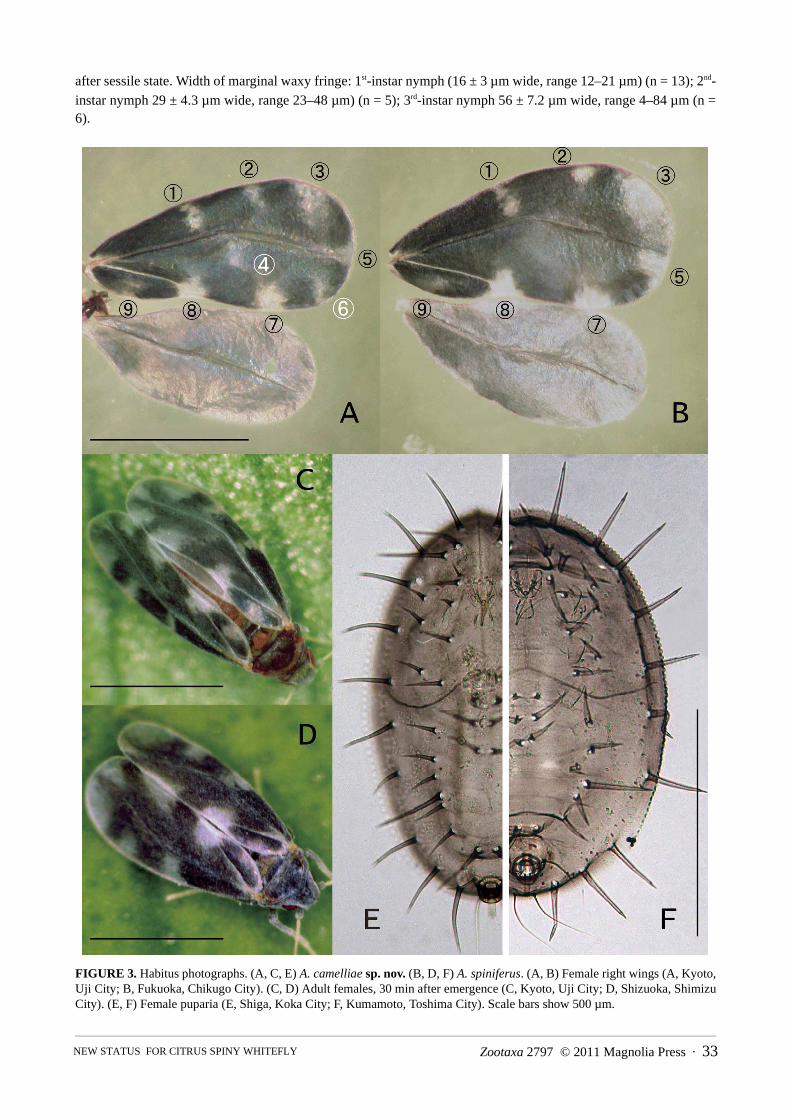
after sessile state. Width of marginal waxy fringe: 1st-instar nymph (16 ± 3 µm wide, range 12–21 µm) (n = 13); 2nd-instar nymph 29 ± 4.3 µm wide, range 23–48 µm) (n = 5); 3rd-instar nymph 56 ± 7.2 µm wide, range 4–84 µm (n =6).
FIGURE 3. Habitus photographs. (A, C, E) A. camelliae sp. nov. (B, D, F) A. spiniferus. (A, B) Female right wings (A, Kyoto,Uji City; B, Fukuoka, Chikugo City). (C, D) Adult females, 30 min after emergence (C, Kyoto, Uji City; D, Shizuoka, ShimizuCity). (E, F) Female puparia (E, Shiga, Koka City; F, Kumamoto, Toshima City). Scale bars show 500 µm.
KANMIYA ET AL.34 · Zootaxa 2797 © 2011 Magnolia Press
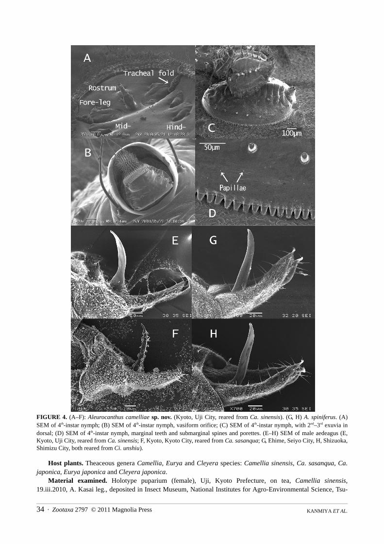
FIGURE 4. (A–F): Aleurocanthus camelliae sp. nov. (Kyoto, Uji City, reared from Ca. sinensis). (G, H) A. spiniferus. (A)SEM of 4th-instar nymph; (B) SEM of 4th-instar nymph, vasiform orifice; (C) SEM of 4th-instar nymph, with 2nd–3rd exuvia indorsal; (D) SEM of 4th-instar nymph, marginal teeth and submarginal spines and porettes. (E–H) SEM of male aedeagus (E,Kyoto, Uji City, reared from Ca. sinensis; F, Kyoto, Kyoto City, reared from Ca. sasanqua; G, Ehime, Seiyo City, H, Shizuoka,Shimizu City, both reared from Ci. unshiu).
Host plants. Theaceous genera Camellia, Eurya and Cleyera species: Camellia sinensis, Ca. sasanqua, Ca.japonica, Eurya japonica and Cleyera japonica.
Material examined. Holotype puparium (female), Uji, Kyoto Prefecture, on tea, Camellia sinensis,19.iii.2010, A. Kasai leg., deposited in Insect Museum, National Institutes for Agro-Environmental Science, Tsu-
Zootaxa 2797 © 2011 Magnolia Press · 35NEW STATUS FOR CITRUS SPINY WHITEFLY
kuba, Japan. Paratypes: 20 puparia, same data as holotype; 16 puparia on tea plants, same locality as holotype,18.v.2009, K. Kanmiya leg.; 20 puparia on tea plants, same locality as holotype, 10.i.2010, K. Yamashita leg.; 18puparia on tea plants, Sayama, Saitama Pref., on tea plants, 9.x.2009, Y.Sato leg.; 15 puparia on tea plants, Kami-ishizu-cho, Ohgaki, Gifu Pref., 13.x.2009, Y. Sato leg.; 18 puparia on tea plants, 3.iii.2009, Kameyama, Mie Pref.,K. Kanmiya leg.; 15 puparia on tea plants, Tanba, Kyoto Pref., 15.ix.2008. Y. Yoshiyasu leg.; 20 puparia on Ca.sasanqua, Nishigyo, Kyoto Pref., 4.iv.2010, Y. Yoshiyasu leg.; 20 puparia on tea plants, Asamiya, Shiga Pref.,4.iv.2009, K. Kanmiya leg.; 20 puparia on tea plants, Asamiya, Shiga Pref., 2.x.2010, A. Kasai leg.; 12 puparia ontea plants, Yagyu, Nara Pref., 4.iv.2009, K. Kanmiya leg.; 10 puparia on Ca. sasanqua, Tsukigase, Nara Pref.,4.iv.2009, K. Kanmiya leg.; 7 puparia on tea plants, Kamishinden, Toyonaka, Osaka Pref., 5.v.2009, K. Kanmiyaleg.; 20 puparia on tea plants, Ajimaoku, Sasayama, Hyogo Pref., 9.ix.2010, J. Yase leg.; 16 puparia on tea plants,Okayama, Okayama Pref., 7.vii.2010, Y. Sato leg.; 15 puparia on tea plants, 14.vii.2009, Ohchi-gun, ShimanePref., Y. Sato leg.; 15 puparia on tea plants, 26.xii.2009, Kitsuki, Oita Pref., Y. Sato leg.
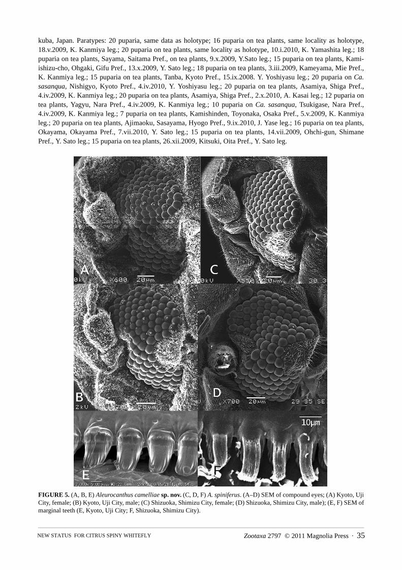
FIGURE 5. (A, B, E) Aleurocanthus camelliae sp. nov. (C, D, F) A. spiniferus. (A–D) SEM of compound eyes; (A) Kyoto, UjiCity, female; (B) Kyoto, Uji City, male; (C) Shizuoka, Shimizu City, female; (D) Shizuoka, Shimizu City, male); (E, F) SEM ofmarginal teeth (E, Kyoto, Uji City; F, Shizuoka, Shimizu City).
KANMIYA ET AL.36 · Zootaxa 2797 © 2011 Magnolia Press
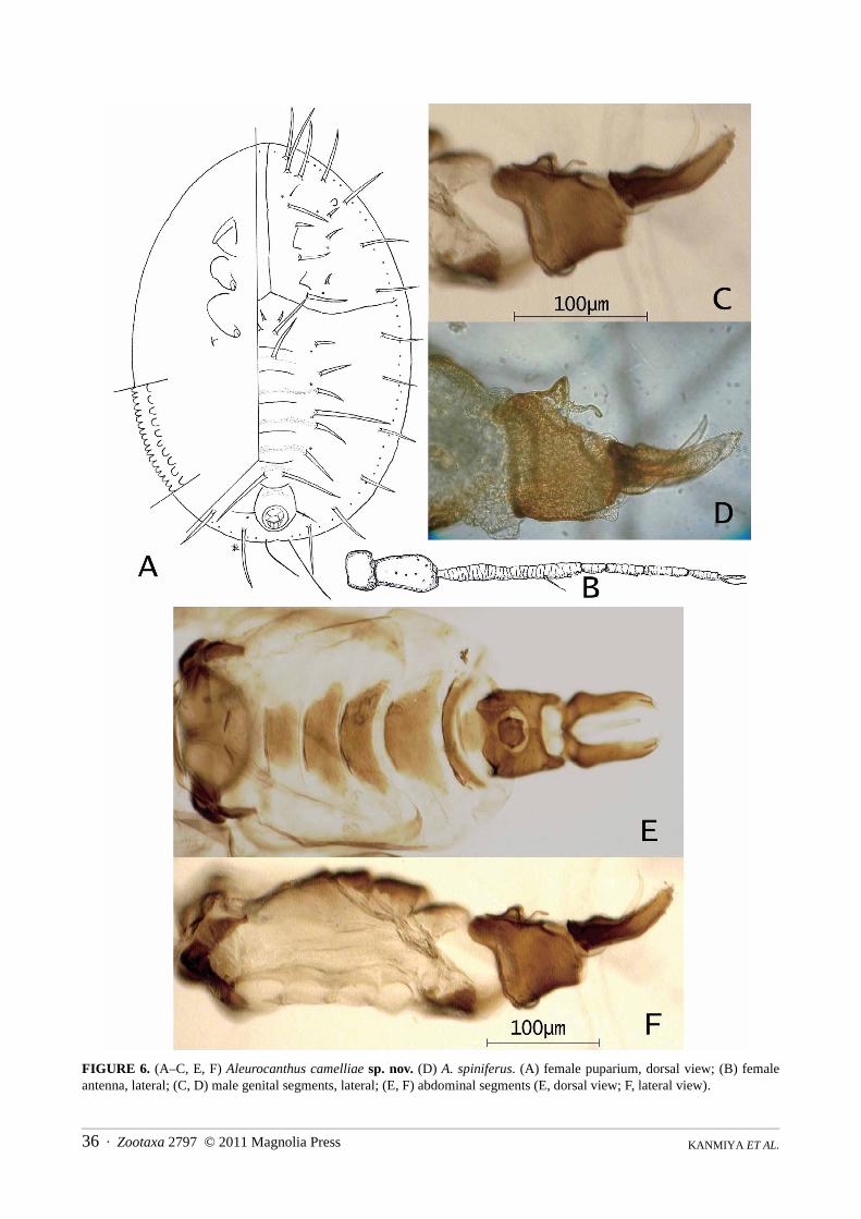
FIGURE 6. (A–C, E, F) Aleurocanthus camelliae sp. nov. (D) A. spiniferus. (A) female puparium, dorsal view; (B) femaleantenna, lateral; (C, D) male genital segments, lateral; (E, F) abdominal segments (E, dorsal view; F, lateral view).
Zootaxa 2797 © 2011 Magnolia Press · 37NEW STATUS FOR CITRUS SPINY WHITEFLY
Specimens depository. Some paratypes of A. camelliae sp. nov. will be deposited in the following institu-tions: The Natural History Museum, London; US National Museum of Natural History, Washington DC; NationalTaiwan University, Taipei; Institute of Zoology, Chinese Academy of Sciences, Beijing; Yokohama Plant Protec-tion Station, Kanagawa.
TABLE 4. Morphological differences in puparial and adult stages between A. camellinae sp. nov. and A. spiniferus.
Comments. Despite the almost identical features of the adult and nymphal stages of A. camellinae sp. nov. andA. spiniferus (Q.), we recognised very few, but clearly distinct, morphological differences in the puparial and adultstages, as listed in Table 4. This new species is rather similar to Aleurocanthus hibisci Corbett, 1935 distributed inMalaysia, Singapore and Reunion Islands, but its pronounced length of cephalothoracic spines and closelyarranged 9th and 10th submarginal spines will be well differentiated from the present new species. Aleurocanthusgordoniae Takahashi, 1942 known from Hong Kong is also peculiar in its theaceous host plant, Gordonia sp., but isdistinguished from the present new species in having vasiform orifice perfectly circular and puparium with reducedspine-chaetotaxy of 8 abdominal pairs (2 submedian + 6 subdorsal) , not 10 pairs.
Results
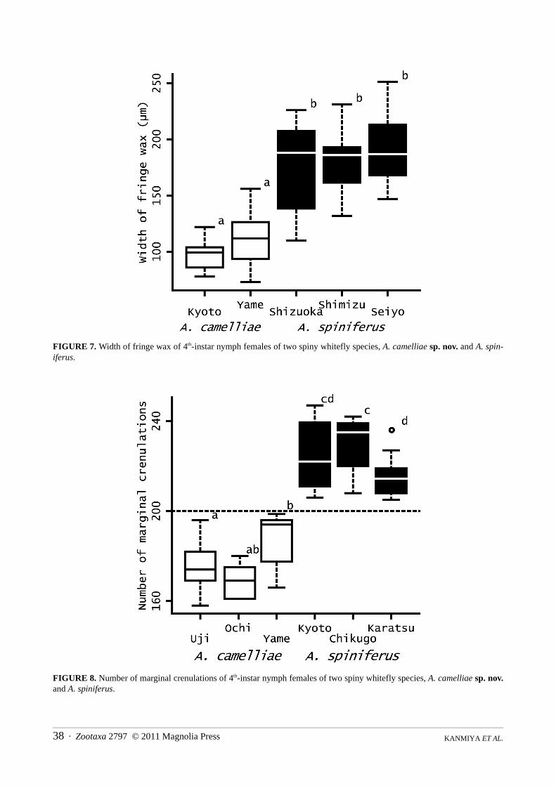
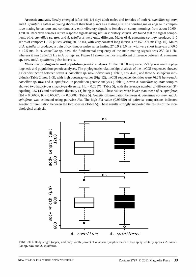
Morphometric analysis. In each case, the width of fringe wax of A. camelliae sp. nov. was significantly narrowerthan that of A. spiniferus (Holm’s sequentially rejective Bonferroni tests, after Wilcoxon’s rank-sum tests, p < 0.05;Fig. 7). Likewise, the number of marginal crenulations of A. camelliae sp. nov. was significantly lower in each casethan that of A. spiniferus (Holm’s sequentially rejective Bonferroni tests, after Wilcoxon’s rank-sum tests, p < 0.05;Fig. 8). However, body length (p = 0.0724) and body width (p = 0.382) did not significantly differ between the spe-cies (Wilcoxon’s rank-sum tests; Fig. 9).
Structure Character Discernible points
A. camelliae A. spiniferus
Food habits Food and oviposition preference
Theaceae, Illiciaceae, Cornaceae, Aquifoliaceae
Rutaceae, Annonaceae, Ebenaceae, Vitaceae, Rosaceae, Flacourtiaceae
Wing Forewing maculation 9 white maculae 7 white maculae
Male abdomen Aedeagus lateral view Distally upcurved on dorsal margin Distally straight on dorsal margin
Ditto Subgenital plate lateral view
Deeply incised on anterior and ventral margins
Weakly depressed on anterior margin, rather convex on ventral margin
Female 4th-instar nymph
Marginal wax secretion Weakly developed, width 11.2–15.8% of puparial width
Well developed, width 17.3–30.0% of puparial width
Ditto Marginal crenulation Teeth lined with loose gaps, less than 200 total (158–196)
Teeth lined with narrow gaps, more than 200 total (205–242)
Ditto Cephalic eyespot Clearly difined, very closely placed to 3rd cephalothoracic submarginal spine
Weakly defined, relatively placed close to 3rd submarginal spine than to the 2nd
Ditto Submedian abdominal spines
Sockets of 2nd to 5th spines lined up roughly linearly
Sockets not in line, 2nd & 4th placed distal and 3rd & 5th proximal
Ditto Microscopic papillae near submarginal spines
Lined outside submarginal spines Situated between submarginal spines
Ditto Abdominal tergite VIII 49.0 ± 10.1 µm long; length/length of vasiform orifice = 1.60 ± 0.26 µm
69.3 ± 8.1 µm long; length/length of vasiform orifice = 1.15 ± 0.09 µm
KANMIYA ET AL.38 · Zootaxa 2797 © 2011 Magnolia Press
FIGURE 7. Width of fringe wax of 4th-instar nymph females of two spiny whitefly species, A. camelliae sp. nov. and A. spin-iferus.
FIGURE 8. Number of marginal crenulations of 4th-instar nymph females of two spiny whitefly species, A. camelliae sp. nov.and A. spiniferus.
Zootaxa 2797 © 2011 Magnolia Press · 39NEW STATUS FOR CITRUS SPINY WHITEFLY
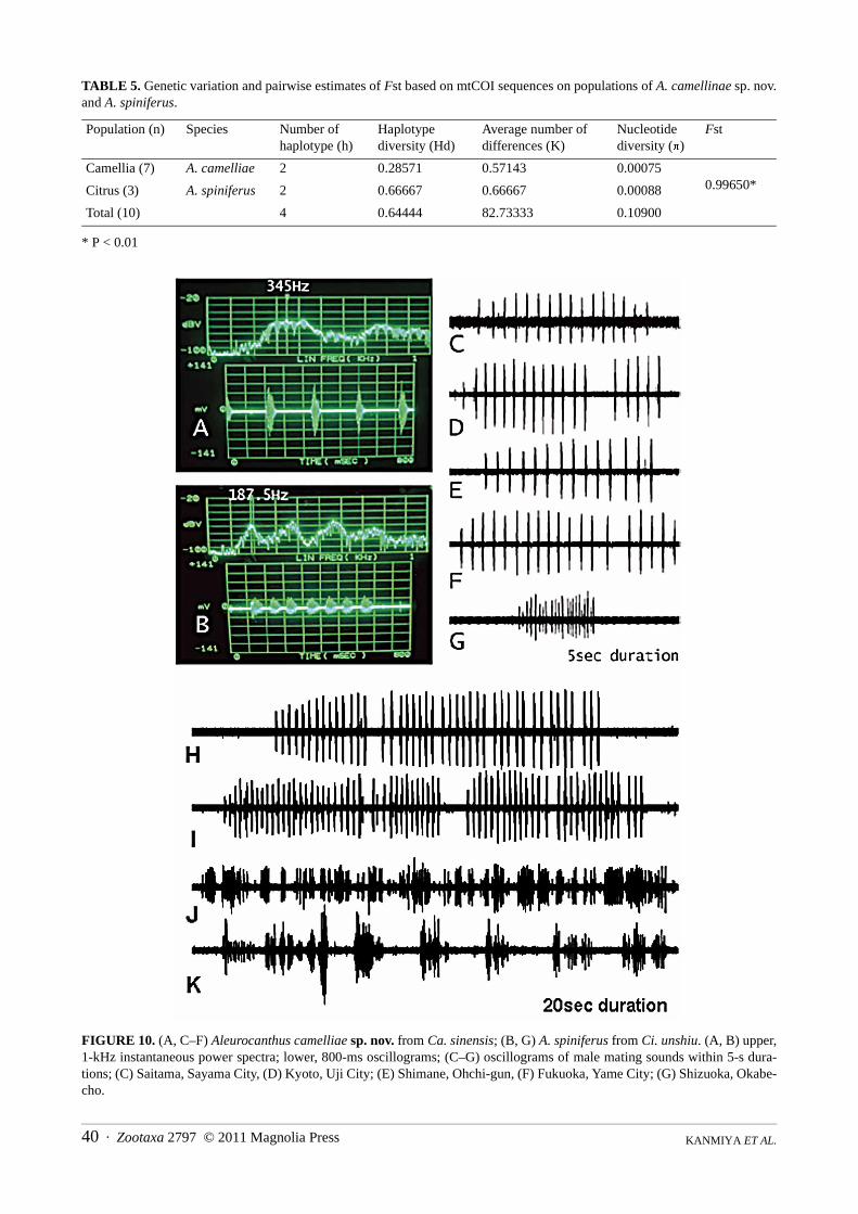
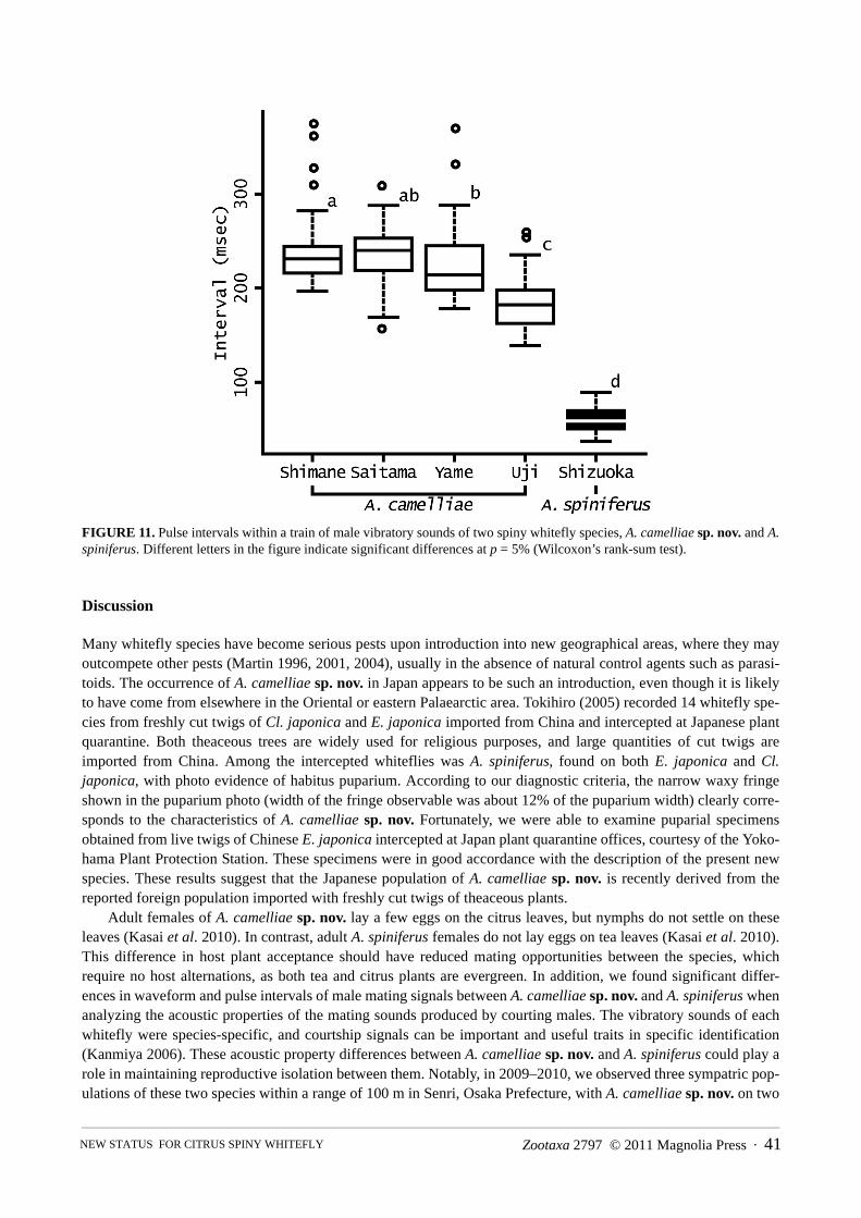
Acoustic analysis. Newly emerged (after 1/8–1/4 day) adult males and females of both A. camelliae sp. nov.and A. spiniferus gather on young shoots of their host plants as a mating site. The courting males engage in compet-itive mating behaviours and continuously emit vibratory signals to females on sunny mornings from about 10:00–12:00 h. Receptive females return response signals using similar vibratory sounds. We found that the signal compo-nents of A. camelliae sp. nov. and A. spiniferus were quite different. Males of A. camelliae sp. nov. produced 1–5series of compact 11–25 pulses lasting 30–52 ms, with very constant long intervals of 157–271 ms (Fig. 10). Malesof A. spiniferus produced a train of continuous pulse series lasting 27.6.9 ± 5.6 ms, with very short intervals of 60.5± 12.5 ms. In A. camelliae sp. nov., the fundamental frequency of the male mating signals was 250–311 Hz,whereas it was 190–205 Hz in A. spiniferus. Figure 11 shows the most significant difference between A. camelliaesp. nov. and A. spiniferus pulse intervals.
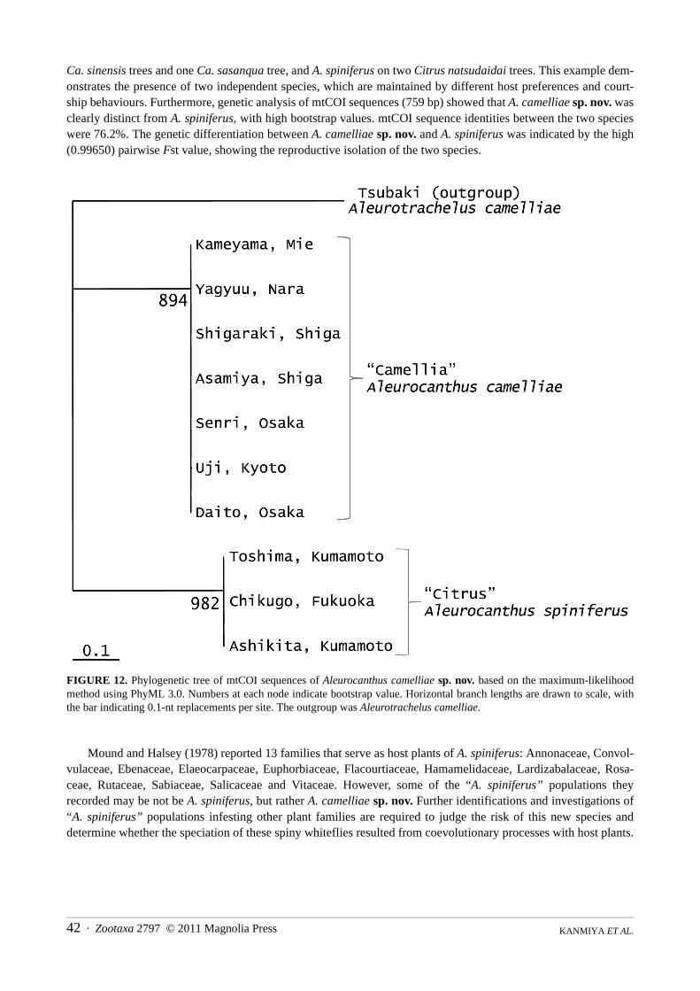
Molecular phylogenetic and population genetic analyses. Of the mtCOI sequence, 759 bp was used in phy-logenetic and population genetic analyses. The phylogenetic relationships analysis of the mtCOI sequences showeda clear distinction between seven A. camelliae sp. nov. individuals (Table 2, nos. 4–10) and three A. spiniferus indi-viduals (Table 2, nos. 1–3), with high bootstrap values (Fig. 12). mtCOI sequence identities were 76.2% between A.camelliae sp. nov. and A. spiniferus. In population genetic analysis (Table 2), seven A. camelliae sp. nov. samplesshowed two haplotypes (haplotype diversity: Hd = 0.28571; Table 5), with the average number of differences (K)equaling 0.57143 and nucleotide diversity (π) being 0.00075. These values were lower than those of A. spiniferus(Hd = 0.66667, K = 0.66667, π = 0.00088; Table 5). Genetic differentiation between A. camelliae sp. nov. and A.spiniferus was estimated using pairwise Fst. The high Fst value (0.99650) of pairwise comparisons indicatedgenetic differentiation between the two species (Table 5). These results strongly supported the results of the mor-phological analysis.
FIGURE 9. Body length (upper) and body width (lower) of 4th-instar nymph females of two spiny whitefly species, A. camel-liae sp. nov. and A. spiniferus.
KANMIYA ET AL.40 · Zootaxa 2797 © 2011 Magnolia Press
TABLE 5. Genetic variation and pairwise estimates of Fst based on mtCOI sequences on populations of A. camellinae sp. nov.and A. spiniferus.
* P < 0.01
FIGURE 10. (A, C–F) Aleurocanthus camelliae sp. nov. from Ca. sinensis; (B, G) A. spiniferus from Ci. unshiu. (A, B) upper,1-kHz instantaneous power spectra; lower, 800-ms oscillograms; (C–G) oscillograms of male mating sounds within 5-s dura-tions; (C) Saitama, Sayama City, (D) Kyoto, Uji City; (E) Shimane, Ohchi-gun, (F) Fukuoka, Yame City; (G) Shizuoka, Okabe-cho.
Population (n) Species Number of haplotype (h)
Haplotype diversity (Hd)
Average number of differences (K)
Nucleotide diversity (π)
Fst
Camellia (7) A. camelliae 2 0.28571 0.57143 0.000750.99650*Citrus (3) A. spiniferus 2 0.66667 0.66667 0.00088
Total (10) 4 0.64444 82.73333 0.10900
Zootaxa 2797 © 2011 Magnolia Press · 41NEW STATUS FOR CITRUS SPINY WHITEFLY
FIGURE 11. Pulse intervals within a train of male vibratory sounds of two spiny whitefly species, A. camelliae sp. nov. and A.spiniferus. Different letters in the figure indicate significant differences at p = 5% (Wilcoxon’s rank-sum test).
Discussion
Many whitefly species have become serious pests upon introduction into new geographical areas, where they mayoutcompete other pests (Martin 1996, 2001, 2004), usually in the absence of natural control agents such as parasi-toids. The occurrence of A. camelliae sp. nov. in Japan appears to be such an introduction, even though it is likelyto have come from elsewhere in the Oriental or eastern Palaearctic area. Tokihiro (2005) recorded 14 whitefly spe-cies from freshly cut twigs of Cl. japonica and E. japonica imported from China and intercepted at Japanese plantquarantine. Both theaceous trees are widely used for religious purposes, and large quantities of cut twigs areimported from China. Among the intercepted whiteflies was A. spiniferus, found on both E. japonica and Cl.japonica, with photo evidence of habitus puparium. According to our diagnostic criteria, the narrow waxy fringeshown in the puparium photo (width of the fringe observable was about 12% of the puparium width) clearly corre-sponds to the characteristics of A. camelliae sp. nov. Fortunately, we were able to examine puparial specimensobtained from live twigs of Chinese E. japonica intercepted at Japan plant quarantine offices, courtesy of the Yoko-hama Plant Protection Station. These specimens were in good accordance with the description of the present newspecies. These results suggest that the Japanese population of A. camelliae sp. nov. is recently derived from thereported foreign population imported with freshly cut twigs of theaceous plants.
Adult females of A. camelliae sp. nov. lay a few eggs on the citrus leaves, but nymphs do not settle on theseleaves (Kasai et al. 2010). In contrast, adult A. spiniferus females do not lay eggs on tea leaves (Kasai et al. 2010).This difference in host plant acceptance should have reduced mating opportunities between the species, whichrequire no host alternations, as both tea and citrus plants are evergreen. In addition, we found significant differ-ences in waveform and pulse intervals of male mating signals between A. camelliae sp. nov. and A. spiniferus whenanalyzing the acoustic properties of the mating sounds produced by courting males. The vibratory sounds of eachwhitefly were species-specific, and courtship signals can be important and useful traits in specific identification(Kanmiya 2006). These acoustic property differences between A. camelliae sp. nov. and A. spiniferus could play arole in maintaining reproductive isolation between them. Notably, in 2009–2010, we observed three sympatric pop-ulations of these two species within a range of 100 m in Senri, Osaka Prefecture, with A. camelliae sp. nov. on two
KANMIYA ET AL.42 · Zootaxa 2797 © 2011 Magnolia Press
Ca. sinensis trees and one Ca. sasanqua tree, and A. spiniferus on two Citrus natsudaidai trees. This example dem-onstrates the presence of two independent species, which are maintained by different host preferences and court-ship behaviours. Furthermore, genetic analysis of mtCOI sequences (759 bp) showed that A. camelliae sp. nov. wasclearly distinct from A. spiniferus, with high bootstrap values. mtCOI sequence identities between the two specieswere 76.2%. The genetic differentiation between A. camelliae sp. nov. and A. spiniferus was indicated by the high(0.99650) pairwise Fst value, showing the reproductive isolation of the two species.
FIGURE 12. Phylogenetic tree of mtCOI sequences of Aleurocanthus camelliae sp. nov. based on the maximum-likelihoodmethod using PhyML 3.0. Numbers at each node indicate bootstrap value. Horizontal branch lengths are drawn to scale, withthe bar indicating 0.1-nt replacements per site. The outgroup was Aleurotrachelus camelliae.
Mound and Halsey (1978) reported 13 families that serve as host plants of A. spiniferus: Annonaceae, Convol-vulaceae, Ebenaceae, Elaeocarpaceae, Euphorbiaceae, Flacourtiaceae, Hamamelidaceae, Lardizabalaceae, Rosa-ceae, Rutaceae, Sabiaceae, Salicaceae and Vitaceae. However, some of the “A. spiniferus” populations theyrecorded may be not be A. spiniferus, but rather A. camelliae sp. nov. Further identifications and investigations of“A. spiniferus” populations infesting other plant families are required to judge the risk of this new species anddetermine whether the speciation of these spiny whiteflies resulted from coevolutionary processes with host plants.
Zootaxa 2797 © 2011 Magnolia Press · 43NEW STATUS FOR CITRUS SPINY WHITEFLY
Acknowledgements
The authors would especially like to thank Jon H. Martin of the Natural History Museum, London, UK for a valu-able advice on the morphological evaluation and for improvement of the manuscript. Sincere thanks are also due Y.Asami, Dean of the Institute of Comparative Studies of International Cultures and Societies, K. Kano, Head of theConservation Science Group of the Institute, and R. Ogata, Executive Director of Mii Campus, Kurume University,for permission to use facilities. Our sincere appreciation is also extended to M. Satoh of Yokohama Plant ProtectionStation for his kindness in providing samples and to Y. Kinoshita and K. Matsuda of Keyence Corp., Osaka. fortheir technical assistance. This study was supported in part by a grant for research and development projects forapplication in promoting new Agriculture, Forestry and Fisheries policies (no. 21002, MAFF).
References
Bink-Moenen, R.M. (1983) Revision of the African whiteflies (Aleyrodidae). Monographieen van de Nederlandse Entomolo-gische Vereniging, 10, 1–210.Byrne, D.N., Bellows, T.S., Jr. & Parrella, M.P. (1990) Whiteflies in agricultural systems. In:Gerling, D. (Ed.) Whiteflies: their Bionomics, Pest Status and Management. Intercept, Andover, Hants, UK, pp. 227–261.
Byrne, D.N., Bellows, Jr. T.S. & Parella, M.P. (1990) Whiteflies in Agricultural systems. In: Gerling, D. (Ed.) Whiteflies: TheirBionomics, Pest Status and Management. Intercept, Andover, Hants, pp. 227–262.
Clausen, C.P. (1978) Biological control of Citrus insects, In: Reuther, W., Calavan, E.C. & Carman, G.E. (Eds.), Citrus Indus-try, Vol. IV, Division of Agriculture and Natural Resources, University of California. Insects injurious to agriculture inJapan. Circ. No. 168, USDA. pp. 276–303.
Corbett, G.H. (1935) Malayan Aleurodidae. Journal of the Federated Malay States Museums, 17, 722–852.David, V. & Subramaniam, T.R. (1976) Study on some Indian Aleyrodidae. Records of Zoological Survey of India, 70, 147–
157.Dubey, A.K. & Sundararaj, R. (2005) Whitefly species of the genus Aleurocanthus Quaintance & Baker (Hemiptera: Aleyrod-
didae) from India, with descriptions of six new species. Oriental Insects, 39, 295–321.Evans, G.A. (2008) The whiteflies (Hemiptera: Aleyrodidae) of the world and their host plants and natural enemies. USDA/
Animal Plant Health Inspection Service (APHIS), 715 pp.Frohlich, D.R., Torres-Jerez, I., Bedford, I.D., Markham, P.G. & Brown, J.K. (1999) A phylogeographical analysis of the Bemi-
sia tabaci species complex based on mitochondrial DNA markers. Molecular Ecology, 8, 1683–1691.Gill, R.J. (1990) The morphology of whiteflies. In: Gerling, D. (Ed.) Whiteflies: their Bionomics, Pest Status and Management.
Intercept, Andover, Hants, UK, pp. 13–46.Guindon, S. & Gascuel, O. (2003) A simple, fast, and accurate algorithm to estimate large phylogenies by maximum likeli-
hood. Systematic Biology, 52, 696–704.Gyeltshen, J., Hodges, A. & Hodges, G.S. (2010) Orange Spiny Whitefly, Aleurocanthus spiniferus (Quaintance) (Insecta:
Hemiptera: Aleyrodidae). EENY 341, 1–4 (University of Florida IFAS Extension). Available from http://edis.ifas.ufl.edu/pdffiles/IN/IN61800.pdf
Han, B.Y. & Cui, L. (2003) Natural population life table of citrus spiny whitefly (Aleurocanthus spiniferus) in tea garden. ActaEcologica Sinica, 23, 1781–1790 (in Chinese with English summary).
Holm, S. (1979) A simple sequentially rejective multiple test procedure. Scandinavian Journal of Statistics, 6, 65–70.Japanese Society of Applied Entomology and Zoology (Ed.) (2006) Major Insect and other Pests of Economic Plants in Japan.
Revised edition. Japanese Society of Applied Entomology and Zoology. Tokyo. 387 pp.Kanmiya, K. (1996) Discovery of male vibratory signal in the greenhouse whitefly, Trialeurodes vaporariorum (Westwood)
(Homoptera: Aleyrodidae). Applied Entomology and Zoology, 31, 255–262.Kanmiya, K. (2006) Mating Behaviour and Vibratory Signals in Whiteflies (Hemiptera: Aleyrodidae). In: Drosopoulous, S. &
Claridge, M.F. (Eds.), Insect Sounds and Communication: Physiology, Behaviour, Ecology and Evolution, CRC Taylor &Francis, Boca Raton, London, New York, pp. 365–379.
Kanmiya, K., Yoshiyasu, Y. & Sato, Y. (2009) Difference of acoustic properties in mating sounds between the tea-infestingpopulation and the citrus-infesting one of the Citrus spiny whitefly, Aleurocanthus spiniferus. Abstract volume, 54th
Annual meeting of Japanese Society of Applied Entomology and Zoology, p. 145.Kasai, A., Yamashita, K. & Yoshiyasu, Y. (2010) Tea-infesting population of the citrus spiny whitefly, Aleurocanthus spiniferus
(Homoptera: Aleyrodidae), does not accept citrus leaves as host plants. Japanese Journal of Applied Entomology andZoology, 54, 140–143 (in Japanese with English summary).
Kuwana, I. & Ishii, T. (1927) On Prospaltella smithi Silv., and Cryptognatha sp., the enemies of Aleurocanthus spiniferusQuaintance, imported from Canton, China. Journal of Okitsu Horticulture Society. 22, 77–80 (in Japanese).
Librado, P. & Rozas, J. (2009) DnaSP v5: A software for comprehensive analysis of DNA polymorphism data. Bioinformatics,25, 1451–1452.
Martin, J.H. (1987) An identification guide to common whitefly pest species of the world (Homoptera, Aleyrodidae). Tropical
KANMIYA ET AL.44 · Zootaxa 2797 © 2011 Magnolia Press
Pest Management, 33, 298–322.Martin, J.H. (1996) Neotropical whiteflies of the subfamily Aleurodicinae established in the western Palaearctic
(Homoptera:Aleyrodidae). Journal of Natural History, 30, 1849–1859.Martin, J.H. (1999) The whitefly fauna of Australia (Sternorrhyncha: Aleyrodidae), a taxonomic account and identification
guide. Technical Paper, CSIRO Entomology, 38, 1–197.Martin, J.H. (2001) Description of an invasive new species of Neotropical aleurodicine whitefly (Hemiptera:Aleyrodidae)−a
case of complete or partial misidentification? Bulletin of Entomological Research, 91,101–107.Martin, J.H. (2004) Whiteflies of Belize (Hemiptera:Aleyrodidae). Part 1 −introduction and account of the subfamily Aley-
rodicinae Quaintance & Baker. Zootaxa, 681, 1–119.Martin, J.H. (2005) Whiteflies of Belize (Hemiptera: Aleyrodidae) Part 2 –a review of the subfamily Aleyrodinae Westwood.
Zootaxa, 1098, 1–116.Martin, J.H. & Mound, L.A. (2007) An annotated check list of the world’s whiteflies (Insecta: Hemiptera: Aleyrodidae) .Zoo-
taxa, 1492, 1–84. Miyatake, Y. (1980) A list of the whiteflies of Japan with their host plant and distribution data (Homoptera:Aleyrodidae). Ros-
tria, 32, 291–330 (in Japanese with English summary).Mound, L.A. & Halsey, S.H. (1978) Whitefly of the World: A Systematic Catalogue of the Aleyrodidae (Homoptera) with Host
Plant and Natural Enemy Data, British Museum (Natural History), London, UK, John Wiley & Sons, Chichester, 340 pp.Nguyen, R., Sailer, R.I. & Hamon, A.B. (1993) Catalog of Aleyrodidae on citrus and their natural enemies (Homoptera–Aley-
rodidae). Florida Department of Agriculture and Consumer Services, Bureau of Entomology. 8, 1–57, Gainesville, FL.Ohgushi, R. (1969) Ecology of Citrus Pests. Rural Culture Association, Tokyo, 244 pp (in Japanese).Quaintance, A.L. (1903) New Oriental Aleurodidae. Canadian Entomologist, 31, 61–64.Quaintance, A.L. & Baker, A.C. (1913) Classification of the Aleyrodidae, Part I. Bureau of Entomology, U. S. Dept. of Agricul-
ture. Technical Series, 27, 1–93.Quaintance, A.L. & Baker, A.C. (1914) Classification of the Aleyrodidae, Part II. Bureau of Entomology, U. S. Dept. of Agri-
culture. Technical Series, 27, 95–109.Quaintance, A.L. & Baker, A.C. (1917) A contribution to our knowledge of the whiteflies of the subfamily Aleurodinae (Aley-
rodidae). Proceedings of the United States National Museum, 51, 335–445.Suh, N.H. (1994) Illustrated Book of Tea pests and Diseases. Wenshan Substation, Tea Research and Extention Station, Taipei,
pp. 28–29(part) (in Chinese).Takahashi, R. (1941) Some foreign Aleyrodidae (Hemiptera), 4 species from Hongkong. Transactions of Natural History Soci-
ety Formosa, 31, 388–393.Takahashi, R. (1942) Aleurocanthus of Thailand and French Indo-China (Aleyrodidae, Homoptera). Kontyu, 56, 57–61.Takahashi, R. & Mamet,R. (1956) Insects of Micronesia: Homoptera:Aleyrodidae. Insects of Micronesia, 6, 1–13.Tamura, K., Dudley, J., Nei, M. & Kumar, S. (2007) MEGA4: Molecular Evolutionary Genetics Analysis (MEGA) software
version 4.0. Molecular Biology and Evolution, 24, 1596–1599.Tokihiro, G. (2005) Whiteflies (Homoptera: Aleyrodidae) intercepted at Japanese Plant Quarantine from fresh-cut twigs of
Cleyera japonica and Eurya japonica (Theaceae) imported from China. Research Bulletin of the Plant Protection ServiceJapan, 41, 87–93.
Ueda, S. & Brown, J.K. (2006) First report of the Q biotype of Bemisia tabaci in Japan by mitochondrial cytochrome oxidase Isequence analysis. Phytoparasitica, 34, 405–411.
Yamashita, K., Hayashida, Y., Haigata, M. & Tani, M. (2005) Occurrence of Citrus spiny whitefly, Aleurocanthus spiniferus(Quaintance), confirmed in tea plants for the first time from Japan. Tea Research Journal, 100, 86–87 (in Japanese).


![[Frontiers in Bioscience 13, 2797-2805, January 1, 2008] HIV ......[Frontiers in Bioscience 13, 2797-2805, January 1, 2008] 2797 HIV-1 transgenic expression in mice induces selective](https://img.pdfslide.us/doc/110x75/61406f551664f1518558c0ca/frontiers-in-bioscience-13-2797-2805-january-1-2008-hiv-frontiers.jpg)