Embed Size (px)
Citation preview
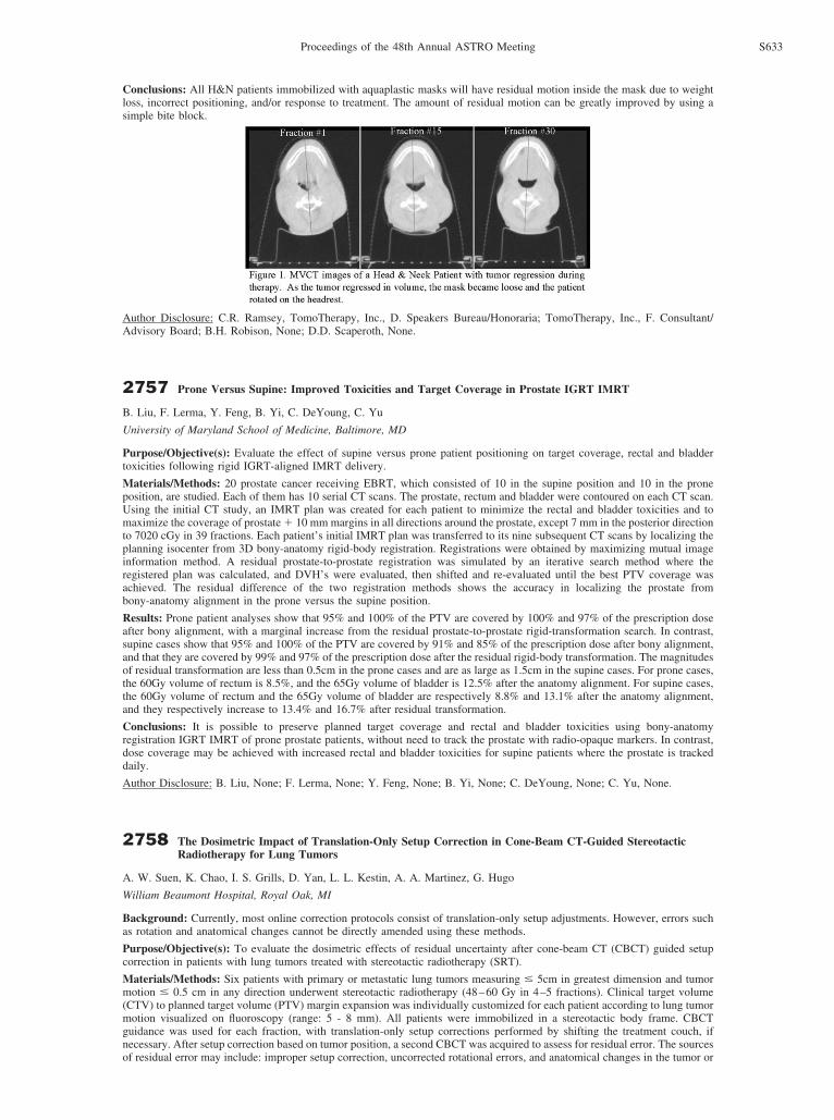
Conclusions: All H&N patients immobilized with aquaplastic masks will have residual motion inside the mask due to weightloss, incorrect positioning, and/or response to treatment. The amount of residual motion can be greatly improved by using asimple bite block.
Author Disclosure: C.R. Ramsey, TomoTherapy, Inc., D. Speakers Bureau/Honoraria; TomoTherapy, Inc., F. Consultant/Advisory Board; B.H. Robison, None; D.D. Scaperoth, None.
2757 Prone Versus Supine: Improved Toxicities and Target Coverage in Prostate IGRT IMRT
B. Liu, F. Lerma, Y. Feng, B. Yi, C. DeYoung, C. Yu
University of Maryland School of Medicine, Baltimore, MD
Purpose/Objective(s): Evaluate the effect of supine versus prone patient positioning on target coverage, rectal and bladdertoxicities following rigid IGRT-aligned IMRT delivery.
Materials/Methods: 20 prostate cancer receiving EBRT, which consisted of 10 in the supine position and 10 in the proneposition, are studied. Each of them has 10 serial CT scans. The prostate, rectum and bladder were contoured on each CT scan.Using the initial CT study, an IMRT plan was created for each patient to minimize the rectal and bladder toxicities and tomaximize the coverage of prostate � 10 mm margins in all directions around the prostate, except 7 mm in the posterior directionto 7020 cGy in 39 fractions. Each patient’s initial IMRT plan was transferred to its nine subsequent CT scans by localizing theplanning isocenter from 3D bony-anatomy rigid-body registration. Registrations were obtained by maximizing mutual imageinformation method. A residual prostate-to-prostate registration was simulated by an iterative search method where theregistered plan was calculated, and DVH’s were evaluated, then shifted and re-evaluated until the best PTV coverage wasachieved. The residual difference of the two registration methods shows the accuracy in localizing the prostate frombony-anatomy alignment in the prone versus the supine position.
Results: Prone patient analyses show that 95% and 100% of the PTV are covered by 100% and 97% of the prescription doseafter bony alignment, with a marginal increase from the residual prostate-to-prostate rigid-transformation search. In contrast,supine cases show that 95% and 100% of the PTV are covered by 91% and 85% of the prescription dose after bony alignment,and that they are covered by 99% and 97% of the prescription dose after the residual rigid-body transformation. The magnitudesof residual transformation are less than 0.5cm in the prone cases and are as large as 1.5cm in the supine cases. For prone cases,the 60Gy volume of rectum is 8.5%, and the 65Gy volume of bladder is 12.5% after the anatomy alignment. For supine cases,the 60Gy volume of rectum and the 65Gy volume of bladder are respectively 8.8% and 13.1% after the anatomy alignment,and they respectively increase to 13.4% and 16.7% after residual transformation.
Conclusions: It is possible to preserve planned target coverage and rectal and bladder toxicities using bony-anatomyregistration IGRT IMRT of prone prostate patients, without need to track the prostate with radio-opaque markers. In contrast,dose coverage may be achieved with increased rectal and bladder toxicities for supine patients where the prostate is trackeddaily.
Author Disclosure: B. Liu, None; F. Lerma, None; Y. Feng, None; B. Yi, None; C. DeYoung, None; C. Yu, None.
2758 The Dosimetric Impact of Translation-Only Setup Correction in Cone-Beam CT-Guided StereotacticRadiotherapy for Lung Tumors
A. W. Suen, K. Chao, I. S. Grills, D. Yan, L. L. Kestin, A. A. Martinez, G. Hugo
William Beaumont Hospital, Royal Oak, MI
Background: Currently, most online correction protocols consist of translation-only setup adjustments. However, errors suchas rotation and anatomical changes cannot be directly amended using these methods.
Purpose/Objective(s): To evaluate the dosimetric effects of residual uncertainty after cone-beam CT (CBCT) guided setupcorrection in patients with lung tumors treated with stereotactic radiotherapy (SRT).
Materials/Methods: Six patients with primary or metastatic lung tumors measuring � 5cm in greatest dimension and tumormotion � 0.5 cm in any direction underwent stereotactic radiotherapy (48–60 Gy in 4–5 fractions). Clinical target volume(CTV) to planned target volume (PTV) margin expansion was individually customized for each patient according to lung tumormotion visualized on fluoroscopy (range: 5 - 8 mm). All patients were immobilized in a stereotactic body frame. CBCTguidance was used for each fraction, with translation-only setup corrections performed by shifting the treatment couch, ifnecessary. After setup correction based on tumor position, a second CBCT was acquired to assess for residual error. The sourcesof residual error may include: improper setup correction, uncorrected rotational errors, and anatomical changes in the tumor or
S633Proceedings of the 48th Annual ASTRO Meeting
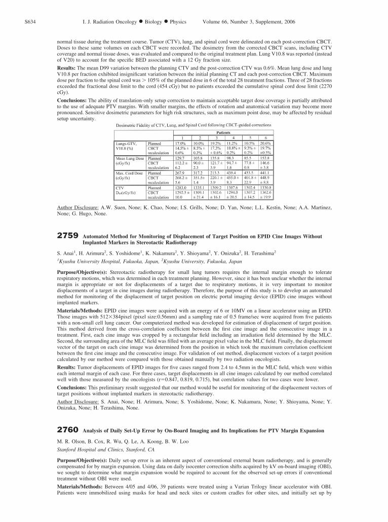
normal tissue during the treatment course. Tumor (CTV), lung, and spinal cord were delineated on each post-correction CBCT.Doses to these same volumes on each CBCT were recorded. The dosimetry from the corrected CBCT scans, including CTVcoverage and normal tissue doses, was evaluated and compared to the original treatment plan. Lung V10.8 was reported (insteadof V20) to account for the specific BED associated with a 12 Gy fraction size.
Results: The mean D99 variation between the planning CTV and the post-correction CTV was 0.6%. Mean lung dose and lungV10.8 per fraction exhibited insignificant variation between the initial planning CT and each post-correction CBCT. Maximumdose per fraction to the spinal cord was � 105% of the planned dose in 6 of the total 28 treatment fractions. Three of 28 fractionsexceeded the fractional dose limit to the cord (454 cGy) but no patients exceeded the cumulative spinal cord dose limit (2270cGy).
Conclusions: The ability of translation-only setup correction to maintain acceptable target dose coverage is partially attributedto the use of adequate PTV margins. With smaller margins, the effects of rotation and anatomical variation may become morepronounced. Sensitive dosimetric parameters for high risk structures, such as maximum point dose, may be affected by residualsetup uncertainty.
Author Disclosure: A.W. Suen, None; K. Chao, None; I.S. Grills, None; D. Yan, None; L.L. Kestin, None; A.A. Martinez,None; G. Hugo, None.
2759 Automated Method for Monitoring of Displacement of Target Position on EPID Cine Images WithoutImplanted Markers in Stereotactic Radiotherapy
S. Anai1, H. Arimura2, S. Yoshidome1, K. Nakamura2, Y. Shioyama2, Y. Onizuka2, H. Terashima2
1Kyushu University Hospital, Fukuoka, Japan, 2Kyushu University, Fukuoka, Japan
Purpose/Objective(s): Stereotactic radiotherapy for small lung tumors requires the internal margin enough to toleraterespiratory motions, which was determined in each treatment planning. However, since it has been unclear whether the internalmargin is appropriate or not for displacements of a target due to respiratory motions, it is very important to monitordisplacements of a target in cine images during radiotherapy. Therefore, the purpose of this study is to develop an automatedmethod for monitoring of the displacement of target position on electric portal imaging device (EPID) cine images withoutimplanted markers.
Materials/Methods: EPID cine images were acquired with an energy of 6 or 10MV on a linear accelerator using an EPID.Those images with 512�384pixel (pixel size:0.56mm) and a sampling rate of 0.5 frame/sec were acquired from five patientswith a non-small cell lung cancer. Our computerized method was developed for estimation of displacement of target position.This method derived from the cross-correlation coefficient between the first cine image and the consecutive image in atreatment. First, each cine image was cropped by a rectangular field including an irradiation field determined by the MLC.Second, the surrounding area of the MLC field was filled with an average pixel value in the MLC field. Finally, the displacementvector of the target on each cine image was determined from the position in which took the maximum correlation coefficientbetween the first cine image and the consecutive image. For validation of out method, displacement vectors of a target positioncalculated by our method were compared with those obtained manually by two radiation oncologists.
Results: Tumor displacements of EPID images for five cases ranged from 2.4 to 4.5mm in the MLC field, which were withineach internal margin of each case. For three cases, target displacements in all cine images calculated by our method correlatedwell with those measured by the oncologists (r�0.847, 0.819, 0.715), but correlation values for two cases were lower.
Conclusions: This preliminary result suggested that our method would be useful for monitoring of the displacement vectors oftarget positions without implanted markers in stereotactic radiotherapy.
Author Disclosure: S. Anai, None; H. Arimura, None; S. Yoshidome, None; K. Nakamura, None; Y. Shioyama, None; Y.Onizuka, None; H. Terashima, None.
2760 Analysis of Daily Set-Up Error by On-Board Imaging and Its Implications for PTV Margin Expansion
M. R. Olson, B. Cox, R. Wu, Q. Le, A. Koong, B. W. Loo
Stanford Hospital and Clinics, Stanford, CA
Purpose/Objective(s): Daily set-up error is an inherent aspect of conventional external beam radiotherapy, and is generallycompensated for by margin expansion. Using data on daily isocenter correction shifts acquired by kV on-board imaging (OBI),we sought to determine what margin expansion would be required to account for the observed set-up errors if conventionaltreatment without OBI were used.
Materials/Methods: Between 4/05 and 4/06, 39 patients were treated using a Varian Trilogy linear accelerator with OBI.Patients were immobilized using masks for head and neck sites or custom cradles for other sites, and initially set up by
S634 I. J. Radiation Oncology ● Biology ● Physics Volume 66, Number 3, Supplement, 2006