Embed Size (px)
Citation preview
2736 Dosimetric Impact of Daily Set-Up Variation in Patients Undergoing Partial Breast Irradiation: Resultsof a Prospective Study Using Cone Beam CT
T. Fatunase, Z. Wang, S. Yoo, R. Prosnitz, F. Yin, L. Marks
Duke University Medical Center, Durham, NC
Purpose/Objective(s): Accurate alignment of the isocenter to target tissues is critical in external beam accelerated partial breastirradiation (APBI). On-board plannar kV/MV imaging can verify daily set-up based on boney landmarks. On-board conebeam-CT (CBCT) provides additional soft-tissue information that may further improve set-up accuracy. The goals of this studywere:
1. To use soft tissue information provided by CBCT imaging to assess the residual error in alignment of patients who havebeen aligned to boney landmarks with on-board kV/MV imaging.
2. To assess the dosimetric impact of this residual error in soft tissue alignment. We hypothesized that the dosimetricconsequences of the residual error will be a �10% reduction in the mean dose to the PTV, and a �10% deviation in volumeat a given dose.
Materials/Methods: Patients undergoing APBI were imaged with an on-board-imager attached to the gantry of a Varian 21EX,under an IRB approved study. Patients were first aligned to skin marks in the treatment position. Orthogonal kV/MV or kV/kVimages were acquired and registered to digitally reconstructed radiographs from the planning CT. Then, a CBCT image dataset was acquired and compared to the planning CT. Two endpoints were addressed:
1. The degree of residual error in the three orthogonal planes was measured by registering the CBCT to the planning CT,based on soft tissue information.
2. The dosimetric consequence of the resulting residual error was assessed by applying the error to the treatment plan withinthe Eclipse planning software. The resultant changes in dose to the lumpectomy cavity (LC), clinical tumor volume (CTV: 1.5cm expansion of LC), and Planning Tumor Volume (PTV: 1 cm expansion of CTV) were calculated and compared to thedosimetric parameters of the original plan. The mean doses were compared using a Student’s t-test.
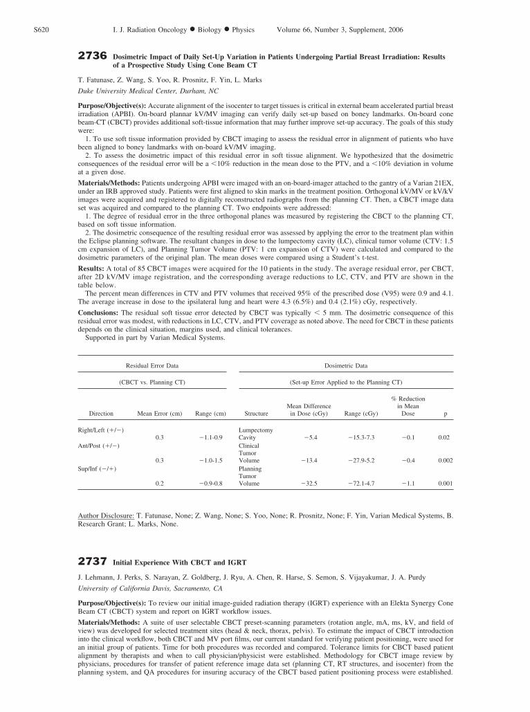
Results: A total of 85 CBCT images were acquired for the 10 patients in the study. The average residual error, per CBCT,after 2D kV/MV image registration, and the corresponding average reductions to LC, CTV, and PTV are shown in thetable below.
The percent mean differences in CTV and PTV volumes that received 95% of the prescribed dose (V95) were 0.9 and 4.1.The average increase in dose to the ipsilateral lung and heart were 4.3 (6.5%) and 0.4 (2.1%) cGy, respectively.
Conclusions: The residual soft tissue error detected by CBCT was typically � 5 mm. The dosimetric consequence of thisresidual error was modest, with reductions in LC, CTV, and PTV coverage as noted above. The need for CBCT in these patientsdepends on the clinical situation, margins used, and clinical tolerances.
Supported in part by Varian Medical Systems.
Author Disclosure: T. Fatunase, None; Z. Wang, None; S. Yoo, None; R. Prosnitz, None; F. Yin, Varian Medical Systems, B.Research Grant; L. Marks, None.
2737 Initial Experience With CBCT and IGRT
J. Lehmann, J. Perks, S. Narayan, Z. Goldberg, J. Ryu, A. Chen, R. Harse, S. Semon, S. Vijayakumar, J. A. Purdy
University of California Davis, Sacramento, CA
Purpose/Objective(s): To review our initial image-guided radiation therapy (IGRT) experience with an Elekta Synergy ConeBeam CT (CBCT) system and report on IGRT workflow issues.
Materials/Methods: A suite of user selectable CBCT preset-scanning parameters (rotation angle, mA, ms, kV, and field ofview) was developed for selected treatment sites (head & neck, thorax, pelvis). To estimate the impact of CBCT introductioninto the clinical workflow, both CBCT and MV port films, our current standard for verifying patient positioning, were used foran initial group of patients. Time for both procedures was recorded and compared. Tolerance limits for CBCT based patientalignment by therapists and when to call physician/physicist were established. Methodology for CBCT image review byphysicians, procedures for transfer of patient reference image data set (planning CT, RT structures, and isocenter) from theplanning system, and QA procedures for insuring accuracy of the CBCT based patient positioning process were established.
Residual Error Data Dosimetric Data
(CBCT vs. Planning CT) (Set-up Error Applied to the Planning CT)
Direction Mean Error (cm) Range (cm) StructureMean Difference
in Dose (cGy) Range (cGy)
% Reductionin Mean
Dose p
Right/Left (�/)0.3 1.1-0.9
LumpectomyCavity 5.4 15.3-7.3 0.1 0.02
Ant/Post (�/)
0.3 1.0-1.5
ClinicalTumorVolume 13.4 27.9-5.2 0.4 0.002
Sup/Inf (/�)
0.2 0.9-0.8
PlanningTumorVolume 32.5 72.1-4.7 1.1 0.001
S620 I. J. Radiation Oncology ● Biology ● Physics Volume 66, Number 3, Supplement, 2006