Embed Size (px)
Citation preview
252
International Maternal & Child Health Care
Subsequently, give oral thiamine 50 mg three times daily until vomiting has stopped.
Other management on discharge from hospitalWithhold iron tablets until vomiting has resolved, but ensure that they are taken subsequently, as iron- defi ciency
anaemia may have been an important consequence of the hyperemesis.
Try to help with any depression that is present and also, if resources to address intimate partner violence are available in the community, make sensitive inquiries of the woman or girl in case this is a contributing factor.
2.7 Medical disorders complicating pregnancy and delivery
2.7.A Heart failure during pregnancy, including rheumatic heart disease
BOX 2.7.A.1 Minimum standards Q Oxygen. Q Furosemide. Q Digoxin. Q Nitroglycerine sublingual tablets. Q Blood transfusion. Q Morphine.
IntroductionSerious cardiac pathology may present either as heart fail-ure where respiratory distress is the most obvious fi nding, or as cardiogenic shock (see shock section later).
Causes of heart failure during pregnancyThere are fi ve main causes of heart failure in pregnancy:1 severe anaemia 2 structural heart disease3 circulatory overload (e.g. excessive IV fl uids) 4 hypertension in severe pre- eclampsia5 hypertrophic cardiomyopathy (HCM) and peripartum
cardiomyopathy.
Heart failure can result from: O left ventricular volume overload (aortic and mitral valve
incompetence) or excessive pulmonary blood fl ow (e.g. congenital heart defects)
O left heart obstruction (aortic stenosis, mitral stenosis, hypertension)
O primary pump failure (severe anaemia, myocarditis, cardiomyopathy or arrhythmia)
O over- transfusion (a particular risk in hospital with IV blood or fl uid infusions, especially in the anaemic mother).
Clinical signsThese include the following:
O respiratory distress (raised rate and some chest wall recession)
O tachycardia out of proportion to respiratory diffi culty
O raised jugular venous pressure O gallop rhythm/murmur O enlarged liver O basal lung crepitations.
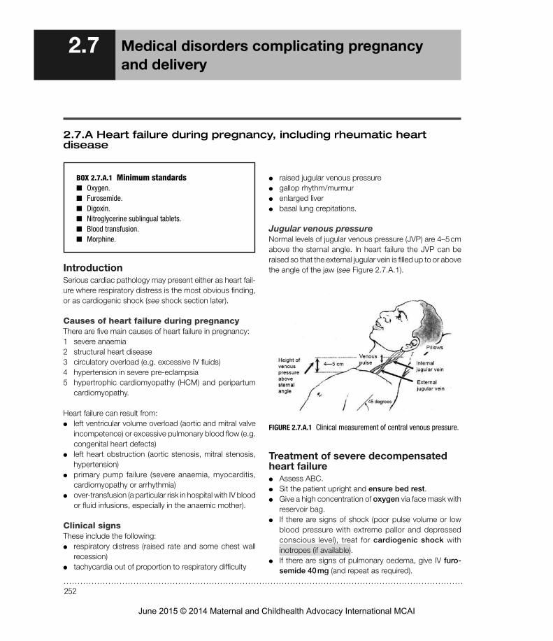
Jugular venous pressureNormal levels of jugular venous pressure (JVP) are 4–5 cm above the sternal angle. In heart failure the JVP can be raised so that the external jugular vein is fi lled up to or above the angle of the jaw (see Figure 2.7.A.1).
FIGURE 2.7.A.1 Clinical measurement of central venous pressure.
Treatment of severe decompensated heart failure
O Assess ABC. O Sit the patient upright and ensure bed rest. O Give a high concentration of oxygen via face mask with
reservoir bag. O If there are signs of shock (poor pulse volume or low
blood pressure with extreme pallor and depressed conscious level), treat for cardiogenic shock with inotropes (if available).
O If there are signs of pulmonary oedema, give IV furo-semide 40 mg (and repeat as required).
June 2015 © 2014 Maternal and Childhealth Advocacy International MCAI

253
Section 2.7
O Provided the patient is not hypotensive (systolic blood pressure > 90 mmHg) and has no serious obstruc-tive valvular disease, give a glyceryl trinitrate tablet 500 micrograms sublingually and repeat up to a total of 3 tablets.
O Give morphine 3 mg over 5 minutes, and consider repeating after 15 minutes. Morphine is effective in reducing the afterload and, in addition, will reduce anxiety and pain both of which are likely to make heart failure worse.
O For patients with persisting heart failure load with digoxin IV, 250–500 micrograms over 10 minutes, then after 6 hours 125–250 micrograms over 10 min-utes, then after a further 6 hours 125–250 micrograms over 10 minutes.
Or give an oral digoxin loading dose (instead of IV) of 375–750 micrograms, then after 6 hours 187.5–375 micro-grams, then after a further 6 hours 187.5–375 micrograms.
O Maintenance digoxin dose IV or oral: — 125–750 micrograms once daily. — Reduce the dose in renal impairment. Be alert for low K+ levels.
O Consider thromboprophylaxis. This treatment must take into account any bleeding risk and the timing of delivery.
O Check for severe anaemia (especially if the haemoglobin concentration is < 5.0 g/dL), for which partial exchange transfusion may be helpful. Partial exchange transfu-sion can be achieved with a cannula in a large vein in the antecubital fossa. Withdraw 25 mL of anaemic blood and infuse 50 mL of new blood over 5 minutes, and repeat up to 10 times. An alternative is careful transfusion of packed cells (hang the bag vertically for 15 minutes) to allow the red blood cells to separate from the plasma. Transfuse only the red blood cell component with 40 mg IV furosemide for each unit of 500 mL infused.
The following are useful investigations if available: O full blood count (to exclude severe anaemia) O serum urea and electrolytes O infection screen, including blood cultures O 12- lead electrocardiogram O chest X- ray O echocardiogram.
Subsequent treatment of heart failure O Dietary sodium restriction. O Loop diuretics (furosemide) for moderate/severe pul-
monary oedema, O Treatment with oral hydralazine or oral nifedipine (modi-
fi ed release version) as a vasodilator (instead of glyceryl trinitrate used in the acute scenario above).
O Treatment with a B- blocker (preferably a B1 cardio- selective B- blocker such as atenolol) can be benefi cial. They are NOT used in the acute presentation of a patient with severe decompensated heart failure. They are contraindicated in asthma and may be associated with intra- uterine growth restriction, but are not associated with congenital malformations.
O Continued treatment with digoxin (as above) should be considered if signifi cant symptoms persist.
O Treatment of any underlying anaemia or poor nutritional status.
O Once the baby is delivered, treatment with hydralazine or nifedipine may be replaced with an ACE inhibitor such as enalapril or lisinopril. ACE inhibitors cause congenital abnormalities and therefore should not be used antenatally. They are contraindicated if the patient is hypovolaemic and in renal artery and aortic stenosis.
Management of heart failure during labour
O The mother must deliver sitting up. O Give oxygen from a face mask throughout labour. O Limit infusion of IV fl uids to decrease the risk of circula-
tory overload, and maintain a strict fl uid balance chart. O Ensure adequate analgesia. O If an oxytocin IV infusion is required, use a higher
concentration at a slower rate while maintaining a fl uid balance chart (e.g. the concentration may be doubled if the number of drops per minute is decreased by half).
— Increase the rate of oxytocin infusion until regular strong contractions are established, and then main-tain infusion at that rate.
O Avoid sustained, bearing- down efforts during the second stage if possible.
O If it is necessary to decrease the woman’s workload dur-ing delivery, perform an episiotomy and assist delivery by vacuum extraction or forceps.
O Ensure active management of the third stage of labour. Oxytocin given on delivery of the baby must be given very slowly IV (5 units diluted in 20 mL of 0.9% saline over 5–10 minutes) to avoid hypotension.
O Do not give ergometrine.
Note: Heart failure is not an indication for Caesarean section.
Management o f anaesthesia if Caesarean section is neededIt is assumed that epidural anaesthesia/analgesia is unlikely to be possible or appropriate in low- resource settings. Avoid spinal anaesthesia if there is a fi xed cardiac output, such as aortic/mitral stenosis or heart failure associated with valvular disease. If you are giving a general anaesthetic, take precautions against aspiration and minimise the risk of an increase in blood pressure associated with intubation by premedication with either morphine (5 mg initially IV) or lignocaine (1 mg/kg IV). If the patient is considered to have insuffi cient cardiovascular stability for general anaes-thetic, undertake Caesarean section under local infi ltration anaesthesia.
In all of the above situations the surgeon should be ready to start operating immediately when anaesthesia is estab-lished, so that the operating time is as short as possible. As above, oxytocin given for active management of third stage must be given very slowly IV (5 units diluted in 20 mL of 0.9% saline over 5–10 minutes) to avoid hypotension.
Post- operative management must ensure adequate analgesia with morphine.
June 2015 © 2014 Maternal and Childhealth Advocacy International MCAI

254
International Maternal & Child Health Care
Cardiac consequences of rheumatic heart disease and their effects on pregnancyIntroductionAfter an episode of acute rheumatic fever, there may be permanent valve damage. Rheumatic heart disease occurs when acute valve inflammation is followed by scarring and fi brosis, resulting in various degrees of short-ening, thickening, rigidity, deformity, retraction and fusion of the valve cusps. The commonest valve lesions are mitral regurgitation, mitral stenosis and aortic regurgitation.Rheumatic heart disease is most severe and progressive in the following:
O patients who initially have severe carditis O patients who have recurrent episodes of acute rheumatic
fever. The prognosis is more favourable if recurrences are prevented. After single episodes, residual cardiac disease may disappear or improve, and valve damage only worsens in a few cases. It is therefore crucial to maintain continuous antibiotic prophylaxis to prevent further valve damage.
O Asymptomatic rheumatic valve disease often becomes clinically relevant during pregnancy. A history of rheu-matic fever should be sought at booking and ideally the heart auscultated for murmers. It should be remembered as a differential diagnosis in a women presenting with shortness of breath, a cardiac arrhythmia or heart failure.
When a cardiology referral and/or surgical intervention is not available
O Medical management is supportive, aiming to maximise cardiac function. The most dangerous period is delivery and shortly afterwards. In general, the more normal the delivery the less stress there is on the heart.
O Regular follow- up and rational drug therapy can make a signifi cant difference. Use routine medications (diuret-ics, B- blocker, digoxin and nitrates) to maximum effect.
O Bed rest and the avoidance of heavy work are essential. O Treat anaemia and any other coexisting conditions. O Advise hospital delivery. O Venesection can be used to decrease venous load for
the patient in life- threatening situations where preload is high.
Note that there is a risk of teratogenicity with ACE inhibitors during the fi rst trimester, and problems with placental and renal function later in pregnancy. There is also diminished placental perfusion with diuretics although these should not be withheld if clinically indicated. ACE inhibitors may be used after delivery and during breastfeeding.
Effects of rheumatic heart diseaseMitral regurgitationMitral regurgitation is the commonest valve lesion.
Clinical featuresThese include the following:
O easy fatigue (caused by low cardiac output) O shortness of breath on exertion (caused by pulmonary
oedema and inability to increase cardiac output) O orthopnoea, paroxysmal nocturnal dyspnoea and
haemoptysis O hyperdynamic apical impulse
O apical impulse displaced laterally and inferiorly O a blowing apical pansystolic murmur radiating to the
left axilla; there may be a third heart sound and a short low- frequency mid- diastolic murmur from increased trans- mitral fl ow
O there may be basal crepitations O chest X- ray demonstrates cardiomegaly and left atrial
enlargement (a double density on the right heart border and elevation of the left main bronchus)
O the ECG demonstrates left atrial enlargement (broad bifi d P waves in lead II and a prominent negative component to the P in V1) and left ventricular hypertrophy
O signs of pulmonary hypertension.
Management O Urgent referral for a cardiology opinion, as surgery is
likely to be necessary (if available). O Annual echocardiograph (if available), as progressive
left heart dilation may result in irreversible left ventricular dysfunction if referral is delayed until symptoms develop.
O Medical treatment for heart failure, but patients who are unwell enough to require this may be more appro-priately treated by mitral valve repair or mitral valve replacement with a mechanical valve or bioprosthesis, if possible locally.
Mitral stenosisFeatures
O Mild stenosis does not cause symptoms, moderate stenosis causes shortness of breath on exertion, and severe stenosis causes easy fatigue, shortness of breath at rest, orthopnoea (shortness of breath on lying down), paroxysmal nocturnal dyspnoea and haemoptysis.
O There is a low- frequency mid- diastolic murmur that is maximal at the apex, accentuated by exercise.
O There is a loud fi rst heart sound and diastolic opening snap.
O The murmur becomes longer as the severity of stenosis increases.
O In severe cases, there are signs of pulmonary hypertension.
O Chest X- ray and ECG show left atrial enlargement when there is moderate mitral stenosis, and chest X- ray shows pulmonary oedema when stenosis is severe.
Management O Symptoms are treated with diuretics and a low- sodium
diet. Digoxin is indicated only in rare cases where there is atrial fi brillation secondary to left atrial enlargement.
O Symptomatic patients and those with pulmonary hyper-tension should be referred for cardiology review (if available), as surgery is often necessary (open or closed mitral commissurotomy, mitral valve replacement and percutaneous catheter balloon mitral commissurotomy).
Aortic regurgitation O This is less common than mitral regurgitation, and fre-
quently occurs in combination with mitral valve disease. O Symptoms occur when left ventricular dysfunction
develops secondary to chronic left ventricular volume overload.
O Once the symptoms have appeared, deterioration is often rapid.
O Symptoms include exercise intolerance, shortness of
June 2015 © 2014 Maternal and Childhealth Advocacy International MCAI

255
Section 2.7
breath on exertion, orthopnoea (shortness of breath on lying down), paroxysmal nocturnal dyspnoea, haemop-tysis and chest pain.
O Examination reveals a blowing decrescendo early dias-tolic murmur that is maximal at the mid to lower left sternal border. The murmur is loudest when sitting forward with the breath held in expiration.
Signs of moderate to severe aortic regurgitation O The murmur lengthens and may be present throughout
diastole. O Hyperdynamic apex. O Apical impulse displaced laterally and inferiorly. O Wide pulse pressure. O Collapsing pulses. O Basal crepitations. O Visible pulsations in the suprasternal notch and neck
vessels. O Systolic murmur at the upper right sternal border (from
increased aortic valve fl ow).
Management O Cardiology assessment, as surgery may be necessary
(if available). Marked cardiomegaly on chest X- ray or multiple ventricular ectopics on the ECG should prompt referral.
O An echocardiogram is needed at least annually (if avail-able), as it is important to assess left ventricular dilation and function to ensure that surgery is performed before irreversible left ventricular dysfunction develops.
O Exercise tolerance may be improved by medical treat-ment for heart failure.
O Surgical options include aortic valve reconstruction, aortic valve replacement with an aortic homograft or mechanical valve, and transferring the patient’s own pulmonary valve to the aortic position (Ross procedure).
Aortic stenosisThe two commonest causes of aortic valve stenosis are progressive wear of a congenital bicuspid aortic valve and rheumatic fever (the most common cause in developing countries, and usually also with aortic regurgitation).
Clinical features O Chest pain (angina from inadequate coronary artery
perfusion). O Fainting, usually with exertion or excitement. O Shortness of breath due to heart failure. O Sudden death. O On examination, delayed upstroke and reduced magni-
tude of the carotid pulse and an ejection systolic heart murmur.
O The ECG shows left ventricular hypertrophy and some-times ST changes of myocardial ischaemia.
O The chest X- ray shows a normal sized heart, dilated aor-tic root and pulmonary venous congestion. Sometimes there is calcifi cation of the aortic valve.
Management O Avoid strenuous exercise. O Avoid endocarditis. O Refer for a specialist opinion if possible. O Diuretics can be helpful, but surgery is usually required.
2.7.B Asthma
BOX 2.7.B.1 Minimum standards Q Oxygen. Q Salbutamol by metered- dose inhaler and nebuliser. Q Aminophylline. Q Magnesium sulphate. Q Adrenaline. Q Prednisolone/hydrocortisone.
Assessment
Features of severe asthma O Too breathless to feed or talk. O Recession/use of accessory muscles. O Respiratory rate > 40 breaths/minute. O Pulse rate > 120 beats/minute.
Features of life- threatening asthma O Conscious level depressed/agitated. O Exhaustion. O Poor respiratory effort. O SaO2 < 85% in air/cyanosis. O Silent chest.
Asthma complicates 3–4% of pregnancies. Pregnancy is
associated with worsening of the symptoms in one- third of affected mothers.
O A chest X- ray is indicated only if there is severe dysp-noea, uncertainty about the diagnosis, asymmetry of chest signs (possible pneumothorax) or signs of severe infection.
O Transcutaneous PCO2, arterial or capillary blood gases (if available) can be helpful in very severe asthma.
O Continuous pulse oximetry is valuable (if available), as hypoxaemia is a major feature of all severe asthma attacks.
O Do not give prostaglandins other than misoprostol (the latter is safe in pregnancy). For the prevention and treatment of postpartum haemorrhage, give oxytocin 10 units IM or ergometrine 500 micrograms IM or both (Syntometrine IM).
O Do not give labetalol for hypertension in patients with asthma.
O The priority of treatment is to maintain good control of the patient’s asthma. This will reduce the likelihood of acute exacerbations which can be life- threatening. In order that control is maintained the following are recommended:
— It should be emphasised that inhaled salbutamol and steroids are not harmful to the fetus and should be continued in pregnancy.
June 2015 © 2014 Maternal and Childhealth Advocacy International MCAI
256
International Maternal & Child Health Care
— The aim should be for the patient to need her sal-butamol inhaler no more than 1–2 times/day. If use in excess of this occurs, the patient should be com-menced on inhaled steroids or have her current dose of inhaled steroids increased.
— If the maximum dose of inhaled steroids is reached, then long acting B2- agonists and slow release theo-phylline should be considered if available.
— If not available, or ineffective, oral prednisolone can be added at the lowest dose required to maintain control. Oral prednisolone is associated with an increased risk of infection and gestational diabetes, and complicates control of established diabetes. Its long term use has other potential side- effects for the mother, such as osteoporosis, but it should not be withheld if required to maintain control of maternal disease.
Emergency treatment of severe asthma O Assess ABC and resuscitate as needed. O Give a high concentration of oxygen via a face mask with
reservoir bag or nasal cannula. Attach a pulse oximeter and maintain SaO2 in the range 94–98%.
O Sit the patient up. O Give nebulised salbutamol 5 mg driven with oxygen
half- hourly to 4- hourly via a nebuliser (or 10–20 puffs of a beta-2- agonist inhaler, such as salbutamol or terb-utaline, giving one puff at a time through a spacer with a mouthpiece or face mask).
O Give oral prednisolone 30–60 mg, or if the patient is vomiting, IV/IM hydrocortisone 100 mg, followed by
100 mg 6- hourly. (Note: steroids will not show benefi ts for a number of hours.)
If the patient is not responding, or their condition is deteriorating:
O Nebulised salbutamol may be given continuously. O In acute severe asthma, 2 g of magnesium sul-
phate IV in 50 mL of Ringer- lactate or Hartmann’s solution over 10–15 minutes can produce signifi cant bronchodilatation.
O As an alternative to magnesium sulphate, and if the patient is not already on oral theophylline or other methylxanthines, give a loading dose of IV aminophyl-line 250 mg over 15 minutes, monitoring the ECG for arrhythmias (if possible), followed by 1 mg/kg/hour by IV infusion.
O IV salbutamol 250 micrograms over 10 minutes is an alternative to magnesium sulphate or aminophylline, followed by IV infusion of 1–5 micrograms/kg/minute (but monitoring ECG and checking K+ levels regularly is necessary; extra potassium may be needed, and monitoring of plasma K+ levels is essential if this drug is given IV).
O In severe cases in the absence of other measures, adrenaline can be effective. It should be given subcu-taneously or IM (dose = 500 micrograms to 1 mg), but may be given IV in life- threatening asthma as follows.
— Place 1 mg of adrenaline in 10 mL of 0.9% saline and give 1 mL of this solution. Wait for 1 minute and then keep on repeating 1 mL doses IV every minute until the patient improves or the whole 1 mg (10 mL) has
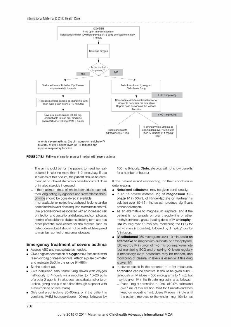
OXYGENProp up in lateral tilt position
Salbutamol inhaler 100 micrograms/puff: 2 puffs over approximately 1
minute
Continue oxygen
Shake salbutamol inhaler: 2 puffs over approximately 1 minute
Repeat x 5 cycles as long as improving, with each cycle given every 5–10 minutes
Is the mother improving? NOYES
Give oral prednisolone 30–60 mg,or if not able to take oral medicine
hydrocortisone 100 mg IV/IM 6-hourly
Nebuliser driven by oxygenSalbutamol 5 mg
Continuous salbutamol by nebuliser or inhaler (if nebuliser not available)
Repeat dose as soon as the last one finishes
IV aminophylline 250 mg as loading dose over 15 minutesThen IV infusion of 1 mg/kg/
hour
Subcutaneous/IMadrenaline 0.5–1 mg
If NOT improving
If NOT improving
In acute severe asthma, 2 g of magnesium sulphate IV in 50 mL of 0.9% saline over 10–15 minutes can improve respiratory function
FIGURE 2.7.B.1 Pathway of care for pregnant mother with severe asthma.
June 2015 © 2014 Maternal and Childhealth Advocacy International MCAI
257
Section 2.7
been given. The risk of cardiac side effects (tachy-cardia, cardiac arrhythmias) is low if adrenaline is given in this way.
O In patients with poor respiratory effort, depressed con-scious level and poor oxygenation despite maximum oxygen therapy:
— Attempt to support ventilation with a bag- valve- mask. — Summon experienced support (an anaesthetist) if available, and consider intubation for mechanical ventilation with IV ketamine or halothane induction.
Indications for intubation and positive pressure ventilation (if available)These include the following:
O increasing exhaustion O progressive deterioration in clinical condition (e.g. a
silent chest) — oxygenation decreasing and/or oxygen requirement increasing
— pCO2 increasing (if measurable from arterial/capil-lary gas)
O sudden deterioration O massive atelectasis O pneumothorax.
If the patient is responding and improving, continue inhaled salbutamol as often as indicated.
Other measures O Reassure the patient, and avoid upsetting them by
performing unnecessary invasive procedures. O Give IV steroids to cover labour/delivery for prevention
of Addisonian crisis in patients with a history of taking signifi cant doses of oral steroids in the recent past, especially if long term.
O Restrict IV fl uids to two- thirds of the normal requirements.
O Give antibiotics only if there are signs of infection (fever and other signs of pneumonia; chest X- ray may be helpful).
O When the patient has recovered, review their mainte-nance treatment and inhaler technique.
How to give drugs such as aminophylline or magnesium sulphate safely IV without syringe drivers or pumps
O Bolus doses: — The safest way to give these is slowly by hand using the syringe.
O IV infusions: — Where volume overload is not an issue, the simplest method is to add the drug (e.g. aminophylline) to 500- mL bags of Ringer- lactate or Hartmann’s solu-tion or other available/appropriate fl uid and run over 12–24 hours.
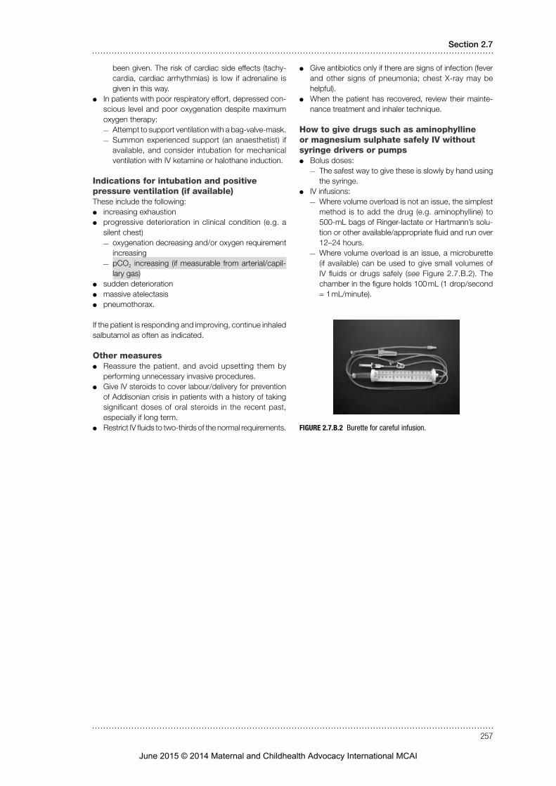
— Where volume overload is an issue, a microburette (if available) can be used to give small volumes of IV fl uids or drugs safely (see Figure 2.7.B.2). The chamber in the fi gure holds 100 mL (1 drop/second = 1 mL/minute).
FIGURE 2.7.B.2 Burette for careful infusion.
June 2015 © 2014 Maternal and Childhealth Advocacy International MCAI
258
International Maternal & Child Health Care
2.7.C Anaphylaxis
BOX 2.7.C.1 Minimum standards Q Adrenaline. Q Hydrocortisone and prednisolone. Q Nebulised adrenaline. Q Antihistamine. Q Nebulised or inhaled salbutamol.
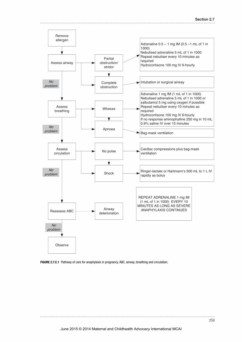
IntroductionAnaphylaxis is an allergic reaction to ingested, inhaled or topical substances, which may present as one or more of stridor, shock or respiratory distress. Common causes include allergy to penicillin, to radiographic contrast media, to blood transfusion, to insect bites and to certain foods, especially nuts. Anaphylaxis can occur with any drug.
Clinical featuresConsider the possibility of anaphylaxis in a patient with any of the symptoms and signs listed in Table 2.7.C.1, especially when any of the following are present:
O a history of previous severe reaction O rapidly progressive or increasingly severe symptoms O a history of asthma, eczema or rhinitis (atopy) O current treatment with beta- blockers.
This condition is potentially life- threatening, and may result in a change in conscious level, collapse, and respiratory or cardiac arrest. Some patients carry their own adrenaline.
Treatment O Remove or stop the allergen if possible. O Adrenaline 1 mg is given IM, unless there is intractable
shock or cardiac arrest on presentation, in which case give adrenaline IV as follows:
— Place 1 mg of adrenaline in 10 mL of 0.9% saline and give 0.5–1 mL of this solution. Wait for 1 minute and then keep on repeating 1 mL doses IV every minute until the patient improves or the whole 1 mg (10 mL) has been given.
O IV/IM hydrocortisone, 100–300 mg (if IV by slow injection) or oral prednisolone 40 mg stat.
O Nebulised adrenaline if there is stridor. O Nebulised salbutamol 5 mg by oxygen driven nebulizer
or adrenaline if there is wheezing. O Antihistamine: chlorphenamine 10–20 mg by slow intra-
venous injection. O Intubation and ventilation (if available) will be required
for severe cases.
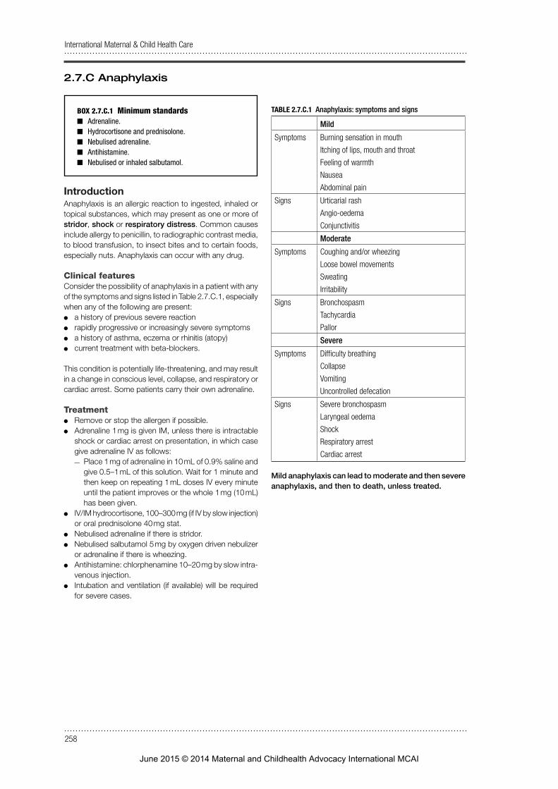
TABLE 2.7.C.1 Anaphylaxis: symptoms and signs
Mild
Symptoms Burning sensation in mouth Itching of lips, mouth and throat Feeling of warmth NauseaAbdominal pain
Signs Urticarial rashAngio- oedemaConjunctivitis
Moderate
Symptoms Coughing and/or wheezing Loose bowel movements Sweating Irritability
Signs BronchospasmTachycardiaPallor
Severe
Symptoms Diffi culty breathingCollapseVomitingUncontrolled defecation
Signs Severe bronchospasmLaryngeal oedemaShockRespiratory arrestCardiac arrest
Mild anaphylaxis can lead to moderate and then severe anaphylaxis, and then to death, unless treated.
June 2015 © 2014 Maternal and Childhealth Advocacy International MCAI
259
Section 2.7
Remove allergen
Assess airway
Assess breathing
Assess circulation
Partial obstruction/
stridor
Complete obstruction
Adrenaline 0.5 – 1 mg IM (0.5 –1 mL of 1 in 1000)Nebulised adrenaline 5 mL of 1 in 1000Repeat nebuliser every 10 minutes as requiredHydrocortisone 100 mg IV 6-hourly
Intubation or surgical airway
Wheeze
Apnoea
Adrenaline 1 mg IM (1 mL of 1 in 1000)Nebulised adrenaline 5 mL of 1 in 1000 or salbutamol 5 mg using oxygen if possibleRepeat nebuliser every 10 minutes as requiredHydrocortisone 100 mg IV 6-hourlyIf no response aminophylline 250 mg in 10 mL 0.9% saline IV over 15 minutes
Bag-mask ventilation
No pulse
Shock
Cardiac compressions plus bag-mask ventilation
Reassess ABC
Ringer-lactate or Hartmann’s 500 mL to 1 L IV rapidly as bolus
Observe
Airway deterioration
No problem
No problem
No problem
No problem
REPEAT ADRENALINE 1 mg IM (1 mL of 1 in 1000) EVERY 10
MINUTES AS LONG AS SEVERE ANAPHYLAXIS CONTINUES
FIGURE 2.7.C.1 Pathway of care for anaphylaxis in pregnancy. ABC, airway, breathing and circulation.
June 2015 © 2014 Maternal and Childhealth Advocacy International MCAI

260
International Maternal & Child Health Care
Two children with pneumonia receiving oxygen from one oxygen concentrator; the only one available in a ward of 26 beds.
Two senior midwives in Liberia trained in advanced obstetrics undertaking an emergency Caesarean section.
June 2015 © 2014 Maternal and Childhealth Advocacy International MCAI
261
Section 2.7
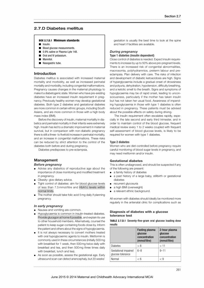
2.7.D Diabetes mellitus
BOX 2.7.D.1 Minimum standards Q Insulin. Q Blood glucose measurements. Q 0.9% saline or Plasma- Lyte 148. Q Oral and IV potassium. Q Mannitol. Q Nasogastric tube.
IntroductionDiabetes mellitus is associated with increased maternal mortality and morbidity, as well as increased perinatal mortality and morbidity, including congenital malformations. Pregnancy causes changes in the maternal physiology to make it a diabetogenic state. Women who have pre- existing diabetes have an increased insulin requirement in preg-nancy. Previously healthy women may develop gestational diabetes. Both type 2 diabetes and gestational diabetes are more common in certain ethnic groups, including South Asians, and are more common in those with a high body mass index (BMI).
Before the discovery of insulin, maternal mortality in dia-betics and perinatal mortality in their infants were extremely high. Insulin has led to a dramatic improvement in maternal survival, but in comparison with non- diabetic pregnancy there is still a three- to fi vefold increase in perinatal mortality, and an increase in congenital malformations. These risks can be reduced by strict attention to the control of the diabetes both before and during pregnancy.
Diabetes predisposes to pre- eclampsia.
Management Before pregnancy
O Advise any diabetics of reproductive age about the importance of close monitoring and modifi ed treatment in pregnancy.
O Obesity: give dietary advice. O Tight control of diabetes: aim for blood glucose levels
of less than 7.5 mmol/litre and HbA1c levels within normal limits.
O The mother should take folic acid 5 mg daily if planning pregnancy.
In early pregnancy O Nausea and vomiting are common. O Hypoglycaemia is common in insulin- treated diabetes.
Provide glucagon at home if possible, and explain its use to other household members. Alternatively, counsel the patient to keep sugar- containing foods close by. Inform the patient and others about the signs of hypoglycaemia.
O It is not always necessary to convert mothers treated with oral hypoglycaemic agents to insulin. Metformin is commonly used in these circumstances (initially 500 mg with breakfast for 1 week, then 500 mg twice daily with breakfast and tea, and then 500 mg three times daily with breakfast, lunch and tea).
O As soon as possible, assess the gestational age. Early ultrasound scan can detect anencephaly, but 20 weeks’
gestation is usually the best time to look at the spine and heart if facilities are available.
During pregnancyType 1 diabetes (insulin dependent)Close control of diabetes is needed. Expect insulin require-ments to increase by up to 50% above pre- pregnant levels. There is an increased risk of congenital abnormalities, macrosomia, polyhydramnios, preterm labour and pre- eclampsia. Plan delivery with care. The risks of infection and development of diabetic ketoacidosis are high. Signs of hyperglycaemia include a gradual onset of drowsiness and polyuria, dehydration, hypotension, diffi culty breathing, and a ketotic smell to the breath. Signs and symptoms of hypoglycaemia may be of rapid onset, leading to uncon-sciousness, particularly if the mother has taken insulin but has not taken her usual food. Awareness of impend-ing hypoglycaemia in those with type 1 diabetes is often reduced in pregnancy. These patients must be advised about the possible effects on safety during driving.
The insulin requirement often escalates rapidly, espe-cially in the late second and early third trimester, and in order to maintain control of the blood glucose, frequent medical review every 1 to 2 weeks coupled with frequent self- assessment of blood glucose levels, is likely to be required for women with type 1 diabetes.
Type 2 diabetesWomen who are diet- controlled before pregnancy require careful monitoring of blood sugar levels in pregnancy, and may need metformin and/or insulin.
Gestational diabetesThis is often undiagnosed, and should be suspected if any of the following are present:
O a family history of diabetes O a past history of a large baby, stillbirth or gestational
diabetes O recurrent glycosuria O a high BMI (overweight) O a relevant ethnic background.
All women with diabetes should ideally be monitored more regularly in the antenatal clinic for complications such as
Diagnosis of diabetes with a glucose tolerance test
TABLE 2.7.D.1 Seventy- five- gram oral glucose loading dose results
Fasting plasma glucose concentration (mmol/litre)
2- hour plasma glucose concentration (mmol/litre)
Diabetes > 8 > 11
Gestational impaired glucose tolerance
6–8 9–11
Normal < 6 < 9
June 2015 © 2014 Maternal and Childhealth Advocacy International MCAI
262
International Maternal & Child Health Care
pre- eclampsia, polyhydramnios and a large or small for gestational age infant.
Management of delivery in women with diabetesFor spontaneous labour, induction of labour and elective Caesarean section:1 Measure glucose on admission and hourly during labour.2 Site an IV line with 500 mL of 0.9% saline containing
10% dextrose and potassium chloride 10 mmol, and give at a rate of 60 mL/hour.
Avoid the routine use of insulin in labour in low resource settings because of lack of experience and lack of blood glucose stick tests. In mothers who were using insulin dur-ing pregnancy and those where blood glucose is > 7 mmol/litre on two successive occasions one hour apart in labour, the insulin requirements shown in Table 2.7.D.2 below can be used.
TABLE 2.7.D.2 Insulin requirements
Blood glucose concentration (mmol/litre)
Hourly subcutaneous injections of insulin
< 2.0 No insulin; dextrose only
2.0–4.0 1 unit
4.1–9.0 2 units
9.1–11.0 3 units
11.1–16.9 4 units
NOTE: for blood glucose, 1 mmol/litre = 18 mg/dL
O If the glucose level is > 17 mmol/litre, expert advice should be sought.
O Aim for a glucose level of 4–9 mmol/litre. O Reduce insulin by half at delivery, and aim to resume the
pre- pregnancy insulin dosage 24 hours after delivery. If the mother is breastfeeding, her insulin requirement may be lower.
O Women who have developed gestational diabetes usually have normal blood glucose levels soon after the delivery of the placenta. Their diabetic medication should be stopped postnatally, and their blood sugar levels should be monitored.
O Mothers who have had gestational diabetes should have a glucose tolerance test at 6 weeks postnatally. They are at risk of developing type 2 diabetes, and appro-priate dietary and lifestyle advice should be provided. A fasting blood glucose test annually should also be recommended.
Diabetic ketoacidosis (DKA)DKA is the commonest endocrine emergency, and should be suspected in patients with any of the following:
O dehydration O abdominal pain O ketone smell on the breath O acidosis O acidotic breathing O unexplained coma.
Patients die from hypokalaemia and cerebral oedema.Patients who are 5% dehydrated or less and are not
clinically unwell usually tolerate oral rehydration and sub-cutaneous insulin.
Patients who are more than 5% dehydrated, or who are vomiting or drowsy or clinically acidotic, need emergency care as follows.
Primary assessment and resuscitation Airway
O If the airway is not open, use an airway- opening manoeuvre, and consider an airway adjunct such as an oropharyngeal airway or intubation (if available and subsequently supported).
O The nares and oropharynx may need gentle suctioning under direct observation.
O If the patient is unconscious and the airway is unpro-tected, the recovery position should be adopted to minimise the risk of aspiration of vomit.
BreathingGive a high concentration of oxygen through a face mask with a reservoir, if the airway is adequate.
If breathing is inadequate, ventilate with oxygen via a bag- valve- mask- reservoir device, and ask for experienced senior help to intubate (if this is available and sustainable).
Circulation O Gain IV access using a short wide- bore cannula (14- to
16G). O External or internal jugular vein access is an option if
peripheral access is impossible. Long saphenous vein cut- down may also be considered.
O Take blood for a full blood count, urea and electrolytes, blood culture, cross- matching, glucose stick test and laboratory blood glucose (if available).
O Give a 500- mL rapid IV bolus of 0.9% saline or Plasma- Lyte 148.
O An antibiotic such as cefotaxime 1 gram IV 6- hourly, or the locally available equivalent, is an appropriate antibiotic for those in whom an infection is likely to have precipitated the DKA. Although, of course, antibiotic therapy must be tailored to the specifi c cause.
Diagnosis O History:
— polydipsia — polyuria — weight loss.
O Clinical: — acidotic respiration — dehydration — drowsiness — abdominal pain and/or vomiting.
O Biochemical: — high blood glucose on fi nger- prick test — ketones and glucose in urine.
Secondary assessment and emergency treatmentThe following in particular need to be assessed.
Degree of dehydration O 3%: dehydration is only just clinically detectable O 3–5%: dry mucous membranes and reduced skin
turgor
June 2015 © 2014 Maternal and Childhealth Advocacy International MCAI

263
Section 2.7
O 5–8%: as above, with sunken eyes and poor capillary return
O > 8% with shock: severely ill with poor perfusion, thready rapid pulse and reduced blood pressure.
Conscious level O Assess AVPU. O Institute hourly neurological observations. O If the patient is less than Alert on admission, or their
conscious level deteriorates, record the Glasgow Coma Scale score.
O Consider instituting cerebral oedema management (if available).
Cerebral oedemaLook for irritability, slow pulse, high blood pressure and papilloedema (a late sign).
InfectionDKA can cause a leucocytosis but not fever. If fever is present, look for and treat infection.
Ileus O Insert a nasogastric tube. O Ensure by clinical assessment, and by abdominal X- ray
if appropriate, that there is no other cause of the acute abdomen, including intestinal obstruction.
Observations O Strict fl uid balance and urine testing of every sample. O Hourly capillary blood glucose measurements. O Twice daily weights. O Initially hourly or more frequent neurological observations. O Report immediately to medical staff (even at night) symp-
toms of headache or any change in either conscious level or behaviour.
O Report any changes in the ECG trace, especially T- wave changes (monitoring for hypokalaemia).
Investigations O When it is safe to do so, weigh the patient. If this is not
possible, use recent clinic weight or an estimated weight. O Blood glucose. O Urea and electrolytes (if available). O Bicarbonate or arterial blood gases (if available). O Haematocrit and full blood count. O Blood culture. O Urine microscopy, culture and sensitivity; check for
ketones. O Monitor the ECG to observe T waves (if available):
— hypokalaemia causes fl at T waves — hyperkalaemia causes peaked T waves.
O Other investigations if indicated (e.g. if fever is present).
Additional emergency treatmentGeneral
O After resuscitation with fl uid boluses, calculate the fl uid requirement (see below).
O Avoid excessive fl uid replacement, as this is a risk factor for cerebral oedema.
O Do not give hypotonic IV solutions (e.g. 0.18% saline with 4% glucose, or 5% glucose): they are risk factors for cerebral oedema.
O Continue to give IV fl uids until the patient is drinking.
O After fl uids are running, calculate the rate of insulin infu-sion (blood glucose levels will already be falling).
O Use a continuous low- dose IV infusion of insulin (there is no need for an initial bolus), or in resource- limited situations use regular subcutaneous injections of short- acting insulin based on a sliding scale according to blood glucose measurements. Details below.
O Continue to give IV fl uids until the patient is tolerating enteral fl uids.
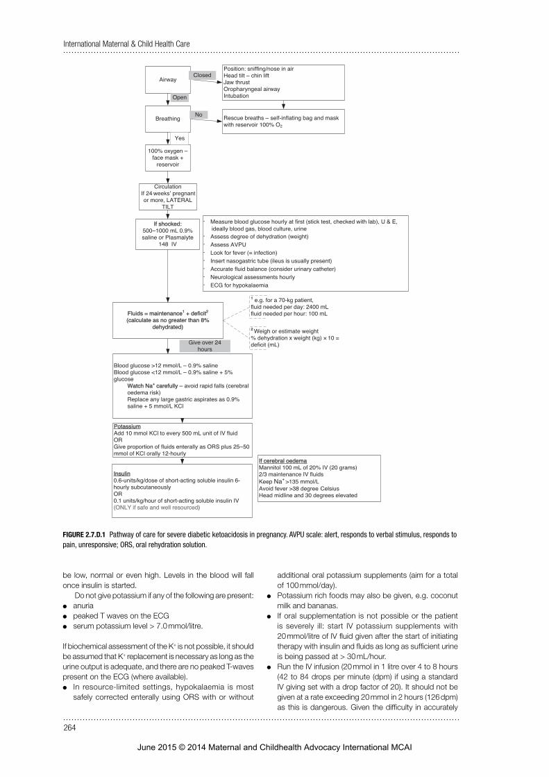
Fluid and electrolyte management O Calculate the patient’s fl uid requirement. This is equal to
maintenance plus defi cit (see Figure 2.7.D.1). — Maintenance — Defi cit (litres) = percentage dehydration × body weight (kg)/100
— Only plan to correct up to an 8% defi cit, as any more risks over- infusion.
O Ignore the volume of fl uids used to resuscitate/treat shock.
O Give the total fl uid requirement over 24 hours:
Glucose > 12 mmol/litre: give 0.9% saline or Plasma- Lyte 148.
Glucose < 12 mmol/litre: give 0.9% saline or Plasma- Lyte 148 containing 5% dextrose (by adding 100 mL of 50% glucose to 900 mL of 0.9% saline or Plasma- Lyte 148).
Sodium 135–155 mmol/litre: correct by rehydration over 24 hours.
Sodium > 155 mmol/litre: correct by rehydration over 48 hours using 0.9% saline or Plasma- Lyte 148.
Expect the sodium level to rise initially as the glucose level falls and water is removed from the circulation.
If the plasma sodium level initially falls (as well as the glucose level), this may precipitate cerebral oedema.
Bicarbonate O Administration of bicarbonate is rarely, if ever,
necessary. O Continuing acidosis usually indicates insuffi cient fl uid
resuscitation. O Consider the use of bicarbonate in patients who are pro-
foundly acidotic (pH < 7.0 if measurable) and shocked. Its only purpose is to improve cardiac contractility in severe shock.
The maximum volume of 8.4% sodium bicarbonate for half- correction of acidosis is calculated according to the following formula, and given over 60 minutes:
Volume (mL 8.4% NaHCO3) =
1/3 × weight (kg) × base deficit (mmol/litre)
2
If blood gas analysis cannot be undertaken: O The kidneys will resolve the acidosis (if they are working)
if the patient receives adequate fl uid and insulin therapy. O If you cannot measure pH, then do not give bicarbonate
except in extremis.
PotassiumIn diabetic ketoacidosis there is always massive depletion of total body potassium, although initial plasma levels may
June 2015 © 2014 Maternal and Childhealth Advocacy International MCAI
264
International Maternal & Child Health Care
Airway
Breathing
100% oxygen – face mask +
reservoir
CirculationIf 24 pregnant or more, LATERAL
TILT
If shocked: 500–1000 mL 0.9% saline or Plasmalyte
148 IV
· Measure blood glucose hourly at first (stick test, checked with lab), U & E, ideally blood gas, blood culture, urine
· Assess degree of dehydration (weight)· Assess AVPU· Look for fever (= infection)· Insert nasogastric tube (ileus is usually present)· Accurate fluid balance (consider urinary catheter)· Neurological assessments hourly· ECG for hypokalaemia
Fluids = maintenance1 + deficit2
(calculate as no greater than 8% dehydrated)
Position: sniffing/nose in airHead tilt – chin liftJaw thrustOropharyngeal airwayIntubation
Rescue breaths – self-inflating bag and mask with reservoir 100% O2
Closed
Open
Yes
No
2 Weigh or estimate weight% dehydration x weight (kg) × 10 = deficit (mL)
1 e.g. for a 70-kg patient, fluid needed per day: 2400 mLfluid needed per hour: 100 mL
Give over 24 hours
Blood glucose >12 mmol/L – 0.9% saline Blood glucose <12 mmol/L – 0.9% saline + 5% glucose· Watch Na+ carefully – avoid rapid falls (cerebral
oedema risk)· Replace any large gastric aspirates as 0.9%
saline + 5 mmol/L KCl
PotassiumAdd 10 mmol KCl to every 500 mL unit of IV fluidORGive proportion of fluids enterally as ORS plus 25–50 mmol of KCl orally 12-hourly
Insulin0.6-units/kg/dose of short-acting soluble insulin 6- hourly subcutaneouslyOR0.1 units/kg/hour of short-acting soluble insulin IV (ONLY if safe and well resourced)
If cerebral oedemaMannitol 100 mL of 20% IV (20 grams)2/3 maintenance IV fluidsKeep Na+ >135 mmol/LAvoid fever >38 degree Head midline and 30 degrees elevated
weeks’
Celsius
FIGURE 2.7.D.1 Pathway of care for severe diabetic ketoacidosis in pregnancy. AVPU scale: alert, responds to verbal stimulus, responds to pain, unresponsive; ORS, oral rehydration solution.
be low, normal or even high. Levels in the blood will fall once insulin is started.
Do not give potassium if any of the following are present: O anuria O peaked T waves on the ECG O serum potassium level > 7.0 mmol/litre.
If biochemical assessment of the K+ is not possible, it should be assumed that K+ replacement is necessary as long as the urine output is adequate, and there are no peaked T- waves present on the ECG (where available).
O In resource- limited settings, hypokalaemia is most safely corrected enterally using ORS with or without
additional oral potassium supplements (aim for a total of 100 mmol/day).
O Potassium rich foods may also be given, e.g. coconut milk and bananas.
O If oral supplementation is not possible or the patient is severely ill: start IV potassium supplements with 20 mmol/litre of IV fl uid given after the start of initiating therapy with insulin and fl uids as long as suffi cient urine is being passed at > 30 mL/hour.
O Run the IV infusion (20 mmol in 1 litre over 4 to 8 hours (42 to 84 drops per minute (dpm) if using a standard IV giving set with a drop factor of 20). It should not be given at a rate exceeding 20 mmol in 2 hours (126 dpm) as this is dangerous. Given the diffi culty in accurately
June 2015 © 2014 Maternal and Childhealth Advocacy International MCAI
265
Section 2.7
monitoring transfusion rates without an electronic pump, a large margin of error should be used.
O Stop IV supplementation when the patient can take oral supplements.
InsulinIn resource- limited settings, give subcutaneous doses of short- acting soluble insulin 6- hourly at 0.6 units/kg/dose (i.e. 0.1 units/kg/hour). Give half the dose if the blood sugar level is falling too fast.
Always have an IV glucose solution (10% or 50%) avail-able to treat any hypoglycaemia that develops.
In well- resourced settings, make up a solution of 1 unit/mL of human soluble insulin (e.g. Actrapid) by adding 50 units of insulin to 50 mL of 0.9% saline or Plasma- Lyte 148 in a syringe pump. Using a Y- connector, attach this to the IV fl uids that are already running. Do not add insulin directly to the fl uid bags. The solution should then run at 0.1 units/kg/hour (0.1 mL/kg/hour).
O If the blood glucose level falls by more than 5 mmol/litre/hour, reduce the infusion rate to 0.05 units/kg/hour.
O If the blood glucose level is less than 12 mmol/litre, and a dextrose- containing fl uid has been started, consider reducing the insulin infusion rate.
O Do not stop the insulin infusion while dextrose is being infused, as insulin is required to switch off ketone production.
O If the blood glucose level falls below 7 mmol/litre, con-sider adding extra glucose to the infusion.
O If the blood glucose level rises out of control, re- evaluate the patient for sepsis or another condition.
O Discontinue the insulin infusion 30 minutes after the first subcutaneous injection, to avoid rebound hyperglycaemia.
Other managementUrine output
O Urinary catheterisation may be useful in patients with impaired consciousness.
O Document all fl uid input and output. O Test all urine samples for glucose and ketones. O If a massive diuresis continues, the fl uid input may need
to be increased.
Gastric aspirate O If large volumes of gastric aspirate occur, replace these
volume for volume with 0.9% saline or Plasma- Lyte 148 plus 5 mmol/litre potassium chloride (KCl).
Biochemistry O Check urea and electrolytes, blood pH/bicarbonate (if
available), and laboratory blood glucose 2 hours after the start of resuscitation, and then at least 4- hourly.
O Do not expect ketones to have disappeared completely before changing to subcutaneous insulin.
Never give an IV insulin infusion without a syringe driver. This is not safe. It is better to use a sliding scale of sub-cutaneous rapid- acting insulin.
Cerebral oedemaCerebral oedema in DKA:
O is unpredictable O occurs more often in new diabetics O has a mortality of around 80%.
Signs and symptomsThese include the following:
O headache O confusion O irritability O reduced conscious level O fi ts O small pupils O increasing blood pressure O slowing pulse O possible respiratory impairment.
Management O Exclude hypoglycaemia. O Give 20 grams of 20% mannitol over 15 minutes as
soon as cerebral oedema is suspected. Repeat every 4–6 hours.
O Restrict IV fl uids to two- thirds maintenance, and replace the defi cit over 72 hours rather than 24 hours.
O Arrange for the patient to be intubated. Keep the PaCO2 in the range 3.5–5.0 kPa (if this is possible and sustainable).
O Keep the sodium (Na+) concentration higher than 135 mmol/litre.
O Keep the head in the midline and 30-degrees elevated.
If there is a fever, treat it actively with environmental meas-ures, or with paracetamol, if more than 38.0°C.
2.7.E Reduced consciousness and coma
IntroductionIn resource- limited countries, severe pre- eclampsia, eclampsia, malaria, meningitis (including TB), HIV infec-tion, head injury and drug ingestion are the most common causes of reduced conscious level and coma in pregnancy.
PathophysiologyRaised intracranial pressure (RICP) is an important com-ponent of the most severe cases. This can occur gradually
or rapidly (e.g. due to intracranial bleeding or cerebral oedema). The initial physiological compensating mecha-nisms include a reduction in the volume of cerebrospinal fl uid and in the volume of venous blood within the cranium. However, when these fail, the cerebral perfusion pressure (CPP) falls and arterial blood fl ow to the brain is reduced.
Cerebral perfusion pressure (CPP) = mean arterial pres-sure (MAP) – intracranial pressure (ICP).
A severely increased pressure within the skull will
June 2015 © 2014 Maternal and Childhealth Advocacy International MCAI
266
International Maternal & Child Health Care
cause pressure effects which are classically recognised in two main sites where brain tissue is pushed against the bone:1 Central syndrome: cerebellar tonsils herniate through
the foramen magnum. This is known as coning. The syndrome consists of slowing pulse, rising blood pres-sure and irregular respiration.
2 Uncal syndrome: the uncus (part of the hippocampal gyrus) is pushed through the tentorium. It may be uni-lateral. This leads to third cranial nerve compression and ipsilateral dilated pupil, followed by oculomotor palsy and failure of lateral gaze. Later effects include hemiplegia.
Raised intracranial pressure (RICP)In a patient with impaired conscious level or with a Glasgow Coma Scale score of < 9, who was previously well and is not post- ictal, the following signs indicate raised ICP:
Absolute signs of raised ICP:
Papilloedema Absence of pulsation of retinal vessels
Signs suggesting raised ICP:
Abnormal oculo- cephalic refl exesDo not test patients with neck injuries in this way
(a) Rotation of the head to the left or right normally causes the eyes to move in the opposite direction; abnormal if there is no response or a random response(b) Flexure of neck usually causes eye gaze deviation upwards; abnormal if there is loss of this refl ex
Abnormal postureMay need to be elicited by a painful stimulus
(a) Decorticate: arms fl exed, legs extended(b) Decerebrate: arms extended, legs extended (see Figure 5.16.A.3)
Abnormal pupillary responses
Unilateral or bilateral suggests raised ICP
Abnormal breathing patterns
Ranges from hyperventilation to Cheyne–Stokes breathing to apnoea
Cushing’s triad Slow pulse, raised blood pressure and abnormal pattern of breathing – a late sign of raised ICP
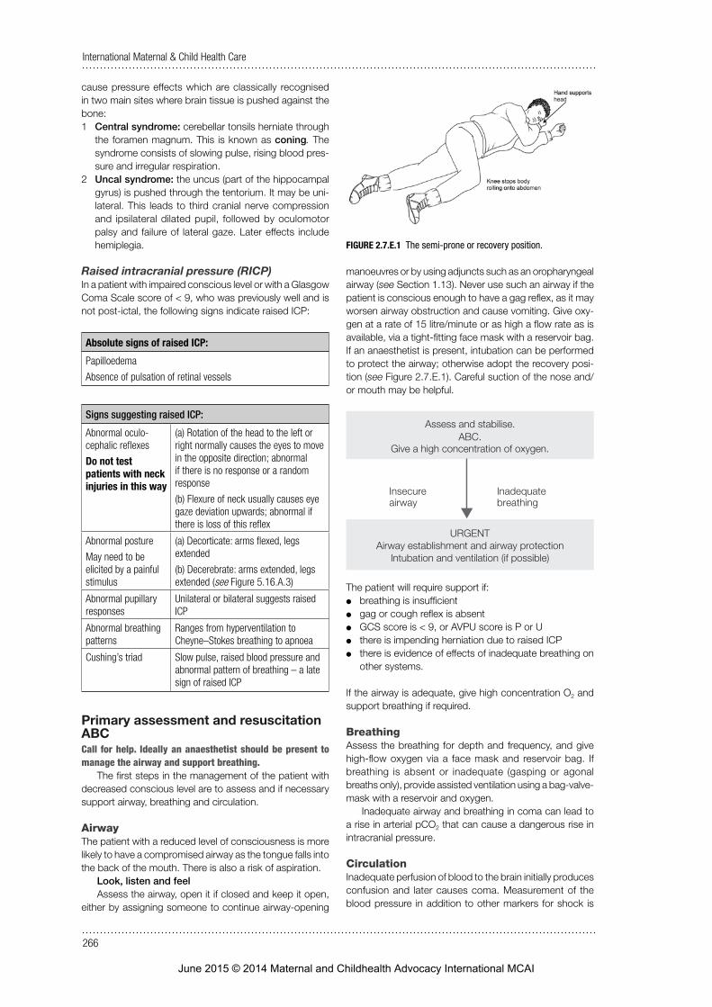
Primary assessment and resuscitation ABCCall for help. Ideally an anaesthetist should be present to manage the airway and support breathing.
The fi rst steps in the management of the patient with decreased conscious level are to assess and if necessary support airway, breathing and circulation.
AirwayThe patient with a reduced level of consciousness is more likely to have a compromised airway as the tongue falls into the back of the mouth. There is also a risk of aspiration.
Look, listen and feelAssess the airway, open it if closed and keep it open,
either by assigning someone to continue airway- opening
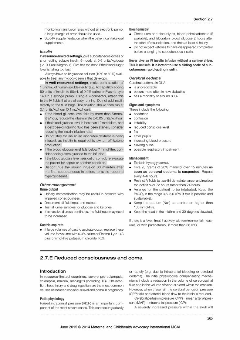
manoeuvres or by using adjuncts such as an oropharyngeal airway (see Section 1.13). Never use such an airway if the patient is conscious enough to have a gag refl ex, as it may worsen airway obstruction and cause vomiting. Give oxy-gen at a rate of 15 litre/minute or as high a fl ow rate as is available, via a tight- fi tting face mask with a reservoir bag. If an anaesthetist is present, intubation can be performed to protect the airway; otherwise adopt the recovery posi-tion (see Figure 2.7.E.1). Careful suction of the nose and/or mouth may be helpful.
Assess and stabilise.ABC.
Give a high concentration of oxygen.
Inadequatebreathing
Insecure airway
URGENT
Airway establishment and airway protection
Intubation and ventilation (if possible)
The patient will require support if: O breathing is insuffi cient O gag or cough refl ex is absent O GCS score is < 9, or AVPU score is P or U O there is impending herniation due to raised ICP O there is evidence of effects of inadequate breathing on
other systems.
If the airway is adequate, give high concentration O2 and support breathing if required.
BreathingAssess the breathing for depth and frequency, and give high- fl ow oxygen via a face mask and reservoir bag. If breathing is absent or inadequate (gasping or agonal breaths only), provide assisted ventilation using a bag- valve-mask with a reservoir and oxygen.
Inadequate airway and breathing in coma can lead to a rise in arterial pCO2 that can cause a dangerous rise in intracranial pressure.
CirculationInadequate perfusion of blood to the brain initially produces confusion and later causes coma. Measurement of the blood pressure in addition to other markers for shock is
FIGURE 2.7.E.1 The semi-prone or recovery position.
June 2015 © 2014 Maternal and Childhealth Advocacy International MCAI

267
Section 2.7
crucial in recognising hypovolaemia after haemorrhage, or unconsciousness after an eclamptic fi t with hypertension.
If the intracranial pressure is high, cerebral perfusion will be compromised if hypotension occurs. However, exces-sive fl uid administration should be avoided.
O Establish IV access quickly. O Take blood samples and send them to the lab for a
full blood count, blood smear for malarial parasites, electrolytes, liver function tests, blood glucose and blood culture.
Neurological failureAssess neurological failure as follows:
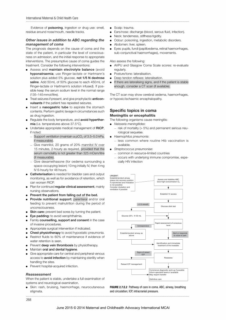
O Use the AVPU scale. O Check blood glucose levels: If the blood sugar level
is low or suspected to be low (< 2.5 mmol/litre or < 45 mg/dL), give 100 mL of 25% glucose IV over 15 minutes (dilute 50 mL of 50% glucose with 50 mL of Ringer- lactate or Hartmann’s solution) and then give 10% dextrose in Ringer- lactate or Hartmann’s solution over 4 hours (add 100 mL of 50% glucose to each 400 mL of Ringer- lactate or Hartmann’s solution infused).
O Check the pupils for signs suggesting raised intrac-ranial pressure (RICP) or opiate overdose.
O Check for neck stiffness which may suggest meningitis. O Look for other signs of raised intracranial pressure, as
outlined above.
Further assessment of conscious level can be aided by the Glasgow Coma Scale score and documentation of pupil function.
TABLE 2.7.E.1 Glasgow Coma Scale (GCS)
Response Score
Eye opening
SpontaneouslyTo verbal stimuliTo painNo response to pain
4321
Best motor response
Obeys verbal commandLocalises to painWithdraws from painAbnormal fl exion to pain (decorticate) Abnormal extension to pain (decerebrate)No response to pain
654321
Best verbal response
Orientated and conversesDisorientated and conversesInappropriate wordsIncomprehensible soundsNo response to pain
54321
Total
A GCS score of < 9 is likely to need airway protection by intubation if skills are available to undertake this safely.
TABLE 2.7.E.2 Pupillary changes
Pupil size and reactivity
Causes
Small reactive pupils
Metabolic disordersMedullary lesion
Pinpoint pupil Metabolic disordersNarcotic/organophosphate ingestion
Fixed mid- sized pupils
Midbrain lesion
Fixed dilated pupils
HypothermiaSevere hypoxaemic/ischaemic brain injuryBarbiturate ingestion (late sign)During and post seizureAnticholinergic drugs
Unilateral dilated pupil
Rapidly expanding ipsilateral lesionTentorial herniationThird cranial nerve lesionEpileptic seizures
Secondary assessment and emergency treatmentSecondary assessment occurs after stabilisation of ABCD. During secondary assessment, continue to monitor the patient, and if there is any change, reassess ABC and treat any residual problems.
Diagnostic pointersAs soon as possible during resuscitation, gain as much information about the history as possible:
O the possibility of eclampsia, which means that magne-sium sulphate may be required
O recent trauma O endemic area for infections such as malaria, sleeping
sickness and encephalitis O pre- existing neurological problem O past history of epilepsy O ingestion of poisons O underlying chronic condition (renal, cardiac, diabetes).
Remember to treat the treatable components. The cause of coma may not be certain, so it is always important to address ABC. If the patient’s condition is unstable or dete-riorating, return to ABC.
Always consider the possibility of eclampsia and the need for magnesium sulphate.
If there is no other clear cause for the coma treat with antibiotics for presumed meningitis (usually a third- generation cephalosporin, or whatever is locally available and appropriate), and in endemic areas, also treat as for cerebral malaria (see Section 2.8.D).
Take the patient’s temperature (core and peripheral). O Fever may be associated with sepsis (but lack of fever
does not exclude sepsis) or poisoning (ecstasy, cocaine or salicylates).
O Hypothermia is found in poisoning with ethanol or barbiturates.
Rash: purpura suggests meningococcal disease; bruises suggest trauma (consider domestic violence).
June 2015 © 2014 Maternal and Childhealth Advocacy International MCAI
268
International Maternal & Child Health Care
Evidence of poisoning, ingestion or drug use: smell, residue around nose/mouth, needle tracks.
Other issues in addition to ABC regarding the management of comaThe prognosis depends on the cause of coma and the state of the patient, in particular the level of conscious-ness on admission, and the initial response to appropriate interventions. The presumptive cause of coma guides the treatment. Consider the following interventions:
O Assess and maintain electrolyte balance (avoid hyponatraemia; use Ringer- lactate or Hartmann’s solution plus added 5% glucose, not 1/5 N dextrose saline. Add 50 mL of 50% glucose to each 450 mL of Ringer- lactate or Hartmann’s solution infused). If pos-sible keep the serum sodium level in the normal range (135–145 mmol/litre).
O Treat seizures if present, and give prophylactic anticon-vulsants if the patient has repeated seizures.
O Insert a nasogastric tube to aspirate the stomach contents. Perform gastric lavage in circumstances such as drug ingestion.
O Regulate the body temperature, and avoid hyperther-mia (i.e. temperatures above 37.5°C).
O Undertake appropriate medical management of RICP, if noted:
— Support ventilation (maintain a pCO2 of 3.5–5.0 kPa, if measurable).
— Give mannitol, 20 grams of 20% mannitol IV over 15 minutes, 2- hourly as required, provided that the serum osmolality is not greater than 325 mOsm/litre (if measurable).
— Give dexamethasone (for oedema surrounding a space- occupying lesion) 10 mg initially IV, then 4 mg IV 6- hourly for 48 hours.
O Catheterisation is needed for bladder care and output monitoring, as well as for avoidance of retention, which can worsen RICP.
O Plan for continued regular clinical assessment, mainly nursing observations.
O Prevent the patient from falling out of the bed. O Provide nutritional support: parenteral and/or oral
feeding to prevent malnutrition during the period of unconsciousness.
O Skin care: prevent bed sores by turning the patient. O Eye padding: to avoid xerophthalmia. O Family counselling, support and consent in the case
of invasive procedures. O Appropriate surgical intervention if indicated. O Chest physiotherapy to avoid hypostatic pneumonia. O Restrict fl uids to 60% of maintenance if evidence of
water retention is seen. O Prevent deep vein thrombosis by physiotherapy. O Maintain oral and dental hygiene. O Give appropriate care for central and peripheral venous
access to avoid infection by maintaining sterility when handling the sites.
O Prevent hospital- acquired infection.
ReassessmentWhen the patient is stable, undertake a full examination of systems and neurological examination.
O Skin: rash, bruising, haemorrhage, neurocutaneous stigmata.
O Scalp: trauma. O Ears/nose: discharge (blood, serous fl uid, infection). O Neck: tenderness, stiffness/rigidity. O Odour: poisoning, ingestion, metabolic disorders. O Abdomen: liver, spleen. O Eyes: pupils, fundi (papilloedema, retinal haemorrhages,
sub- conjunctival haemorrhages), movements.
Also assess the following: O AVPU and Glasgow Coma Scale scores: re- evaluate
regularly. O Posture/tone: lateralisation. O Deep tendon refl exes: lateralisation. O If there are lateralising signs, and if the patient is stable
enough, consider a CT scan (if available).
The CT scan may show cerebral oedema, haemorrhages, or hypoxic/ischaemic encephalopathy.
Specifi c topics in comaMeningitis or encephalitisThe following organisms cause meningitis:
O Neisseria meningitides: — risk of mortality (> 5%) and permanent serious neu-rological sequelae.
O Haemophilus pneumonia: — less common where routine Hib vaccination is available.
O Streptococcus pneumoniae: — common in resource- limited countries — occurs with underlying immune compromise, espe-cially HIV infection
Assess and stabilise ABCGive high-concentration O2
URGENT:Establish/protect airway (place into recovery position if unprotected and intubation is not possible)Consider intubation and ventilation if possible
Establish IV access
Glucose stick test
Rapid assessment of conscious level
Establish/protect airway as above
Identification and immediate treatment of the treatable
Reassess
Raised ICP management
Commence diagnostic work-up if possibleInform specialist teams if availableMay require transfer
Definitive care
Glucose 25% IV 50 mL
< 2.5 mmol/L
Unresponsive
Alert or responds to voice or pain
Signs of raised ICP
FIGURE 2.7.E.2 Pathway of care in coma. ABC, airway, breathing and circulation; ICP, intracranial pressure.
June 2015 © 2014 Maternal and Childhealth Advocacy International MCAI

269
Section 2.8
— may follow a head injury if there is damage to the dura and/or meninges.
There is a risk of coning and death if a diagnostic lumbar puncture is performed in a patient with signifi cantly raised intracranial pressure.
Diagnosis of meningitis or encephalitisClassic signs and symptoms include the following:
O headache
O vomiting O neck stiffness O opisthotonus O photophobia O rash O altered consciousness.
Poisoning (see Section 7.4).Malaria in pregnancy (see Section 2.8.D).Eclamptic coma (see Section 2.5.E).
2.8 Infections complicating pregnancy, delivery and after birth
2.8.A Pneumonia
BOX 2.8.A.1 Minimum standards Q Oxygen. Q Antibiotics (IV and oral). Q Chest X- ray.
Clinical fi ndingsA high fever is usually associated with pneumonia and bacterial tracheitis. In the absence of stridor and wheeze, breathing diffi culties in association with a signifi cant fever are likely to be due to pneumonia.
Examination of the chest may show reduced air entry, bronchial breathing and crepitations. Pleuritic chest pain, neck stiffness and abdominal pain may be present if there is pleural infl ammation. Pleural effusions and empyema are complications.
Always consider HIV infection and TB.
Emergency treatment of pneumonia O Assess ABC. O Give oxygen through nasal cannulae or mask depending
on fl ow rate required to maintain saturation (if available) as below.
O Attach a pulse oximeter (if available). O Maintain SaO2 in the range 94–98%, with nasal cannulae
at a fl ow rate usually up to 5 litres/minute or if necessary by face mask with higher fl ow rates.
O Give antibiotics for 7 days: — ampicillin 2 grams IV/IM 6- hourly plus gentamicin 80 mg IV/IM 8- hourly or 5 mg/kg IV/IM every 24 hours for most cases of community- acquired pneumonia
— cefuroxime 500 mg IV/IM 8- hourly or fl ucloxacillin 500 mg IM or IV slowly every 6 hours for suspected or bacteriologically diagnosed Staphylococcus aureus
— erythromycin 500 mg every 6 hours orally for Chlamydia or Mycoplasma pneumoniae
— or whatever is available locally and appropriate. O Sit the patient upright. O Maintain hydration.
— Extra fl uid may be needed to compensate for fl uid loss from fever.
— Fluid restriction may be needed because of inappro-priate ADH secretion, revealed by oliguria < 30 mL per hour or rising blood urea levels.
O Chest X- ray is indicated. O Large pleural effusions/empyemas should be diagnosed
where possible by ultrasound, and pleural drainage undertaken under ultrasound cover (do not place a chest drain into the heart, liver or an undiagnosed tumour or hydatid cyst) (see Section 8.3). Remember that in advanced pregnancy the diaphragm is elevated.
— Effusions/empyemas adjacent to the heart on the left side may cause pericarditis and cardiac arrhythmias. (Listen regularly for a pericardial rub, and ideally monitor an ECG if available until the patient is stable.)
June 2015 © 2014 Maternal and Childhealth Advocacy International MCAI