Embed Size (px)
Citation preview
Articles © The authors | Journal compilation © J Curr Surg and Elmer Press Inc™ | www.jcs.elmerpress.comThis is an open-access article distributed under the terms of the Creative Commons Attribution License, which permits unrestricted use, distribution, and reproduction
in any medium, provided the original work is properly cited146
Case Report J Curr Surg. 2015;5(1):146-150
ressElmer
Giant Mucinous Cystadenoma of the Appendix With Low Grade Dysplasia Presented as Acute Appendicitis: A Case
Report and Literature Review
Aleksandr A. Reznichenko
Abstract
Mucinous cystadenoma of the appendix is an unusual neoplasm, which represents one of the histological subtypes of mucocele. Giant appendiceal cystadenomas are extremely rare and most commonly presented with palpable abdominal mass, chronic right lower quad-rant (RLQ) pain, or could be asymptomatic. We present a 51-year-old female with clinical picture of acute appendicitis and a giant muci-nous cystadenoma of the appendix with low grade dysplasia.
Keywords: Mucocele; Mucinous cystadenoma; Appendicitis; Ap-pendectomy; Colectomy; Pseudomixoma peritonei
Introduction
Mucinous cystadenoma of the appendix is one of the histologi-cal types of mucocele and it accounts for 63-84% of all cases. Clinical picture of these lesions is unspecific, and most com-monly they present with palpable abdominal mass, chronic right lower quadrant (RLQ) pain, or could be asymptomatic. Computer tomography (CT) is the gold standard for the di-agnosis. Treatment requires surgery. Extent of the operation depends on the dimension and histological type of the lesion.
Giant mucinous cystadenomas are extremely rare. It is not clear, what should be called a “giant cystadenoma”. The vast majority of giant appendiceal cystadenomas were 10 cm or larger. We accounted 18 patients in the literature, who were diagnosed with mucinous cystadenoma of the appendix with the size of the neoplasm exceeding 10 cm.
We analyzed clinical and radiological features of giant mu-cinous cystadenomas, as well as differences in surgical strategy.
We report an unusual case of a giant mucinous cystade-noma of the appendix in a 51-year-old female, who presented with clinical picture of acute appendicitis.
Case Report
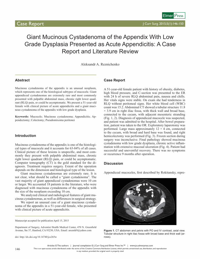
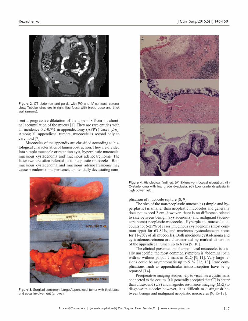
A 51-year-old female patient with history of obesity, diabetes, high blood pressure, and C-section was presented to the ER with 24 h of severe RLQ abdominal pain, nausea and chills. Her vitals signs were stable. On exam she had tenderness in RLQ without peritoneal signs. Her white blood cell (WBC) count was 15.2. Abdominal CT showed a tubular structure 11.8 × 3.9 cm in right iliac fossa, with thick wall and broad base, connected to the cecum, with adjacent mesenteric stranding (Fig. 1, 2). Diagnosis of appendiceal mucocele was suspected, and patient was admitted to the hospital. After bowel prepara-tion, patient was taken to the OR. Exploratory laparotomy was performed. Large mass approximately 12 × 4 cm, connected to the cecum, with broad and hard base was found, and right hemicolectomy was performed (Fig. 3). Frozen section during surgery was inconclusive. Final pathology showed mucinous cystadenoma with low grade dysplasia, chronic active inflam-mation with extensive mucosal ulceration (Fig. 4). Patient had successful and uneventful recovery. There was no symptoms or recurrence 9 months after operation.
Discussion
Appendiceal mucoceles, first described by Rokitansky, repre-
Manuscript accepted for publication April 15, 2015
Department of Surgery, Adventist Health Medical Center, 470 N. Greenfield Avenue, Ste 37, Hanford, CA 93230, USA. Email: [email protected]
doi: http://dx.doi.org/10.14740/jcs265w
Figure 1. CT abdomen and pelvis with PO and IV contrast, axial view. Tubular structure in right iliac fossa with broad base and thick wall (ar-rows).
Articles © The authors | Journal compilation © J Curr Surg and Elmer Press Inc™ | www.jcs.elmerpress.com 147
Reznichenko J Curr Surg. 2015;5(1):146-150
sent a progressive dilatation of the appendix from intralumi-nal accumulation of the mucus [1]. They are rare entities with an incidence 0.2-0.7% in appendectomy (APPY) cases [2-6]. Among all appendiceal tumors, mucocele is second only to carcinoid [7].
Mucoceles of the appendix are classified according to his-tological characteristics of lumen obstruction. They are divided into simple mucocele or retention cyst, hyperplastic mucocele, mucinous cystadenoma and mucinous adenocarcinoma. The latter two are often referred to as neoplastic mucoceles. Both mucinous cystadenoma and mucinous adenocarcinoma may cause pseudomixoma peritonei, a potentially devastating com-
plication of mucocele rupture [8, 9].The size of the non-neoplastic mucoceles (simple and hy-
perplastic) is smaller than neoplastic mucoceles and generally does not exceed 2 cm; however, there is no difference related to size between benign (cystadenoma) and malignant (adeno-carcinoma) neoplastic mucoceles. Hyperplastic mucocele ac-counts for 5-25% of cases, mucinous cystadenoma (most com-mon type) for 63-84%, and mucinous cystoadenocarcinoma for 11-20% of all mucoceles. Both mucinous cystadenoma and cystoadenocarcinoma are characterized by marked distention of the appendiceal lumen up to 6 cm [9, 10].
The clinical presentation of appendiceal mucoceles is usu-ally unspecific; the most common symptom is abdominal pain with or without palpable mass in RLQ [9, 11]. Very large le-sions could be asymptomatic up to 51% [12, 13]. Rare com-plications such as appendicular intussusception have being reported [14].
Preoperative imaging studies help to visualize a cystic mass connected to the cecum. It is generally accepted that CT is better than ultrasound (US) and magnetic resonance imaging (MRI) to diagnose mucocele: however, it is difficult to distinguish be-tween benign and malignant neoplastic mucoceles [9, 15-17].
Figure 3. Surgical specimen. Large Appendiceal tumor with thick base and cecal involvement (arrows).
Figure 4. Histological findings. (A) Extensive mucosal ulceration. (B) Cystadenoma with low grade dysplasia. (C) Low grade dysplasia in high power field.
Figure 2. CT abdomen and pelvis with PO and IV contrast, coronal view. Tubular structure in right iliac fossa with broad base and thick wall (arrows).
Articles © The authors | Journal compilation © J Curr Surg and Elmer Press Inc™ | www.jcs.elmerpress.com148
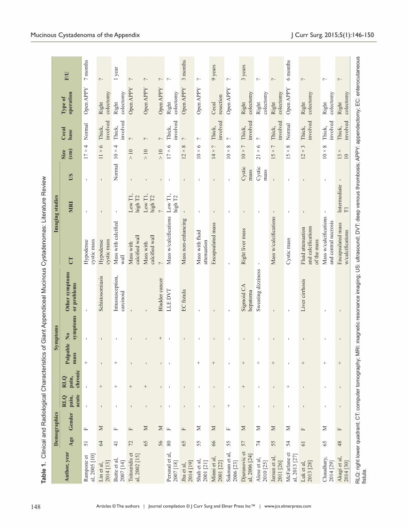
Mucinous Cystadenoma of the Appendix J Curr Surg. 2015;5(1):146-150Ta
ble
1. C
linic
al a
nd R
adio
logi
cal C
hara
cter
istic
s of
Gia
nt A
ppen
dice
al M
ucin
ous
Cys
tade
nom
as: L
itera
ture
Rev
iew
Aut
hor,
year
Dem
ogra
phic
sSy
mpt
oms
Imag
ing
stud
ies
Size
(c
m)
Cec
al
base
Type
of
oper
atio
nF/
UA
geG
ende
rR
LQ
pa
in,
acut
e
RL
Q
pain
, ch
roni
c
Palp
able
m
ass
No
sym
ptom
sO
ther
sym
ptom
s or
pro
blem
sC
TM
RI
US
Ram
pone
et
al, 2
005
[10]
51F
--
+-
-H
ypod
ense
cy
stic
mas
s-
-17
× 4
Nor
mal
Ope
n A
PPY
7 m
onth
s
Lin
et a
l, 20
14 [1
3]64
M-
+-
-Sc
hist
osom
iasi
sH
ypod
ense
cy
stic
mas
s-
-11
× 6
Thic
k,
invo
lved
Rig
ht
cole
ctom
y?
But
te e
t al,
2007
[14]
41F
-+
+-
Intu
ssus
cept
ion,
ca
rcin
oid
Mas
s with
cal
cifie
d
wal
l-
Nor
mal
10 ×
4Th
ick,
in
volv
edR
ight
co
lect
omy
1 ye
ar
Tsito
urid
is e
t al
, 200
2 [1
5]72
F-
+-
--
Mas
s with
ca
lcifi
ed w
all
Low
T1,
hi
gh T
2-
> 10
?O
pen
APP
Y?
65M
-+
--
-M
ass w
ith
calc
ified
wal
lLo
w T
1,
high
T2
->
10?
Ope
n A
PPY
?
56M
--
-+
Bla
dder
can
cer
??
->
10?
Ope
n A
PPY
?Pe
rsau
d et
al,
2007
[18]
80F
--
--
LLE
DV
TM
ass w
/cal
cific
atio
nsLo
w T
1,
high
T2
-17
× 6
Thic
k,
invo
lved
Rig
ht
cole
ctom
y?
Jha
et a
l, 20
14 [1
9]65
F-
--
EC fi
stul
aM
ass n
on-e
nhan
cing
--
12 ×
8?
Ope
n A
PPY
3 m
onth
s
Shah
et a
l, 20
01 [2
1]55
M-
-+
--
Mas
s with
flui
d at
tenu
atio
n-
-10
× 6
?O
pen
APP
Y?
Min
ni e
t al,
2001
[22]
66M
--
+-
-En
caps
ulat
ed m
ass
--
14 ×
?Th
ick,
in
volv
edC
ecal
re
sect
ion
9 ye
ars
Sakm
an e
t al,
2006
[23]
55F
+-
--
--
--
10 ×
8?
Ope
n A
PPY
?
Dju
rano
vic
et
al, 2
006
[24]
57M
-+
+-
Sigm
oid
CA
he
pato
ma
Rig
ht li
ver m
ass
-C
ystic
m
ass
10 ×
7Th
ick,
in
volv
edR
ight
co
lect
omy
3 ye
ars
Ale
se e
t al,
2010
[25]
74M
--
+-
Swea
ting
dizz
ines
s-
-C
ystic
m
ass
21 ×
6?
Rig
ht
cole
ctom
y?
Jars
un e
t al,
2011
[26]
55M
--
+-
-M
ass w
/cal
cific
atio
ns-
-15
× 7
Thic
k,
invo
lved
Rig
ht
cole
ctom
y?
McF
arla
ne e
t al
, 201
3 [2
7]54
M-
+-
--
Cys
tic m
ass
--
15 ×
8N
orm
alO
pen
APP
Y6
mon
ths
Luk
et a
l, 20
13 [2
8]61
F-
-+
-Li
ver c
irrho
sis
Flui
d at
tenu
atio
n an
d ca
lcifi
catio
ns
of th
e m
ass
--
12 ×
3Th
ick,
in
volv
edR
ight
co
lect
omy
?
Cha
udha
ry,
2014
[29]
65M
--
+-
-M
ass w
/cal
cific
atio
ns
and
cent
ral n
ecro
sis
--
10 ×
8Th
ick,
in
volv
edR
ight
co
lect
omy
?
Aka
gi e
t al,
2014
[30]
48F
--
+-
-En
caps
ulat
ed m
ass
w
/cal
cific
atio
nsIn
term
edia
te
T1-
13 ×
10
Thic
k,
invo
lved
Rig
ht
cole
ctom
y?
RLQ
: rig
ht lo
wer
qua
dran
t; C
T: c
ompu
ter t
omog
raph
y; M
RI:
mag
netic
reso
nanc
e im
agin
g; U
S: u
ltras
ound
; DVT
: dee
p ve
nous
thro
mbo
sis;
APP
Y: a
ppen
dect
omy;
EC
: ent
eroc
utan
eous
fis
tula
.
Articles © The authors | Journal compilation © J Curr Surg and Elmer Press Inc™ | www.jcs.elmerpress.com 149
Reznichenko J Curr Surg. 2015;5(1):146-150
Appendiceal mucocele requires surgical resection. The ex-tent of surgery remains controversial and depends on the size and histology type. For simple and hyperplastic mucoceles of smaller size, APPY is adequate. Cecal resection is indicated for cystadenomas with large base, and hemicolectomy is rec-ommended for cystadenocarcinomas [9, 10, 18, 19]. There is no consensus regarding frozen section of the removed speci-men during surgery as it is not always accurate [20].
Giant mucinous cystadenomas of the appendix are ex-tremely rare and only few cases have been reported. There is no clear definition of giant cystadenoma. According to the lit-erature, the vast majority of giant appendiceal cystadenomas were described to be bigger than 10 cm in size [10, 13-15, 18-30]. We included 18 patients into our literature review, who were diagnosed with mucinous cystadenoma of the appendix with the size of the neoplasm exceeding 10 cm. Patients with cystoadenocarcinoma were not included. Patients with mu-cocele 10 cm and larger but without elements of mucinous cystadenoma were not included. We analyzed clinical and ra-diological characteristics of giant mucinous cystadenoma, as well as different surgical approaches. Results of this analysis are summarized in Table 1.
Giant mucinous cystadenoma of the appendix was seen slightly more often in men, but it was not statistically signifi-cant. Majority of the patients were older than 50 years (89%). The most common symptom was palpable mass (55%), fol-lowed by chronic RLQ pain (33%). Absence of symptoms was noticed in one patient (5%). Acute onset of RLQ pain was pre-sent in the only one patient (5%), who was emergently taken to the OR and the diagnosis of appendiceal tumor was made in-traoperatively [23]. We present another case of giant mucinous cystadenoma of the appendix with the clinical presentation of acute appendicitis. Our case is noticeable because, despite an unusual clinical presentation, the diagnosis of mucocele was suspected prior to surgery and patient was well prepared for the operation.
Imaging studies were important in evaluation of appendi-ceal neoplasms. CT was more informative among other tests (MRI and US) and most commonly was showing large hy-podense encapsulated mass with fluid attenuation and calcified wall.
Decision about extent of surgical resection was made most of the time during surgery based on the thickness and involve-ment of the cecal base in the pathological process. Laparo-scopic approach was not utilized, because of risk of mucocele rupture. Open APPY was considered appropriate for mucinous cystadenomas without involvement of the cecum. Cecectomy or right hemicolectomy were performed in patients with thick appendiceal base and involvement of the cecum, with right hemicolectomy being the preferred approach. We agree with this choice, particularly when frozen section is not really help-ful, like it was in our case.
Conclusion
Giant mucinous cystadenomas of the appendix are extremely rare. They most commonly present with palpable mass, but not in acute settings. We present an unusual case of giant muci-
nous cystadenoma, with clinical picture of acute appendicitis. We proposed to define a giant mucinous cystadenoma as an appendiceal neoplasm exceeding 10 cm in size. CT remains the gold standard for the diagnosis. Extent of surgical resec-tion depends on the histology type, as well as thickness and involvement of the cecal base.
Abbreviations
RLQ: right lower quadrant; CT: computer tomography; MRI: magnetic resonance imaging; US: ultrasound; DVT: deep ve-nous thrombosis; APPY: appendectomy; EC: enterocutaneous fistula; WBC: white blood cell count; PO: per os; IV: intrave-nous
References
1. Rokitansky CF. A manual of pathologica anatomy. Vol 2. English translation of the Vienna edition (1942). Philadel-phia: Blancard and Lea, 1855;89.
2. Zagrodnik DF, Rose DM. Mucinous cystadenoma of the appendix. Can J Surg. 2009;52(2):158-170.
3. Demetrashvili Z, Chkhaidze M, Khutsishvili K, Topchish-vili G, Javakhishvili T, Pipia I, Qerqadze V. Mucocele of the appendix: case report and review of literature. Int Surg. 2012;97(3):266-269.
4. Zanati SA, Martin JA, Baker JP, Streutker CJ, Marcon NE. Colonoscopic diagnosis of mucocele of the appen-dix. Gastrointest Endosc. 2005;62(3):452-456.
5. Woodruff R, McDonald JR. Benign and malignant cystic tumors of the appendix. Surg Gynecol Obstet. 1940;71:750-755.
6. Aho AJ, Heinonen R, Lauren P. Benign and malignant mucocele of the appendix. Histological types and prog-nosis. Acta Chir Scand. 1973;139(4):392-400.
7. Deans GT, Spence RA. Neoplastic lesions of the appen-dix. Br J Surg. 1995;82(3):299-306.
8. Higa E, Rosai J, Pizzimbono CA, Wise L. Mucosal hy-perplasia, mucinous cystadenoma, and mucinous cystad-enocarcinoma of the appendix. A re-evaluation of appen-diceal "mucocele". Cancer. 1973;32(6):1525-1541.
9. Jandui Gomes De Abreu Filho, Erivaldo Fernandes De Lira. Mucocele of the appendix: appendectomy or colec-tomy. J Coloproctol. 2011;31(3).
10. Rampone B, Roviello F, Marrelli D, Pinto E. Giant ap-pendiceal mucocele: report of a case and brief review. World J Gastroenterol. 2005;11(30):4761-4763.
11. Stocchi L, Wolff BG, Larson DR, Harrington JR. Sur-gical treatment of appendiceal mucocele. Arch Surg. 2003;138(6):585-589; discussion 589-590.
12. Dhage-Ivatury S, Sugarbaker PH. Update on the surgical approach to mucocele of the appendix. J Am Coll Surg. 2006;202(4):680-684.
13. Lin C, Li X, Guo Y, Hu G, Zhang Y, Yang K, Gan Y, et al. Simultaneous giant mucinous cystadenoma of the appen-dix and intestinal schistosomiasis: 'case report and brief review'. World J Surg Oncol. 2014;12(385.
Articles © The authors | Journal compilation © J Curr Surg and Elmer Press Inc™ | www.jcs.elmerpress.com150
Mucinous Cystadenoma of the Appendix J Curr Surg. 2015;5(1):146-150
14. Butte JM, Torres J, Henriquez IM, Pinedo G. Appendicu-lar mucosal intussusception into the cecum secondary to an intramural mucinous cystoadenoma of the appendix. J Am Coll Surg. 2007;204(3):510.
15. Tsitouridis I, Kouklakis G, Kalambakas A, Papastergiou C, Emmanouilidou M, Goutsaridou F, Tsantiridis C. Gi-ant appendiceal mucoceles of cystadenomas type: CT and MRI evaluation. Report of three cases and review of lit-erature. Ann of Gastroenterology. 2002;15(1):53-57.
16. Vagholkar K, Jain U, Mahadik A, Iyengar M. Mucocele of the appendix. JMSCR. 2014;2(12):3163-3170.
17. Wang H, Chen YQ, Wei R, Wang QB, Song B, Wang CY, Zhang B. Appendiceal mucocele: A diagnostic dilemma in differentiating malignant from benign lesions with CT. AJR Am J Roentgenol. 2013;201(4):W590-595.
18. Persaud T, Swan N, Torreggiani WC. Giant mucinous cys-tadenoma of the appendix. Radiographics. 2007;27(2):553-557.
19. Jha NK, Sinha DK, Anand A, Rai MK, Gandhi A, Ya-dav J, Yadav SK. Mucinous cystadenoma of the appendix with enterocutaneous fistula: a therapeutic dilemma. Gas-troenterol Rep (Oxf). 2015;3(1):86-89.
20. Harris SH, Khan R, Ansari MM, Maheshwari V. Inciden-tally discovered giant mucocele of the appendix. J Coll Physicians Surg Pak. 2014;24 Suppl 3(S196-197.
21. Shah I, Gupta R, Sanjeev N, Gupta CL. A rare presenta-tion of cystadenoma of appendix as giant retroperitoneal mass. Practitioner. 2001;8(4):250-251.
22. Minni F, Petrella M, Morganti A, Santini D, Marrano D. Giant mucocele of the appendix: report of a case. Dis Co-
lon Rectum. 2001;44(7):1034-1036.23. Sakman G, Parsak C, Akcam T, Alabaz O. Four giant mu-
coceles of the appendix vermiformis. The Internet J of Surgery. 2006;9(1).
24. Djuranovic SP, Spuran MM, Kovacevic NV, Ugljesic MB, Kecmanovic DM, Micev MT. Mucinous cystadenoma of the appendix associated with adenocarcinoma of the sig-moid colon and hepatocellular carcinoma of the liver: re-port of a case. World J Gastroenterol. 2006;12(12):1975-1977.
25. Alese OB, Irabor DO. Mucinous cystadenoma of the ap-pendix: a case report. Afr Health Sci. 2010;10(1):99-100.
26. Gaby Adriana Alarcon Jarsun, Ariel Shuchleib Cung, Leon Ylgovsky Weintraub, Alvaro Padilla Rodriguez, Alberto Chousleb Kalach, Samuel Shuchleib Chaba An Med (Mex), 2011;56(4):210-217.
27. McFarlane ME, Plummer JM, Bonadie K. Mucinous cystadenoma of the appendix presenting with an el-evated carcinoembryonic antigen (CEA): Report of two cases and review of the literature. Int J Surg Case Rep. 2013;4(10):886-888.
28. Luk YS, Lam LRS, Shum JSF, Khoo JLS. Mucinous cys-tadenoma of the appendix: a rare cause of right lower ab-dominal mass. Hong Kong J Radiol. 2013;16:e31-34.
29. Chaudhary P. Mucinous cystadenoma of the appen-dix: A diagnostic dilemma. J of Surgical Academia. 2014;4(1):60-62.
30. Akagi I, Yokoi K, Shimanuki K, Satake S, Takeda K, Shimizu T, Kondo R, et al. Giant appendiceal mucocele: report of a case. J Nippon Med Sch. 2014;81(2):110-113.