Embed Size (px)
Citation preview

* CHAPTER 11
Management of thalassaemia
Renzo Galanello, Raffaella Origa
IRON2009_CAP.11(264-285):EBMT2008 4-12-2009 16:25 Pagina 264

1. IntroductionThe clinical manifestations of thalassaemia major typically start in the first yearof life. The reduced amount (beta+) or absence (beta0) of beta globin chains resultsin a relative excess of unbound alpha globin chains that precipitate in erythroidprecursors in the bone marrow, leading to their premature death and hence toineffective erythropoiesis (1).Peripheral haemolysis in thalassaemia major is less prominent and occurs wheninsoluble alpha globin chains induce membrane damage in the peripheralerythrocytes. Ineffective erythropoiesis and haemolysis cause severe anaemia, whichis the first clinical manifestation of thalassaemia major. Anaemia stimulates theproduction of erythropoietin with consequent intensive but ineffective expansionof the bone marrow (up 25 to 30 times normal), which in turn causes typical bonedeformities of the skull, facies, short and long bones and growth abnormalities.Prolonged and severe anaemia and increased erythropoietic drive also result inhepatosplenomegaly and extramedullary erythropoiesis. A regular transfusionregimen is the most effective method for alleviating anaemia in patients withthalassaemia. However, this regimen eventually results in progressive iron overload,which if not adequately treated causes severe complications, including liver andendocrine gland damage and heart dysfunction, which is the most common causeof death (2).The complete clinical picture of thalassaemia major includes features that are dueboth to the disease itself and to the therapy (both transfusions and iron chelation).If untreated or poorly treated, anaemia can lead to failure to thrive, tissue anoxia,congestive heart failure and eventually early death.In this chapter we will discuss some aspects of the management of thalassaemiamajor, particularly transfusion therapy, how to manage transfusion complicationsother than iron overload (which is discussed elsewhere in this book) and othersupportive therapies, endocrinopathies and their treatment, osteoporosis, fertility,assisted reproduction and management of pregnancy.Checklists for the clinical and laboratory evaluation of patients with betathalassaemia major at different ages (Tables 1, 2 and 3) and a summary ofindications for splenectomy (Table 4) are given.Non-conventional therapies, such as haematopoietic stem cell transplantation,which, if successful, can offer a complete cure for patients with beta thalassaemia,and gene therapy, which offers a potentially curative approach, will be discussedin other chapters.
DISORDERS OF ERYTHROPOIESIS, ERYTHROCYTES AND IRON METABOLISM265
CHAPTER 11 • Management of thalassaemia
IRON2009_CAP.11(264-285):EBMT2008 4-12-2009 16:25 Pagina 265

2. Transfusion therapy
2.1 GoalsThe goals of transfusion therapy are the correction of anaemia, suppression oferythropoiesis and inhibition of gastrointestinal iron absorption, which occurs intransfused patients as a consequence of increased, although ineffective,erythropoiesis.
2.2 Whom to transfuseThe decision to start transfusion in patients with a confirmed diagnosis of thalassaemiashould be based on the presence of severe anaemia (Hb < 7 g/dL on 2 occasions,more than two weeks apart, excluding all other contributory causes such asinfections). However, even in patients with haemoglobin > 7 g/dL, other factors
THE HANDBOOK 2009 EDITION266
Before each transfusion• Compatibility testing
At each transfusion• Complete physical exam• Complete blood count or Hb level
Every 3 months• Biochemistry
GlucoseCreatinineAlanine aminotransferase (ALT)
• Serum ferritin• Urine iron excretion
Every 6 months• Complete growth assessment
Standing heightSitting heightWeightCranial circumference
Yearly• Evaluation of transfusion therapy
Mean pre-and post-transfusion HbMean daily Hb fallMean transfusion intervalRed cell requirement (mL/kg/year)Iron intake (mg/kg/day)
Yearly (continued)• Biochemistry
Calcium, phosphorus, sodium, potassium,zincAlkaline phosphataseTotal and unconjiugated bilirubinγ−GTLDHTotal protein and albumin
• VirologyAnti-HIVAnti-HCV (if anti-HCV negative)HCVRNA (if anti-HCV positive and HCV-RNAnegative)
• ECG• Bidimensional echocardiography• Chelation toxicity monitoring
Ophthalmology examinationAudiology examination
Every two years• Abdominal ultrasound scan
As indicated• Bone age• Endocrine function evaluation• Additional examinations
Table 1: Clinical and laboratory evaluation checklist for children with betathalassaemia major
IRON2009_CAP.11(264-285):EBMT2008 4-12-2009 16:25 Pagina 266
should be considered, including age at presentation of first symptoms, facialchanges, poor growth, evidence of bony expansion and increasing splenomegaly.Where transfusion is started because of poor growth or bony changes, it should notbe assumed that lifelong transfusion will be necessary. When possible, the decisionto start regular transfusions should not be delayed until after the third year, as therisk of developing multiple red cell antibodies increases, with subsequent difficultyin finding suitable blood units.
2.3 Transfusion regimenThe superiority of regularly repeated transfusions, as compared to transfusions onlyfor symptomatic anaemia, was first recognised by Orsini in France and later by Wolman
DISORDERS OF ERYTHROPOIESIS, ERYTHROCYTES AND IRON METABOLISM
CHAPTER 11 • Management of thalassaemia
267
Before each transfusion• Compatibility testing
At each transfusion• Complete physical exam• Complete blood count or Hb level
Every 3 months• Biochemistry
GlucoseCreatinineAlanine aminotransferase (ALT)
• Serum ferritin• Urine iron excretion
Every 6 months• Complete growth assessment
Standing heightSitting heightWeightCranial circumference
• Pubertal assessment (Tanner stages)• ECG
Yearly• Evaluation of transfusion therapy
Mean pre-and post-transfusion HbMean daily Hb fallMean transfusion intervalRed cell requirement (mL/kg/year)Iron intake (mg/kg/day)
Yearly (continued)• Biochemistry
Calcium, phosphorus, sodium, potassium,zincAlkaline phosphataseTotal and unconjiugated bilirubinγ−GTLDHTotal protein and albumin
• VirologyAnti-HIVAnti-HCV (if anti-HCV negative)HCVRNA (if anti-HCV positive and HCVRNAnegative)
• Endocrine function evaluationTSHParathyroid hormoneFSH, LH, testosterone, oestradiolOral glucose tolerance test
• Bidimensional echocardiography• Abdominal ultrasound scan• Chelation toxicity monitoring
Ophthalmology examinationAudiology examination
As indicated• Bone age• Additional examinations
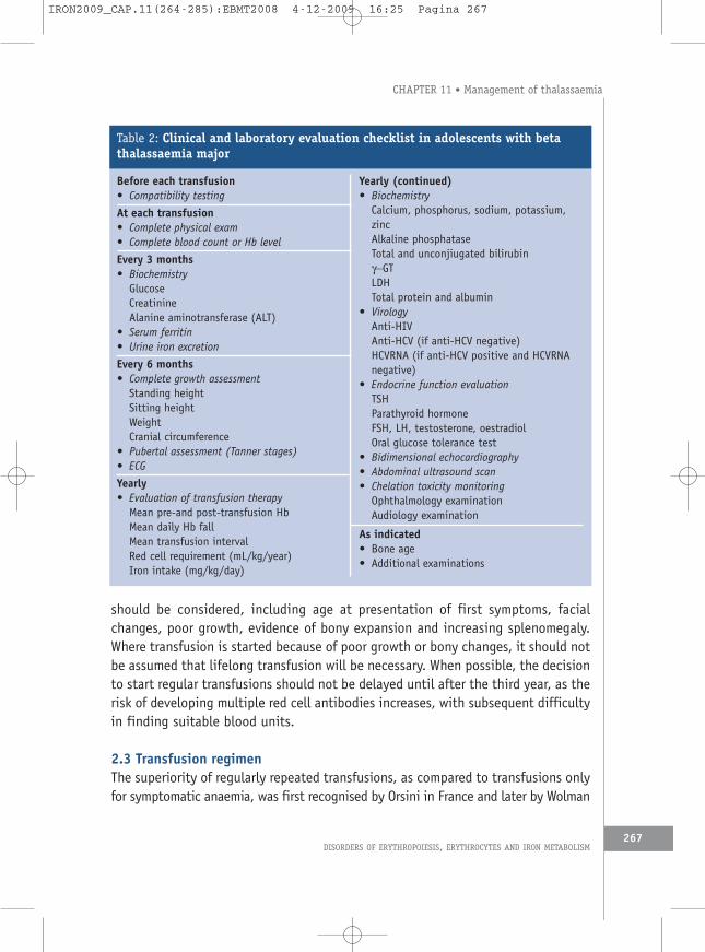
Table 2: Clinical and laboratory evaluation checklist in adolescents with betathalassaemia major
IRON2009_CAP.11(264-285):EBMT2008 4-12-2009 16:25 Pagina 267

and Piomelli in US, who suggested a transfusion program aimed at monitoring a basalHb level sufficient to eliminate hypoxia (3, 4). Several different regimens have beenproposed over the years, but at present the majority of centres choose to transfuseat a pre-transfusion Hb level of 9 to 10 g/dL, and to reach a post-transfusion levelof 13 to 14 g/dL. This prevents growth impairment, organ damage and bonedeformities, allowing normal activity and quality of life, and is associated with
THE HANDBOOK 2009 EDITION268
Before each transfusion• Compatibility testing
At each transfusion• Complete physical exam• Complete blood count or Hb level
Every 3 months• Biochemistry
GlucoseCreatinineAlanine aminotransferase (ALT)
• Serum ferritin• Urine iron excretion
Every 6 months• ECG
Yearly• Evaluation of transfusion therapy
Mean pre-and post-transfusion HbMean daily Hb fallMean transfusion intervalRed cell requirement (mL/kg/year)Iron intake (mg/kg/day)
Yearly (continued)• Biochemistry
Calcium, phosphorus, sodium, potassium,zincAlkaline phosphataseTotal and unconjiugated bilirubinγ−GTLDHTotal protein and albumin
• VirologyAnti-HIVAnti-HCV (if anti-HCV negative)HCVRNA (if anti-HCV positive andHCVRNA negative)
• Endocrine function evaluationTSHParathyroid hormoneFSH, LH, testosterone, oestradiolOral glucose tolerance test
• Bone density evaluation• Bidimensional echocardiography• Abdominal ultrasound scan• Chelation toxicity monitoring
Ophthalmology examinationAudiology examination
As indicated• 24-hour Holter monitor• Cardiac stress test• Heart iron assessment (MRI)• Complete liver function assessment
including coagulation• Alpha-fetoprotein• Liver biopsy• Liver iron assessment (SQUID, MRI)• Additional examinations
Table 3: Clinical and laboratory evaluation checklist for adults with betathalassaemia major
IRON2009_CAP.11(264-285):EBMT2008 4-12-2009 16:25 Pagina 268

relatively low rates of blood requirement and of iron accumulation (5). The frequencyof transfusion is usually every two to five weeks. Shorter intervals might further reducethe overall blood requirement, but are incompatible with an acceptable quality oflife. The amount of blood to be transfused depends on several factors including theweight of the patient, and the target increase in Hb level. Appropriate graphs andformulae to calculate the amount of blood to be transfused are available (6, 7). Ingeneral, the amount of transfused RBC should not exceed 15 to 20 mL/kg/day, infusedat a maximum rate of 5 mL/kg/hour to avoid a fast increase in blood volume.
2.4 Evaluation of transfusion therapyTo evaluate the effectiveness of transfusion therapy, some indices should berecorded at each transfusion, including pre- and post-transfusion Hb, amount andhaematocrit (Hct) of the unit, daily Hb fall, and interval between transfusions. Thesemeasurements enable two important parameters to be calculated: red cellrequirement and iron intake. Dedicated computerised programs are available tomonitor transfused thalassaemia patients (8). If the annual red cell requirementexceeds 1.5 times that of splenectomised patients splenectomy should be considered,provided that other reasons for increased consumption, such as haemolyticreactions (see below), have been excluded. For patients maintaining a pre-transfusion Hb of 9.5 g/dL, the increase in transfusion requirement is representedby a consumption of more than 200 mL of RBC/kg/year (assuming that the Hct ofthe unit of red cells is 75%) (Table 4) (6).
2.5 Characteristics of blood products for transfusionCareful selection of healthy voluntary donors is a prerequisite for obtaining safe bloodunits for patients with thalassaemia. To avoid transfusion reactions from antileukocyteand antiplatelet antibodies and transmission of viral agents present in leukocytessuch as cytomegalovirus, patients with thalassaemia should receive leukoreducedpacked red cells. Removal of leukocytes and platelets is obtained by filtration ofwhole blood (9). Reduction to 1x10⁶ or fewer leukocytes per unit is considered thecritical threshold for eliminating adverse reactions attributed to contaminating whitecells (Council of Europe, RE 2006).
DISORDERS OF ERYTHROPOIESIS, ERYTHROCYTES AND IRON METABOLISM
CHAPTER 11 • Management of thalassaemia
269
• Blood consumption > 200-220 mL/kg/year• Symptoms of splenic enlargement• Leukopenia and/or thrombocytopenia• Increasing iron stores despite good chelation
Table 4: Indications for splenectomy
IRON2009_CAP.11(264-285):EBMT2008 4-12-2009 16:25 Pagina 269

In the rare patients sensitised to plasma proteins (see later) washed red cells maybe beneficial.Extended red cell antigen typing including at least Rh antigens, Duffy, Kidd and Kellis recommended before starting transfusions to avoid alloimmunisation against redcells. Despite increasingly sophisticated blood additives, RBCs suffer from ex vivostorage and undergo biochemical, structural, enzymatic, morphological and functionaldeterioration. Although many of these changes are reversible following transfusion,it is debatable what is the limit to RBC storage beyond which transfusion isunfavourable: the current practice is to transfuse red cells in appropriate additivesolutions for less than two weeks (10, 11).
2.6 Complications of transfusionsAlthough red cell transfusions are lifesavers for patients with thalassaemia, they areresponsible for a series of complications and expose the patients to a variety of risks.Adverse events associated with red cell transfusions are summarised in Table 5. Ironoverload is the most relevant complication associated with transfusion therapy. Theconsequences of secondary iron overload and its treatment are reported elsewherein this book.
2.6.1 InfectionsThe risk of infections is one of the most relevant potential complications of bloodtransfusion. Blood-borne infections can be viral, bacterial or protozoal (9) (Table 6).The hepatotropic viruses include hepatitis B virus (HBV), hepatitis C virus (HCV) andhepatitis G virus. Transmission of these viruses in multitransfused patients varies widelyin different parts of the world and is directly correlated with their frequency in eachpopulation. The reported prevalence of serologic evidence of HBV infection is 19%in French, 34% in Italian and 56 to 66% in Indian patients (12, 13). Administrationof recombinant HBV vaccine is recommended for all non-infected patients. HepatitisC virus infection is common in patients transfused before 1989, when screening ofblood units was widely introduced in clinical practice (14). A 75% prevalence of HCVantibody positivity has been reported in Italian patients, 23% in French, 21% in Indianand about 30% in North American patients (15-17). Chronic HCV infection developsin 70-80% of infected patients and risk of progression to cirrhosis and/or hepatocellular
THE HANDBOOK 2009 EDITION270
• Iron overload• Infections• Immunisations (allergic reactions, alloimmunisation)
Table 5: Transfusion-dependent complications
IRON2009_CAP.11(264-285):EBMT2008 4-12-2009 16:25 Pagina 270

carcinoma is increased if an elevated iron load is present (18, 19). The hepatitis Gvirus, commonly found in polytransfused patients, seems to be associated with a verylow risk of chronic liver disease (20). The prevalence of HIV positivity in patientsfrom Mediterranean countries is 1.6%. Most of these patients were infected before1987, when blood donor screening became systematic. The implementation ofnucleic acid testing (NAT) together with the existing antigen/antibody-based assaysfor donor screening has further reduced the residual risk of recipient’s viral infectionby shortening the window period that is the temporal gap spanning from the timeof infection to seroconversion. In Italy, before NAT introduction, the residual risk fortransfusion-transmitted HCV and HIV was 2.7 per 106 blood units and 2.2 per 106
blood units, respectively and has currently dropped to 0.2 per 106 blood units forHCV and 0.4 per 106 for HIV (21). The current risk of HBV transmission has beencalculated to be 1 per 180,000 blood units in US (22).The risk of bacterial contamination of RBC for transfusion is 2.6 per 100,000 units(22). The most common bacteria include Yersinia, Serratia and Pseudomonas. Fever,shivers, nausea, vomiting, dyspnoea and hypotension are the most commonsymptoms occurring during or shortly after transfusion. Differential diagnosisincludes haemolytic and non-haemolytic reactions. Supportive and antibiotictherapy should be initiated as required. In US death from transfusion-transmittedinfections is the second leading cause of death. In countries where it is endemic,malaria can be acquired from transfusions. Transmission of the variant Creutzfeldt-Jacob disease is questionable.
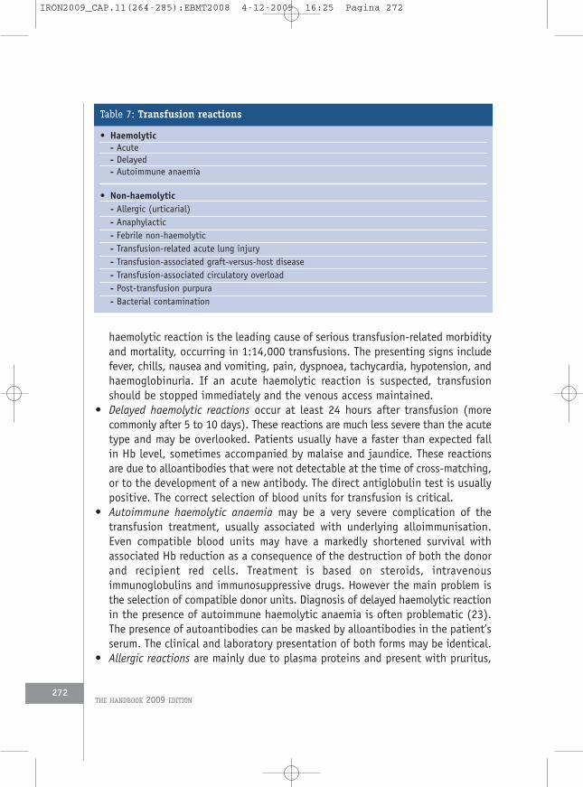
2.6.2 Transfusion reactionsAdverse reactions to RBC transfusions usually occur during or after transfusion andcan be haemolytic or non-haemolytic (23) (Table 7).• Acute haemolytic reactions can be due to transfusion of incompatible units
arising from errors in blood typing or in patient identification. An acute
DISORDERS OF ERYTHROPOIESIS, ERYTHROCYTES AND IRON METABOLISM
CHAPTER 11 • Management of thalassaemia
271
• KnownViral- HIV, HCV, HBV, HTLV 1, West Nile virusBacterialParasitic
• TheoreticalCreutzfeldt-Jakob diseaseEmerging and new pathogens
Table 6: Transmission of infectious agents with red cell transfusions
IRON2009_CAP.11(264-285):EBMT2008 4-12-2009 16:25 Pagina 271
haemolytic reaction is the leading cause of serious transfusion-related morbidityand mortality, occurring in 1:14,000 transfusions. The presenting signs includefever, chills, nausea and vomiting, pain, dyspnoea, tachycardia, hypotension, andhaemoglobinuria. If an acute haemolytic reaction is suspected, transfusionshould be stopped immediately and the venous access maintained.
• Delayed haemolytic reactions occur at least 24 hours after transfusion (morecommonly after 5 to 10 days). These reactions are much less severe than the acutetype and may be overlooked. Patients usually have a faster than expected fallin Hb level, sometimes accompanied by malaise and jaundice. These reactionsare due to alloantibodies that were not detectable at the time of cross-matching,or to the development of a new antibody. The direct antiglobulin test is usuallypositive. The correct selection of blood units for transfusion is critical.
• Autoimmune haemolytic anaemia may be a very severe complication of thetransfusion treatment, usually associated with underlying alloimmunisation.Even compatible blood units may have a markedly shortened survival withassociated Hb reduction as a consequence of the destruction of both the donorand recipient red cells. Treatment is based on steroids, intravenousimmunoglobulins and immunosuppressive drugs. However the main problem isthe selection of compatible donor units. Diagnosis of delayed haemolytic reactionin the presence of autoimmune haemolytic anaemia is often problematic (23).The presence of autoantibodies can be masked by alloantibodies in the patient’sserum. The clinical and laboratory presentation of both forms may be identical.
• Allergic reactions are mainly due to plasma proteins and present with pruritus,
THE HANDBOOK 2009 EDITION272
• Haemolytic- Acute- Delayed- Autoimmune anaemia
• Non-haemolytic- Allergic (urticarial)- Anaphylactic- Febrile non-haemolytic- Transfusion-related acute lung injury- Transfusion-associated graft-versus-host disease- Transfusion-associated circulatory overload- Post-transfusion purpura- Bacterial contamination
Table 7: Transfusion reactions
IRON2009_CAP.11(264-285):EBMT2008 4-12-2009 16:25 Pagina 272
erythema and urticaria. If the respiratory tract is involved there may behoarseness, stridor, wheezing, retrosternal pain, dyspnoea, anxiety and, rarely,cyanosis. Treatment is based on antihistaminics for the mild forms and epinephrinefor the severe forms. Premedication with antihistaminics and/or steroids may helpto prevent allergic reactions. Patients with repeated allergic reactions maybenefit from washed blood units.The most severe forms of allergic reactions are the anaphylactic reactions. Theymay occur in polytransfused patients with IgA deficiency. Besides the above-reported symptoms, there is cardiovascular instability including hypotension,tachycardia, cardiac arrhythmia and loss of consciousness. Repeated washing ofRBC is warranted in these patients. To detect patients at risk, evaluation ofimmunoglobulin level is recommended before starting a transfusion program.
• Febrile non-haemolytic reactions are due to the presence in the blood unit ofpyrogenic cytokines originating from leukocytes during blood storage, or ofacquired antibodies against donor leukocyte antigens. They occur during orwithin a few hours after transfusion and manifest with fever, chills, sensationof cold, headache, nausea and vomiting. Treatment consists of discontinuing thetransfusion and administering antipyretics and steroids. Premedication with thesame drugs is often used to prevent febrile reactions. Removal of leukocytes bypost-storage blood filtration has dramatically reduced the occurrence of febrilereactions in patients with thalassaemia (24). The use of pre-storage filtrationhas been shown to reduce the generation of cytokines and may be effective inpreventing febrile reactions. Washing of RBC can be used to remove cytokinesaccumulated during storage.
• Transfusion-related acute lung injury (TRALI) is a rare but severe reactionoccurring within hours of the transfusion. It is characterised by dyspnoea,cyanosis, tachycardia, fever and hypotension. A chest X-ray usually showspulmonary oedema. TRALI is caused by specific anti-neutrophil or anti-HLAantibodies. Treatment includes stopping the transfusion, oxygen administration,steroids and diuretics. In the severe forms mechanical ventilation is required.
• Transfusion-associated graft-versus-host disease may be a risk in immunosuppressedpatients who receive RBC transfusions from family donors, sharing some HLAhaplotypes with the recipient. Symptoms become evident weeks after transfusionand affect skin, liver and gastrointestinal tract.
• Circulatory overload may be a consequence of fast and/or excessive volumeadministration. It presents as congestive heart failure during or immediately aftertransfusion, with dyspnoea, tachycardia, cyanosis, hypertension and pulmonaryoedema. Patients with heart disease are at risk of transfusion-associatedcirculatory overload.
DISORDERS OF ERYTHROPOIESIS, ERYTHROCYTES AND IRON METABOLISM
CHAPTER 11 • Management of thalassaemia
273
IRON2009_CAP.11(264-285):EBMT2008 4-12-2009 16:25 Pagina 273

• Post-transfusion purpura is a rare reaction characterised by thrombocytopenia andsometimes bleeding. The thrombocytopenia is self-limited with recovery withinone to a few weeks. The mechanism is not clear but appears to be immune-mediated. Reaction to bacterial blood unit contamination has been discussed above.
3. Growth deficiency and endocrinopathiesAround 30% of patients with thalassaemia major are affected by growth disorders(25, 26). Growth is usually normal until the age of 9 and afterwards there is a decreaseof growth rate which leads to a final height lower than that expected on a geneticbasis. The factors contributing to stunted growth are not completely understood butinclude chronic anaemia, hypersplenism, folate deficiency, direct iron toxicity,endocrine disorders such as hypogonadism, hypothyroidism and growth hormone (GH)insufficiency, chronic liver disease and deferoxamine toxicity. In regularly transfusedand well-chelated patients, deferoxamine at high doses or at therapeutic doses inpatients with hypersensitivity, can be toxic to osteogenesis, collagen synthesis andbone turnover, leading to reduced growth (especially of the trunk), protrusion of thesternum, valgus deformity of knees and elbows, swelling of wrists and knees and slidingof the femoral head (27, 28). Radiologically, platyspondylisis and rachitic-likelesions in the metaphyses of the long bones are present (Figure 1). The effectivetreatment of growth disorders depends on an accurate assessment of their cause.Studies evaluating the secretion of GH have yielded contradictory results, limitingthe therapeutic use of GH to those patients proven to have GH deficiency, the onlyones to show a satisfactory response to treatment (29-31). In the case of signs ofdeferoxamine toxicity, a reduction in deferoxamine dose or its substitution with anoral chelator, can prevent progression of bone lesions and improve growth. In themost severe cases, surgery can be necessary to correct valgus deformity of the kneesor sliding of the femoral head.
3.1 Delayed puberty and hypogonadismDelayed puberty (defined as the complete lack of pubertal development by the ageof 13 in girls and by the age of 14 in boys) and hypogonadism (the absence oftesticular enlargement in boys and of breast development in girls by the age of 16)are the most common endocrinological complications of iron overload. Delayed ordefective function of the hypothalamic/pituitary axis occurs in approximately 50%of both male and female patients (25, 26, 32, 33). Clinically, sexual infantilism orinterruption of puberty are associated with a marked delay in growth. The responseof LH and FSH to a stimulation test with GnRH is poor, and only basal levels of sexualsteroids (testosterone and oestradiol) may be obtained. A stimulation test with human
THE HANDBOOK 2009 EDITION274
IRON2009_CAP.11(264-285):EBMT2008 4-12-2009 16:25 Pagina 274

chorionic gonadotrophin (hCG) in males and with human menopausal gonadotrophin(hMG) in females leads to a normal or almost normal increase in testosterone or17beta-oestradiol. Appropriate treatment depends on factors such as age, severityof iron overload, chronic liver disease, thrombophilia status and the presence ofpsychological problems. All these issues must be discussed by the doctor in chargeof the patient’s care, with the endocrinologist and the patient himself. Hormonesubstitution is usually started after pubertal assessment according to Tanner. Fordelayed puberty in girls, therapy may begin with the administration of ethinyloestradiol (2.5-5 µg daily) for 6 months, followed by hormonal reassessment. Ifspontaneous puberty does not occur within 6 months after the end of the treatment,ethinyl oestradiol should be used at increasing dosages (from 5-10 µg daily) foranother 12 months. If breakthrough uterine bleeding does not occur, a lowoestrogen-progesterone hormone replacement is recommended. For delayed pubertyin males, intramuscular depot-testosterone esters at a dose of 50-100 mg twice amonth should be given until complete virilisation has been achieved (34, 35). Topicaltestosterone gel can also be used (36).When there is a lack of pubertal progression over a year or longer (arrested
DISORDERS OF ERYTHROPOIESIS, ERYTHROCYTES AND IRON METABOLISM
CHAPTER 11 • Management of thalassaemia
275
Figure 1: Abnormalities of metaphyseal growth and platyspondylisis in 3patients with thalassaemia major
A.S. 15 years S.M. 20 years A.S. 23 years
IRON2009_CAP.11(264-285):EBMT2008 4-12-2009 16:25 Pagina 275

puberty), treatment should consist of testosterone esters in males and oestrogenprogesterone replacement therapy in females.
3.2 Fertility, assisted reproduction and management of pregnancyMore and more adult thalassaemic patients, feeling well and planning their future,are asking for help with fertility. These patients must be properly advised about therisks by the primary physician, who should be assisted by a multidisciplinary teamincluding an expert in thalassaemia, a gynaecologist, a cardiologist, and anendocrinologist. Induction of ovulation or spermatogenesis must be performed underrigorous control and never before a global evaluation has been made of the patient,including detailed assessment of cardiac status (if the facility exists, cardiacmagnetic resonance imaging should be performed), liver function, viral infections,and endocrinopathies, with particular emphasis on diabetes control and thrombophiliastatus (37). Males with thalassaemia can fail to enter puberty or manifesthypogonadism later in life after successful completion of their sexual development.The presence of abnormal sperm concentration and abnormalities in the quality,morphology and motility of spermatozoa have also been documented in patientspresenting no evidence of gonadal dysfunction (38). In a number of males sufferingfrom azoospermia or asthenospermia, spermatogenesis may be induced bycombination therapy with hCG and hMG intramuscularly or subcutaneously.Moreover, the recent advent of micromanipulation techniques such asintracytoplasmatic sperm injection (ICSI) has improved conception rates (35).Females with thalassaemia may have primary or secondary amenorrhea, whichleads to failure of the reproductive axis with chronic anovulation. Despite severehaemosiderosis, ovarian function is preserved in most patients, and they are stillable to increase the oestradiol level following stimulation with gonadotrophins, andfurthermore produce ova.The dose of hMG needed to induce ovulation varies from patient to patient and fromone treatment cycle to another in the same patient. The use of relatively high dosesof gonadotrophins can result in ovarian hyperstimulation syndrome (OHS) whoseclinical manifestations are the result of increased vascular permeability and shiftof fluids from blood vessels to the extravascular space. Besides the risk of OHS, themain consequence of the ovarian stimulation is the occurrence of a high numberof twin pregnancies with their potential risks.However evidence-based guidelines have been developed for proper managementof thalassaemic patients throughout pregnancy and, thanks to a multidisciplinaryapproach, involving all specialists in the medical care of thalassaemia, an increasingnumber of women with thalassaemia major may have a successful pregnancy(Figure 2) (38, 39).
THE HANDBOOK 2009 EDITION276
IRON2009_CAP.11(264-285):EBMT2008 4-12-2009 16:25 Pagina 276

Although deferoxamine therapy has not been implicated as producing any deleteriouseffect on the human foetus, the current recommendation is still its discontinuationonce pregnancy is detected. When strictly necessary, deferoxamine could bereintroduced after the embryonal period (40). Deferiprone is teratogenic in animalsand should be discontinued when pregnancy is planned and, due to the risk ofagranulocytosis, should be avoided for the whole pregnancy. Reproduction studiesin animals have revealed no evidence of impaired fertility or harm to the fetus dueto deferasirox. In absence of adequate and well-controlled studies in pregnant women,its assumption is not indicated in pregnancy.Since chronic anaemia in pregnant women with thalassaemia may result in foetalhypoxia, which predisposes to poor foetal outcome including death, premature labourand intrauterine growth retardation, the mean pre-transfusional Hb level should bemaintained at 10-10.5 g/dL, particularly during the third trimester. Cardiac functionshould be carefully monitored throughout pregnancy, in addition to monitoring ofendocrine function and the diabetes status. The choice of Caesarean section fordelivery remains controversial. Although cephalopelvic disproportion is consideredas the commonest indication, in a number of cases Caesarean section is performedmainly for safety reasons. However, since more data has been accumulated supportingthe safety of vaginal delivery (36) Caesarean section can no longer be consideredthe preferred method of delivery.
DISORDERS OF ERYTHROPOIESIS, ERYTHROCYTES AND IRON METABOLISM
CHAPTER 11 • Management of thalassaemia
277
0
1
2
3
4
5
6
7
8
9
10
1998 1999 2000 2001 2002 2003 2004 2005 2006 2007 2008
Father with thalassaemia
Mother with thalassaemia
Years
Newb
orns
/yea
rsFigure 2: Newborns from patients with thalassaemia major at Ospedale Regionaleper le Microcitemie - Day Hospital Talassemia dell'Età Evolutiva, Cagliari
IRON2009_CAP.11(264-285):EBMT2008 4-12-2009 16:25 Pagina 277
3.3 HypothyroidismThis complication is attributed to deposition of iron in the follicular epithelium withconsequent fibrosis. The reported prevalence has varied, but recent studies show aprevalence between 3 and 10% (25, 26, 32, 33). It is uncommon in optimally treatedpatients. Typically, the thyroid gland is not palpable and antithyroid antibodies arenegative. Secondary hypothyroidism [caused by a decreased thyroid stimulatinghormone (TSH) secretion] is very rare. Preclinical hypothyroidism is characterised bynormal thyroxine (T4) and free thyroxine (FT4), normal basal TSH and TSH slightlyincreased after the Thyrotropin-releasing Hormone TRH test. A careful follow-up withan intensification of chelation therapy is required in such cases. Subclinicalhypothyroidism is defined as a normal serum T4 and FT4 level with a slightlyincreased TSH level. It is debatable whether patients with subclinical hypothyroidismshould be treated. If treatment is considered unnecessary, close monitoring ismandatory. Therapy can be recommended for patients with TSH levels greater than10 U/mL, thyroid abnormalities, vague symptoms that might be attributable tohypothyroidism, a history of radioactive iodine treatment or surgery for Gravesdisease, a psychiatric disorder, or pregnancy. In overt hypothyroidism, characterisedby low T4 and FT4 values with signs and symptoms such as mental and physicalsluggishness, weight gain, cold, sleepiness, bradycardia and constipation, treatmentwith increasing doses of L-thyroxine starting with 25 µg per day is indicated. Theaim of the replacement therapy is to restore a situation of clinical euthyroidism, withnormal concentrations of FT4 and TSH. Abnormal thyroid function may be reversibleat an early stage through intensive combined chelation, and good compliance (35).
3.4 HypoparathyroidismHypoparathyroidism is a late complication of iron overload, characterised byhypocalcaemia with normal or slightly increased serum phosphate. It affects fewerthan 10% of thalassaemia patients, especially after 16 years of age (25, 26, 32, 33).No clear relationship between hypoparathyroidism and serum ferritin levels has beenestablished, suggesting either an individual sensitivity to iron toxicity or early damageof the parathyroid gland (35). Low calcium levels can cause tingling and pricklingfeeling in the arms and legs, and sometimes cramps and muscle spasms. In severeforms, generalised seizures and heart dysfunction can occur (41). Severehypocalcaemia with tetany requires intravenous administration of calcium undercareful electrocardiographic monitoring, followed by oral vitamin D. In milderforms, calcitriol is the drug of choice, because of its short half-life and rapid action.A dosage of 0.25-1 µg twice daily is usually sufficient to normalise calcium andphosphate. Because of the risk of hypercalcaemia and hypercalciuria, serum calciumlevel and 24-hour urinary calcium and phosphate measurements should be carefully
THE HANDBOOK 2009 EDITION278
IRON2009_CAP.11(264-285):EBMT2008 4-12-2009 16:25 Pagina 278

monitored, especially at the beginning of treatment and if elevated doses ofvitamin D are administered. In patients with persistently high serum phosphate levels,a phosphate binder (except aluminium) may be considered.
3.5 Diabetes and impaired glucose toleranceImpaired glucose tolerance and diabetes mellitus are relatively common complicationsin patients who have been inadequately iron chelated, although they have beenobserved also in well transfused and well chelated patients. Diabetes prevalence variesin different reports from 3 to 10%, usually appearing after the age of 10 (25, 26,32, 33, 42). Diabetes in thalassaemia is rarely complicated by ketoacidosis.Impaired glucose tolerance is an asymptomatic condition defined by elevatedlevels of blood glucose (> 140 < 200 mg/dL) two hours after a 75 g oral glucosechallenge. Glucose intolerance is characterised by an increase of insulin productionin response to its impaired action. This stage, called insulin resistance, can beimproved by an appropriate diet, weight reduction when applicable and possiblyintensive iron chelation therapy. Acarbose at the dose of 100 mg (orally withbreakfast, lunch and evening meals) could have an indirect effect delaying theabsorption of glucose, complex carbohydrates and disaccharides, and has been usedwith good results for impaired glucose tolerance or non-insulin dependent diabetesmellitus and hyperinsulinism (43). Where there is overt diabetes mellitus, patientsrequire daily subcutaneous injections of insulin to normalise blood sugar levels. Sincetreatment of diabetes in patients with thalassaemia major is an additional burden,support from doctors and psychologists is needed. Investigation of the kidney functionand imaging of the fundi should be carried out to evaluate the presence anddegree of diabetic complications. However, the incidence of retinopathy andnephropathy in patients with diabetes and thalassaemia is lower than in patientsaffected by juvenile diabetes, probably consequently to normal or below normal serumlevels of cholesterol and triglycerides and to the frequent presence of hypogonadism.Intensive iron chelation therapy with deferoxamine and deferiprone seems to beassociated with an improvement in glucose intolerance in terms of glucose and insulinsecretion, particularly in patients in early stages of glucose intolerance (44).
3.6 OsteoporosisAs the mean life expectancy increases, bone disease is becoming a serious causeof morbidity in patients with thalassaemia (45-48) (Figure 3). Reduction in bonemass, associated with bone pain and fracture, is multifactorial, being stronglyassociated with hypogonadism, diabetes mellitus, cardiac dysfunction and chronichepatitis, and is already present in childhood (49, 50). A reduction in bone massis generally diagnosed by measuring bone density in the area of the spine and the
DISORDERS OF ERYTHROPOIESIS, ERYTHROCYTES AND IRON METABOLISM
CHAPTER 11 • Management of thalassaemia
279
IRON2009_CAP.11(264-285):EBMT2008 4-12-2009 16:25 Pagina 279

hip, using DEXA and, if indicated, by other laboratory investigations. The World HealthOrganisation (WHO) defines osteopenia as a bone density between -1 and -2.5 SDbelow normal and osteoporosis as a bone density -2.5 below normal (51). Sinceosteoporosis is a progressive disease, prevention is the basis of the management.No smoking, a calcium-rich diet, correction of hypogonadism by sex hormonereplacement therapy and exercise should be recommended. Oral calcium supplementsshould be used with caution because of the risk of renal stones. When vitamin Ddeficiency is diagnosed, supplementation is necessary to avoid increased parathyroidhormone production and mobilisation of calcium from the bones and resorption inthe kidneys, and to maintain normal serum calcium levels (secondary hyper-parathyroidism). An association between increased circulating levels of pro-resorptive cytokines and an altered bone turnover was recently found, suggestingtheir involvement in the pathogenesis of osteoporosis in thalassaemia major (52).Bisphosphonates, potent inhibitors of osteoclast activation and differentiation, areproven be effective in postmenopausal osteoporosis and in other conditionscharacterised by an increased bone resorption, and could play an important role inthe management of thalassaemia-associated osteoporosis. Several bisphosphonateshave been used in thalassaemia patients for the treatment of osteoporosis withvariable results. To date, alendronate, pamidronate, and zoledronate seem to beeffective in increasing bone mineral density and normalising bone turnover, but more
THE HANDBOOK 2009 EDITION280
Figure 3: Marked osteoporosis with collapsed vertebra, in a patient withthalassaemia major
Ospedale Regionale per le Microcitemie - Day Hospital Talassemia dell'Età Evolutiva, Cagliari
D.M. 28 years
IRON2009_CAP.11(264-285):EBMT2008 4-12-2009 16:25 Pagina 280

trials are necessary to evaluate their efficacy in reducing fracture risks in largerthalassemic populations (53, 54).
4. ConclusionsThe treatment of patients with beta thalassaemia has considerably changed overthe past few decades, with advances in red cell transfusion and the introductionof iron chelation therapy. A well-functioning blood bank, able to find a sufficientamount of blood, and to guarantee the safest blood in order to minimise the risksof the transfusions (infection and immunisation) is of primary importance. Carefuldonor selection and screening of blood units with modern technologies havedrastically reduced the risk of transfusion-transmitted infections. Extended red cellantigen typing and matching that includes at least C, c, E, e and Kell has decreasedthe risk of immune complications. The use of filtered packed red cells has madetransfusion reactions much less common than in the past. Strategies to reduce cardiacdisease by improving chelation regimens have included development of novel oraliron chelators to improve compliance and improved assessment of cardiac iron status.With magnetic resonance deferiprone, an oral alternative chelator available for morethan a decade, was shown to unload myocardial iron faster than deferoxamine, anda novel oral chelator - deferasirox - was recently approved in most countries.This progress has greatly increased the probability for a thalassaemic child to reachadult age with a good quality of life. The expectancy of a long survival of good qualityencourages the patients to plan their future, having a job, a family and often children.Despite the significant advances, other progresses are expected to further improvethe survival and the quality of life, not only in terms of bone marrow transplantationand gene therapy, but also in terms of conventional treatment.Efforts should be made by the Western countries, and by the international healthand economic organisations to achieve adequate management of thalassaemia inall parts of the world.
References1. Weatherall DJ, Clegg JB. The Thalassemia Syndromes. Fourth Edition. Blackwell Science.
2001.2. Borgna-Pignatti C, Galanello R. Thalassemias and related disorders: Quantitative disorders
of hemoglobin synthesis. In: Wintrobe’s Clinical Hematology. 11th edition. LippincottWilliams & Wilkins - Philadelphia 2004; 42: 1319-1365.
3. Orsini A, Boyer G. La talassemia a Marsiglia; dati sulla frequenza ed osservazioni su alcuniaspetti clinici terapeutici ed assistenziali. Il problema sociale della microcitemia e delMorbo di Cooley. Rome 1961.
4. Piomelli S, Danoff SJ, Becker MH et al. Prevention of bone malformations and cardiomegaly
DISORDERS OF ERYTHROPOIESIS, ERYTHROCYTES AND IRON METABOLISM
CHAPTER 11 • Management of thalassaemia
281
IRON2009_CAP.11(264-285):EBMT2008 4-12-2009 16:25 Pagina 281

in Cooley’s anemia by early hypertransfusion regimen. Ann NY Acad Sci 1969; 165: 427-436.5. Cazzola M, Borgna-Pignatti C, Locatelli F et al. A moderate transfusion regimen may reduce
iron loading in beta-thalassemia major without producing excessive expansion oferythropoiesis. Transfusion 1997; 37: 135-140.
6. Thalassemia International Federation (T.I.F.). Guidelines for the clinical management ofthalassemia. 2000.
7. http://www.thalassemia.org.cy8. http://www.thalassemia.it9. Norfolk DR, Williamson LM. Leucodepletion of blood products by filtration. Blood Rev
1995; 9: 7-14.10. Koch CG, Li L, Sessler DI, Figueroa P et al. Duration of red-cell storage and complications
after cardiac surgery. N Engl J Med 2008; 358: 1229-1239.11. Dzik W. Fresh blood for everyone? Balancing availability and quality of stored RBCs.
Transfus Med 2008; 18: 260-265.12. Jaiswal SP, Chitnis DS, Jain AK et al. Prevalence of hepatitis viruses among multi-
transfused homogenous thalassaemia patients. Hepatol Res 2001; 1: 247-253.13. Williams TN, Wonke B, Donohue SM. A study of hepatitis B and C prevalence and liver
function in multiply transfused thalassemics and their parents. Indian Pediatr 1992; 29:1119-1124.
14. Kuo G, Choo QL, Alter HJ et al. An assay for circulating antibodies to a major etiologicvirus of human non-A, non-B hepatitis. Science 1989; 244: 362-364.
15. de Montalembert M, Costagnola DG, Lefrere JJ et al. Prevalence of markers for humanimmunodeficiency virus types I and II, human T-lymphotrophic virus type I,cytomegalovirus, and hepatitis B and C virus in multiply transfused thalassemiapatients.The French Study Group on Thalassemia. Transfusion 1992; 32: 509-512.
16. Prati D, Zanella A, Farma E et al. A multicentre prospective study on the risk of acquiringliver disease in anti-hepatitis C virus negative patients affected from homozygous betathalassemia. Blood 1998; 92: 3460-3464.
17. Wonke B, Hoffbrand AV, Brown D et al. Antibody to hepatitis C virus in multiplytransfused patients with thalassemia major. J Clin Pathol 1990; 43: 638-640.
18. Prati D. Benefits and complications of regular blood transfusion in patients with betathalassemia major. Vox Sang 2000; 79: 129-137.
19. Angelucci E, Muretto P, Nicolucci A et al. Effects of iron overload and HCV positivity indetermining progression of liver fibrosis in thalassemia following bone marrowtransplantation. Blood 2002; 100: 17-21.
20. Sampietro M, Corbetta N, Cerino M et al. Prevalence and clinical significance of hepatitisG virus infection in adult beta-thalassemia major patients. Br J Haematol 1997; 97: 904-907.
21. Hillyer CD, Josephson CD, Blajchman MA et al. Bacterial contamination of bloodcomponents: Risks, strategies, and regulation: Ioint ASH and AABB educational sessionin transfusion medicine. Hematology (Am Soc Hematol Educ Program) 2003: 575-89.
22. Velati C, Romanò L, Fomiatti L et al. Impact of nucleic acid testing for hepatitis B virus,hepatitis C virus, and human immunodeficiency virus on the safety of blood supply in
THE HANDBOOK 2009 EDITION282
IRON2009_CAP.11(264-285):EBMT2008 4-12-2009 16:25 Pagina 282

Italy: A 6-year survey. Transfusion 2008; 48: 2205-2213.23. Davenport RD. Management of transfusion reactions. In: Mintz PD ed. Transfusion
Therapy: Clinical Principles and Practice. Bethesda MD: AABB Press 1999.24. Federowicz I, Barrett B, Andersen J et al. Characterization of reactions after transfusion
of prestorage leukoreduced cellular blood components. Blood 1995: 86: 608a.25. De Sanctis V, Eleftheriou A, Malaventura C. Thalassaemia International Federation Study
Group on Growth and Endocrine Complications in Thalassaemia. Prevalence of endocrinecomplications and short stature in patients with thalassaemia major: A multicenterstudy by the Thalassaemia International Federation (TIF). Pediatr Endocrinol Rev 2004;2: 249-255.
26. Toumba M, Sergis A, Kanaris C et al. Endocrine complications in patients with ThalassaemiaMajor. Pediatr Endocrinol Rev 2007; 5: 642-648.
27. Caruso-Nicoletti M, De Sanctis V, Capra M et al. Short stature and body proportion inthalassaemia. J Pediatr Endocrinol Metab 1998; 11: 811-816.
28. De Virgiliis S, Congia M, Frau F et al. Deferoxamine-induced growth retardation inpatients with thalassemia major. J Pediatr 1988; 113: 661-669.
29. Cavallo L, De Sanctis V, Cisternino M et al. Final height in short polytransfusedthalassemia major patients treated with recombinant growth hormone. J Endocrinol Invest2005; 28: 363-366.
30. Karydis I, Karagiorga-Lagana M, Nounopoulos C et al. Basal and stimulated levels of growthhormone, insulin-like growth factor-I (IGF-I), IGF-I binding and IGF-binding proteinsin beta-thalassemia major. J Pediatr Endocrinol Metab 2004; 17: 17-25.
31. Wu KH, Tsai FJ, Peng CT. Growth hormone (GH) deficiency in patients with betathalassemiamajor and the efficacy of recombinant GH treatment. Ann Hematol 2003; 82: 637-640.
32. Borgna-Pignatti C, Cappellini MD, DE Stefano P et al. Survival and complications inthalassemia. Ann N Y Acad Sci 2005; 1054: 40-47.
33. Cunningham MJ, Macklin EA, Neufeld EJ et al. Thalassemia Clinical Research Network.Complications of beta-thalassemia major in North America. Blood 2004; 104: 34-39.
34. De Sanctis V. Growth and puberty and its management in thalassaemia. Horm Res2002; 58: 72-79.
35. Guidelines for the clinical management of thalassaemia. 2° revised edition. ThalassaemiaInternational Federation (TIF), 2008.
36. De Sanctis V, Vullo C, Urso L et al. Clinical experience using the Androderm testosteronetransdermal system in hypogonadal adolescents and young men with beta-thalassemiamajor. J Pediatr Endocrinol Metab 1998; 11: 891-900.
37. Scrod’s N, Petrikkos L, Toumba M et al. Update on fertility in thalassaemia major. PediatrEndocrinol Rev 2004; 2: 296-302.
38. Perera D, Pizzey A, Campbell A et al. Sperm DNA damage in potentially fertile homozygousbeta-thalassaemia patients with iron overload. Hum Reprod 2002; 17: 1820-1825.
39. Skordis N, Petrikkos L, Skordos G et al. Update on Fertility in Thalassaemia major. 3rdInternational Conference on Thalassemia. New Delhi 2004.
40. Toumba M, Kanaris C, Simamonian K et al. Outcome and management of pregnancy inwomen with thalassaemia in Cyprus. East Mediterr Health J 2008; 14: 628-635.
DISORDERS OF ERYTHROPOIESIS, ERYTHROCYTES AND IRON METABOLISM
CHAPTER 11 • Management of thalassaemia
283
IRON2009_CAP.11(264-285):EBMT2008 4-12-2009 16:25 Pagina 283

41. De Sanctis V, Vullo C, Bagni B et al. Hypoparathyroidism in beta-thalassemia major. Clinicaland laboratory observations in 24 patients. Acta Haematol 1992; 88: 105-108.
42. Gamberini MR, Fortini M, De Sanctis V et al. Diabetes mellitus and impaired glucosetolerance in thalassaemia major: incidence, prevalence, risk factors and survival in patientsfollowed in the Ferrara Center. Pediatr Endocrinol Rev 2004; 2: 276-278.
43. Mangiagli A, Campisi S, De Sanctis V et al. Study Group of the Italian Pediatric and DiabetesSociety (SIEDP) on Endocrine Complications in Non-Endocrine Disease. Effects ofacarbose in patients with beta-thalassaemia major and abnormal glucose homeostasis.Pediatr Endocrinol Rev 2004; 2: 285-291.
44. Farmaki K, Angelopoulos N, Anagnostopoulos G et al. Effect of enhanced iron chelationtherapy on glucose metabolism in patients with beta-thalassaemia major. Br J Haematol2006; 134: 438-444.
45. Jensen CE, Tuck SM, Agnew JE et al. High prevalence of low bone mass in thalassemiamajor. Br J Hematol 1998; 103: 911-915.
46. Vichinsky EP. The morbidity of bone disease in thalassemia. Ann N Y Acad Sci 1998; 850,344-348.
47. Wonke B, Jensen C, Hanslip JJ et al. Genetic and acquired predisposing factors andtreatment of osteoporosis in thalassemia major. J Pediatr Endocrinol Metab 1998; 11:795-801.
48. Chan YL, Pang LM, Chik KW et al. Patterns of bone diseases in transfusion-dependenthomozygous thalassaemia major: Predominance of osteoporosis and desferrioxamine-induced bone dysplasia. Pediatr Radial 2002; 32: 492-497.
49. Voskaridou E, Terpos E. New insights into the pathophysiology and management ofosteoporosis in patients with beta thalassaemia. Br J Haematol 2004; 127: 127-139.
50. Origa R, Fiumana E, Gamberini MR et al. Osteoporosis in beta-thalassemia: Clinical andgenetic aspects. Ann N Y Acad Sci 2005; 1054: 451-456.
51. World Health Organization: Assessment of fracture risk and its application to screeningfor postmenopausal osteoporosis. In: Technical report series 843. Geneva, WHO; 1994.
52. Morabito N, Russo GT, Gaudio A et al. The “lively” cytokines network in beta-ThalassemiaMajor-related osteoporosis. Bone 2007; 40: 1588-1594.
53. Gaudio A, Morabito N, Xourafa A et al. Bisphosphonates in the treatment of thalassemia-associated osteoporosis. J Endocrinol Invest 2008; 31: 181-184.
54. Voskaridou E, Christoulas D, Kostantinidou M et al. Continuous improvement of bonemineral density two years post zoledronic acid discontinuation in patients withthalassemia-induced osteoporosis: Long-term follow-up of a randomized, placebo-controlled trial. Haematologica 2008; 93: 1588-1590.
Multiple Choice Questionnaire
To find the correct answer, go to http://www.esh.org/iron-handbook2009answers.htm
1. The decision to start transfusion in patients with
THE HANDBOOK 2009 EDITION284
IRON2009_CAP.11(264-285):EBMT2008 4-12-2009 16:25 Pagina 284

a confirmed diagnosis of thalassaemia may be based on:a) Haemoglobin < 7 g/dL on 2 occasions . . . . . . . . . . . . . . . . . . . . . . . . . . . . . . . . . . . . . . .
b) Poor growth . . . . . . . . . . . . . . . . . . . . . . . . . . . . . . . . . . . . . . . . . . . . . . . . . . . . . . . . . . . . . . . . . . . .
c) Evidence of bony expansion . . . . . . . . . . . . . . . . . . . . . . . . . . . . . . . . . . . . . . . . . . . . . . . . . .
d) All of the above . . . . . . . . . . . . . . . . . . . . . . . . . . . . . . . . . . . . . . . . . . . . . . . . . . . . . . . . . . . . . . . .
2. The residual risk for transfusion-transmitted HCV infectionafter the introduction of NAT has dropped to:a) 0 . . . . . . . . . . . . . . . . . . . . . . . . . . . . . . . . . . . . . . . . . . . . . . . . . . . . . . . . . . . . . . . . . . . . . . . . . . . . . . . .
b) 0.2x10⁶ blood units . . . . . . . . . . . . . . . . . . . . . . . . . . . . . . . . . . . . . . . . . . . . . . . . . . . . . . . . . . .
c) 1x10⁶ blood units . . . . . . . . . . . . . . . . . . . . . . . . . . . . . . . . . . . . . . . . . . . . . . . . . . . . . . . . . . . . . .
d) 2.7x10⁶ blood units . . . . . . . . . . . . . . . . . . . . . . . . . . . . . . . . . . . . . . . . . . . . . . . . . . . . . . . . . . .
3. Which one of the following is true regarding pregnancy in womenwith thalassaemia major?a) Is not possible in the case of primary amenorrhea . . . . . . . . . . . . . . . . . . . . . . . . .
b) Is not possible if the partner suffers from thalassaemia . . . . . . . . . . . . . . . . . . .
c) Should be discouraged after 35 years . . . . . . . . . . . . . . . . . . . . . . . . . . . . . . . . . . . . . . . .
d) Should be managed by a multidisciplinary team . . . . . . . . . . . . . . . . . . . . . . . . . . . .
4. Which one of the following sentences about growthin thalassaemia major is not true?a) Growth is usually normal until the age of 12 years . . . . . . . . . . . . . . . . . . . . . . . . .
b) Factors contributing to stunted growth are not completelyunderstood . . . . . . . . . . . . . . . . . . . . . . . . . . . . . . . . . . . . . . . . . . . . . . . . . . . . . . . . . . . . . . . . . . . . .
c) Deferoxamine bone toxicity is reversible . . . . . . . . . . . . . . . . . . . . . . . . . . . . . . . . . . . .
d) Growth hormone (GH) insufficiency can contribute to abnormalgrowth . . . . . . . . . . . . . . . . . . . . . . . . . . . . . . . . . . . . . . . . . . . . . . . . . . . . . . . . . . . . . . . . . . . . . . . . . .
5. Which one of these bisphosphonates does not seem to beeffective in increasing bone mineral density in thalassaemia major?a) Clodronate . . . . . . . . . . . . . . . . . . . . . . . . . . . . . . . . . . . . . . . . . . . . . . . . . . . . . . . . . . . . . . . . . . . . . .
b) Pamidronate . . . . . . . . . . . . . . . . . . . . . . . . . . . . . . . . . . . . . . . . . . . . . . . . . . . . . . . . . . . . . . . . . . . .
c) Zolendronate . . . . . . . . . . . . . . . . . . . . . . . . . . . . . . . . . . . . . . . . . . . . . . . . . . . . . . . . . . . . . . . . . . .
d) Alendronate . . . . . . . . . . . . . . . . . . . . . . . . . . . . . . . . . . . . . . . . . . . . . . . . . . . . . . . . . . . . . . . . . . . .
DISORDERS OF ERYTHROPOIESIS, ERYTHROCYTES AND IRON METABOLISM
CHAPTER 11 • Management of thalassaemia
285
IRON2009_CAP.11(264-285):EBMT2008 4-12-2009 16:25 Pagina 285


![Index [cds.cern.ch]...Index 803 electrochemical properties, 287 five-level model, 285, 286 ISA, 286 nonlinear absorption, 285 photophysical properties, 285–287 solubility, 285 structure,](https://img.pdfslide.us/doc/110x75/6064d77b5ba3771e9668db51/index-cdscernch-index-803-electrochemical-properties-287-ive-level-model.jpg)