Embed Size (px)
Citation preview
SummaryArrhythmogenic right ventricular cardiomyopathy (ARVC) is described as a disease that has an autosomal dominant trait with
reduced penetrance, that appears in dogs between two and eight years of age. Boxers are predisposed to ARVC. Some of the symptoms of ARVC are fainting, attacks of tachycardia - especially vetnricular tachycardia and sudden cardiac death. The aim of the study was to estimate the usefulness of 24-h Holter monitoring and the level of troponin I (cTnl) in examining the treatment of Boxers with ARVC. 24-h Holter monitoring and plasma concentrations of cTnI were carried out every three months after introducing anti-arrhythmic treatment (metoprolol prolongatum, sotalol or amiodaron). There was a significant correlation between the number of ventricular premature complexes (VPC) over a 24-h period and the level of cTnl. No correlation was found between the appearance of monitoring ventricular tachycardia (VT) and the level of cTnl. The presented results show the possibility to use cTnl to evaluate the efficacy of anti-arrhythmia treatment in dogs with ARVC.
Keywords: Arrhythmogenic right ventricular cardiomyopathy, Holter, Troponin
Aritmojenik Sağ Ventrikül Kardiyomiyopatili Boxerlarda 24 Saatlik Holter Monitorizasyon ve Troponin I Seviyesi
ÖzetAritmojenik sağ ventrikül kardiyomiyopatisi (ARVC) azalmış penetrasyon ile otozomal dominant geçişe sahip bir hastalık olarak
tanımlanır, köpeklerde 2 ve 8 yaş arasında görülür. Boxerlar ARVC yatkındırlar. ARVC semptomları bayılma , taşikardi atakları - özellikle ventriküler taşikardi ve ani kardiyak ölümdür. Bu çalışmanın amacı, aritmojenik sağ ventrikül kardiyomiyopatili Boxerlarda 24 saatlik Holter monitorizasyonunun ve tedavinin monitorize edilmesinde Troponin I düzeyinin yararlılıklarını değerlendirmektir. Antiaritmik tedavinin (metoprolol prolongatum, sotalol veya amiodaron) başlatılmasından sonra her 3 ayda bir Holter monitorizasyonu ve cTnI plazma konsantrasyonu uygulandı. 24 saatlik süreçte cTn1 düzeyi ile VPC sayısı arasında önemli bir korelasyon vardı. cTnI düzeyi ile VT görülmesi arasında hiçbir korelasyon bulunamadı. Sunulan sonuçlar ARVC’li olan köpeklerde antiaritmik tedavisinin etkinliğini değerlendirmede cTnI’in kullanılabilirliğini göstermektedir.
Anahtar sözcükler: Aritmojenik sağ ventrikül kardiyomiyopatisi, Holter, Troponin
24-hour Holter Monitoring and Troponin I Level in Boxers with Arrhythmogenic Right Ventricular Cardiomyopathy
Agnieszka NOSZCZYK-NOWAK * Urszula PASLAWSKA * Alicja CEPIEL * Maciej STASZCZYK * Adrian JANISZEWSKI * Jozef NICPON *
* Wroclaw University of Environmental and Life Sciences, Faculty of Veterinary Medicine, Department of Internal Medicine and Clinic of Diseases of Horses, Dogs and Cats, Grunwaldzki Sq. 47, 50-366 Wroclaw, POLAND
Makale Kodu (Article Code): KVFD-2012-8041
Arrhythmogenic right ventricular cardiomyopathy (ARVC) is one of the heart diseases that can be found both in humans and animals. The first clinical description of this disease was described in 1736 1, however, a more specific description of the etiology and development of the diseases was defined in the 80s of the twentieth century.
ARVC is described as a progressive replacement of myo-cardium cells with adipose cells and connective tissue cells as well as the emergence of ventricular heart rate disorders that lead to sudden cardiac death (SCD) 2-4.
Boxers are predisposed to have ARVC. ARVC is described as a disease that has an autosomal dominant trait with
INTRODUCTION
İletişim (Correspondence) +48 71 3201011 [email protected]
Journal Home-Page: http://vetdergi.kafkas.edu.tronline SubmiSSion: http://vetdergikafkas.org RESEARCH ARTICLE
Kafkas Univ Vet Fak Derg19 (Suppl-A): A99-A104, 2013DOI: 10.9775/kvfd.2012.8041
A10024-hour Holter Monitoring and ...
reduced penetrance that appears in dogs two to eight years old 5. This breed is used as a model for research on ARVC 6. Single cases of ARVC were noticed in breeds such as Syberian Husky, Labrador Retriever and English Bulldog 7-9. Cats do not show any predisposition correlated with sex, breed or age 5. In spite of the development of many new diagnostic techniques like MRI, the foundation to recognize the ARVC is still finding fatty infiltrations in the myocardium of the right ventricle and the developing fibrosis tissue. As a result cardiomyocytes undergo apoptosis. The process initiates in the epicardium of the right ventricle. The highest level of fat (above 40%) is observed in the upper-lateral wall of the right ventricle and the infundibulum 2. Fatty infiltrations in dogs with ARVC are also observed in the left ventricle, intraventricular septum and even in the walls of the right and left atria 2,10. The level of fat and fibrous tissue is not correlated with the age, body weight or size of the heart. Lymphocytic infiltrations are also observed. According to Basso et al. they can be found in 60% of Boxers with ARVC and contain CD45, CD45RO-, CD43-positive lymphocytes 2. Myocardium is obtained for histopathology during endomyocardial biopsies or posthumously. Dogs with ARVC show a reduction in the number of desmosomes in the right ventricle and adherens junctions and gap junctions in the right and left ventricle 11.
The whole process initiates in the epicardium of the right ventricle and may progress to the left ventricle and both atrias. Structural disorders begin in the desmosomes of the cardiomyocytes. The gold standard for ARVC diagnosis is demonstration of transmural fibrofatty replacement in cardiac tissue obtained by autopsy or surgery.
The aim of the study was to estimate the usefulness of 24-h Holter monitoring and the level of troponin I in monitoring the treatment of arrhythmogenic right ventricular cardiomyopathy in Boxers.
MATERIAL and METHODS
The prospective study was performed on 11 Boxer dogs, aged from 5 to 11 years, of both sexes (7 male, 4 female). The observation time was between 4 and 19 months.
All of the dogs were clinically examined, had an echo-cardiography, electrocardiography, 24-h Holter monitoring, morphological and biochemistry blood tests performed. The echocardiography examination was conducted on the ALOKA 4000+ echocardiograph. A sector 5 MHz and 7.5 MHz probe was used. A right parasternal approach was used to carry out the echocardiography, and a long axis view of the left ventricle in motion mode was used for the observation. All dogs underwent ECG in a right lateral position on the BTL SD08 electrocardiograph machine equipped with a net filter and different frequencies of muscular filters. The electrodes were placed on the: right
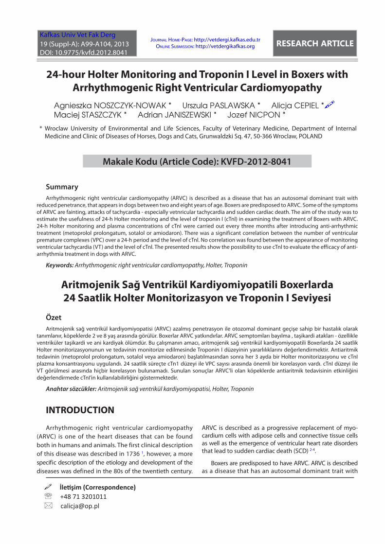
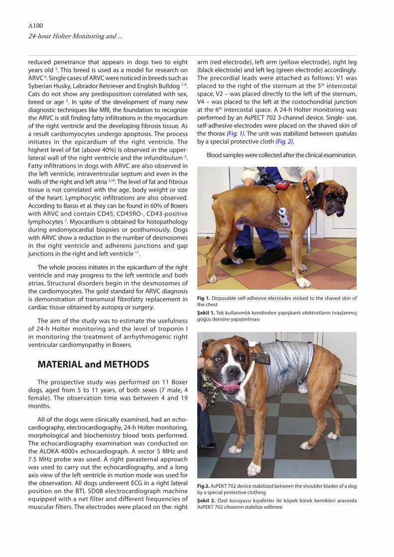
arm (red electrode), left arm (yellow electrode), right leg (black electrode) and left leg (green electrode) accordingly. The precordial leads were attached as follows: V1 was placed to the right of the sternum at the 5th intercostal space, V2 – was placed directly to the left of the sternum, V4 – was placed to the left at the costochondrial junction at the 6th intercostal space. A 24-h Holter monitoring was performed by an AsPECT 702 3-channel device. Single- use, self-adhesive electrodes were placed on the shaved skin of the thorax (Fig. 1). The unit was stabilized between spatulas by a special protective cloth (Fig. 2).
Blood samples were collected after the clinical examination.
Fig 1. Disposable self-adhesive electrodes sticked to the shaved skin of the chest
Şekil 1. Tek kullanımlık kendinden yapışkanlı elektrotların tıraşlanmış göğüs derisine yapıştırılması
Fig 2. AsPEKT 702 device stabilized between the shoulder blades of a dog by a special protective clothing
Şekil 2. Özel koruyucu kıyafetler ile köpek kürek kemikleri arasında AsPEKT 702 cihazının stabilize edilmesi
A101
NOSZCZYK-NOWAK, PASLAWSKA, CEPIELSTASZCZYK, JANISZEWSKI, NICPON
Using minimum stasis, cephalic venous blood was obtained with a 21 G disposable butterfly needle and Vacutainer system into serum (6 mL), EDTA (2 mL) tubes. In the morphological blood tests the total level of red blood cells, white blood cells, thrombocytes (PLT), haematocrit (Ht) and haemoglobin (Hb) were measured. Morphological blood tests were performed on an Animal Blood Center abc VET analyzer. Biochemistry tests included the evaluation of the activity of alanine aminotransferase (ALT), asparate aminotransferase (AST), level of urea, creatinine, bilirubin, total protein and albumins, as well as ion levels: Na+, K+, Mg2+, total Ca and inorganic phosphorus. Biochemistry tests were performed on the MaxMat Pl analyzer. An automated immunoluminescence test was used to evaluate the concentration of Troponin I (cTnI).
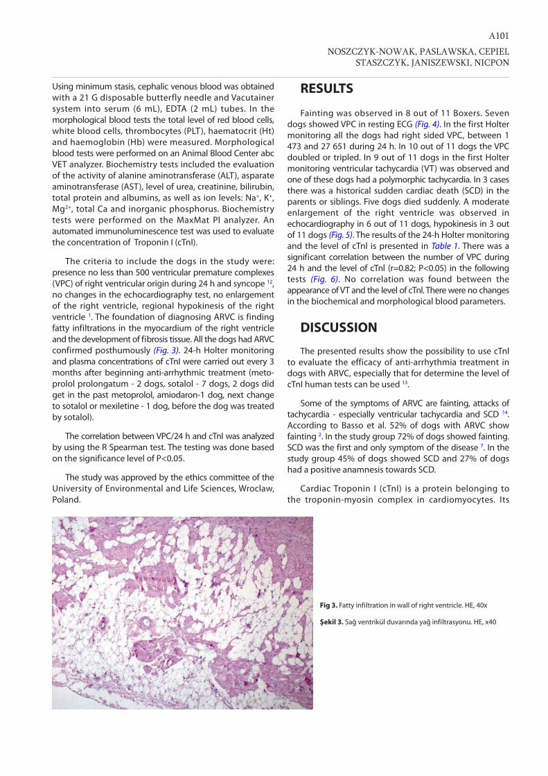
The criteria to include the dogs in the study were: presence no less than 500 ventricular premature complexes (VPC) of right ventricular origin during 24 h and syncope 12, no changes in the echocardiography test, no enlargement of the right ventricle, regional hypokinesis of the right ventricle 1. The foundation of diagnosing ARVC is finding fatty infiltrations in the myocardium of the right ventricle and the development of fibrosis tissue. All the dogs had ARVC confirmed posthumously (Fig. 3). 24-h Holter monitoring and plasma concentrations of cTnI were carried out every 3 months after beginning anti-arrhythmic treatment (meto-prolol prolongatum - 2 dogs, sotalol - 7 dogs, 2 dogs did get in the past metoprolol, amiodaron-1 dog, next change to sotalol or mexiletine - 1 dog, before the dog was treated by sotalol).
The correlation between VPC/24 h and cTnI was analyzed by using the R Spearman test. The testing was done based on the significance level of P<0.05.
The study was approved by the ethics committee of the University of Environmental and Life Sciences, Wroclaw, Poland.
RESULTS
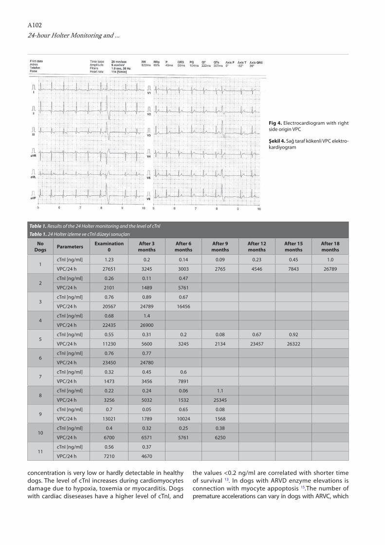
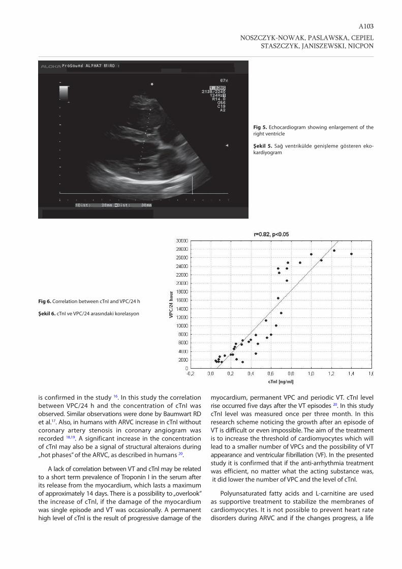
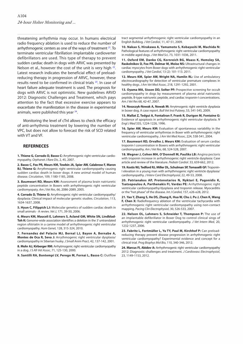
Fainting was observed in 8 out of 11 Boxers. Seven dogs showed VPC in resting ECG (Fig. 4). In the first Holter monitoring all the dogs had right sided VPC, between 1 473 and 27 651 during 24 h. In 10 out of 11 dogs the VPC doubled or tripled. In 9 out of 11 dogs in the first Holter monitoring ventricular tachycardia (VT) was observed and one of these dogs had a polymorphic tachycardia. In 3 cases there was a historical sudden cardiac death (SCD) in the parents or siblings. Five dogs died suddenly. A moderate enlargement of the right ventricle was observed in echocardiography in 6 out of 11 dogs, hypokinesis in 3 out of 11 dogs (Fig. 5). The results of the 24-h Holter monitoring and the level of cTnl is presented in Table 1. There was a significant correlation between the number of VPC during 24 h and the level of cTnl (r=0.82; P<0.05) in the following tests (Fig. 6). No correlation was found between the appearance of VT and the level of cTnl. There were no changes in the biochemical and morphological blood parameters.
DISCUSSION
The presented results show the possibility to use cTnl to evaluate the efficacy of anti-arrhythmia treatment in dogs with ARVC, especially that for determine the level of cTnI human tests can be used 13.
Some of the symptoms of ARVC are fainting, attacks of tachycardia - especially ventricular tachycardia and SCD 14. According to Basso et al. 52% of dogs with ARVC show fainting 2. In the study group 72% of dogs showed fainting. SCD was the first and only symptom of the disease 7. In the study group 45% of dogs showed SCD and 27% of dogs had a positive anamnesis towards SCD.
Cardiac Troponin I (cTnI) is a protein belonging to the troponin-myosin complex in cardiomyocytes. Its
Fig 3. Fatty infiltration in wall of right ventricle. HE, 40x
Şekil 3. Sağ ventrikül duvarında yağ infiltrasyonu. HE, x40
A10224-hour Holter Monitoring and ...
concentration is very low or hardly detectable in healthy dogs. The level of cTnI increases during cardiomyocytes damage due to hypoxia, toxemia or myocarditis. Dogs with cardiac diseseases have a higher level of cTnI, and
the values <0.2 ng/ml are correlated with shorter time of survival 13. In dogs with ARVD enzyme elevations is connection with myocyte appoptosis 15.The number of premature accelerations can vary in dogs with ARVC, which
Table 1. Results of the 24 Holter monitoring and the level of cTnl
Tablo 1. 24 Holter izleme ve cTnl düzeyi sonuçları
No Dogs Parameters Examination
0After 3 months
After 6 months
After 9 months
After 12 months
After 15 months
After 18 months
1cTnI [ng/ml] 1.23 0.2 0.14 0.09 0.23 0.45 1.0
VPC/24 h 27651 3245 3003 2765 4546 7843 26789
2cTnI [ng/ml] 0.26 0.11 0.47
VPC/24 h 2101 1489 5761
3cTnI [ng/ml] 0.76 0.89 0.67
VPC/24 h 20567 24789 16456
4cTnI [ng/ml] 0.68 1.4
VPC/24 h 22435 26900
5cTnI [ng/ml] 0.55 0.31 0.2 0.08 0.67 0.92
VPC/24 h 11230 5600 3245 2134 23457 26322
6cTnI [ng/ml] 0.76 0.77
VPC/24 h 23450 24780
7cTnI [ng/ml] 0.32 0.45 0.6
VPC/24 h 1473 3456 7891
8cTnI [ng/ml] 0.22 0.24 0.06 1.1
VPC/24 h 3256 5032 1532 25345
9cTnI [ng/ml] 0.7 0.05 0.65 0.08
VPC/24 h 13021 1789 10024 1568
10cTnI [ng/ml] 0.4 0.32 0.25 0.38
VPC/24 h 6700 6571 5761 6250
11cTnI [ng/ml] 0.56 0.37
VPC/24 h 7210 4670
Fig 4. Electrocardiogram with right side origin VPC
Şekil 4. Sağ taraf kökenli VPC elektro-kardiyogram
A103
is confirmed in the study 16. In this study the correlation between VPC/24 h and the concentration of cTnI was observed. Similar observations were done by Baumwart RD et al.17. Also, in humans with ARVC increase in cTnI without coronary artery stenosis in coronary angiogram was recorded 18,19. A significant increase in the concentration of cTnI may also be a signal of structural alteraions during „hot phases” of the ARVC, as described in humans 20.
A lack of correlation between VT and cTnl may be related to a short term prevalence of Troponin I in the serum after its release from the myocardium, which lasts a maximum of approximately 14 days. There is a possibility to „overlook” the increase of cTnl, if the damage of the myocardium was single episode and VT was occasionally. A permanent high level of cTnl is the result of progressive damage of the
myocardium, permanent VPC and periodic VT. cTnI level rise occurred five days after the VT episodes 20. In this study cTnI level was measured once per three month. In this research scheme noticing the growth after an episode of VT is difficult or even impossible. The aim of the treatment is to increase the threshold of cardiomyocytes which will lead to a smaller number of VPCs and the possibility of VT appearance and ventricular fibrillation (VF). In the presented study it is confirmed that if the anti-arrhythmia treatment was efficient, no matter what the acting substance was, it did lower the number of VPC and the level of cTnl.
Polyunsaturated fatty acids and L-carnitine are used as supportive treatment to stabilize the membranes of cardiomyocytes. It is not possible to prevent heart rate disorders during ARVC and if the changes progress, a life
NOSZCZYK-NOWAK, PASLAWSKA, CEPIELSTASZCZYK, JANISZEWSKI, NICPON
Fig 5. Echocardiogram showing enlargement of the right ventricle
Şekil 5. Sağ ventrikülde genişleme gösteren eko-kardiyogram
Fig 6. Correlation between cTnI and VPC/24 h
Şekil 6. cTnI ve VPC/24 arasındaki korelasyon
A10424-hour Holter Monitoring and ...
threatening arrhythmia may occur. In humans electrical radio frequency ablation is used to reduce the number of arrhythmogenic centers as one of the ways of treatment 21. To terminate ventricular fibrillation implantable cardioverter defibrillators are used. This type of therapy to prevent sudden cardiac death in dogs with ARVC was presented by Nelson et al., however the cost of the unit is very high 22. Latest research indicates the beneficial effect of preload-reducing therapy in progression of ARVC, however, these results need to be confirmed in clinical trials 23. In case of heart failure adequate treatment is used. The prognosis for dogs with ARVC is not optimistic. New guidelines ARVD 2012: Diagnostic Challenges and Treatment, which pays attention to the fact that excessive exercise appears to exacerbate the manifestation in the disease in experimental animals, were published this year 24.
Monitoring the level of cTnl allows to check the efficacy of anti-arrhythmia treatment by lowering the number of VPC, but does not allow to forecast the risk of SCD related with VT and VF.
REFERNECES
1. Thiene G, Corrado D, Basso C: Arrythmogenic right ventricular cardio-myopathy. Orphanet J Rare Dis, 2, 45, 2007.
2. Basso C, Fox PR, Meurs KM, Towbin JA, Spier AW, Calabrese F, Maron BJ, Thiene G: Arrythmogenic right ventricular cardiomyopathy causing sudden cardiac death in boxer dogs: A new animal model of human disease. Circulation, 109, 1180-1185, 2008.
3. Baumwart RD, Meurs KM: Assessment of plasma brain natriuretic peptide concentration in Boxers with arrhythmogenic right ventricular cardiomyopaty. Am J Vet Res, 66, 2086-2089, 2005.
4. Corrado D, Thiene G: Arrythmogenic right ventricular cardiomyopathy/dysplasia: Clinical impact of molecular genetic studies. Circulation, 113, 1634-1637, 2008.
5. Hyun C, Filippich LJ: Molecular genetics of sudden cardiac death in small animals - A reviev. Vet J, 171, 39-50, 2006.
6. Meurs KM, Mauceli E, Lahmers S, Acland GM, White SN, Lindblad-Toh K: Genome-wide association identifies a deletion in the 3’ untranslated region ofstriatin in a canine model of arrhythmogenic right ventricular cardiomyopathy. Hum Genet, 128, 315-324, 2010.
7. Fernandez del Palacio MJ, Bernal LJ, Bayon A, Bernabe A, Montes de Oca R, Seva J: Arrythmogenic right ventricular dysplasia/ cardiomyopathy in Siberian husky. J Small Anim Pract, 42, 137-142, 2001.
8. Mohr AJ, Kirberger RM: Arrhytmogenic right ventricular cardiomyopathy in a dog. J S Afr Vet Assoc, 71, 125-130, 2000.
9. Santilli RA, Bontempi LV, Perego M, Fornai L, Basso C: Outflow
tract segmental arrhythmogenic right ventricular cardiomyopathy in an English Bulldog. J Vet Cardiol, 11, 47-51, 2009.
10. Nakao S, Hirakawa A, Yamamoto S, Kobayashi M, Machida N: Pathological features of arrhythmogenic right ventricular cardiomyopathy in middle-aged dogs. J Vet Med Sci, 73, 1031-1036, 2011.
11. Oxford EM, Danko CG, Kornreich BG, Maass K, Hemsley SA, Raskolnikov D, Fox PR, Delmar M, Moïse NS: Ultrastructural changes in cardiac myocytes from Boxer dogs with arrhythmogenic right ventricular cardiomyopathy. J Vet Cardiol, 13 (2): 101-113. 2011.
12. Meurs KM, Spier AW, Wright NA, Hamlin RL: Use of ambulatory electrocardiography for detection of ventricular premature complexes in healthy dogs. J Am Vet Med Assoc, 218, 1291-1292, 2001.
13. Oyama MA, Sisson DD, Solter PF: Prospective screening for occult cardiomyopathy in dogs by measurement of plasma atrial natriuretic peptide, B-type natriuretic peptide, and cardiac troponin-I concentrations. Am J Vet Res 68, 42-47, 2007.
14. Noszczyk-Nowak A, Nowak M: Arrytmogenic right ventricle dysplasia in boxer dog. A case report. Bull Vet Inst Pulawy, 53, 541-545, 2009.
15. Mallat Z, Tedgui A, Fontaliran F, Frank R, Durigon M, Fontaine G: Evidence of apoptosis in arrhythmogenic right ventricular dysplasia. N Engl J Med 335, 1224-1226, 1996.
16. Spier AW, Meurs KM: Evaluation of spontaneous variability in the frequency of ventricular arrhythmias in Boxer with arrhythmogenic right ventricular cardiomiopathy. J Am Vet Med Assoc, 224, 538-541, 2004.
17. Baumwart RD, Orvalho J, Meurs KM: Evaluation of serum cardiac troponin I concentration in Boxers with arrhythmogenic right ventricular cardiomyopathy. Am J Vet Res, 68, 524-528, 2007.
18. Wegner J, Cohen MH, O’Donnell M, Pauliks LB: Angina pectoris with troponin increase in arrhythmogenic right ventricle dysplasia: Case article and review of the literature. Pediatr Cardiol. 33, 659-662, 2012.
19. Kostis WJ, Tedford RJ, Miller DL, Schulman SP, Tomaselli GF: Troponin- I elevation in a young man with arrhythmogenic right ventricle dysplasia/cardiomyopathy. J Interv Card Electrophysiol, 22, 49-53, 2008.
20. Patrianakos AP, Protonotarios N, Nyktari E, Pagonidis K, Tsatsopoulou A, Parthenakis FI, Vardas PE: Arrhythmogenic right ventricular cardiomyopathy/dysplasia and troponin release. Myocarditis or the “hot phase” of the disease. Int J Cardiol, 157, e26-e28, 2012.
21. Yao Y, Zhang S, He DS, Zhang K, Hua W, Chu J, Pu J, Chen K, Wang F, Chen X: Radiofrequency ablation of the ventricular tachycardia with arrhythmogenic right ventricular cardiomyopathy using non-contact mapping. Pacing Clin Electrophysiol, 30, 526-533, 2007.
22. Nelson OL, Lahmers S, Schneider T, Thompson P: The use of an implantable defibrillator in Boxer Dog to control clinical sings of arrhythmogenic right ventricular cardiomyopathy. J Vet Intern Med, 20, 1232-1237, 2006.
23. Fabritz L, Fortmüller L, Yu TY, Paul M, Kirchhof P: Can preload-reducing therapy prevent disease progression in arrhythmogenic right ventricular cardiomyopathy? Experimental evidence and concept for a clinical trial. Prog Biophys Mol Bio, 110, 340-346, 2012.
24. Marcus FI, Abidov A: Arrhythmogenic right ventricular cardiomyopathy 2012: Diagnostic challenges and treatment. J Cardiovasc Electrophysiol, 23, 1149-1153, 2012.


![Blast Boxers / POG [UK]](https://img.pdfslide.us/doc/110x75/568bdc2b1a28ab2034b13634/blast-boxers-pog-uk.jpg)