Embed Size (px)
Citation preview
23: Shock
Shock
• State of collapse and failure of the cardiovascular system
• Leads to inadequate circulation• Without adequate blood flow, cells cannot
get rid of metabolic wastes • The result of hypoperfusion to cells that
causes the organ, then organ systems, to fail
Perfusion
• The cardiovascular system’s circulation of blood and oxygen to all the cells in different tissues and organs of the body
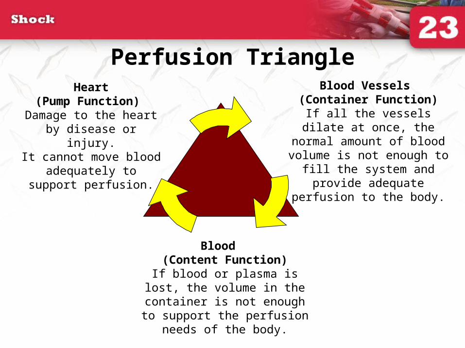
Heart (Pump Function)
Damage to the heart by disease or injury.
It cannot move blood adequately to support
perfusion.
Blood Vessels (Container Function)
If all the vessels dilate at once, the normal amount of blood
volume is not enough to fill the system and provide adequate
perfusion to the body.
Blood (Content Function)
If blood or plasma is lost, the volume in the container is not
enough to support the perfusion needs of the body.
Perfusion Triangle
Capillary Sphincters
• Regulate the blood flow through the capillary beds.
• Sphincters are under the control of the automatic nervous system.
• Regulation of blood flow is determined by cellular need.
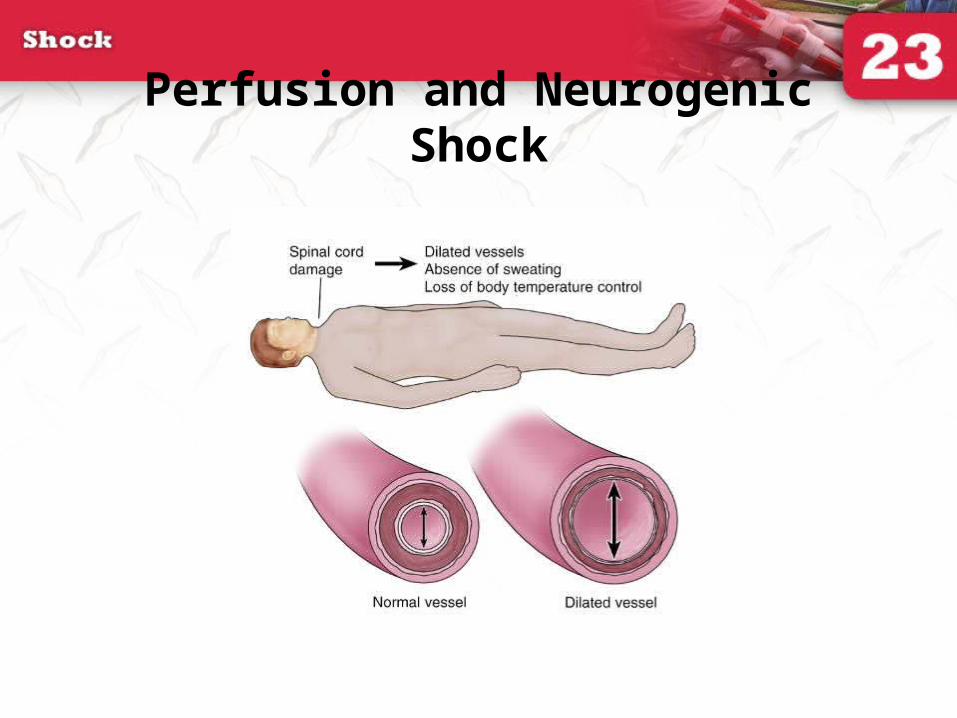
Perfusion and Neurogenic Shock

Cardiovascular Causes of Shock (1 of 4)
• Pump failure (cardiogenic shock)– Inadequate function of the heart or pump failure– Causes a backup of blood into the lungs– Results in pulmonary edema– Pulmonary edema leads to impaired ventilation

Cardiovascular Causes of Shock (2 of 4)
• Poor vessel function (neurogenic shock)– Damage to the cervical spine may affect control
of the size and muscular tone of blood vessels.– The vascular system increases.
• Blood in the body cannot fill the enlarged system.
• Neurogenic shock occurs.
Cardiovascular Causes of Shock (3 of 4)
• Content failure (hypovolemic shock)
– Results from fluid or blood loss
– Blood is lost through external and internal bleeding.
– Severe thermal burns cause plasma loss.
– Dehydration aggravates shock.

Cardiovascular Causes of Shock (4 of 4)
• Combined vessel and content failure– Some patients with severe bacterial infections,
toxins, or infected tissues contract septic shock.
– Toxins damage vessel walls, causing leaking and impairing ability to contract.
– Leads to dilation of vessels and loss of plasma, causing shock
Noncardiovascular Causes of Shock (1 of 3)
• Respiratory insufficiency
– Patient with a severe chest injury or airway obstruction may be unable to breathe adequate amounts of oxygen.
– Insufficient oxygen in the blood will produce shock.

Noncardiovascular Causes of Shock (2 of 3)
• Anaphylactic shock– Occurs when a person reacts violently to a
substance. – Four categories of common causes:
• Injections• Stings• Ingestion• Inhalation
Noncardiovascular Causes of Shock (3 of 3)
• Psychogenic shock– Caused by sudden reaction of the nervous
system that produces a temporary, generalized vascular dilation
– Commonly referred to as fainting or syncope– Can be brought on by serious causes: irregular
heartbeat, brain aneurysm– Can be brought on by fear, bad news,
unpleasant sights
Progression of Shock
• Compensated shock
– When the body compensates for blood loss
• Decompensated shock
– The late stage of shock when blood pressure is falling
• Irreversible shock
– The terminal stage

Compensated Shock
• Agitation• Anxiety• Restlessness• Feeling of impending
doom• Altered mental status• Weak pulse
• Clammy skin• Pallor• Shallow, rapid breathing• Shortness of breath• Nausea or vomiting• Delayed capillary refill• Marked thirst
Decompensated Shock
• Falling blood pressure (<90 mm Hg in an adult)
• Labored, irregular breathing
• Ashen, mottled, cyanotic skin
• Thready or absent pulse• Dull eyes, dilated pupils• Poor urinary output
Irreversible Shock
• This is the terminal stage of shock.
• A transfusion of any type will not be enough to save a patient’s life.

When to Expect Shock
• Multiple severe fractures• Abdominal or chest injuries• Spinal injuries• Severe infection• Major heart attack• Anaphylaxis
You are the Provider
• You and your partner respond to an MVC involving two cars. En route you follow BSI.
• You arrive to a 25-year-old man. • Law enforcement informs you that the other car left
the scene. Patient was restrained and is sitting outside car. He is pale.
• The airbag has deployed and the steering wheel has some damage.

Scene Size-up
• In addition to BSI, what are some considerations at the scene?
• What is the mechanism of injury?

You are the Provider (continued)
• You approach the patient and introduce yourself. He appears visibly upset but lets you take his vital signs.– Pulse: 115 beats/min– Respirations: 26 breaths/min– Blood pressure: 110 mm Hg
• He has a laceration on his knee where it hit the dashboard.
Initial Assessment
• Describe the steps of your initial assessment and findings:– General impression– Airway– Breathing– Circulation– Transport decision
You are the Provider (continued)
• Spinal immobilization needed.• Pallor is a sign of shock. • He is A on the AVPU scale.• Airway is open.• Breathing is rapid.• Inspect and palpate chest for DCAP-BTLS. • Observe for accessory muscle use.
You are the Provider (continued)
• Patient has rapid pulse.• Clammy skin.• Knee laceration• Priority transport
Focused History and Physical Exam
• Would you perform a rapid physical exam or focused physical exam?
• What is your reasoning?
Detailed Physical Exam
• If time permits, perform en route to the hospital.

Ongoing Assessment
• Perform reassessment.• Take vital signs every 5 minutes.
You are the Provider
(continued)
• You reassess the patient in the ambulance and he has a pulse of 122 beats/min, respirations of 30 breaths/min, and a blood pressure of 106/68 mm Hg.
• What do his vital sign changes indicate?
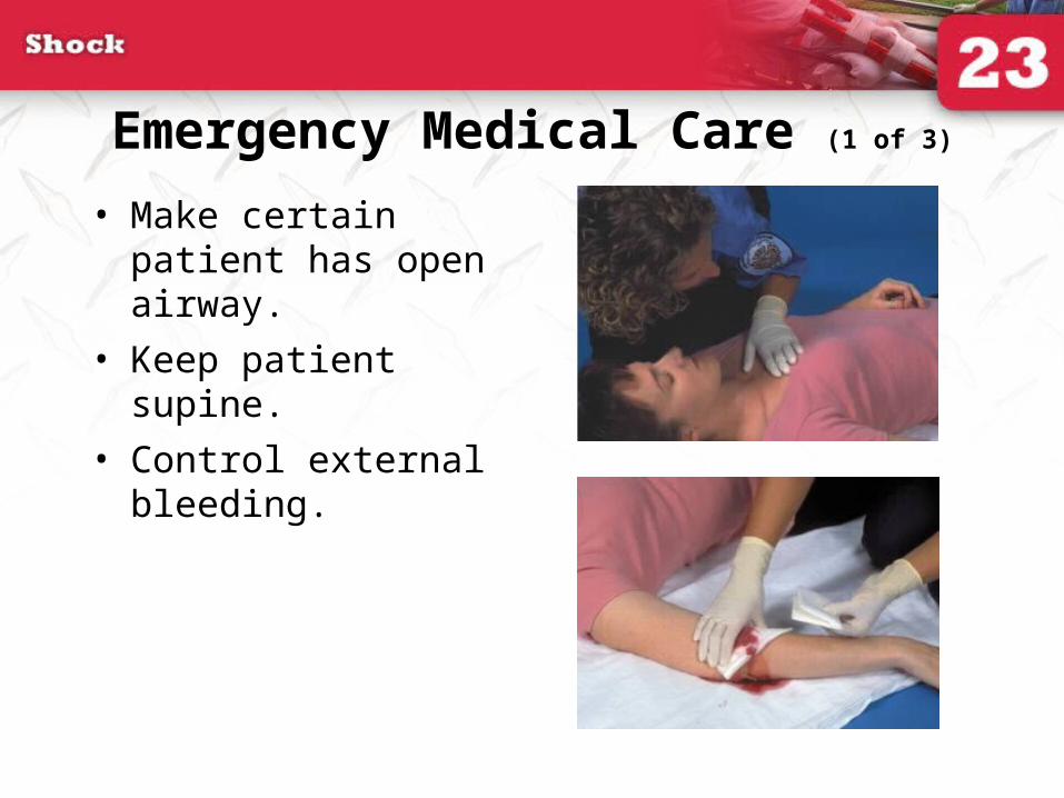
Emergency Medical Care (1 of 3)
• Make certain patient has open airway.
• Keep patient supine.
• Control external bleeding.

Emergency Medical Care (2 of 3)
• Splint any broken bones or joint injuries.
• Always provide oxygen.
• Place blankets under and over patient.
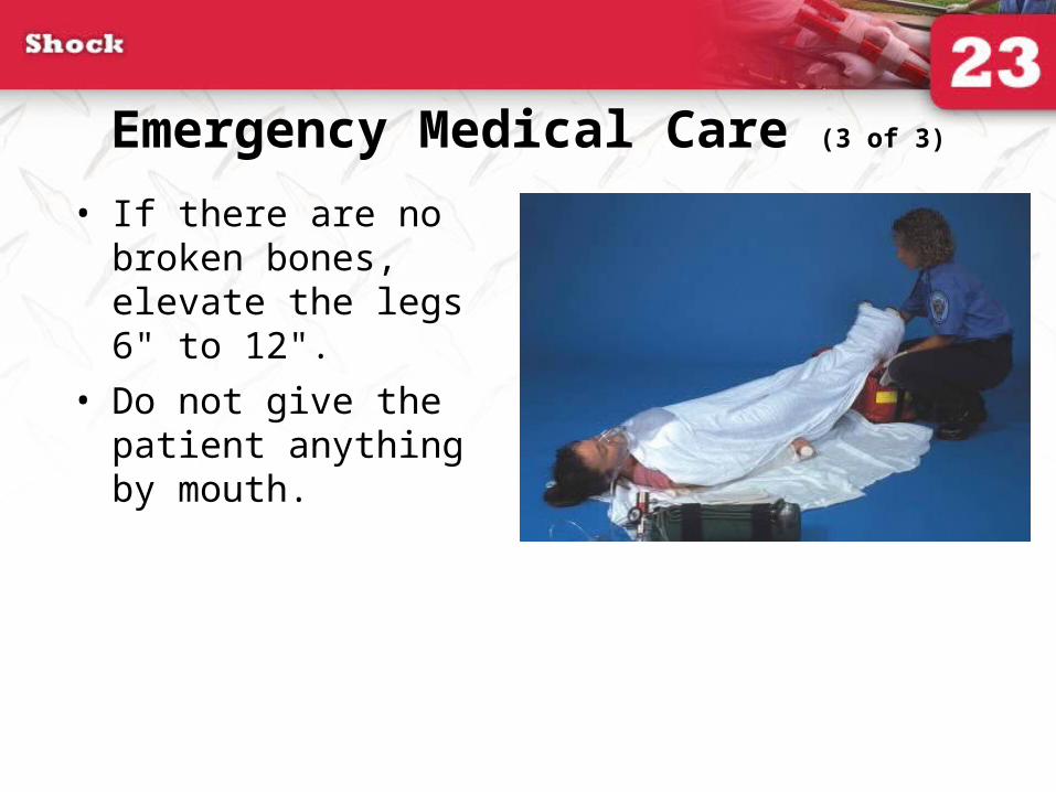
Emergency Medical Care (3 of 3)
• If there are no broken bones, elevate the legs 6" to 12".
• Do not give the patient anything by mouth.
Pneumatic Antishock Garment
• Some localities allow EMTs to apply a pneumatic antishock garment (PASG) for some patients in decompensated shock.
• Know your local protocol regarding their usage.

Treating Cardiogenic Shock
• Patient may breathe better in a sitting or semi-sitting position.
• Administer high-flow oxygen.• Assist ventilations as necessary.• Have suction nearby in case the patient
vomits.• Transport promptly.
Treating Neurogenic Shock
• Maintain airway and assist breathing as needed.
• Keep patient warm.
• Transport promptly.
Treating Hypovolemic Shock
• Control obvious bleeding.
• Splint any bone or joint injuries.
• If no fractures, raise legs 6" to 12".
• Secure and maintain airway.
• Give oxygen as soon as you suspect shock.
• Transport rapidly.
Treating Septic Shock
• Transport as promptly as possible while giving all general support available.
• Give high-flow oxygen during transport.
• Use blankets to conserve body heat.
Treating Respiratory Insufficiency
• Secure and support the airway.• Clear airway of any obstructions.• Ventilate if needed with a BVM device.• Administer oxygen.• Transport promptly.
Treating Anaphylactic Shock
• Administer epinephrine.
• Provide prompt transport.
• Provide all possible support.
– Oxygen
– Ventilatory assistance
Treating Psychogenic Shock
• It is usually self-resolving.
• Assess patient for injuries from fall.
• If patient has difficulties after regaining consciousness, suspect another problem.