Embed Size (px)
Citation preview

22CHAPTER
INHERITANCE
UNIT 2 CONTENT
SCIENCE INQUIRY SKILLS ❯ select, construct and use appropriate
representations, including models of DNA replication, transcription and translation, Punnett squares, pedigrees and karyotypes, to communicate conceptual understanding, solve problems and make predictions
SCIENCE UNDERSTANDING
Types of inheritance ❯ probable frequencies of genotype and
phenotype of offspring can be predicted using Punnett squares and by taking into consideration patterns of inheritance, including the effects of dominance, co-dominance, autosomal or sex-linked alleles, and multiple alleles: Huntington’s disease, phenylketonuria (PKU), ABO blood groups, red-green colour blindness/haemophilia show different inheritance patterns
Year
11
Sylla
bus,
Hum
an B
iolo
gy, ©
Sch
ool C
urri
culu
m a
nd S
tand
ards
Aut
hori
ty, 2
014,
Gov
ernm
ent o
f Wes
tern
Aus
tral
ia
Shut
ters
tock
/Vtld
tlm
ISBN 9780170351126 | CHAPTER 22 | INHERITANCE 303
From very early times, farmers realised that many characteristics of domesticated plants and animals are passed from parent to offspring. Exactly how these characteristics, or traits, were transmitted was not clear. It was thought by many that offspring were simply a blend of the characteristics of the two parents. However, this was not the case. The first clear explanation of patterns of inheritance was provided by an Austrian monk, Gregor Mendel, in 1865. After spending two years at the University of Vienna, he returned to his monastery as a schoolteacher. Here he was able to link his two loves, nature and mathematics, through a careful study of the reproductive behaviour of pea plants. After 10 years of research, Mendel put forward two principles relating to inheritance:1 that the various hereditary characteristics were controlled
by factors (which we now call genes) and that these occurred in pairs
2 that during the formation of the gametes (in humans, the eggs and the sperm), the pairs of factors separate. Each gamete receives only one set of factors, or genes, the other set going to another gamete. Gametes unite at fertilisation, allowing different combinations of genes to come together.Mendel’s findings went unnoticed for 35 years before
their significance was fully appreciated. At the same time as his work was being rediscovered, scientists were making considerable advances in cytology, the study of cells. A young American graduate student, Walter Sutton, was able to link the work of Mendel to that of the cytologists. His observations of the behaviour of chromosomes during meiosis (the type of cell division that takes place to produce gametes, see Chapter 15), and Mendel’s speculation on the separation of the hereditary factors during the formation of gametes, led Sutton to suggest that the hereditary factors, or genes, were located in the chromosomes. This important hypothesis, contained in a research paper he published in 1903, led to the chromosome theory of heredity.
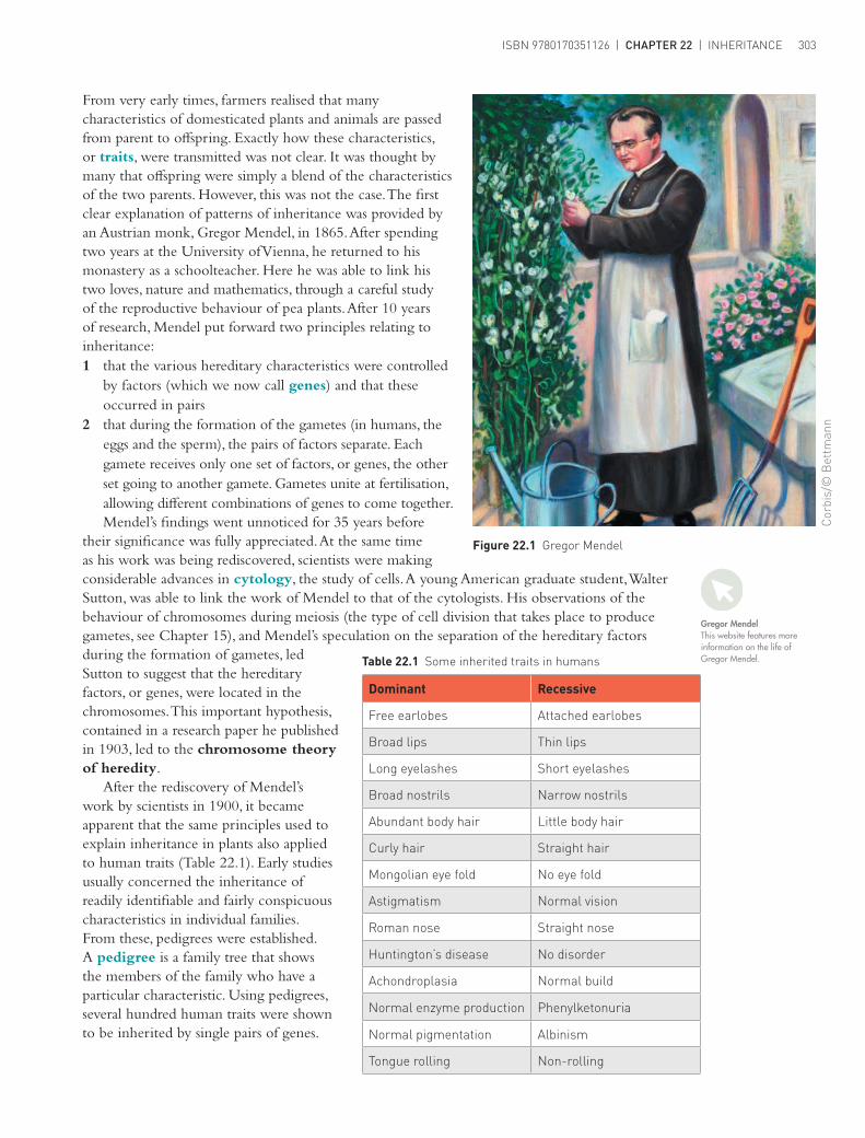
After the rediscovery of Mendel’s work by scientists in 1900, it became apparent that the same principles used to explain inheritance in plants also applied to human traits (Table 22.1). Early studies usually concerned the inheritance of readily identifiable and fairly conspicuous characteristics in individual families. From these, pedigrees were established. A pedigree is a family tree that shows the members of the family who have a particular characteristic. Using pedigrees, several hundred human traits were shown to be inherited by single pairs of genes.
Figure 22.1 Gregor Mendel
Cor
bis/
© B
ettm
ann
Gregor MendelThis website features more information on the life of Gregor Mendel.Table 22.1 Some inherited traits in humans
Dominant Recessive
Free earlobes Attached earlobes
Broad lips Thin lips
Long eyelashes Short eyelashes
Broad nostrils Narrow nostrils
Abundant body hair Little body hair
Curly hair Straight hair
Mongolian eye fold No eye fold
Astigmatism Normal vision
Roman nose Straight nose
Huntington’s disease No disorder
Achondroplasia Normal build
Normal enzyme production Phenylketonuria
Normal pigmentation Albinism
Tongue rolling Non-rolling
Characteristicstudied
Seed shape
Seed colour
Seed-coat colour
Pod shape
Pod colour
Flower position
Stem length
Round
Yellow
Coloured
Inflated
Green
Axial
Long
Wrinkled
Green
White
Constricted
Yellow
Terminal
Short
Dominantcharacter
Recessivecharacter
UNIT 2 | HUMAN PERSPECTIVES UNITS 1 & 2 | ISBN 9780170351126304
Mendel’s discoveriesTo understand how the characteristics in Table 22.1 are passed from one generation to another it is necessary to review the early work of Mendel. Although he worked with plants, his discoveries apply equally well to animals.
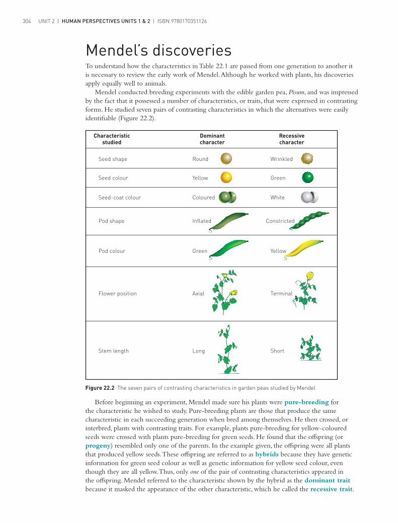
Mendel conducted breeding experiments with the edible garden pea, Pisum, and was impressed by the fact that it possessed a number of characteristics, or traits, that were expressed in contrasting forms. He studied seven pairs of contrasting characteristics in which the alternatives were easily identifiable (Figure 22.2).
Before beginning an experiment, Mendel made sure his plants were pure-breeding for the characteristic he wished to study. Pure-breeding plants are those that produce the same characteristic in each succeeding generation when bred among themselves. He then crossed, or interbred, plants with contrasting traits. For example, plants pure-breeding for yellow-coloured seeds were crossed with plants pure-breeding for green seeds. He found that the offspring (or progeny) resembled only one of the parents. In the example given, the offspring were all plants that produced yellow seeds. These offspring are referred to as hybrids because they have genetic information for green seed colour as well as genetic information for yellow seed colour, even though they are all yellow. Thus, only one of the pair of contrasting characteristics appeared in the offspring. Mendel referred to the characteristic shown by the hybrid as the dominant trait because it masked the appearance of the other characteristic, which he called the recessive trait.
Figure 22.2 The seven pairs of contrasting characteristics in garden peas studied by Mendel

ISBN 9780170351126 | CHAPTER 22 | INHERITANCE 305
When Mendel allowed the hybrid plants to self-pollinate, a second generation of plants was produced. In this generation the characteristics reappeared in the ratio of about three with the dominant trait for every one with the recessive trait (3:1). From these results Mendel concluded that the hereditary factors, or genes, were unchanged as they passed from one generation to the next. He further reasoned that each pea plant had two hereditary factors for each characteristic under study. During the formation of gametes, these factors are separated (or, as Mendel called it, segregated), each gamete receiving only one factor, or gene, for each trait. This is known as the principle of segregation. As offspring are formed by the union of a male and a female gamete, each offspring receives one gene for each characteristic from each parent.
Monohybrid crossesA cross is the mating of two organisms. In a monohybrid cross only one pair of contrasting characteristics is studied. For example, yellow and green pod colour in peas or tongue rolling and non-rolling in humans. It is much easier to refer to the genes by a letter than by name. For a particular characteristic, the genes for an individual are, therefore, represented by two letters, one for the gene that originated from the female parent, and one for the gene that originated from the male parent. If the gene is a dominant one it is shown as a capital letter; if it is for a recessive characteristic a lower-case letter is used. For the garden pea, green pod colour is dominant to yellow, so the gene for green pod colour is represented with a capital G and the recessive gene for yellow pod colour with a lower-case g.
For pure-breeding plants of green pod colour the symbols used are GG; pure-breeding plants of yellow pod colour are gg. Hybrids, with one of each gene type, have the symbols Gg. The alternative forms of the gene for pod colour, in this case G and g, are called alleles. For a pair of contrasting characteristics the two alleles may occur in one of three possible combinations: GG, gg or Gg. In two of these three gene combinations, or genotypes, the alleles are the same (GG and gg). These are described as homozygous, whereas the hybrid, Gg, with one of each allele, is termed heterozygous. The three genotypes listed produce only two types of pod colour in garden peas – green for GG and Gg, and yellow for gg. This physical appearance, or what the pods look like, is called the phenotype. These and other terms used to describe inheritance are defined in Table 22.2.
Table 22.2 Terms relating to Mendelian genetics
Term MeaningGene The factor that determines an inherited characteristic; located in the chromosomes
AlleleAn alternative form of a gene (e.g. the gene for pod colour in peas has two alleles, green and yellow); an individual normally has only one or two alleles of each gene
DominantAn allele that masks the effect of another allele (e.g. a pea plant with alleles for green and yellow pods will produce green pods because green is dominant to yellow)
RecessiveAn allele that is masked by the effect of an alternative allele (e.g. the allele for yellow pod colour is masked by the allele for green pod colour)
HomozygousThe situation where an individual has the same alleles for a particular characteristic; also called pure-breeding (e.g. a pea plant with two alleles for green pod colour is homozygous green)
HeterozygousThe situation where an individual possesses different alleles for a particular characteristic; also called hybrid (e.g. a pea plant with alleles for both green and yellow-coloured pods is heterozygous)
PhenotypeThe physical appearance of an individual as determined by the expression of the alleles for that characteristic (e.g. a pea plant with alleles for green-coloured and for yellow-coloured pods will have the phenotype green pods)
GenotypeThe genetic make-up of an individual as determined by the alleles for the characteristic being considered (e.g. a pea plant with an allele for green-coloured pods and one for yellow-coloured pods will have the heterozygous genotype)
Parents:
Gametes:
First filialgeneration (F1):
all
all
all
HH
Hh
h
hh
H
(1/2 +
i.e.
Hh
H
HH
(1/2H
Hh
+
+ +
1/2 ]+
++Second filialgeneration (F2):
F1:
Gametes: h1/2 ]h
1/2Hh
1/4
HH1/4
Hh1/4
hh1/4
Hh1/4 Hh1/4
UNIT 2 | HUMAN PERSPECTIVES UNITS 1 & 2 | ISBN 9780170351126306
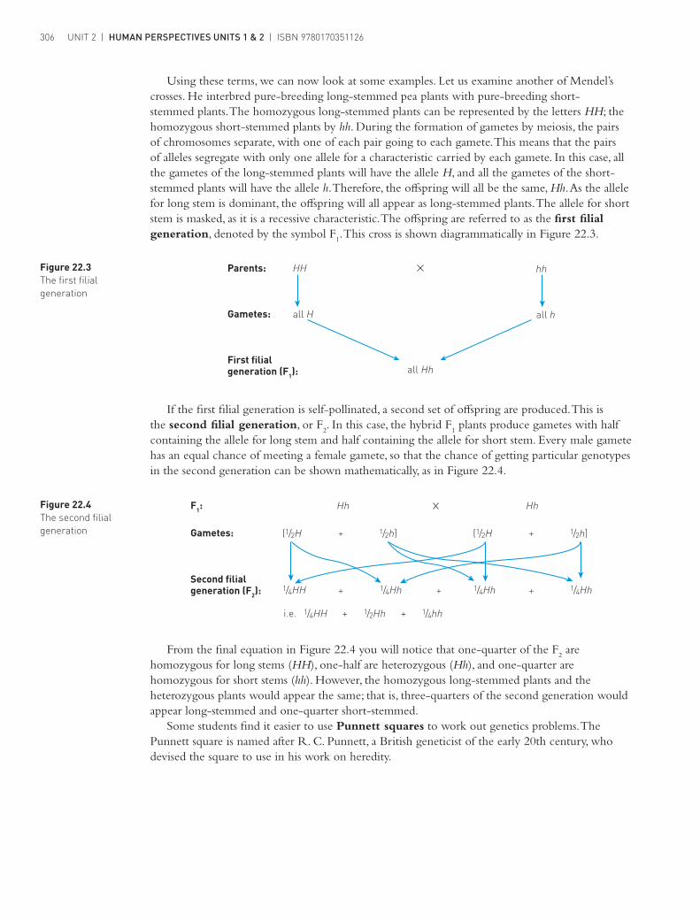
Using these terms, we can now look at some examples. Let us examine another of Mendel’s crosses. He interbred pure-breeding long-stemmed pea plants with pure-breeding short-stemmed plants. The homozygous long-stemmed plants can be represented by the letters HH; the homozygous short-stemmed plants by hh. During the formation of gametes by meiosis, the pairs of chromosomes separate, with one of each pair going to each gamete. This means that the pairs of alleles segregate with only one allele for a characteristic carried by each gamete. In this case, all the gametes of the long-stemmed plants will have the allele H, and all the gametes of the short-stemmed plants will have the allele h. Therefore, the offspring will all be the same, Hh. As the allele for long stem is dominant, the offspring will all appear as long-stemmed plants. The allele for short stem is masked, as it is a recessive characteristic. The offspring are referred to as the first filial generation, denoted by the symbol F
1. This cross is shown diagrammatically in Figure 22.3.
Figure 22.3 The first filial generation
Figure 22.4 The second filial generation
If the first filial generation is self-pollinated, a second set of offspring are produced. This is the second filial generation, or F
2. In this case, the hybrid F
1 plants produce gametes with half
containing the allele for long stem and half containing the allele for short stem. Every male gamete has an equal chance of meeting a female gamete, so that the chance of getting particular genotypes in the second generation can be shown mathematically, as in Figure 22.4.
From the final equation in Figure 22.4 you will notice that one-quarter of the F2 are
homozygous for long stems (HH), one-half are heterozygous (Hh), and one-quarter are homozygous for short stems (hh). However, the homozygous long-stemmed plants and the heterozygous plants would appear the same; that is, three-quarters of the second generation would appear long-stemmed and one-quarter short-stemmed.
Some students find it easier to use Punnett squares to work out genetics problems. The Punnett square is named after R. C. Punnett, a British geneticist of the early 20th century, who devised the square to use in his work on heredity.

ISBN 9780170351126 | CHAPTER 22 | INHERITANCE 307
The Punnett square method for the example on page 306 is as follows.
Parents HH × hh
GametesFemale
½H ½H
Male½h ¼Hh ¼Hh
½h ¼Hh ¼Hh
F1 genotype All Hh
F1 phenotype All long-stemmed plants
The F2 is produced by self-pollinating the F
1 (that is, Hh× Hh):
GametesFemale
½H ½h
Male½H ¼HH ¼Hh
½h ¼Hh ¼hh
Each ‘box’ within the inner ‘square’ represents the proportion of that genotype that will occur in the offspring. Adding the number of boxes within the square, we obtain:
F2 genotype ¼HH + ½Hh + ¼hh
F2 phenotype ¾ long-stemmed and ¼ short-stemmed
Each pea plant produces a great many seeds and, therefore, many potential offspring, so it is appropriate to talk about the proportion of each genotype or phenotype that should occur in the offspring of a cross. In the case of humans, where each couple has only a small number of offspring, the proportions of expected genotypes and phenotypes in the offspring represent probabilities. In the example above, the probability that any one seed will grow into a long-stemmed plant is ¾, or 0.75, or 75%. The probability that a seed will produce a plant that is long-stemmed and homozygous for the long-stem allele is ¼ (0.25 or 25%).
In humans, an example of a dominant characteristic that follows the rules of Mendelian genetics is Huntington’s disease (formerly referred to as Huntington’s chorea). As the disease is controlled by a dominant allele, the condition is very likely to be passed on from one generation to the next. On the other hand, phenylketonuria (PKU) is an example of a recessive characteristic found in human populations. PKU often skips a generation, as is the case with most recessive characteristics. Both these conditions are discussed in more detail in the next chapter.
Sex determinationAn examination of birth records for Australia this century would indicate that girls and boys are born in approximately equal numbers. However, a given family does not necessarily contain the same number of boys as girls. For centuries, people have tried to explain how the sex of a child is determined, and it was not until scientists began to examine the nuclei of cells that they realised the chromosome sets in the nuclei of cells of men and women were slightly different. In women, the 46 chromosomes in the nucleus of each cell were in 23 matched pairs, whereas in men the 46 chromosomes were in only 22 matched pairs, the 23rd pair consisting of two unmatched chromosomes (Figure 22.5, page 308).

Male parentXY
Sperm1/2 X 1/2 Y
Children1/2 XX + 1/2 XY
or1/2 female and 1/2 male
Female parentXX
Eggsall X+
Figure 22.6 Chromosomal basis for sex determination
1
6
13
19 20
Female chromosomes
Male chromosomes
22 XX
14 15 16 17 18
7 8 9 10 11 12
2 3 4 5
1
6
13
19 20 21 22 YX
14 15 16 17 18
7 8 9 10 11 12
2 3 4 5
21
Figure 22.5 Human chromosomes: as chromosomes become visible only during cell division, each appears as a double strand ready for division; the strands are joined at one point, so that each double strand is referred to as a single chromosome.
1
6
13
19 20
Female chromosomes
Male chromosomes
22 XX
14 15 16 17 18
7 8 9 10 11 12
2 3 4 5
1
6
13
19 20 21 22 YX
14 15 16 17 18
7 8 9 10 11 12
2 3 4 5
21UNIT 2 | HUMAN PERSPECTIVES UNITS 1 & 2 | ISBN 9780170351126308
Examination of the 23rd pair of chromosomes in males indicated that one of the pair was similar to the chromosomes of the 23rd pair in females, but the other was much smaller. The large chromosome became known as the X chromosome and the smaller the Y chromosome. Females, therefore, had two X chromosomes, and males one X and one Y. These are called sex chromosomes. The chromosomal basis for sex determination is shown schematically in Figure 22.6. The 22 pairs of non-sex chromosomes, called autosomes, are not normally involved in sex determination and are not shown. All the eggs produced by a female are of one type with respect
to the sex chromosomes: they all possess one X chromosome. On the other hand, the male’s sperm are of two types: about half contain an X chromosome and about half a Y chromosome. From this information it should be clear that it is the chromosomal complement of the father’s sperm that determines the sex of the child. If an X-bearing sperm fertilises the egg, the zygote (fertilised egg) will develop into a female; if a Y-bearing sperm fertilises the egg, the zygote will develop into a male.
Sex-linked characteristicsExamine the X and Y chromosomes illustrated in Figure 22.5 again. Notice how the Y chromosome is very small compared with the X chromosome. Obviously, the Y chromosome could not have the same number of genes in it as the X and, therefore, most of the genes in the X chromosome will lack matching alleles in males. For females, however, the normal pairing of alleles will exist. When characteristics located on the X chromosome are studied, it is found that the pattern of inheritance is different in the two sexes. Characteristics that show different patterns in the two sexes are called sex-linked characteristics or X-linked characteristics. Two common traits of this type are red–green colour blindness and haemophilia.
The ability to discriminate between the colours red and green is controlled by a gene located in the X chromosome. Individuals who are unable to distinguish between the two colours possess the recessive allele of this gene. As the gene is located in the X chromosome, this form of colour blindness is found more frequently in males than in females.

ISBN 9780170351126 | CHAPTER 22 | INHERITANCE 309
In the case of the children of a colourblind man and a woman homozygous for normal vision, all would have normal vision. However, the daughters could produce sons who were colourblind. The daughters are carriers for colour blindness because, although they are not colourblind themselves, they can have children who are colourblind. This is shown in the Punnett squares below.
Parents XbY × XBXB
GametesFemale
½XB ½XB
Male½Xb ¼XBXb ¼XBXb
½Y ¼XBY ¼XBY
Children ¼XBY + ¼XBY + ¼XBXb + ¼XBXb
½ normal visioned males + ½ normal visioned females but carriers
(That is, all children, male and female, will have normal vision.)
Note how in crosses for sex-linked characteristics the symbols include the sex chromosome (either X or Y ) and the symbol for the allele being studied. The colourblind man is hemizygous for the recessive allele. (As there is no allelic counterpart for males with sex-linked traits, the term ‘hemizygous’ is used instead of homozygous or heterozygous.) The woman is homozygous for the normal allele (XBXB), while all the daughters are heterozygous for the normal allele (XBXb), as they inherit the recessive allele from their father. Such people, who carry a recessive allele but do not show the recessive phenotype, are known as carriers. The sons are normal, as they inherit a normal gene from their mother and the Y chromosome with no allelic counterpart from their father.
If a carrier daughter had children with a man with normal vision, the pattern of inheritance for their children would result in daughters who may be carriers and sons who have a 50% chance of having the recessive allele.
Parents XBY × XBXb
GametesFemale
½XB ½Xb
Male½XB ¼XBXB ¼XBXb
½Y ¼XBY ¼XbY
Children ¼XBY + ¼XbY + ¼XBXB + ¼XBXb
(normal male)
(colourblind male)
(normal female)
(normal female but a carrier)
Is it possible for females to be red–green colourblind? If a male with red–green colour blindness has children with a woman who carried the recessive allele, then they have a 50% chance that their daughters will be colourblind and a 50% chance that their sons will also have the trait:
Parents XbY × XBXb
GametesFemale
½XB ½Xb
Male½Xb ¼XBXb ¼XbXb
½Y ¼XBY ¼XbY
Children ¼XBY + ¼XbY + ¼XBXb + ¼XbXb
(normal male)
(colourblind male)
(normal female but a carrier)
(colourblind female)

UNIT 2 | HUMAN PERSPECTIVES UNITS 1 & 2 | ISBN 9780170351126310
Haemophilia is another sex-linked characteristic. It is a relatively rare disease in which the blood clots slowly or not at all. The defective allele is recessive to that controlling normal clotting of the blood and is carried on the X chromosome. Males, therefore, can be either normal or haemophiliacs, as they have only one X chromosome. Females can be homozygous normal; heterozygous and therefore carriers of the condition; or haemophiliacs. This last case is extremely rare.
The pattern of inheritance for haemophilia is similar to that already studied for red–green colour blindness. Haemophiliac fathers pass the recessive gene to their daughters. Carrier mothers may pass a defective gene to their sons, who will be haemophiliacs, or to their daughters, who will then also carry the gene. The most famous family pedigree for haemophilia is that of the European royal families descended from Queen Victoria.
EXTENSIONQueen Victoria was a carrier of the gene for haemophilia and passed the condition on to some of her children.
Find out:
❯❯ which of her children were carriers of the condition and if any of her children were haemophiliacs
❯❯ how Queen Victoria came to be a carrier for haemophilia, given that none of her ancestors showed the trait.
Besides haemophilia and red–green colour blindness, there are a number of disorders in humans that are inherited through recessive genes in the X chromosome. These conditions include diabetes insipidus, in which the kidneys are unable to concentrate urine, and Duchenne muscular dystrophy, a progressive wasting disease of the voluntary muscles.
Diabetes insipidus is a sex-linked disorder in which the affected individual passes very large quantities of urine and gradually becomes dehydrated. Death may result unless water is available to replace that lost. The Duchenne form of muscular dystrophy is a wasting disease of the leg muscles and later the arms, shoulders and chest. At times it may be due to a mutation – either in a woman, making her a carrier, or in a boy, giving him the disease. Duchenne muscular dystrophy usually becomes apparent around the age of 3–5 years, when muscle weakness becomes evident. As the years pass, more and more muscle tissue wastes away and is replaced by fatty substances. By around 12–14 years the child is confined to a wheelchair and later is bedridden. With respiratory failure, death becomes inevitable. Generally speaking, boys with the Duchenne form of muscular dystrophy have little chance of living beyond 20–25 years.
Co-dominanceThere are many situations in genetics where the alleles of a particular gene are neither dominant nor recessive. One case is the inheritance of flower colour in four-o’clock plants (Mirabilis jalapa). It was found that when homozygous red-flowered plants were crossed with homozygous white-flowered plants the offspring all had pink flowers. Neither red nor white was dominant: both colours were expressed in the heterozygous offspring as pink. It appeared as though the red and white colours had blended together to form a flower colour in between the two parental colours. As neither parental colour was dominant, and as a form of blending appeared to have taken place, the term co-dominance was used to describe the situation. This pattern of inheritance can also be called incomplete dominance, although some people do make a fine distinction between the two.

Parents:
Gametes:
F1 genotype:F1 phenotype:
All All pink
FR FR
FR
F W F W
F W
F W
FRAll All
ISBN 9780170351126 | CHAPTER 22 | INHERITANCE 311
The Punnett square method can be used for this example:
Parents FRFR × FWFW
GametesFemale
½FW ½FW
Male½FR ¼FRFW ¼FRFW
½FR ¼FRFW ¼FRFW
F1 genotype All FRFW
F1 phenotype All pink
Crossing F1 individuals produces the following F
2:
GametesFemale
½FR ½FW
Male½FR ¼FRFR ¼FRFW
½FW ¼FRFW ¼FWFW
F1 genotype ¼ FRFR + ½ FRFW + ¼ FWFW
F1 phenotype ¼ red + ½ pink + ¼ white
Figure 22.7 Co-dominance
It was found that, when two of these heterozygous pink-flowered plants were crossed, the offspring produced were in the ratio of ¼ red-flowered plants, ½ pink-flowered plants and ¼ white-flowered plants. These results indicated that the alleles for red and white had not mixed in the pink offspring: each allele had retained its identity. Let us consider this situation using FR to represent the red allele and FW to represent the white allele, as shown in Figure 22.7.
UNIT 2 | HUMAN PERSPECTIVES UNITS 1 & 2 | ISBN 9780170351126312
This situation is not limited to plants. Humans also show co-dominance; an example is the BM and BN alleles for the M and N antigens in human blood. Neither allele is dominant to the other, and so three phenotypes are possible: type M blood, type N blood and type MN blood. People with type M blood have the genotype BMBM, those with blood type N have the genotype BNBN and those with blood type MN have the heterozygous BMBN genotype. Again, during crosses between various blood types, the identity of the separate alleles is maintained. However, as neither allele is dominant to the other, heterozygous individuals have both characters because they have both antigens.
The conventions used for naming alleles are outlined in Table 22.3.
Table 22.3 Conventions for representing alleles
❯❯ DOMINANT alleles are represented by UPPER-CASE letters.
❯❯ recessive alleles are represented by lower-case letters.
❯❯ CO-DOMINANT alleles are represented by UPPER-CASE letters.
❯❯ Use the same letter for each allele of a gene; use superscripts for co-dominant alleles (e.g. IA, IB).
❯❯ Do not use letters where upper and lower case are not easily distinguished (e.g. Ww, Ss, Cc).
❯❯ For X-linkage the X and Y chromosomes must be shown; use superscripts for the alleles (e.g. X H).
Multiple allelesSometimes there are more than two alleles for a particular characteristic. In such cases they are called multiple alleles and the position of that gene on a chromosome is called multi-allelic. An excellent example of multiple alleles is seen in the way ABO blood groups are inherited in humans.
A person may belong to blood group A, group B, group AB or group O. This is known as the ABO blood group system. Blood groups are inherited and the ABO blood grouping system is based on the fact that an individual can possess any two of three alternative alleles: IA, IB or i. These three alleles are found at the same position on the long arm of chromosome number nine. Blood groups within the ABO system are determined by the inheritance of these alleles, which are responsible for two different protein antigens found on the membranes of red blood cells. The allele that causes production of the antigen A is usually represented by IA, and the allele that causes the production of the antigen B is represented by IB. The third allele does not produce detectable amounts of either antigen A or B and is represented by i.
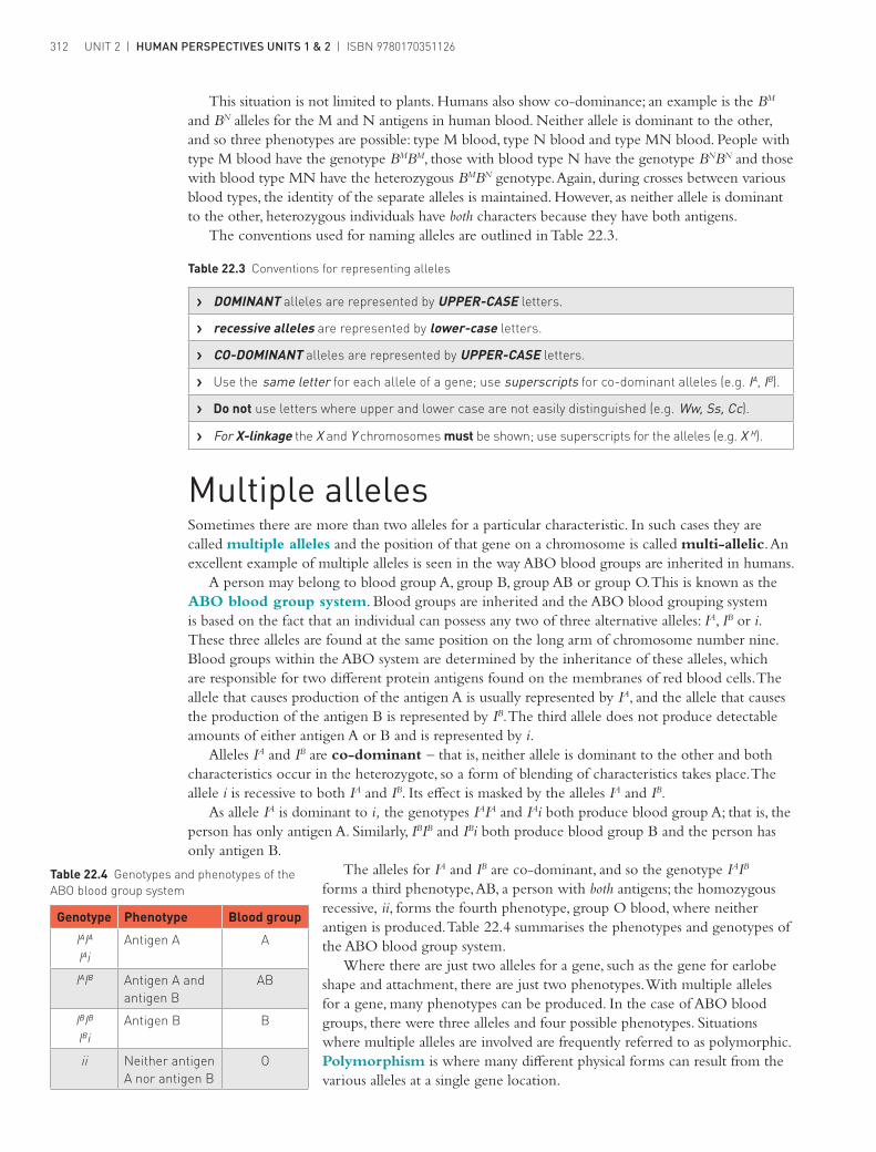
Alleles IA and IB are co-dominant – that is, neither allele is dominant to the other and both characteristics occur in the heterozygote, so a form of blending of characteristics takes place. The allele i is recessive to both IA and IB. Its effect is masked by the alleles IA and IB.
As allele IA is dominant to i, the genotypes IAIA and IAi both produce blood group A; that is, the person has only antigen A. Similarly, IBIB and IBi both produce blood group B and the person has only antigen B.
The alleles for IA and IB are co-dominant, and so the genotype IAIB forms a third phenotype, AB, a person with both antigens; the homozygous recessive, ii, forms the fourth phenotype, group O blood, where neither antigen is produced. Table 22.4 summarises the phenotypes and genotypes of the ABO blood group system.
Where there are just two alleles for a gene, such as the gene for earlobe shape and attachment, there are just two phenotypes. With multiple alleles for a gene, many phenotypes can be produced. In the case of ABO blood groups, there were three alleles and four possible phenotypes. Situations where multiple alleles are involved are frequently referred to as polymorphic. Polymorphism is where many different physical forms can result from the various alleles at a single gene location.
Table 22.4 Genotypes and phenotypes of the ABO blood group system
Genotype Phenotype Blood group
IAIA
IAiAntigen A A
IAIB Antigen A and antigen B
AB
IBIB
IBiAntigen B B
ii Neither antigen A nor antigen B
O

ISBN 9780170351126 | CHAPTER 22 | INHERITANCE 313
Knowledge of the inheritance of blood groups was sometimes used to determine the parentage of a particular child. For example, it would be impossible for a child whose blood group was AB to have parents whose blood groups were A and O. However, the child’s parents could have been blood groups A and B. This is shown in the crosses below.
Parental blood group A × O or A × O
Genotype IAIA ii IAi ii
Gametes all IA all i (IA or i) all i
F1 genotypes all IAi IAi or ii
F1 phenotypes all group A group A or group O
Parental blood group A × B or A × B
Genotype IAIA IBIB IAi IBIB
Gametes all IA all IB (IA or i) all IB
F1 genotypes all IAIB IAIB or IBi
F1 phenotypes all group AB group AB or group B
Parental blood group A × B or A × B
Genotype IAIA IBi IAi IBi
Gametes all IA (IB or i) (IA or i) (IB or i)
F1 genotypes IAIB or IAi IAIB or IAi or IBi or ii
F1 phenotypes group AB or group A group AB or group A
or group B or group O
Table 22.5 Blood groups and parentage
Blood groups of parents
Possible blood groups of children
Blood groups impossible in children from these parents
O × O O A, B, AB
O × A O or A B, AB
O × B O or B A, AB
O × AB A or B O, AB
A × A O or A B, AB
A × B O or A or B or AB None
A × AB A or B or AB O
B × B O or B A, AB
B × AB A or B or AB O
AB × AB A or B or AB O

UNIT 2 | HUMAN PERSPECTIVES UNITS 1 & 2 | ISBN 9780170351126314
Inheritance of mitochondrial DNAMitochondria are the organelles in the cell where the aerobic phase of respiration occurs to release energy for use by the cell (see Chapter 6). Most of a cell’s DNA is located in the nucleus but a small amount is in the mitochondria. This is called mitochondrial DNA or mtDNA.
Human eggs and sperm both have mitochondria, but while an egg has many hundreds, a sperm has only about 100 – just enough to provide the energy for the sperm to swim to the egg. After a sperm has penetrated the egg at fertilisation, the mitochondria in the sperm are rapidly destroyed. This means that our nuclear DNA comes from the nucleus of the egg and the sperm but the mitochondrial DNA comes only from the egg. In other words, we inherit nuclear DNA from both parents but we inherit mitochondrial DNA from our mothers. You got your mitochondrial DNA from your mother; she got it from her mother, who got it from hers, and so on.
Genetic diseases within populationsThe incidence in the population of severe recessive disorders that are not linked to a sex chromosome is low, as it is highly unlikely that a carrier from one family will mate with another carrier of the same recessive condition. However, in some regions of the world where populations have been geographically or culturally isolated, marriages between close relatives are common. The probability of having a child with a genetic disease then increases. Marriages of this type are generally between cousins. In these cases there is often a high incidence of a particular genetic disease, as the related parents have received some of their genes from a common ancestor and, therefore, have a greater chance of being carriers of an allele for the same abnormal condition.
Marriages between cousins were once common among people inhabiting the countries around the Mediterranean Sea. In these people, the incidence of thalassaemia, a recessive disease in which anaemia results from defects in the formation of haemoglobin, is relatively high. As thalassaemia is most frequent in countries along the Mediterranean coast, in Australia it occurs in those of Mediterranean origin, especially immigrants from Italy and Greece, or their children. People with thalassaemia require frequent blood transfusions throughout their life and special drugs to remove the excess iron that tends to build up in the body.
Figure 22.8a A boy with thalassaemia receives a blood transfusion
Figure 22.8b A Wright-stained blood smear from a patient with thalassaemia
a b
Cor
bis/
© D
r. G
ladd
en W
illis
/Vis
uals
Unl
imite
d
Scie
nce
Pho
to L
ibra
ry/M
auro
Fer
mar
iello

ISBN 9780170351126 | CHAPTER 22 | INHERITANCE 315
Another inherited condition is sickle-cell anaemia. It occurs mainly in black Africans, or in people of black African ancestry. In the tropical zone of Africa, up to 40% of the population of some tribes carry the allele for sickle-cell anaemia. The disease occurs when a person inherits the allele from both parents. It results in the red blood cells being a crescent-like, or sickle, shape (Figure 22.8). The disease is usually fatal, as the sickle-shaped cells do not carry as much oxygen as normal red blood cells. They also stick together and block small blood vessels. Heterozygotes normally show no ill effects unless oxygen is in short supply. When this occurs, their red blood cells show mild ‘sickling’. These individuals are carriers and suffer from sickle-cell trait. (Thus, sickle cell anaemia is an example of co-dominance.) The sickle-cell trait gives certain advantages to those who have it. It provides a degree of immunity to malaria, a disease that is prevalent in those parts of the world where the sickle-cell gene is found.
Figure 22.9 Blood of a person with sickle-cell disease. Instead of the usual biconcave disks the red blood cells are often shaped like sickles.
Get
ty Im
ages
/Pho
to R
esea
rche
rs/O
mik
ron
A third genetic disorder, inherited in an autosomal recessive pattern, is Tay-Sachs disease (TSD). This hereditary disorder of lipid metabolism occurs most frequently in individuals of Jewish descent from eastern Europe (the Ashkenazi Jewish population). It is a fatal disorder caused by a missing enzyme that results in the accumulation of a fatty substance in the nervous system. A baby who has Tay-Sachs develops normally for the first few months, but then deterioration causing mental and physical disabilities begins. Death usually occurs in early childhood.
The same mutation that causes Tay-Sachs in Ashkenazi Jews also occurs in the Cajun population of southern Louisiana in the United States. Cajuns are an ethnic group who have been reproductively isolated for several hundred years because of language differences. It has been suggested that the mutation may have entered the Cajun population when a Jewish family assimilated into Cajun society.

UNIT 2 | HUMAN PERSPECTIVES UNITS 1 & 2 | ISBN 9780170351126316
Science inquiry
ACTIVITY 22.1 MarsiansMarsians are an imaginary group of people from the red planet, Mars. Their skin colour is determined by two alleles – one for red skin colour and one for white skin colour. Red is dominant to white on Mars. In this activity we will investigate whether Marsians follow the principles of Mendelian genetics by simulating a cross between two heterozygous Marsians.
YOU WILL NEED
For each pair: ❯ 2 containers – 2 L ice-cream containers work well ❯ 20 red beads or counters to simulate the dominant red allele (R) in each gamete ❯ 20 white beads or counters to simulate the recessive white allele (r) in each gamete ❯ felt pen, tally sheet, pencil
WHAT TO DO1 Label one container ‘Male parent’ and the other ‘Female parent’.2 Place 10 of the red beads (gamete with R allele) in each container, then 10 of the white beads
(gamete with r allele).3 Prepare a tally sheet similar to the one below.
Genotypes in the Marsian offspring
RR Rr rR rr
1
2
3
4
5
6
7
8
9
10
4 Shake the containers well. Draw out one bead (gamete) from each container in turn and place a tick in the relevant box on the tally sheet to show the combination of alleles in the offspring.
5 When you have completed 10 draws, place the beads back into the container. Your partner should repeat steps 1 to 4. Together you should now have two completed tally sheets.

ISBN 9780170351126 | CHAPTER 22 | INHERITANCE 317
STUDYING YOUR DATA1 Because the first three columns all contain the dominant allele (R), individuals with these
genotypes will all appear red. Tally up the number of red offspring.2 Individuals with the genotype rr will appear white. How many white offspring do you have?3 What is the ratio of the phenotypes, red to white?4 Combine your data with that of the other groups in the class to obtain a bigger sample. What is
the ratio now?
INTERPRETING YOUR DATA1 Has this activity shown that inheritance of skin colour in Marsians follows the principles of
Mendelian genetics?2 How close were your results to the expected result of 3:1?3 Was the ratio calculated by combining all groups in the class closer to the expected? Explain
why this was the case.4 If the red and white alleles had been incompletely dominant, with the heterozygous colour
being pink, what ratio would you now obtain? What ratio would you have expected?

UNIT 2 | HUMAN PERSPECTIVES UNITS 1 & 2 | ISBN 9780170351126318
Review questions 1 Define the following terms:
a pure-breedingb progenyc hybridd dominante recessive.
2 Briefly describe what is meant by the principle of segregation. 3 Using examples, distinguish between:
a homozygous and heterozygousb phenotype and genotypec allele and gene.
4 a What is the first filial generation?b Distinguish between the first filial generation and the second filial generation.
5 a What is a monohybrid? Give an example to explain your answer.b What is co-dominance? Give an example in which co-dominance influences the
characteristics of offspring. 6 Explain how the sex of a child is determined at the time of fertilisation. 7 Describe the difference in appearance between the X and Y chromosomes. 8 a What are autosomes?
b How many autosomes occur in (i) each normal human cell and (ii) each sperm or egg? 9 a What are sex-linked (or X-linked) characteristics?
b Give examples of such characteristics.10 a What are multiple alleles?
b Give examples of characteristics that are determined by multiple alleles.11 a What is sickle-cell anaemia? Describe why it is usually lethal.
b What is sickle-cell trait? Does sickle-cell trait have any advantages for people who have it?
Apply your knowledge 1 In garden peas, round seed shape is dominant to wrinkled seed shape. Pure-breeding round
seed plants were crossed with pure-breeding wrinkled seed plants. Determine the expected genotypes and phenotypes of the F1 and F2, and the expected proportions.
2 In humans, normal melanin production is dominant to albino, which produces white hair and pink eyes. The first child born to a married couple with normal pigmentation is an albino. Calculate the probability that the second child will also be an albino. Give a clear explanation for your results.
3 In guinea pigs, black fur colour is dominant over white fur colour. How could an animal breeder test whether a black guinea pig is homozygous or heterozygous?
4 In humans, free earlobes are dominant over attached earlobes. A woman heterozygous for free earlobes marries a man with attached earlobes. Use a Punnett square to determine their chance of producing children with attached earlobes.
5 In many families, a Roman-shaped nose is dominant to a straight nose. If a man from a family pure-breeding for a Roman nose has children with a woman from a family pure-breeding for a straight nose, what would they look like? If one of the children has children with a person from a family with a long history of straight noses, what types of noses would you expect the grandchildren to possess and in what proportions?
6 When plants that are pure-breeding for wrinkled seeds are crossed with plants that are pure-breeding for round seeds, the ratio of genotypes in the F2 is 1:2:1 and the ratio of phenotypes is 3:1. Explain what causes the genotypic ratio to differ from the phenotypic ratio.

ISBN 9780170351126 | CHAPTER 22 | INHERITANCE 319
7 How is the inheritance of flower colour in four-o’clock plants an exception to Mendel’s principle of dominance?
8 In what situations would you be able to deduce a person’s genotype by determining their phenotype? Give examples using ABO blood groups.
9 Describe why it is impossible for parents who have the blood groups A and AB to produce children with blood group O.
10 Charlie Chaplin, a famous comedian of the silent screen, was taken to court in 1944 by a young starlet, Joan Barry. She claimed that Chaplin was the father of her child, and the court ruled in her favour. Blood group data were not admissible evidence at the time of that trial. However, if you were the judge, how would you have decided? The baby was blood group B, the mother A and Chaplin O. Give genetic reasons for your decision.
11 If a human male with blood group M has children with a female with blood group N, what blood groups would they possess? If one of the children has children with a person with blood group M, what blood groups could the grandchildren possess? Construct the crosses for each of these matings. List the genotypes and phenotypes that would be expected, and the probability of obtaining each genotype and phenotype.
12 A woman from a family with no history of haemophilia marries a man who is a haemophiliac. What is the probability that they will produce:a sons with normal blood clottingb sons with haemophiliac daughters who are carriers of haemophiliad daughters who will be haemophiliacs?
13 Red–green colour blindness is a sex-linked characteristic. Under what circumstances would a couple produce daughters who all had normal vision and sons who were all colourblind? Describe the genotypes of both parents and all the children.
14 A woman has a brother with Duchenne muscular dystrophy. What information could be given to the woman about the risk of her having a child with Duchenne muscular dystrophy?